Article

Seasonal Dynamics of Gonadal Antioxidant Biomarkers Associated with Reproductive Indices in Capoeta umbla and Capoeta trutta

1
Department of Veterinary Medicine, Vocational School of Food, Agriculture and Livestock, Bingöl University, 12000 Bingöl, Türkiye
2
Department of Occupational Health and Safety, Vocational School of Tercan, Erzincan Binali Yıldırım University, 24100 Erzincan, Türkiye
3
Department of Veterinary Medicine, Çayırlı Vocational School, Erzincan Binali Yıldırım University, 24500 Erzincan, Türkiye
4
Department of Plant and Animal Production, Vocational School of Food, Agriculture and Livestock, Bingöl University, 12000 Bingöl, Türkiye
5
Department of Food Processing, Vocational School of Food, Agriculture and Livestock, Bingöl University, 12000 Bingöl, Türkiye
*
Author to whom correspondence should be addressed.
Fishes 2026, 11(7), 423; https://doi.org/10.3390/fishes11070423
Submission received: 1 May 2026 / Revised: 8 July 2026 / Accepted: 10 July 2026 / Published: 17 July 2026
(This article belongs to the Special Issue Reproductive Physiology of Fishes)

Abstract

Seasonal variability and reproductive investment influence oxidative metabolism in fish; however, the relationship between reproductive dynamics and antioxidant responses in freshwater cyprinids remains insufficiently understood. This study investigated seasonal and biological factors affecting antioxidant biomarkers in the gonads of Capoeta umbla and Capoeta trutta inhabiting the Karasu River (Türkiye). A total of 152 individuals were examined between April 2023 and March 2024, and reproductive indices (total body weight, gonad weight, and gonadosomatic index) were evaluated together with oxidative stress biomarkers, including malondialdehyde (MDA), superoxide dismutase (SOD), catalase (CAT), glutathione reductase (GR), and glutathione peroxidase (GPx). Multivariate analyses (PERMANOVA, SIMPER, and principal coordinate analysis) were used to determine the factors shaping oxidative responses. Females exhibited higher reproductive investment, while reproductive activity increased during spring and summer, accompanied by elevated MDA levels and antioxidant enzyme activities. PERMANOVA identified season as the primary factor explaining variation in oxidative biomarkers (p = 0.001), followed by reproductive parameters, and PCO revealed clear seasonal separation among groups. These findings indicate that oxidative balance in gonadal tissues is strongly associated with seasonal reproductive cycles and environmental variability in wild freshwater cyprinids.
Key Contribution: This study investigates gonadal antioxidant dynamics in Capoeta umbla and Capoeta trutta over a one-year period, integrating seasonal, reproductive, and sex-related factors. It shows that seasonality is the main driver of antioxidant variation, with reproductive investment (GSI, TGW) closely linked to increased oxidative stress and antioxidant activity. SOD is identified as the key biomarker shaping these patterns. This study also reveals species-specific tendencies in oxidative regulation and highlights gonadal antioxidant biomarkers as effective indicators of reproductive status and environmental adaptation in freshwater fish.

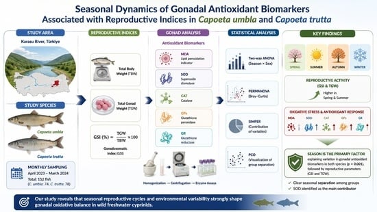
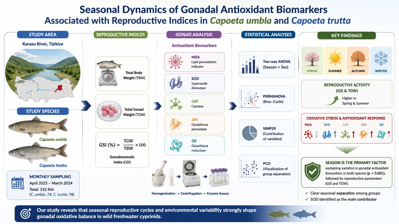
Graphical Abstract

1. Introduction

Fish reproduction represents a highly energy-intensive physiological process characterized by rapid cellular proliferation, gametogenesis, and hormonally regulated gonadal development, all of which are coordinated through complex neuroendocrine pathways and require substantial metabolic investment [1,2]. These processes require high metabolic activity, particularly in gonadal tissues, which inevitably leads to increased production of reactive oxygen species (ROS) as by-products of mitochondrial respiration [3]. Although ROS play essential roles in normal cellular signaling, steroidogenesis, and gamete maturation, excessive ROS production can disrupt redox homeostasis and result in oxidative stress [4,5,6]. Oxidative stress can induce lipid peroxidation, protein oxidation, DNA damage, and enzyme inactivation, ultimately impairing the structural integrity and function of reproductive tissues and reducing gamete quality [4,7]. In fish, oxidative damage in gonadal tissues has been associated with impaired oocyte maturation, reduced sperm motility, and decreased reproductive success, highlighting the importance of antioxidant defense mechanisms in maintaining reproductive health [7].
Fish gonads are particularly vulnerable to oxidative stress due to their high content of polyunsaturated fatty acids and elevated oxygen consumption during gametogenesis [8]. To protect against oxidative damage, fish possess an antioxidant defense system consisting of enzymatic antioxidants, including superoxide dismutase (SOD), catalase (CAT), glutathione peroxidase (GPx), and glutathione reductase (GR), which function cooperatively to eliminate ROS and maintain redox homeostasis [9,10]. In contrast, malondialdehyde (MDA), a by-product of lipid peroxidation, is widely used as an indicator of oxidative damage and oxidative stress in fish tissues, particularly in reproductive organs [11]. Variations in antioxidant enzyme activities and MDA levels reflect changes in physiological status, reproductive activity, and environmental conditions [12,13].
The gonadosomatic index (GSI) and total gonad weight (TGW) are widely used indicators of reproductive status and energy allocation in fish and reflect gonadal maturation and reproductive investment [14,15]. During gonadal development, increased metabolic activity and oxygen consumption may enhance ROS production, resulting in changes in antioxidant enzyme activities and oxidative stress levels [16,17]. Furthermore, reproductive physiology differs significantly between male and female fish due to differences in gametogenesis, hormonal regulation, and reproductive investment, leading to sex-related differences in oxidative stress responses and antioxidant defense mechanisms [18,19]. Seasonal variations in environmental factors such as temperature, photoperiod, and food availability regulate reproductive cycles and influence oxidative stress responses in fish [12,20]. Previous studies have demonstrated that antioxidant enzyme activities and lipid peroxidation levels fluctuate significantly during different reproductive stages and seasons, reflecting physiological adjustments associated with gonadal maturation and reproductive activity [21,22,23,24]. Additionally, the precise mechanisms underlying the relationship between total body weight (TBW) and the gonadal antioxidant system, particularly in fish, have not yet been fully elucidated. In cyprinid fishes, somatic condition and body energy reserves are closely associated with reproductive investment and physiological resilience [20]. Individuals with higher body condition generally possess greater energetic resources that can support gametogenesis, gonadal development, and the maintenance of antioxidant defenses during periods of elevated metabolic demand [23,25]. Conversely, reduced somatic reserves may limit antioxidant capacity by prioritizing energy allocation toward essential maintenance functions rather than reproductive processes. Therefore, body mass-related variation may indirectly influence oxidative balance in reproductive tissues through differences in energy availability and metabolic activity [10]. Nonetheless, existing literature indicates that antioxidant enzyme activities in gonadal tissues are closely associated with an individual’s condition and energy reserves. As body weight and reproductive investment increase, gonadal metabolic activity may rise, leading to enhanced production of ROS, which in turn can induce regulatory and/or compensatory adjustments in the antioxidant defense system [25]. In fish specifically, the allocation of energy between somatic growth and reproductive investment can influence gonadal oxidative balance, resulting in positive or negative correlations in antioxidant enzyme activities [26,27]. Within this context, a detailed investigation of this relationship in fish could provide a more comprehensive understanding of the interplay between reproductive physiology, energy allocation, and oxidative stress, offering novel perspectives on the topic.
Capoeta umbla (C. umbla) and Capoeta trutta (C. trutta) are freshwater cyprinid fish species widely distributed throughout the Euphrates–Tigris river basin and adjacent inland water systems, where they play significant ecological and economic roles in local fisheries [28,29,30,31]. These species exhibit well-defined seasonal reproductive cycles characterized by pronounced changes in gonadal development, TGW, and GSI, primarily regulated by environmental factors such as temperature, photoperiod, and food availability [32,33]. Due to these distinct reproductive patterns, both species provide suitable biological models for investigating reproductive physiology and its associated biochemical processes, including oxidative stress and antioxidant defense mechanisms [34,35]. Previous studies have highlighted the importance of antioxidant systems in maintaining gonadal function and reproductive success in Capoeta species from cyprinid fishes [17,36,37,38]; however, information regarding seasonal variations in antioxidant enzyme activities in gonadal tissues and their relationships with TBW, TGW, GSI, and sex in C. umbla and C. trutta remains limited. Within this context, the present study aims to provide a comprehensive evaluation of seasonal variations in gonadal antioxidant defense mechanisms in both species. It is hypothesized that seasonal gonadal development, TGW, and GSI significantly influence antioxidant enzyme activities and lipid peroxidation levels in both male and female fish. Specifically, we sought to (i) quantify seasonal changes in antioxidant enzyme activities (SOD, CAT, GPx, and GR) and lipid peroxidation (MDA) in males and females, (ii) determine sex-specific differences in oxidative stress responses, (iii) assess correlations between antioxidant biomarkers and reproductive indices including TGW, GSI, and TBW to understand the influence of energy allocation on gonadal oxidative balance, and (iv) identify the most influential antioxidant parameters and seasonal patterns using multivariate analyses (PERMANOVA, PCO, and SIMPER). This approach allows for a detailed understanding of the interplay between reproductive physiology, energy allocation, and oxidative stress throughout a one-year reproductive cycle in C. umbla and C. trutta.

2. Materials and Methods

2.1. Study Area and Sampling

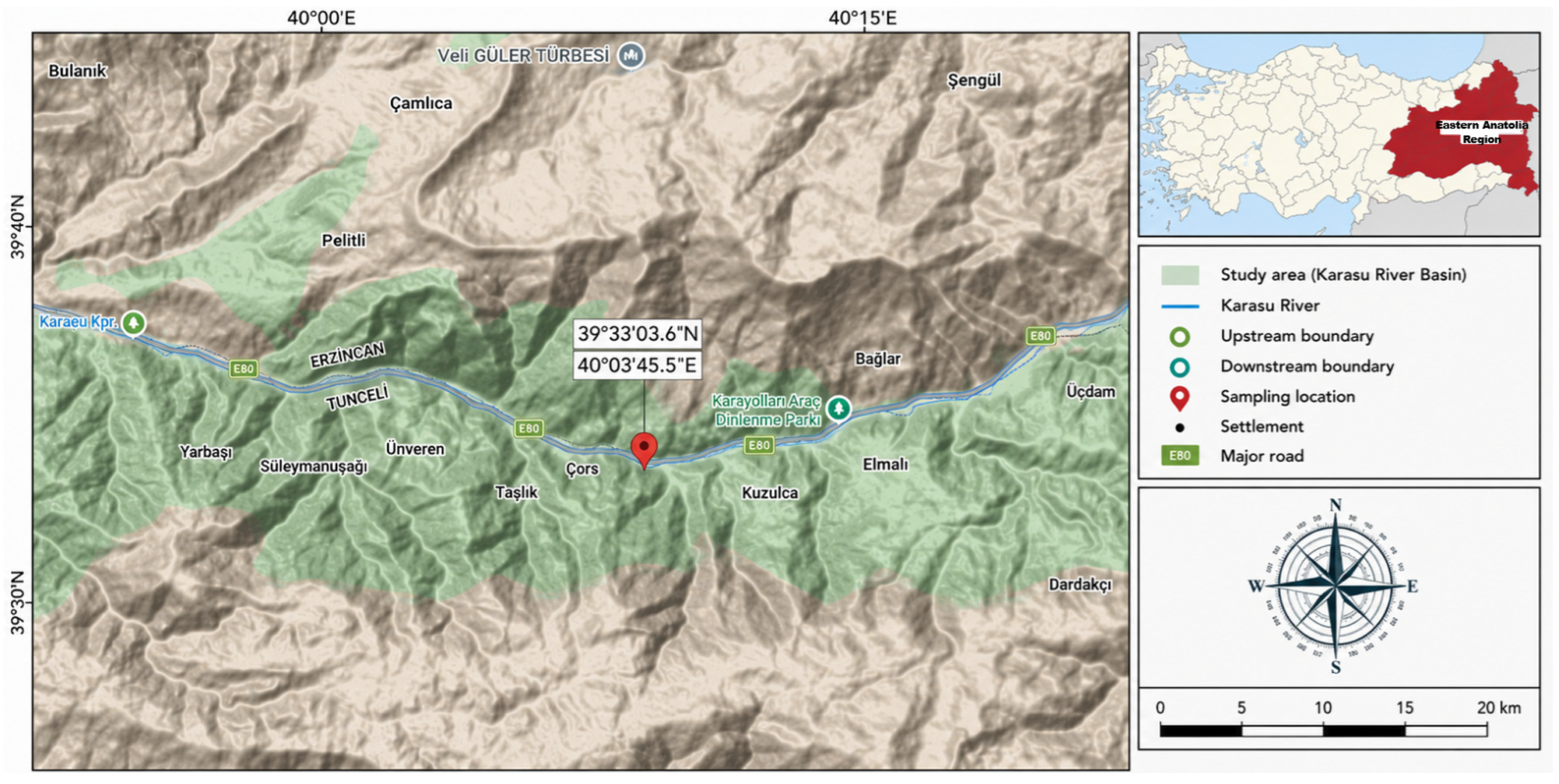
The Karasu River, located in the Eastern Anatolia Region of Türkiye, originates from Dumlu Mountain in Erzurum Province and converges with the Murat River near Keban, forming the Euphrates River [39]. Specimens of C. umbla and C. trutta were obtained directly from local fishermen conducting wild-capture activities at a fixed sampling location (39°33′03.6″ N, 40°03′45.5″ E) along the Karasu River within the administrative boundaries of Erzincan Province (Figure 1). The specimens were collected during routine fishing activities using gill nets and were captured directly from the natural river habitat. The wild origin of specimens was further verified through direct communication with local fishermen, who confirmed that all individuals were captured from natural river habitats and not from aquaculture facilities. Since neither C. umbla nor C. trutta is commercially cultured in Türkiye, the collected individuals were considered wild-caught specimens. Fish were collected on the same day of capture and transported to the laboratory under cold-chain conditions. Sampling was conducted monthly from April 2023 through March 2024 to ensure comprehensive representation of seasonal variation. Local fishermen captured the specimens using gill nets with mesh sizes of 16 mm × 16 mm, 22 mm × 22 mm, and 32 mm × 32 mm according to their routine fishing practices. Concurrently, the water temperature, oxygen, salinity, electrical conductivity (EC), and pH values were measured in situ at each sampling event, and the seasonal mean temperature values were subsequently calculated for analysis.

2.2. Laboratory Studies

To minimize potential post-mortem alterations affecting oxidative stress biomarkers, all specimens were processed within the shortest possible time after capture. Fish were collected on the sampling day, placed on ice immediately after capture, and transported to the laboratory under cold-chain conditions for processing. The elapsed time between capture and laboratory processing was approximately 3 h. Following transportation, the total weight of each specimen was recorded, and individuals were subsequently separated according to sex. Sex and sexual maturity status were determined through macroscopic examination of the gonads after dissection. Individuals were classified as sexually mature based on the presence of developed gonads characteristic of reproductive adults, including enlarged ovaries with visible oocytes in females and developed testes containing milt in males. Specimens possessing paired, elongated, and whitish testes were identified as males, whereas individuals with relatively larger, lobulated ovaries ranging in color from yellow to orange and containing visible oocytes were identified as females. Individuals with immature or poorly developed gonads were excluded from the study. Gonadal morphology and coloration were assessed in accordance with established ichthyological criteria reported in previous studies [40,41]. For the calculation of the GSI and subsequent multivariate statistical analyses, TBW (g) was measured using a digital precision balance (Weightlab WL-3002L, Shanghai, China; accuracy ± 0.01 g; maximum capacity 3000 g). The fish samples were collected monthly, and seasonal mean values were calculated from the corresponding monthly averages. Although many individuals were sampled throughout the study period, a subset of randomly selected specimens was used for GSI calculations by measuring TBW and TGW. For the assessment of gonadal antioxidant biomarker parameters, individuals were randomly subsampled from the total catch for each sampling period, with a minimum of 10 specimens selected to represent each season, depending on overall sample availability. The subsampling procedure was performed by considering seasonal sampling intensity and the minimum tissue quantity required for biochemical assays. All tissue samples were stored at −80 °C until biochemical assays were performed.

2.3. Gonadosomatic Index (GSI)

GSI was calculated to evaluate the relative contribution of gonads to the TBW of each individual, serving as an indicator of reproductive condition and seasonal gonadal development. The GSI of male and female individuals of C. umbla and C. trutta were calculated separately for each season. The index was calculated using the following formula [42]:
G S I ( % ) = T G W ( g ) T B W ( g ) × 100
where TBW is the total body weight (g), and TGW is the total gonad weight (g).
GSI values, calculated for sexually mature individuals, were employed in subsequent statistical analyses to assess seasonal variations in reproductive status and to investigate their associations with antioxidant enzyme activities.

2.4. Antioxidant Enzyme Analysis

2.4.1. Homogenate Preparation

The gonadal tissues were carefully rinsed with physiological saline (0.9% NaCl) to eliminate potential contaminants and residual blood. The tissues were subsequently dissected into small fragments using a scalpel and suspended in 50 mM KH2PO4 buffer (Sigma-Aldrich, St. Louis, MO, USA; pH = 7.4). The tissue samples were then homogenized in liquid nitrogen to effectively release cellular components. The resulting homogenates were centrifuged at 27,000× g for 60 min using a refrigerated centrifuge (Avanti J-26 XP, Beckman Coulter, Brea, CA, USA). Following centrifugation, the supernatant was carefully collected using a Pasteur pipette, while any sediment remaining at the bottom of the tube was discarded. The clarified supernatant fraction was used for the determination of antioxidant enzyme activities [43,44].

2.4.2. Determination of Lipid Peroxidation and Enzyme Activities

Malondialdehyde (MDA) levels, used as an indicator of lipid peroxidation (LPO), were quantified in all tissue samples using the thiobarbituric acid (TBA;Sigma-Aldrich, St. Louis, MO, USA)) assay as described previously [45]. This method relies on the reaction of MDA with TBA to form a colored adduct, which was subsequently measured spectrophotometrically using a UV–Visible spectrophotometer (UV-1800, Shimadzu Corporation, Kyoto, Japan).
Glutathione reductase (GR) activity was assessed at 25 °C using a modified protocol [46], based on the reduction of oxidized glutathione to its reduced form and quantified by monitoring NADPH (Sigma-Aldrich, St. Louis, MO, USA) consumption during the reaction. Superoxide dismutase (SOD) activity was determined at 20 °C and 560 nm, with the assay principle relying on the inhibition of the reaction between superoxide anions and reduced nitroblue tetrazolium (NBT; Sigma-Aldrich, St. Louis, MO, USA) [47]. Catalase (CAT) activity was measured by monitoring the decomposition rate of hydrogen peroxide (H2O2; Merck, Darmstadt, Germany) at 20 °C and 240 nm [48]. Glutathione peroxidase (GPx) activity was determined spectrophotometrically at 340 nm based on NADPH oxidation [49].
Prior to biochemical analyses, the total protein concentrations of tissue homogenates were determined according to Lowry et al. [50], and all antioxidant enzyme activities (CAT, SOD, GPx, and GR) as well as MDA levels were normalized to protein content following the methods described above. Enzyme activities are expressed as units per mg protein (U/mg protein), while MDA concentrations are reported as nmol MDA per mg protein (nmol MDA/mg protein). All assays were performed in technical replicates for each sample, and mean values were used for subsequent statistical analyses. Experimental conditions were maintained consistently across all samples to minimize analytical variability.

2.5. Statistical Analysis

Relationships among the examined variables were quantified using Spearman’s rank correlation coefficient for those that did not meet parametric assumptions.
Multivariate statistical analyses were performed to evaluate variations in antioxidant biomarker levels using PRIMERv7 with the PERMANOVA+ add-on (version 7.0.21; PRIMER-e Ltd., Auckland, New Zealand). Bray–Curtis similarity coefficients were calculated for PERMANOVA, while Principal Coordinate (PCO) Analysis and hierarchical cluster analysis were applied to assess and visualize similarity patterns among samples. Antioxidant enzyme biomarker data were analyzed in relation to season, GSI, TGW, TBW, sex (for both species), and interspecific differences. For comparative purposes, the dataset was stratified into groups according to whether values for each factor were above or below their respective mean thresholds. Factor groups were defined based on mean body weight and GSI values to reflect biological variation within the fish population and to obtain comparable group sizes for statistical analyses [51,52].
For C. umbla, factor groups were defined as follows:
TBW: Group 1 (TBW1, n = 39): <536.00 g; Group 2 (TBW2, n = 35): ≥536.00 g
TGW: Group 1 (TGW1, n = 21): ≤8.00 g; Group 2 (TGW2, n = 15): >8.00–20.00 g; Group 3 (TGW3, n = 13): ≥20.00–31.00 g; Group 4 (TGW4, n = 13): ≥32.00–65.00 g; Group 5 (TGW5, n = 12): ≥65.00–125.00 g
GSI: Group 1 (GSI1, n = 23): ≤1.99%; Group 2 (GSI2, n = 28): >1.99–5.50%; Group 3 (GSI3, n = 8): ≥5.50–11.00%; Group 4 (GSI4, n = 15): ≥11.00–19.30%
For C. trutta, factor groups were defined as follows:
TBW: Group 1 (TBW1, n = 39): <410.00 g; Group 2 (TBW2, n = 39): ≥410.00 g
TGW: Group 1 (TGW1, n = 34): ≤5.00 g; Group 2 (TGW2, n = 25): >5.00–9.40 g; Group 3 (TGW3, n = 19): ≥9.40–51.01 g
GSI: Group 1 (GSI1, n = 37): ≤1.38%; Group 2 (GSI2, n = 23): >1.38–2.25%; Group 3 (GSI3, n = 18): ≥2.25–12.61%
Antioxidant biomarkers exhibiting the greatest variability among samples were analyzed within these factor groups. Similarity Percentage Analysis (SIMPER), using a 70% cut-off, was performed to identify biomarkers contributing most to within- and between-group similarity. The most influential factor groups were determined using the PERMANOVA Main test (unrestricted permutation of raw data; number of permutations = 999), and group effects within each factor were further assessed using the PERMANOVA pairwise test (unrestricted permutation of raw data; number of permutations = 999).
All statistical analyses were conducted using Statistica version 12.0 (TIBCO Software Inc., Palo Alto, CA, USA). The effects of season and sex, as well as their interaction, on antioxidant biomarkers (MDA, CAT, GR, GPx, and SOD) were assessed using a two-way factorial analysis of variance (ANOVA), with season and sex considered as fixed factors. Each biomarker was analyzed independently. When significant main or interaction effects were detected, Tukey’s honestly significant difference (HSD) test was applied for multiple pairwise comparisons among groups. Differences were considered statistically significant at p < 0.05.

3. Results

3.1. Seasonal Variations in Reproductive Indices and Antioxidant Biomarker Responses in the Gonad Tissues of Capoeta umbla and Capoeta trutta

A total of 152 individuals belonging to two cyprinid species, C. umbla and C. trutta, collected from the Karasu River between April 2023 and March 2024 were examined. Annual and seasonal variations in TBW, TGW, and GSI were determined, and seasonal changes in antioxidant biomarkers (MDA, CAT, GR, GPx, and SOD) in gonad tissues were evaluated in relation to species, sex, and reproductive indices.
Annual mean values of TBW, TGW, and GSI for both species are presented in Table 1. In C. umbla, females showed higher mean TBW (559.53 g) compared with males (464.26 g). Similarly, TGW and GSI values were considerably higher in females (39.16 g and 6.88%, respectively) than in males (13.21 g and 1.48%, respectively). The overall averages for this species were 535.07 g for TBW, 30.86 g for TGW, and 5.49% for GSI. In C. trutta, TBW values of females (413.5 g) and males (405.56 g) were relatively similar. However, females exhibited higher TGW (12.06 g) and GSI (2.87%) compared with males (6.07 g and 1.50%, respectively). The overall mean values for TBW, TGW, and GSI were 409.83 g, 9.29 g, and 2.24%, respectively.
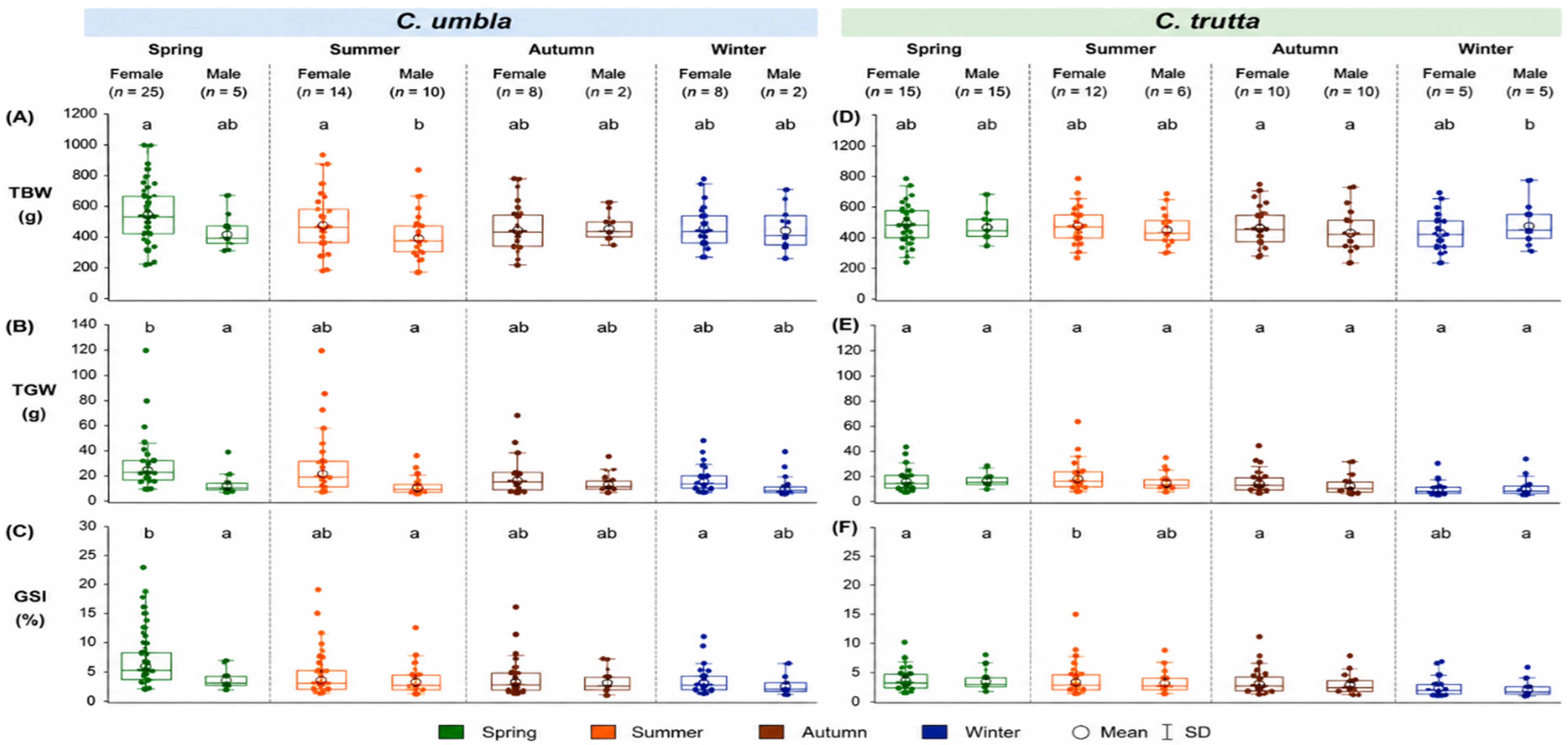
Seasonal variations in TBW, TGW, and GSI for both species are shown in Table 2 and Figure 2. In C. umbla, female TBW values were highest during summer (595.29 g) and lowest during autumn (512.00 g), whereas male TBW values were highest in spring (484.00 g). Seasonal changes in TGW and GSI were more pronounced in females. The highest TGW and GSI values were recorded in spring (50.59 g and 9.10%, respectively), followed by summer, and gradually decreased toward winter. Male TGW and GSI values remained comparatively low throughout the sampling period. In C. trutta, seasonal changes in TBW were less pronounced. Female TBW values peaked in summer (449.75 g) and reached the lowest levels in autumn (372.00 g). Male TBW values were highest in winter (494.00 g). Seasonal variation in TGW and GSI was more evident in females, with maximum values observed in summer (23.07 g and 5.33%, respectively). In contrast, males exhibited relatively smaller seasonal fluctuations in these parameters. Statistical comparisons indicated significant differences among seasons for several reproductive parameters within species (p < 0.05).
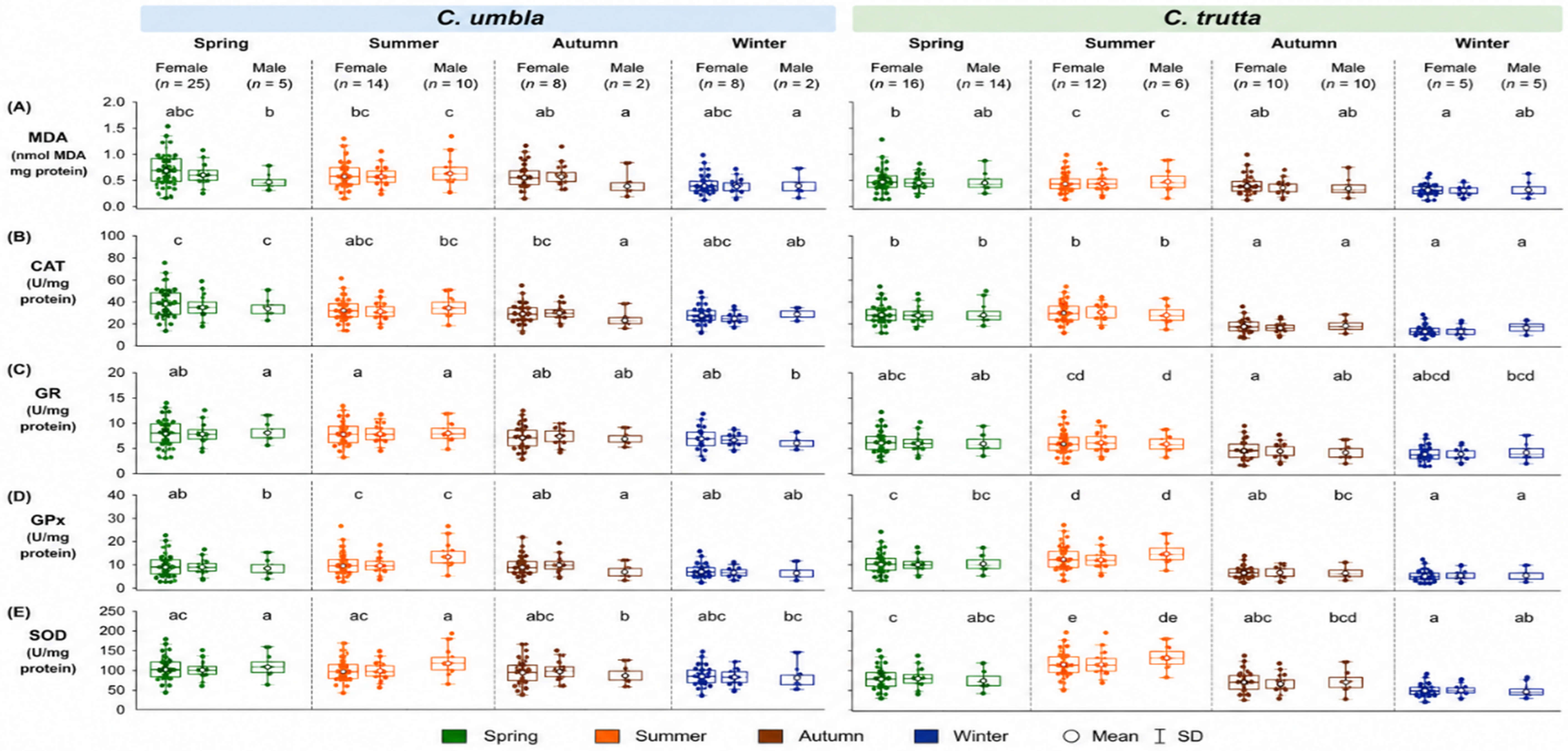
Seasonal variations in antioxidant biomarkers (MDA, CAT, GR, GPx, and SOD) in gonad tissues of both species are presented in Table 3 and Figure 3, based on Tukey’s HSD post-hoc comparisons (p < 0.05). In C. umbla, gonadal MDA levels showed significant seasonal variation, with summer values in both males (1.07 nmol MDA/mg protein) and females (1.14 nmol MDA/mg protein) being significantly higher than winter values (males: 0.53; females: 0.50 nmol MDA/mg protein), while differences between summer and spring were not statistically emphasized based on Tukey groupings. Autumn values were intermediate and did not differ markedly from adjacent seasons. CAT activity was significantly higher in spring compared with autumn and winter in both sexes (spring males: 41.95; females: 40.04 U/mg protein), whereas summer values were not significantly different from spring in males but showed partial overlap with lower autumn–winter groups in females. GR activity showed that spring and summer values generally formed higher statistical groups compared with winter, indicating significant seasonal differences between these periods. GPx activity was significantly higher in summer than in spring and winter in both males (14.06 U/mg protein) and females (16.69 U/mg protein), while winter values were significantly lower. SOD activity showed significant seasonal structuring, with higher values in spring and summer compared with winter, particularly in females where winter values (75.18 U/mg protein) were significantly lower than spring (119.60 U/mg protein). In C. trutta, MDA levels showed clear and significant seasonal variation, with summer values in both males (1.39 nmol MDA/mg protein) and females (1.30 nmol MDA/mg protein) being significantly higher than winter values (males: 0.63; females: 0.63 nmol MDA/mg protein), while autumn and spring values formed intermediate statistical groups. CAT activity was significantly higher in summer compared with winter and autumn in both sexes (summer males: 45.64; females: 45.70 U/mg protein), whereas spring values did not differ significantly from summer in most comparisons. GR activity showed significant seasonal differences, with summer values (males: 9.93; females: 9.91 U/mg protein) being significantly higher than winter values, while autumn showed intermediate grouping. GPx activity exhibited pronounced seasonal differences, with summer values significantly higher than all other seasons in both sexes (males: 18.59; females: 20.11 U/mg protein), and winter values being the lowest. SOD activity also showed significant seasonal structuring, with summer values significantly higher than winter in both sexes, while spring and autumn formed intermediate groups depending on sex.
The seasonal dynamics of antioxidant biomarkers corresponded with variations in reproductive indices (TGW and GSI). Periods characterized by elevated TGW and GSI values, particularly during spring and summer, were associated with increased activities of antioxidant enzymes such as GPx, CAT, and SOD. Conversely, lower reproductive indices during autumn and winter coincided with reduced enzyme activities and lower oxidative stress marker levels. Overall, the results revealed significant species-, sex-, and season-dependent variations in reproductive parameters and antioxidant biomarker responses in gonad tissues of C. umbla and C. trutta inhabiting the Karasu River ecosystem.
The physicochemical conditions of the Karasu River, showing pronounced seasonal variations, are summarized in Table 4. Water temperature increased from 5.48 °C in spring to 8.60 °C in summer, reflecting typical seasonal warming. Dissolved oxygen (DO) exhibited an inverse trend, decreasing from 12.20 mg/L in spring to 10.80 mg/L in summer, consistent with the temperature-dependent solubility of oxygen in freshwater systems. In autumn, water temperature decreased to 6.50 °C, and DO rose to 11.50 mg/L, whereas winter conditions showed the lowest temperature (3.75 °C) and the highest DO concentration (13.00 mg/L), demonstrating the expected inverse relationship between temperature and oxygen saturation. Salinity remained relatively constant at 0.39 ppt throughout the year, and EC varied moderately from 300 µS/cm in winter to 360 µS/cm in summer, indicating minimal osmotic stress and stable ionic composition. pH values ranged from 7.4 in winter to 8.1 in summer, reflecting slightly alkaline conditions with minor seasonal fluctuations that are unlikely to cause significant stress to freshwater fish. Overall, these seasonal patterns are characteristic of a temperate freshwater river system. Temperature-driven changes in DO, together with stable salinity and conductivity, provide a suitable environment for aquatic organisms. These parameters are particularly relevant for interpreting seasonal variations in metabolic activity and oxidative stress in C. umbla and C. trutta, as temperature and DO directly influence fish respiration and the production of ROS.

3.2. Primary Factors Affecting Antioxidant Biomarker Dynamics in C. umbla and C. trutta

Multivariate analyses of antioxidant biomarkers identified season as the principal factor explaining variation in both C. umbla and C. trutta. Reproductive variables (GSI in C. umbla and TGW in C. trutta) also showed significant effects, whereas TBW, sex, and species did not significantly influence the overall antioxidant biomarker composition.
The effects of season, body weight, gonad weight, GSI groups, sex, and species on antioxidant biomarkers in C. umbla and C. trutta were evaluated using PERMANOVA and SIMPER analyses. PERMANOVA indicated that season was the primary driver of antioxidant biomarker variation in both C. umbla (Pseudo-F = 19.78, Pperm = 0.001) and C. trutta (Pseudo-F = 31.55, Pperm = 0.001). In C. umbla, GSI represented the second most important explanatory factor (Pseudo-F = 11.22, Pperm = 0.001), whereas TGW was the second strongest factor in C. trutta (Pseudo-F = 7.97, Pperm = 0.001). By contrast, TBW, sex, and interspecific differences were not statistically significant (Pperm > 0.05). Consistently, SIMPER analysis showed high similarity between species, reaching 83% for C. umbla and 82% for C. trutta. In both species, SOD was the dominant contributor to this similarity.
Seasonal pairwise comparisons for C. umbla indicated that the lowest seasonal variation occurred between winter and autumn (Pperm = 0.08, t = 1.65), whereas the highest variation was observed between autumn and summer (Pperm = 0.001, t = 5.99). SIMPER analysis revealed that antioxidant biomarker profiles were highly conserved within each season, with the greatest similarity observed in winter and autumn (both 91%), followed by summer (87%) and spring (86%). Across all seasons, SOD consistently represented the main contributor to similarity, accounting for 68% in spring, 67% in autumn, and 67% in winter, with a slightly lower contribution in summer (64%). Seasonal dissimilarity was greatest between spring and autumn (23%), followed by autumn and summer (22%), whereas the lowest dissimilarity occurred between spring and summer (14%). In these seasonal contrasts, SOD remained the principal biomarker driving the observed differences, contributing 59% and 64%, respectively. CAT was the second most influential biomarker contributing to seasonal variation, with its highest contribution observed between spring and autumn (25%).
Seasonal pairwise comparisons conducted for C. trutta indicated that the lowest variation occurred between spring and autumn (Pperm = 0.05, t = 2.88), whereas the highest variation was observed between winter and summer (Pperm = 0.001, t = 12.66). According to SIMPER analysis, antioxidant biomarker profiles were largely conserved within each season. The highest within-season similarity was recorded in winter (92%) and summer (91%), followed by autumn (87%) and spring (61%). Across all seasons, SOD was the major contributor to within-group similarity, accounting for 61% in spring, 68% in autumn, 64% in winter, and 67% in summer. Between-season comparisons revealed that the greatest dissimilarity occurred between summer and winter (34%), whereas the lowest dissimilarity was observed between autumn and winter (15%). In these contrasts, SOD and CAT were the principal biomarkers driving the observed differences, contributing 68% and 64% for summer and winter dissimilarity, respectively. In contrast, GPx also emerged as a secondary contributor, particularly in the autumn-winter comparison, where it accounted for 13% of the dissimilarity.
Within-species analysis indicated that season was the primary factor influencing variation in antioxidant biomarkers in both C. umbla and C. trutta. In C. umbla, this was followed by GSI, whereas in C. trutta, TGW was the second most influential factor. Examination of additional factors revealed that in C. umbla, TGW (Pseudo-F = 7.25, Pperm = 0.001), TBW (Pseudo-F = 1.01, Pperm = 0.34), and sex (Pseudo-F = 0.05, Pperm = 0.95) contributed progressively less to variation. In C. trutta, following the primary seasonal factor and TGW, variation was influenced to a lesser extent by GSI (Pseudo-F = 7.15, Pperm = 0.003), TBW (Pseudo-F = 1.08, Pperm = 0.30), and sex (Pseudo-F = 0.27, Pperm = 0.71). Since the effects of factors beyond the two most influential variables were relatively weak in both species, PCO plots were generated only for the two primary factors.
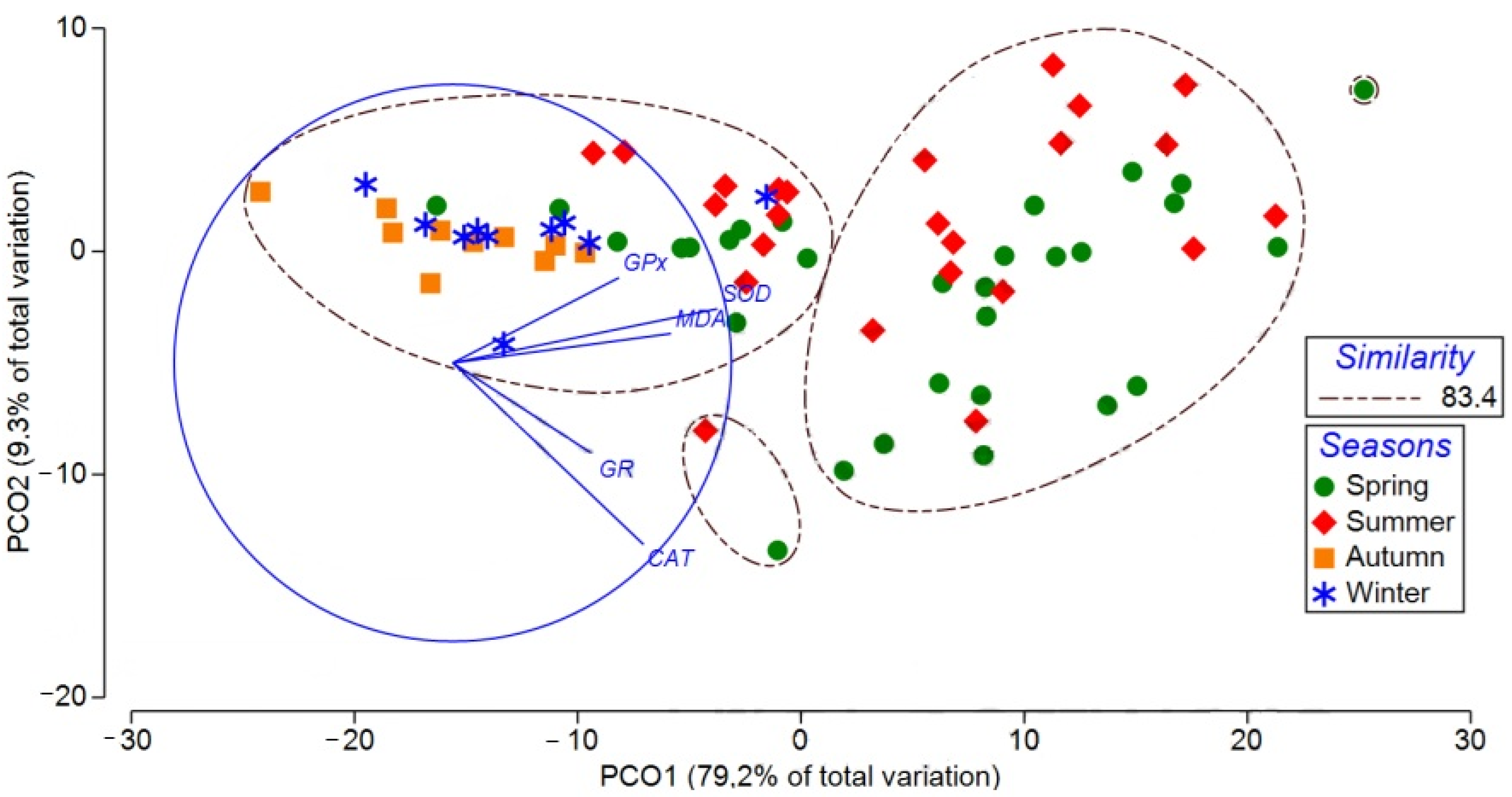
The two-dimensional PCO ordination of the Bray–Curtis resemblance matrix based on antioxidant biomarkers in the gonad tissues of C. umbla revealed a clear seasonal structuring of the samples (Figure 4). The first two axes explained a substantial proportion of the total variation (PCO1: 79.2%; PCO2: 9.3%), accounting for 88.5% overall. Seasonal groups were mainly separated along the PCO1 axis, where spring and summer samples were predominantly distributed on the positive side, whereas autumn and winter samples clustered on the negative side of the axis. Spring and summer groups showed a relatively wider dispersion within the ordination space, while autumn and winter samples formed a more compact cluster, indicating differences in antioxidant biomarker profiles among seasons. The vector projections of antioxidant biomarkers indicated their relative contributions to the observed multivariate pattern. SOD, GPx, and MDA were strongly aligned with the negative direction of PCO1, suggesting that variation in these biomarkers was primarily associated with the autumn–winter samples. In contrast, CAT and GR were oriented toward the negative direction of PCO2, indicating their contribution to the secondary gradient of variation and to the separation of samples located in the lower region of the ordination space. Despite the observed seasonal differentiation, the relatively high similarity level (83.4%) indicates that antioxidant biomarker responses remained broadly comparable across seasons, although the distribution along PCO1 suggests a clear seasonal modulation of oxidative status in the gonad tissues of C. umbla.
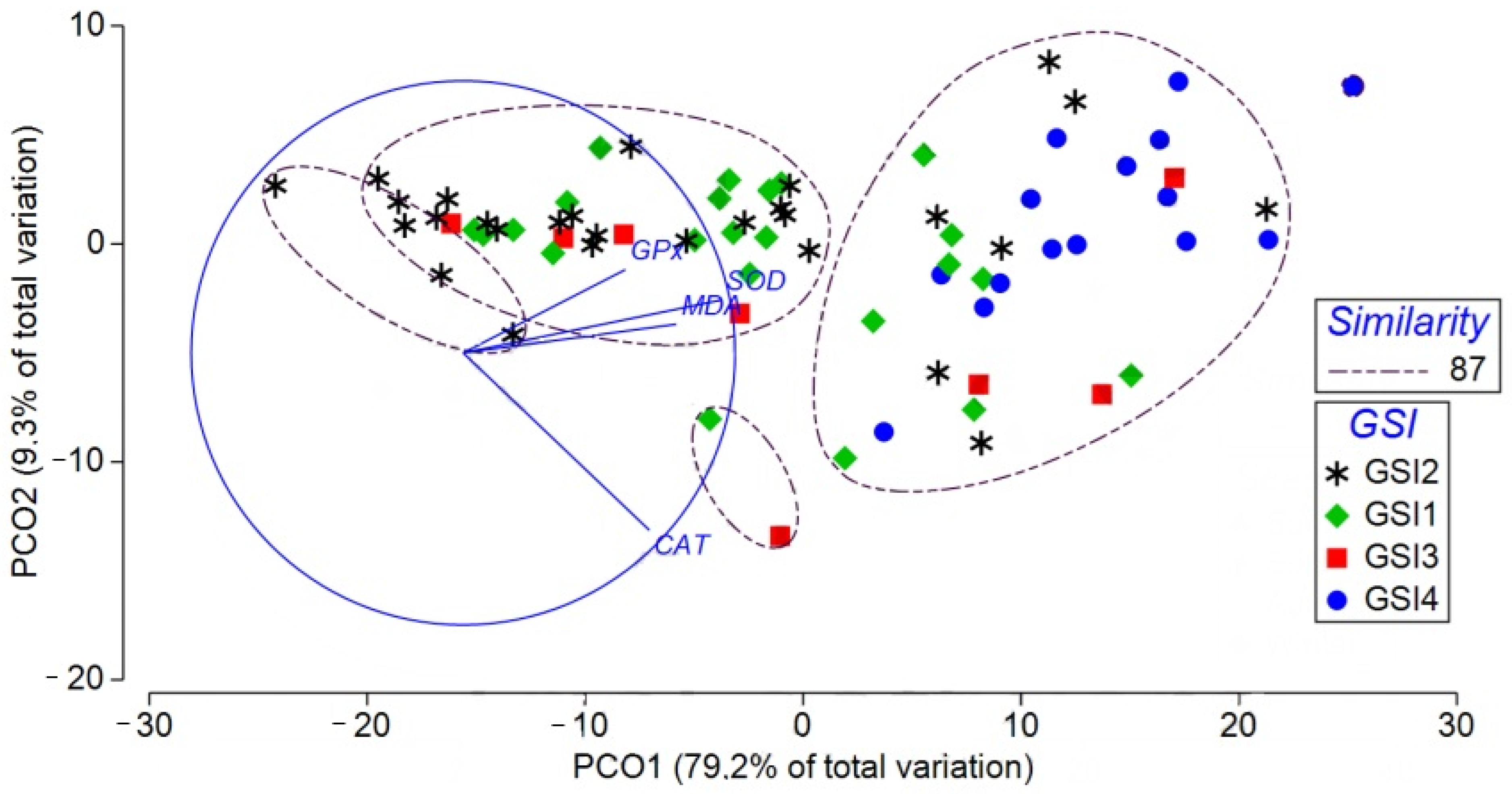
The two-dimensional PCO ordination of the Bray–Curtis resemblance matrix based on antioxidant biomarkers in the gonad tissues of C. umbla revealed a clear distribution of samples according to GSI factor groups (GSI1: ≤1.99%; GSI2: >1.99–5.50%; GSI3: ≥5.50–11.00%; GSI4: ≥11.00–19.30%) (Figure 5). The first two axes explained a substantial proportion of the total variation (88.5%), with PCO1 accounting for 79.2% and PCO2 for 9.3%. Sample separation was primarily structured along the PCO1 axis, where individuals belonging to the higher reproductive stages (GSI3 and GSI4) were predominantly located on the positive side of the axis, while samples from the lower GSI groups (GSI1 and GSI2) were mainly distributed on the negative side, indicating differences in antioxidant biomarker profiles among reproductive conditions. The vector projections of antioxidant biomarkers highlighted their relative contributions to the observed multivariate structure. SOD, GPx, and MDA were strongly aligned with the negative direction of PCO1, suggesting that variation in these biomarkers was more closely associated with the lower GSI groups (GSI1–GSI2). In contrast, CAT exhibited a pronounced orientation toward the negative direction of PCO2, indicating its contribution to the secondary gradient of variation and to the differentiation of samples positioned in the lower region of the ordination space. GR showed a similar orientation but with a comparatively weaker influence. Overall, the ordination pattern, together with the relatively high similarity level (87%), indicates that antioxidant biomarker responses vary with reproductive status in C. umbla, with specific enzymes contributing differentially to the multivariate separation among GSI groups.
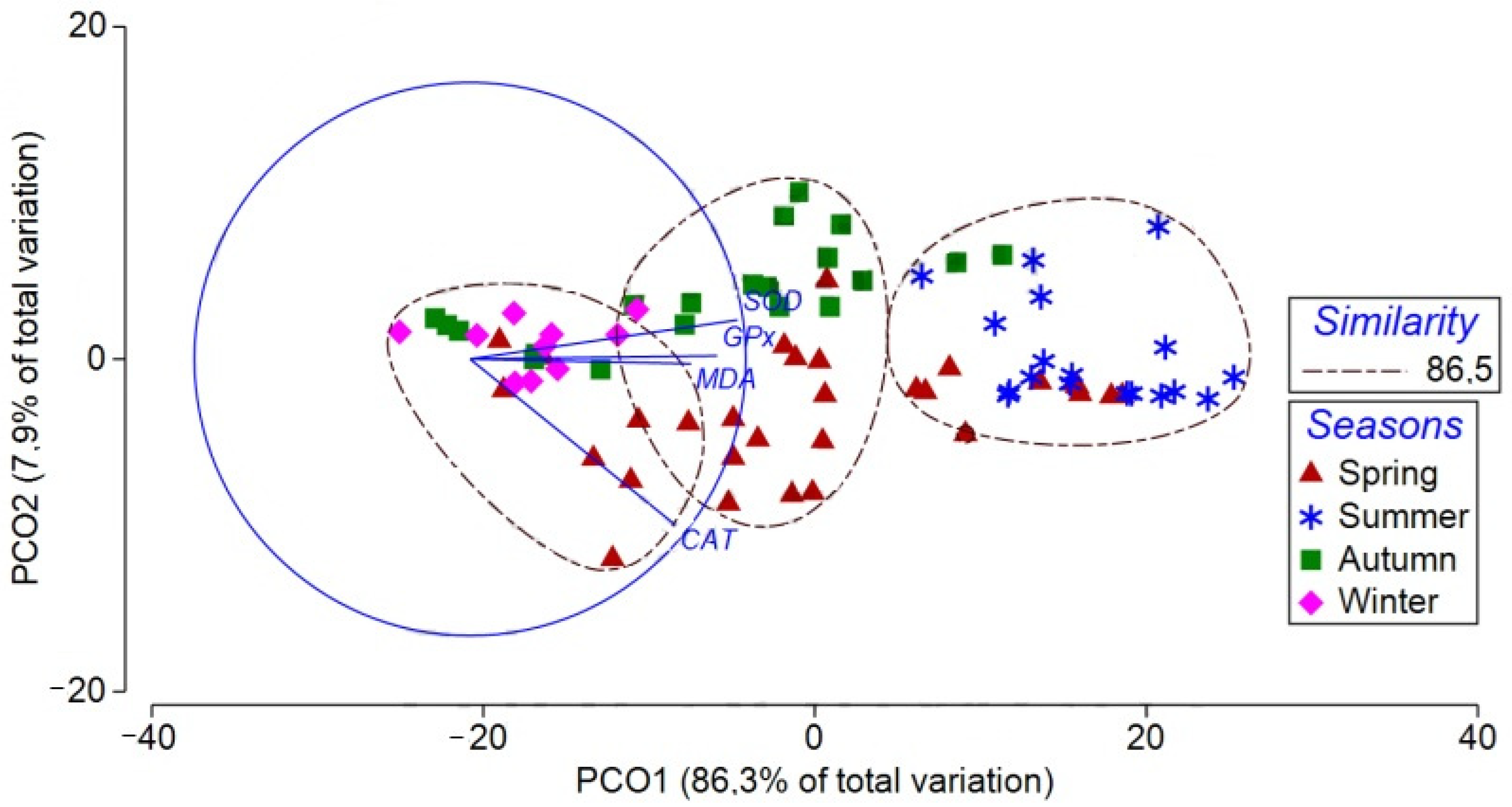
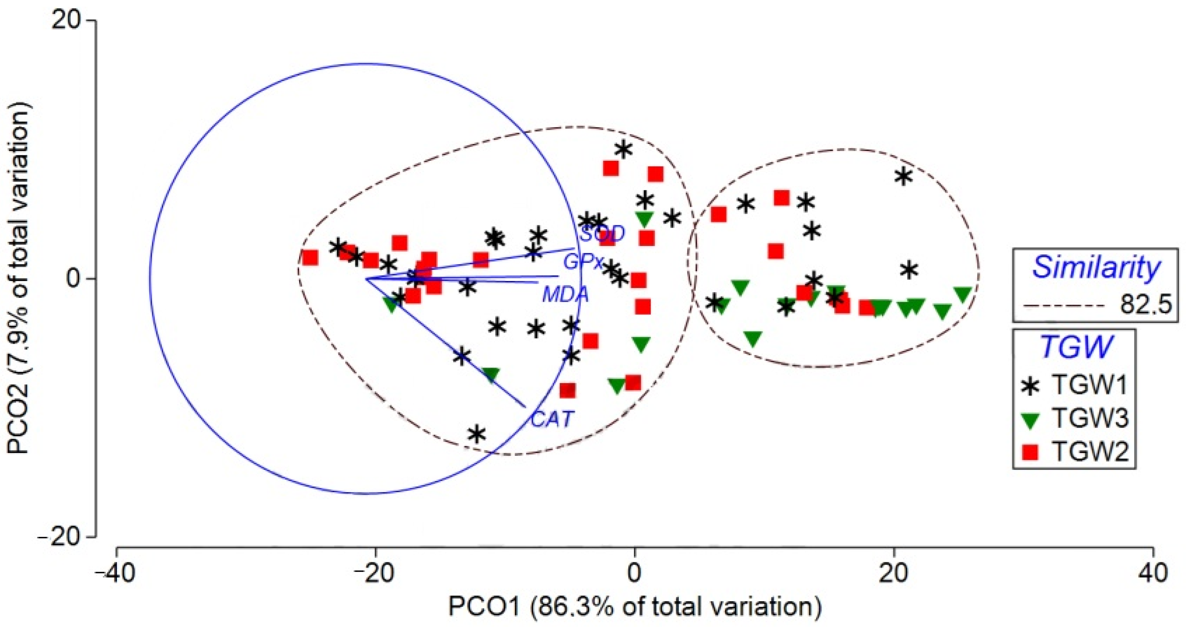
The two-dimensional PCO ordination based on the Bray–Curtis resemblance matrix of antioxidant biomarkers in the gonad tissues of C. trutta revealed a clear seasonal structuring of the samples (Figure 6). The first two axes accounted for a substantial proportion of the total variation, explaining 93.2% overall (PCO1: 86.3%; PCO2: 7.9%). Seasonal groups were primarily differentiated along the PCO1 axis, with summer samples clustering on the positive side and winter samples distributed on the negative side. In contrast, spring and autumn samples were positioned closer to the center of the ordination space, exhibiting partial overlap and indicating intermediate antioxidant profiles. The vector projections of antioxidant biomarkers highlighted their relative contributions to the observed multivariate pattern. SOD, GPx, and MDA were strongly aligned with the negative direction of PCO1, indicating that variation in these biomarkers was closely associated with the winter cluster. CAT was oriented toward the negative direction of PCO2, suggesting its contribution to the secondary gradient of variation, whereas GR was positioned near the center of the ordination plot, reflecting a comparatively weaker influence on seasonal separation. The high overall similarity (86.5%) nevertheless indicates that antioxidant biomarker responses remained broadly comparable among seasons, although the clear distribution along PCO1 suggests a pronounced seasonal modulation of oxidative status in the gonad tissues of C. trutta.
The two-dimensional PCO analysis of the resemblance matrix revealed patterns in antioxidant biomarkers in gonad tissues of C. trutta across different TGW factor groups (TGW1: ≤5.00 g; TGW2: >5.00–9.40 g; TGW3: ≥9.40–51.01 g) in Figure 7. The first two axes explained a large proportion of the total variation (PCO1: 86.3%; PCO2: 7.0%), indicating that most of the variability in antioxidant biomarker profiles was captured within the two-dimensional space. Samples belonging to TGW3 were primarily distributed on the positive side of PCO1, whereas TGW1 samples were mainly located on the negative side, suggesting a gradient in antioxidant responses associated with increasing gonad weight. In contrast, TGW2 samples were positioned around the central region of the ordination plot, showing partial overlap with other groups. The vector projections of antioxidant biomarkers indicated that SOD, GPx, and MDA were aligned with the negative direction of PCO1, suggesting that variation in these biomarkers contributed substantially to the separation of samples along this axis. CAT was more closely associated with the negative direction of PCO2, indicating its role in the secondary gradient of variation. In contrast, GR was located near the center of the ordination space, implying a relatively limited contribution to group differentiation. Despite these patterns, the relatively high similarity level (82.5%) indicates that antioxidant biomarker responses among TGW factor groups remained broadly comparable, although the distribution along PCO1 suggests that gonad weight may partly influence the multivariate antioxidant profile in C. trutta.

4. Discussion

This study provides a comprehensive evaluation of the influences of seasonal variation, TGW, TBW, GSI, sex, and species on the antioxidant biomarker profiles (MDA, SOD, CAT, GR, and GPx) in the gonad tissues of two cyprinid species, C. umbla and C. trutta, inhabiting the Karasu River (Türkiye). Multivariate analyses revealed that seasonality was the predominant driver of variation in antioxidant responses, reflecting the strong impact of environmental fluctuations on oxidative stress and enzymatic defense mechanisms in reproductive tissues. Secondary influences were exerted by reproductive indices (GSI, TGW) and species-specific traits, suggesting a complex interplay between intrinsic physiological states and extrinsic environmental conditions.
Multivariate analyses, including PCO ordination, PERMANOVA, and SIMPER, demonstrated that the primary structuring of the samples was driven by seasonal variation, reflecting the strong influence of environmental fluctuations on oxidative stress and enzymatic defense mechanisms in reproductive tissues. Season was identified as the primary driver, which is biologically plausible because reproductive physiology in temperate freshwater fish is strongly regulated by seasonal cycles. Therefore, the seasonal pattern detected by PERMANOVA likely reflects the combined influence of environmental variation and endogenous reproductive rhythms. These findings are consistent with previous reports indicating that oxidative metabolism in fish gonads exhibits pronounced seasonal dynamics, closely associated with photoperiod, temperature, and reproductive activity [20,24,53,54,55].

4.1. Seasonal Modulation of Antioxidant Biomarkers

In both C. umbla and C. trutta, MDA levels increased during spring and summer, indicating enhanced lipid peroxidation during periods of elevated reproductive activity and metabolic demand. This seasonal elevation suggests that increased energy expenditure associated with gonadal development may contribute to higher ROS production, triggering lipid oxidative damage and the activation of antioxidant defense mechanisms. Concurrently, reproductive indices such as TBW, GSI, and TGW were generally higher in both sexes during these reproductive periods, consistent with observations in other cyprinid and freshwater teleost species [56,57].
The seasonal variation observed in antioxidant biomarkers can also be interpreted in relation to the seasonal progression of gonadal maturation observed in this study. During spring and summer, when reproductive activity intensifies, gonadal tissues undergo extensive cellular remodeling associated with gametogenesis, including spermatogenic proliferation in males and oocyte growth and vitellogenic processes in females. These cellular transitions require increased ATP production and elevated mitochondrial activity, which may enhance electron transport chain activity and consequently increase ROS generation. The observed increases in MDA levels together with elevated SOD, CAT, and GPx activities during these periods likely represent an adaptive response to increased oxidative pressure associated with reproductive investment.
In particular, SOD represents the first enzymatic defense line against superoxide radicals by converting them into hydrogen peroxide, whereas CAT and GPx subsequently contribute to hydrogen peroxide detoxification. Therefore, the concurrent elevation of these enzymes during periods of advanced gonadal development suggests coordinated antioxidant regulation to maintain redox balance during energetically demanding reproductive processes. Conversely, reduced antioxidant activity during autumn and winter may reflect decreased metabolic requirements following spawning and may reflect reduced gametogenic activity, corresponding to lower oxidative challenges in gonadal tissues.
Critically, these biochemical patterns closely paralleled seasonal physicochemical dynamics. Water temperatures increased from 5.48 °C to 8.60 °C during spring and summer, while DO declined from 12.2 to 10.8 mg/L. Such thermal and oxygen gradients are likely to elevate metabolic rates and mitochondrial ROS production, thereby intensifying oxidative stress, as temperature and oxygen availability are well-established modulators of ROS generation and antioxidant responses in fish (e.g., temperature changes directly influence respiratory metabolism and electron transport leakage leading to ROS production) [58], and the highest summer temperature (8.6 °C) coincided with significant increases in antioxidant biomarkers in C. umbla and C. trutta [53]. Stable low salinity (0.39 ppt) and moderate EC (300–360 µS/cm) indicate minimal osmotic challenge, highlighting that oxidative responses are primarily metabolically driven rather than stress-induced by ionic fluctuations, consistent with evidence that modest salinity variation has limited direct oxidative impact compared to temperature and oxygen shifts in freshwater species [58]. Minor pH fluctuations (7.4–8.1) further suggest subtle but physiologically relevant shifts in enzymatic activity, as pH changes can affect acid-base balance and influence both ionoregulation and antioxidant enzyme activity, contributing to variations in ROS scavenging efficiency and redox homeostasis in aquatic organisms [58]. Additionally, these findings are supported by studies in Carassius auratus, a well-characterized model species for fish reproduction, where gonadal maturation is primarily expressed by gonad size and GSI, peaking during the spawning season in both males and females [59]. Similarly, gametogenesis in multiple freshwater teleosts is closely associated with elevated oxidative stress markers and increased antioxidant enzyme activities, reflecting the substantial energetic and metabolic demands of oogenesis and spermatogenesis [60,61,62,63,64].
Multivariate analyses, particularly PCO ordination, revealed that variation in antioxidant biomarkers was closely associated with reproductive investment, with SOD, GPx, and MDA contributing substantially to the differentiation of individuals according to GSI and TGW patterns. These associations highlight the close relationship between gonadal development, metabolic demand, and oxidative regulation during reproductive cycles. In contrast, CAT and GR contributed mainly to secondary gradients of variation, suggesting enzyme-specific roles in maintaining redox homeostasis under changing physiological conditions. Seasonal structuring of antioxidant profiles was evident in both species, with C. umbla showing clear differentiation between spring–summer and autumn–winter metabolic patterns. The contribution of SOD, GPx, and MDA to this seasonal separation suggests that seasonal changes in reproductive activity and environmental conditions influence oxidative balance. Similarly, C. trutta exhibited seasonal shifts in antioxidant profiles, particularly during periods of increased reproductive activity, reflecting the coordinated regulation of enzymatic defenses in response to elevated metabolic requirements. These patterns indicate that seasonal reproductive cycles represent a major physiological driver shaping oxidative responses, while individual biomarkers contribute differently to the overall variation in redox status.
These observations are consistent with previous evidence demonstrating that reproductive effort and increased metabolic activity enhance ROS production, thereby necessitating intensified antioxidant responses during gametogenesis [65,66,67,68]. Furthermore, the observed seasonal variation in antioxidant activities corresponds with established links between oxidative stress and environmental factors, including temperature and photoperiod. Specifically, elevated activities of GPx, CAT, and SOD, together with increased MDA levels during spring and summer, reflect an adaptive upregulation of antioxidant defenses in response to higher metabolic rates and ROS production. Conversely, the lower antioxidant enzyme activities and MDA concentrations observed in autumn and winter likely reflect seasonal metabolic downregulation. This reduction in metabolic activity not only mitigates oxidative challenges but may also constrain reproductive investment, highlighting an energy allocation trade-off between self-maintenance and reproduction [65,69].
In support of these patterns, previous studies have reported that the alteration of physiological states throughout the annual cycle (including pre-spawning mobilization, spawning-related energy loss, post-spawning energy reserve accumulation, and reduced metabolic activity during wintering across a broad temperature range) necessitates substantial plasticity in fish defense systems [70]. Consistently, life-history theory posits that reproduction entails high energetic demands, and reductions in energy allocation may consequently limit reproductive investment [65,69]. Moreover, increased metabolic activity during reproduction can elevate ROS production, whereas lower metabolic rates reduce oxidative load, further emphasizing the mechanistic link between energy metabolism and oxidative stress [71].
Finally, our findings in C. umbla and C. trutta agree with patterns reported in related cyprinids, such as Cyprinus carpio [64] and Luciobarbus esocinus [72], where antioxidant enzyme activities peak during reproductive and metabolically demanding periods. Overall, our study suggests that the orchestration of oxidative metabolism and antioxidant defenses is finely tuned and closely synchronized with seasonal reproductive physiology, serving as an adaptive mechanism to meet the heightened energetic and metabolic demands of reproduction [62,64].

4.2. Influence of Reproductive Status on Oxidative Biomarkers

Our findings clearly indicate that reproductive status, as measured by GSI and TGW, plays a pivotal role in modulating oxidative biomarker responses in the gonad tissues of C. umbla and C. trutta. Individuals in advanced reproductive stages (higher GSI and TGW) exhibited elevated activities of key antioxidant enzymes, including SOD, CAT, and GPx, along with increased levels of MDA. These results suggest that antioxidant defenses are upregulated in response to the enhanced metabolic demands associated with gametogenesis and oocyte maturation, reflecting the well-established relationship between reproductive effort and oxidative stress in vertebrates [7,65].
The observed positive correlation between reproductive indices and antioxidant activity supports the hypothesis that reproductive investment is inherently linked to oxidative stress management. High reproductive effort increases cellular metabolic activity and ROS production, particularly in gonadal tissues undergoing rapid proliferation and steroidogenesis, necessitating a strengthened antioxidant system to prevent oxidative damage [64]. In line with our findings, studies in teleosts such as Cyprinus carpio and Oncorhynchus mykiss have demonstrated that antioxidant enzyme activities, including SOD, GPx, and related components of the antioxidant defense system, change significantly during the reproductive (spawning) period, reflecting modulation of oxidative status in response to reproductive demands [64]. Studies in teleosts such as Oreochromis niloticus [73] and Carassius auratus [74] have demonstrated that individuals with greater gonad mass or higher GSI exhibit significantly higher activities of key antioxidant enzymes. This increase, particularly in SOD and GPx activities, reflects a protective response to oxidative challenges associated with reproduction [64]. Interestingly, GR activity exhibited relatively low variability across GSI and TGW groups in both C. umbla and C. trutta, suggesting that this enzyme primarily maintains baseline redox homeostasis rather than responding dynamically to reproductive stage. Although GR activity is an important component of the antioxidant system, existing studies in teleost fish have not shown dramatic modulation of GR specifically with reproductive status compared with other antioxidant enzymes. For example, in Oreochromis niloticus, differences in GR activity were observed between testis and ovary, indicating tissue-specific roles rather than strong reproductive stage-dependent variation [75]. In contrast, CAT, SOD, and GPx displayed more pronounced responses, with activities increasing in association with higher reproductive investment. Such enzyme-specific modulation is consistent with studies in freshwater teleosts, demonstrating that CAT, SOD, and GPx activities markedly increase during gametogenesis and oocyte maturation, effectively counteracting the elevated ROS burden associated with reproduction [64,76].
Multivariate analyses, including PCO ordination, further confirmed that reproductive status is a key structuring factor for antioxidant profiles. Individuals with higher GSI (GSI3–GSI4) clustered separately from those with lower reproductive investment, with SOD, GPx, and MDA driving the differentiation. This pattern aligns with the conceptual framework of life-history trade-offs, whereby organisms allocate resources toward reproduction at the cost of increased oxidative challenge, necessitating upregulated antioxidant defenses to maintain cellular integrity and reproductive success [77,78]. Overall, these results demonstrate that oxidative biomarker dynamics in gonadal tissues are closely linked to reproductive condition, highlighting the importance of antioxidant defenses in supporting energetically demanding reproductive processes. The differential responses of specific enzymes (e.g., CAT, GPx, SOD) emphasize that reproductive modulation of oxidative status is selective and enzyme-specific, reflecting the physiological prioritization of redox balance during periods of high reproductive investment.

4.3. Species-Specific Patterns and Ecological Implications

The present study showed that seasonal variation was the main factor shaping antioxidant biomarker responses in the gonad tissues of C. umbla and C. trutta, two cyprinid species inhabiting the Karasu River (Türkiye). Reproductive parameters, particularly GSI and TGW, also contributed to this variability, suggesting that oxidative balance in these species is closely linked to both environmental seasonality and reproductive dynamics.
Despite belonging to the same genus and inhabiting the same river system, C. umbla and C. trutta exhibited differences in the relative importance of reproductive variables influencing antioxidant biomarker dynamics. In C. umbla, the GSI emerged as the second most influential factor after season, whereas TGW played a more prominent role in C. trutta. The relatively higher GSI and TGW values observed in C. umbla, particularly in females (GSI: 3.69–9.10%; TGW: 19.28–50.59 g), suggest a greater reproductive investment that may require more efficient physiological regulation of oxidative stress during gametogenesis. Reproductive processes such as vitellogenesis, steroidogenesis, and rapid cellular proliferation are known to elevate metabolic activity and increase the production of ROS, thereby necessitating stronger antioxidant defense mechanisms in reproductive tissues. This relationship has been observed in multiple fish species, where oocyte maturation and gametogenesis are accompanied by heightened enzymatic antioxidant activity to counteract ROS-induced oxidative stress [7,27,79,80]. Similar relationships between reproductive activity and increased antioxidant responses have been reported in various freshwater fish species. For instance, during ovarian maturation in grass carp (Ctenopharyngodon idellus), antioxidant enzyme activities such as SOD, CAT, and POD were elevated, reflecting enhanced oxidative metabolism associated with reproductive investment [81]. Seasonal variations in gonadal antioxidant defenses have been well-documented in cyprinid species, indicating a close link between reproductive activity and oxidative stress management. In Cyprinion macrostomus, Kırıcı et al. [24] reported significant increases in SOD, CAT, and GPx activities during periods corresponding to peak reproductive investment, suggesting that antioxidant systems are upregulated to buffer the heightened production of ROS during gametogenesis. Similarly, in Salmo trutta subspecies, Aras et al. [62] observed that gonadal and gill antioxidant enzyme activities peaked in synchrony with the reproductive cycle, supporting the notion that seasonal modulation of oxidative defenses represents an adaptive physiological adjustment to reproductive demands. These findings are consistent with our observations in C. umbla and C. trutta, where elevated GSI and TGW values coincided with increased activities of key antioxidant enzymes such as SOD, CAT, and GPx. Collectively, these patterns highlight the ecological significance of maintaining oxidative balance during reproduction, as failure to regulate ROS production could compromise gamete quality and ultimately affect population sustainability in riverine ecosystems like the Karasu River. Moreover, dietary antioxidants such as astaxanthin have been shown to modulate oxidative stress responses and support reproductive performance in fish, further emphasizing the link between reproductive maturation and antioxidant defense systems [76]. Collectively, these findings corroborate our results in C. umbla and C. trutta, where periods of elevated GSI and TGW coincided with increased activities of antioxidant enzymes, highlighting the ecological importance of oxidative balance in sustaining reproductive success.
Although PERMANOVA analysis did not reveal statistically significant interspecific differences in the overall antioxidant biomarker composition, the contrasting importance of GSI and TGW suggests subtle species-specific tendencies in reproductive energetics and oxidative stress regulation, potentially reflecting divergent life-history strategies within the Karasu River ecosystem. Comparative research indicates that oxidative stress and antioxidant defense dynamics are closely linked to life-history traits and environmental variation in fish, with seasonal metabolic adjustments and reproductive cycles shaping antioxidant responses across taxa [82]. From an ecological perspective, these findings indicate that reproductive investment may influence how closely related fish species regulate oxidative balance, highlighting the importance of integrating physiological biomarkers with reproductive metrics when assessing the health and adaptive capacity of freshwater fish populations in dynamic riverine environments. Seasonal fluctuations in antioxidant capacity have been widely reported in cyprinid and other temperate fishes, where antioxidant enzyme activities (e.g., SOD, CAT, GPx) increase during spring–summer reproductive periods and decline in winter, reflecting coordinated physiological responses to environmental cues and reproductive demands [70]. Moreover, antioxidant systems are sensitive to environmental stressors such as temperature changes and contaminants, and the ability to adjust redox balance is increasingly viewed as pivotal for resilience in ecosystems facing anthropogenic pressures and climatic variability [58]. The oxidative status of gonad tissues, modulated seasonally and by reproductive investment, thus emerges as a sensitive biomarker for assessing fish health and reproductive potential in ecological monitoring programs.
Although the present study provides valuable insights into seasonal and reproductive modulation of oxidative responses in wild Capoeta populations, some limitations associated with field-based sampling should be acknowledged. Fish collected from natural river systems are exposed to a combination of environmental variables that cannot be fully controlled, including small-scale habitat differences, fluctuations in water quality, and episodic anthropogenic inputs such as agricultural runoff. These factors may interact with physiological condition and metabolic responses, potentially contributing to individual variation in antioxidant biomarkers. Therefore, future studies combining field observations with controlled laboratory experiments, including standardized reproductive and environmental conditions, would help to further distinguish the relative contributions of intrinsic reproductive processes and external environmental drivers.

5. Conclusions

This study provides compelling evidence that both seasonal variation and reproductive status are key determinants of antioxidant biomarker dynamics in the gonad tissues of C. umbla and C. trutta inhabiting the Karasu River. Seasonal fluctuations, particularly during spring and summer, were consistently associated with elevated lipid peroxidation and enhanced activities of major antioxidant enzymes (SOD, CAT, GPx, and GR), reflecting the metabolic demands of gametogenesis. Reproductive indices, especially GSI and TGW, were identified as significant modulators of oxidative balance, highlighting the tight coupling between reproductive investment and antioxidant defense mechanisms. Although overall biomarker composition did not differ significantly between species, the relative contribution of reproductive parameters differed, with GSI showing greater influence in C. umbla and TGW in C. trutta, suggesting subtle differences in reproductive allocation strategies rather than divergent oxidative profiles. From an ecological perspective, these findings underscore the adaptive significance of antioxidant regulation in supporting reproductive success under dynamic environmental conditions. Our findings suggest that seasonal modulation of antioxidant defenses may contribute to maintaining physiological conditions that support normal reproductive processes. However, the present study did not directly evaluate gamete quality or reproductive performance; therefore, any implications for reproductive success should be considered speculative and require further investigation. Moreover, the consistent seasonal and reproductive modulation of antioxidant defenses indicates that oxidative status of gonad tissues can serve as a sensitive biomarker for assessing fish health and reproductive potential, with direct implications for monitoring the impacts of environmental change and anthropogenic stressors in freshwater ecosystems. Overall, integrating oxidative stress biomarkers with reproductive indices provides complementary physiological information that may improve ecological monitoring of freshwater fish populations. Future studies should investigate how combined environmental pressures, such as climate variability and pollution, interact with reproductive investment to influence oxidative balance, ultimately shaping population dynamics and ecosystem resilience in freshwater systems. Consequently, maintaining oxidative balance is likely to support normal reproductive physiology during periods of high metabolic demand. However, the consequences for gamete quality, reproductive success, and population sustainability were beyond the scope of the present study and warrant further investigation.

Author Contributions

Conceptualization, N.Ş.Ö. and M.K.; methodology, N.Ş.Ö., F.C. and M.K.; software, T.Ö.S. and N.Ş.Ö.; validation, M.G., C.T. and N.D.; formal analysis, N.Ş.Ö. and M.K.; investigation, M.K., C.T. and F.C.; resources, T.Ö.S. and M.G.; data curation, N.Ş.Ö. and F.C.; writing—original draft preparation, M.K. and N.Ş.Ö.; writing—review and editing, M.K., N.Ş.Ö., M.G., F.C., C.T., T.Ö.S. and N.D.; visualization, M.K. and N.Ş.Ö.; supervision, T.Ö.S., M.G. and N.D.; project administration, M.K. and N.Ş.Ö. All authors have read and agreed to the published version of the manuscript.

Funding

This research was supported by the Bingöl University Scientific Research Projects Coordination Unit (Project No. BAP-GTHMYO.2025.001).

Institutional Review Board Statement

The fish used in this study were not caught under contract, were not requested to be of a specific size or condition and were not provided live to the authors. All fish were purchased dead from a licensed commercial fish market as part of routine commercial fishing activities, without informing the fishermen or vendors of any research purpose, in the same manner as fish purchased for personal consumption. In accordance with national regulations and EU Directive 2010/63/EU, ethical approval is not required for studies involving vertebrate animals when the animals are purchased dead, and their capture was not expressly conducted for research purposes. Consistent with this framework, our institutional Ethics Committee formally reviewed the study and issued an official confirmation letter stating that ethical approval was not required.

Data Availability Statement

The original contributions presented in this study are included in the article. Further inquiries can be directed to the corresponding author.

Conflicts of Interest

The authors declare no conflicts of interest.

References

  1. Levavi-Sivan, B.; Bogerd, J.; Mañanós, E.L.; Gómez, A.; Lareyre, J.J. Perspectives on fish gonadotropins and their receptors. Gen. Comp. Endocrinol. 2010, 165, 412–437. [Google Scholar] [CrossRef] [Scilit] [PubMed]
  2. Zohar, Y.; Muñoz-Cueto, J.A.; Elizur, A.; Kah, O. Neuroendocrinology of reproduction in teleost fish. Gen. Comp. Endocrinol. 2010, 165, 438–455. [Google Scholar] [CrossRef] [Scilit] [PubMed]
  3. Murphy, M.P. How mitochondria produce reactive oxygen species. Biochem. J. 2009, 417, 1–13. [Google Scholar] [CrossRef] [Scilit] [PubMed]
  4. Sies, H. Oxidative stress: Oxidants and antioxidants. Exp. Physiol. 1997, 82, 291–295. [Google Scholar] [CrossRef] [Scilit] [PubMed]
  5. Agarwal, A.; Gupta, S.; Sharma, R.K. Role of oxidative stress in female reproduction. Reprod. Biol. Endocrinol. 2005, 3, 28. [Google Scholar] [CrossRef] [Scilit] [PubMed]
  6. Sandoval-Vargas, L.; Silva Jiménez, M.; Risopatrón González, J.; Villalobos, E.F.; Cabrita, E.; Valdebenito Isler, I. Oxidative stress and use of antioxidants in fish semen cryopreservation. Rev. Aquac. 2021, 13, 365–387. [Google Scholar] [CrossRef] [Scilit]
  7. Lushchak, V.I. Environmentally induced oxidative stress in aquatic animals. Aquat. Toxicol. 2011, 101, 13–30. [Google Scholar] [CrossRef] [Scilit] [PubMed]
  8. Petrocheilou, D.; Petriki, O.; Kaloyianni, M.; Bobori, D.C. Oxidative stress biomarkers in Carassius gibelio from lakes of varying ecological quality. Hydrobiology 2026, 5, 4. [Google Scholar] [CrossRef] [Scilit]
  9. Lopes, P.A.; Pinheiro, T.; Santos, M.C.; da Luz Mathias, M.; Collares-Pereira, M.J.; Viegas-Crespo, A.M. Response of antioxidant enzymes in freshwater fish populations (Leuciscus alburnoides complex) to inorganic pollutants exposure. Sci. Total Environ. 2001, 280, 153–163. [Google Scholar] [CrossRef] [Scilit] [PubMed]
  10. Gradinariu, L.; Cretu, M.; Vizireanu, C.; Dediu, L. Oxidative stress biomarkers in fish exposed to environmental concentrations of pharmaceutical pollutants: A review. Biology 2025, 14, 472. [Google Scholar] [CrossRef] [Scilit] [PubMed]
  11. El-Shenawy, N.S.; El-Hak, H.N.G.; Ghobashy, M.A.; Mansour, F.A.; Soliman, M.F. Using antioxidant changes in liver and gonads of Oreochromis niloticus as biomarkers for the assessment of heavy metals pollution at Sharkia province, Egypt. Reg. Stud. Mar. Sci. 2021, 46, 101863. [Google Scholar] [CrossRef] [Scilit]
  12. Chainy, G.B.N.; Paital, B.; Dandapat, J. An overview of seasonal changes in oxidative stress and antioxidant defence parameters in some invertebrate and vertebrate species. Scientifica 2016, 2016, 6126570. [Google Scholar] [CrossRef] [Scilit] [PubMed]
  13. Kırıcı, M.; Tüzün, B.; Yonar, M.E.; Poustforoosh, A.; Mişe Yonar, S.; Şeker, E.; İspir, Ü. Change of micronucleus frequencies, antioxidant parameters and in silico analysis in carp (Cyprinus carpio) fry exposed to ibuprofen. J. Fish Biol. 2026, 108, 502–516. [Google Scholar] [CrossRef] [Scilit] [PubMed]
  14. Eyo, V.O.; Ekanem, A.P.; Jimmy, U.I.U. A comparative study of the gonado-somatic index (GSI) and gonad gross morphology of African catfish (Clarias gariepinus) fed unical aqua feed and Coppens commercial feed. Croat. J. Fish. 2014, 72, 63–69. [Google Scholar] [CrossRef] [Scilit]
  15. Mahalder, B.; Ahasan, N.; Siddique, M.A.B.; Shohan, M.H.; Ahammad, A.S.; Aziz, M.S.B.; Haque, M.M. Gonadosomatic index (GSI) of stinging catfish in relation to water quality and climatic variables, and its trend analysis using SARIMA model. Aquac. Fish Fish. 2025, 5, e70137. [Google Scholar] [CrossRef] [Scilit]
  16. Antinozzi, C.; Di Luigi, L.; Sireno, L.; Caporossi, D.; Dimauro, I.; Sgrò, P. Protective role of physical activity and antioxidant systems during spermatogenesis. Biomolecules 2025, 15, 478. [Google Scholar] [CrossRef] [Scilit] [PubMed]
  17. Liu, H.; Wang, Y.; Fang, J.; Zhang, M.; Shi, X.; Zhang, J.; Kong, X. Effects of temperature on growth performance, antioxidant capacity, ovary development and gene expression in Micropterus salmoides. Aquac. Rep. 2025, 45, 103255. [Google Scholar] [CrossRef] [Scilit]
  18. Agarwal, A.; Aponte-Mellado, A.; Premkumar, B.J.; Shaman, A.; Gupta, S. The effects of oxidative stress on female reproduction: A review. Reprod. Biol. Endocrinol. 2012, 10, 49. [Google Scholar] [CrossRef] [Scilit] [PubMed]
  19. Zhang, Q.F.; Li, Y.W.; Liu, Z.H.; Chen, Q.L. Reproductive toxicity of inorganic mercury exposure in adult zebrafish: Histological damage, oxidative stress, and alterations of sex hormone and gene expression in the hypothalamic–pituitary–gonadal axis. Aquat. Toxicol. 2016, 177, 417–424. [Google Scholar] [CrossRef] [Scilit] [PubMed]
  20. Wang, W.; Su, S.; Dong, P.; Feng, W.; Li, J.; Zhang, C.; Tang, Y. Effects of seasonal photoperiod on growth, lipid metabolism, and antioxidant response in the Huanghe carp (Cyprinus carpio haematopterus). Fishes 2023, 8, 595. [Google Scholar] [CrossRef] [Scilit]
  21. Kolaylı, S.; Arikan, M.; Uzunosmanoğlu, D.; Vanİzor, B.; Kiran, E.; Sağban, R. Comparative studies on antioxidant enzyme activities and lipid peroxidation in different fish species. Turk. J. Zool. 1997, 21, 171–173. [Google Scholar] [CrossRef] [Scilit]
  22. Parisi, C.; Guerriero, G. Antioxidative defense and fertility rate in the assessment of reprotoxicity risk posed by global warming. Antioxidants 2019, 8, 622. [Google Scholar] [CrossRef] [Scilit] [PubMed]
  23. Parolini, M.; Iacobuzio, R.; De Felice, B.; Bassano, B.; Pennati, R.; Saino, N. Age- and sex-dependent variation in the activity of antioxidant enzymes in the brown trout (Salmo trutta). Fish Physiol. Biochem. 2019, 45, 145–154. [Google Scholar] [CrossRef] [Scilit] [PubMed]
  24. Kırıcı, M.; Şen Özdemir, N.; Caf, F.; Koyun, M. Seasonal changes in antioxidant defense system indicators in the tissues of Cyprinion macrostomus (Heckel, 1843) caught from Göynük Stream (Bingöl, Turkey). Ege J. Fish. Aquat. Sci. 2022, 39, 174–181. [Google Scholar] [CrossRef] [Scilit]
  25. Alonso-Alvarez, C.; Bertrand, S.; Devevey, G.; Prost, J.; Faivre, B.; Sorci, G. Increased susceptibility to oxidative stress as a proximate cost of reproduction. Ecol. Lett. 2004, 7, 363–368. [Google Scholar] [CrossRef] [Scilit]
  26. Caballero-Huertas, M.; Frigola-Tepe, X.; Viñas, J.; Muñoz, M. Somatic condition and reproductive potential as a tandem in European sardine: An analysis with an environmental perspective in the northern Adriatic (Gulf of Trieste). Fishes 2022, 7, 105. [Google Scholar] [CrossRef] [Scilit]
  27. Ladisa, C.; Ma, Y.; Habibi, H.R. Metabolic changes during growth and reproductive phases in the liver of female goldfish (Carassius auratus). Front. Cell Dev. Biol. 2022, 10, 834688. [Google Scholar] [CrossRef] [Scilit] [PubMed]
  28. Demirok, N.K.; Ünlü, E. Karyotypes of cyprinid fish Capoeta trutta and Capoeta capoeta umbla (Cyprinidae) from the Tigris River. Turk. J. Zool. 2001, 25, 389–393. [Google Scholar]
  29. Türkmen, M.; Erdoğan, O.; Yıldırım, A.; Akyurt, İ. Reproduction tactics, age and growth of Capoeta capoeta umbla (Heckel, 1843) from the Aşkale region of the Karasu River, Turkey. Fish. Res. 2002, 54, 317–328. [Google Scholar] [CrossRef] [Scilit]
  30. Turan, D.; Kottelat, M.; Ekmekçi, F.G.; İmamoğlu, H.O. A review of Capoeta tinca, with descriptions of two new species from Turkey (Teleostei: Cyprinidae). Rev. Suisse Zool. 2006, 113, 421–436. [Google Scholar] [CrossRef] [Scilit]
  31. Kaya, C.; Turan, D.; Ünlü, E. The latest status and distribution of fishes in upper Tigris River and two new records for Turkish freshwaters. Turk. J. Fish. Aquat. Sci. 2016, 16, 545–562. [Google Scholar] [CrossRef] [PubMed]
  32. Oymak, S.; Musa, D.; Ünlü, E. Reproductive biology and histological changes in the gonads of barb, Capoeta trutta (Heckel, 1843) in Atatürk Dam Lake, Turkey. Aquat. Sci. Eng. 2008, 23, 1–11. [Google Scholar]
  33. Poria, M.; Abdoli, A.; Kazemian, M.; Nouri, F.; Ghanbari, K.; Ejraei, F. Study of reproductive characteristics of Capoeta trutta in Gamasyab River, Kermanshah Province, Iran. Int. J. Biosci. 2014, 4, 39–46. [Google Scholar]
  34. Atamanalp, M.; Kokturk, M.; Kırıcı, M.; Ucar, A.; Kırıcı, M.; Parlak, V.; Alak, G. Interaction of microplastic presence and oxidative stress in freshwater fish: A regional scale research, East Anatolia of Türkiye (Erzurum, Erzincan, Bingöl). Sustainability 2022, 14, 12009. [Google Scholar] [CrossRef] [Scilit]
  35. Taysi, M.R.; Kırıcı, M.; Kırıcı, M.; Tuzun, B.; Poustforoosh, A. Antioxidant enzyme activities, molecular docking studies, MM-GBSA, and molecular dynamics of chlorpyrifos in freshwater fish Capoeta umbla. J. Biomol. Struct. Dyn. 2024, 42, 163–176. [Google Scholar] [CrossRef] [Scilit] [PubMed]
  36. Sökmen, T.Ö.; Güneş, M.; Kırıcı, M. Determination of heavy metal levels in water, sediment and Capoeta umbla tissues of Karasu River (Erzincan). Turk. J. Agric. Nat. Sci. 2018, 5, 578–588. [Google Scholar] [CrossRef] [Scilit]
  37. Öz, Ö.B.; Barata, S. Effects of reproduction on oxidative stress and some antioxidant levels in tissues of Capoeta umbla (Heckel, 1843). J. Mater. Electron. Devices 2021, 7, 7–14. [Google Scholar]
  38. Vašková, J.; Klepcová, Z.; Špaková, I.; Urdzík, P.; Štofilová, J.; Bertková, I.; Rabajdová, M. The importance of natural antioxidants in female reproduction. Antioxidants 2023, 12, 907. [Google Scholar] [CrossRef] [Scilit] [PubMed]
  39. Öz, Ö.B.; Köprücü, S. The effects of reproduction on oxidative stress, vitamins E, A, C and β-carotene in Capoeta umbla (Heckel, 1843). J. Mater. Electron. Devices 2021, 6, 1–6. [Google Scholar]
  40. Bagenal, T.B.; Tesch, F.W. Age and growth. In Methods for Assessment of Fish Production in Freshwaters; IBP Handbook; Blackwell Science Publications: Oxford, UK, 1978; Volume 3, pp. 101–136. [Google Scholar]
  41. Brown-Peterson, N.J.; Wyanski, D.M.; Saborido-Rey, F.; Macewicz, B.J.; Lowerre-Barbieri, S.K. A standardized terminology for describing reproductive development in fishes. Mar. Coast. Fish. 2011, 3, 52–70. [Google Scholar] [CrossRef] [Scilit]
  42. Kabir, M.A.; Quddus, M.M.A. Fecundity and gonadosomatic index of Labeo calbasu (Hamilton) from a stocking pond of a hatchery at Faridpur, Bangladesh. Bangladesh J. Zool. 2013, 41, 43–48. [Google Scholar] [CrossRef] [Scilit]
  43. Beutler, E. Red Cell Metabolism: Manual of Biochemical Methods; Academic Press: New York, NY, USA, 1971. [Google Scholar]
  44. Kırıcı, M.; Turk, C.; Caglayan, C.; Kırıcı, M. Toxic effects of copper sulphate pentahydrate on antioxidant enzyme activities and lipid peroxidation of freshwater fish Capoeta umbla (Heckel, 1843) tissues. Appl. Ecol. Environ. Res. 2017, 15, 1685–1696. [Google Scholar] [CrossRef] [Scilit]
  45. Placer, Z.A.; Cushman, L.L.; Johnson, B.C. Estimation of product of lipid peroxidation (malondialdehyde) in biochemical systems. Anal. Biochem. 1966, 16, 359–364. [Google Scholar] [CrossRef] [Scilit] [PubMed]
  46. Carlberg, I.; Mannervik, B. Purification and characterization of the flavoenzyme glutathione reductase from rat liver. J. Biol. Chem. 1975, 250, 5475–5480. [Google Scholar] [CrossRef] [Scilit]
  47. Sun, Y.; Oberley, L.W.; Li, Y. A simple method for clinical assay of superoxide dismutase. Clin. Chem. 1988, 34, 497–500. [Google Scholar] [CrossRef] [Scilit]
  48. Aebi, H. Catalase in vitro. In Methods in Enzymology; Academic Press: New York, NY, USA, 1984; Volume 105, pp. 121–126. [Google Scholar] [CrossRef] [Scilit] [PubMed]
  49. Beutler, E. Red Cell Metabolism: Manual of Biochemical Methods; Grune & Stratton: New York, NY, USA, 1975. [Google Scholar]
  50. Lowry, O.H.; Rosebrough, N.J.; Farr, A.L.; Randall, R.J. Protein measurement with Folin phenol reagent. J. Biol. Chem. 1951, 193, 265–275. [Google Scholar] [CrossRef] [Scilit]
  51. Anderson, R.O.; Neumann, R.M. Length, weight, and associated structural indices. In Fisheries Techniques, 2nd ed.; Murphy, B.E., Willis, D.W., Eds.; American Fisheries Society: Bethesda, MD, USA, 1996; pp. 283–300. [Google Scholar]
  52. Şen Özdemir, N.; Koyun, M.; Caf, F.; Kırıcı, M. Factors affecting nutritional quality in terms of the fatty acid composition of Cyprinion macrostomus. Grasas Aceites 2023, 74, e508. [Google Scholar] [CrossRef] [Scilit]
  53. Kırıcı, M.; Özdemir, N.Ş.; Güneş, M.; Sökmen, T.Ö.; Caf, F.; Türk, C.; Demir, N. Seasonal dominance over morphometric effects in regulating antioxidant defense in two freshwater Capoeta species. Diversity 2026, 18, 157. [Google Scholar] [CrossRef] [Scilit]
  54. Chesnokova, I.I.; Rudneva, I.I. Interspecies peculiarities of some enzyme activities in the gonads of Black Sea fish. J. Evol. Biochem. Physiol. 2019, 55, 372–379. [Google Scholar] [CrossRef] [Scilit]
  55. Kırıcı, M.; Özdemir, N.Ş.; Caf, F.; Koyun, M. Seasonal changes in antioxidant enzyme activities of Garra rufa (Heckel, 1843) in Göynük Stream (Bingöl, Türkiye). Ege J. Fish. Aquat. Sci. 2023, 40, 174–181. [Google Scholar] [CrossRef] [Scilit]
  56. Mohamad, I.; Bhat, F.A.; Balkhi, M.H.; Shah, T.H.; Bhat, B.A.; Wali, A. Relationship among body weight, body length, ovary weight and the fecundity of Cyprinus carpio var. communis in Kashmir Himalaya. J. Pharmacogn. Phytochem. 2018, 7, 2018–2020. [Google Scholar]
  57. Yang, Z.; Wang, J. From gonads to generations: Mechanistic insights into reproductive disruption by polystyrene nanoplastics and co-contaminants in fish. Environ. Chem. Ecotoxicol. 2025, 7, 1871–1883. [Google Scholar] [CrossRef] [Scilit]
  58. Menon, S.V.; Kumar, A.; Middha, S.K.; Paital, B.; Mathur, S.; Johnson, R.; Asthana, M. Water physicochemical factors and oxidative stress physiology in fish: A review. Front. Environ. Sci. 2023, 11, 1240813. [Google Scholar] [CrossRef] [Scilit]
  59. Kobayashi, M.; Iwata, E.; Sorensen, P.W. Environmental and physiological regulation of reproduction in the goldfish: Gonadal development, maturation, and spawning behavior: A review. Animals 2026, 16, 775. [Google Scholar] [CrossRef] [Scilit] [PubMed]
  60. Abele, D.; Heise, K.; Portner, H.O.; Puntarulo, S. Temperature-dependence of mitochondrial function and production of reactive oxygen species in the intertidal mud clam Mya arenaria. J. Exp. Biol. 2002, 205, 1831–1841. [Google Scholar] [CrossRef] [Scilit] [PubMed]
  61. Monserrat, J.M.; Martínez, P.E.; Geracitano, L.A.; Amado, L.L.; Martins, C.M.G.; Pinho, G.L.L.; Bianchini, A. Pollution biomarkers in estuarine animals: Critical review and new perspectives. Comp. Biochem. Physiol. C 2007, 146, 221–234. [Google Scholar] [CrossRef] [Scilit] [PubMed]
  62. Aras, N.M.; Bayir, A.; Sirkecioglu, A.N.; Bayir, M.; Aksakal, E.; Haliloglu, H.I. Seasonal changes in antioxidant defence system of liver and gills of Salmo trutta caspius, Salmo trutta labrax and Salmo trutta macrostigma. J. Fish Biol. 2009, 74, 842–856. [Google Scholar] [CrossRef] [Scilit] [PubMed]
  63. Regoli, F.; Giuliani, M.E. Oxidative pathways of chemical toxicity and oxidative stress biomarkers in marine organisms. Mar. Environ. Res. 2014, 93, 106–117. [Google Scholar] [CrossRef] [Scilit] [PubMed]
  64. Shaliutina-Kolešová, A.; Rui, N.; Ashtiani, S.; Rodina, M.; Cosso, J.; Linhart, O. Oxidative stress and antioxidant enzyme defence system in seminal plasma of common carp (Cyprinus carpio) and rainbow trout (Oncorhynchus mykiss) during spawning season. Czech J. Anim. Sci. 2018, 63, 78–84. [Google Scholar] [CrossRef] [Scilit]
  65. Monaghan, P.; Metcalfe, N.B.; Torres, R. Oxidative stress as a mediator of life history trade-offs: Mechanisms, measurements and interpretation. Ecol. Lett. 2009, 12, 75–92. [Google Scholar] [CrossRef] [Scilit] [PubMed]
  66. Rizzo, A.; Roscino, M.T.; Binetti, F.; Sciorsci, R.L. Roles of reactive oxygen species in female reproduction. Reprod. Domest. Anim. 2012, 47, 344–352. [Google Scholar] [CrossRef] [Scilit] [PubMed]
  67. Costantini, D. Oxidative Stress and Hormesis in Evolutionary Ecology and Physiology: A Marriage Between Mechanistic and Evolutionary Approaches; Springer: Berlin, Germany, 2014. [Google Scholar]
  68. Aitken, R.J.; Bromfield, E.G.; Gibb, Z. Oxidative stress and reproductive function: The impact of oxidative stress on reproduction: A focus on gametogenesis and fertilization. Reproduction 2022, 164, F79–F94. [Google Scholar] [CrossRef] [Scilit] [PubMed]
  69. Dowling, D.K.; Simmons, L.W. Reactive oxygen species as universal constraints in life-history evolution. Proc. R. Soc. B 2009, 276, 1737–1745. [Google Scholar] [CrossRef] [Scilit] [PubMed]
  70. Morozov, A.A.; Chuiko, G.M.; Yurchenko, V.V. Annual variations in hepatic antioxidant defenses and lipid peroxidation in a temperate fish, Abramis brama (L.). Int. Aquat. Res. 2017, 9, 249–257. [Google Scholar] [CrossRef] [Scilit]
  71. Alonso-Alvarez, C.; Canelo, T.; Romero-Haro, A.Á. The oxidative cost of reproduction: Theoretical questions and alternative mechanisms. BioScience 2017, 67, 258–270. [Google Scholar] [CrossRef] [Scilit]
  72. Öz, Ö.B.; Şahin, H. Investigation of oxidative stress and some antioxidant levels of Luciobarbus esocinus (Heckel, 1843). J. Mater. Electron. Devices 2021, 7, 19–24. [Google Scholar]
  73. Habibah, A.N.; Sharifi, A.R.; Wessels, S.; Wilting, J.; Hoerstgen-Schwark, G.; Holtz, W. Growth and gonadal development of female Nile tilapia (Oreochromis niloticus) exposed to sex reversing thermal treatment. Aquaculture 2021, 531, 735865. [Google Scholar] [CrossRef] [Scilit]
  74. Basu, A.; Bej, A.; Mondal, M.; Mitry, I. Histological analysis of the gonadal development in fantail goldfish (Carassius auratus). J. Entomol. Zool. Stud. 2024, 12, 145–151. [Google Scholar] [CrossRef] [Scilit]
  75. Hamed, R.R.; Saleh, N.S.M.; Shokeer, A.; Guneidy, R.A.; Abdel-Ghany, S.S. Glutathione and its related enzymes in the gonad of Nile tilapia (Oreochromis niloticus). Fish Physiol. Biochem. 2016, 42, 353–364. [Google Scholar] [CrossRef] [Scilit] [PubMed]
  76. Shastak, Y.; Pelletier, W. Captivating colors, crucial roles: Astaxanthin’s antioxidant impact on fish oxidative stress and reproductive performance. Animals 2023, 13, 3357. [Google Scholar] [CrossRef] [Scilit] [PubMed]
  77. Costantini, D. Oxidative stress in ecology and evolution: Lessons from avian studies. Ecol. Lett. 2008, 11, 1238–1251. [Google Scholar] [CrossRef] [Scilit] [PubMed]
  78. Metcalfe, N.B.; Alonso-Alvarez, C. Oxidative stress as a life-history constraint: The role of reactive oxygen species in shaping phenotypes from conception to death. Funct. Ecol. 2010, 24, 984–996. [Google Scholar] [CrossRef] [Scilit]
  79. Agarwal, A.; Tvrda, E.; Sharma, R. Relationship amongst teratozoospermia, seminal oxidative stress and male infertility. Reprod. Biol. Endocrinol. 2014, 12, 45. [Google Scholar] [CrossRef] [Scilit] [PubMed]
  80. Song, C.; Sun, C.; Liu, B.; Xu, P. Oxidative stress in aquatic organisms. Antioxidants 2023, 12, 1223. [Google Scholar] [CrossRef] [Scilit] [PubMed]
  81. Shu, T.S.; Yang, J.; Yu, Z.; Xiao, K.; Huang, H.; Dai, L.; Yin, Z.; Jiang, W. Influences of water velocity on ovarian maturation and antioxidant capacity in adult grass carp (Ctenopharyngodon idellus). Front. Mar. Sci. 2024, 11, 1441426. [Google Scholar] [CrossRef] [Scilit]
  82. Birnie-Gauvin, K.; Costantini, D.; Cooke, S.J.; Willmore, W.G. A comparative and evolutionary approach to oxidative stress in fish: A review. Fish Fish. 2017, 18, 928–942. [Google Scholar] [CrossRef] [Scilit]
Figure 1. Location of the sampling site in the Karasu River (Eastern Anatolia, Türkiye). The light green shaded area represents the Karasu River Basin (study area), the blue line indicates the Karasu River, and the red marker identifies the sampling location and the, green and teal circles indicate the upstream and downstream boundaries of the sampling reach.
Fishes 11 00423 g001
Figure 2. Seasonal variation in morphometric and reproductive indices of female and male C. umbla and C. trutta. (A) Total body weight (TBW) of C. umbla; (B) Total gonad weight (TGW) of C. umbla; (C) Gonadosomatic index (GSI) of C. umbla; (D) Total body weight (TBW) of C. trutta; (E) Total gonad weight (TGW) of C. trutta; (F) Gonadosomatic index (GSI) of C. trutta. Boxplots show the median (thick line), the interquartile range (25–75% quartiles; boxes), and the 1.5 × interquartile range (IQR; whiskers). Individual points represent individual fish, and mean ± SD values are also shown. Different letters and letter groups (a, b) indicate statistically significant differences among seasons within the same species and sex (p < 0.05). TBW = total body weight (g); TGW = total gonad weight (g); GSI = gonadosomatic index (%); n = sample size.
Fishes 11 00423 g002
Figure 3. Seasonal variation in antioxidant biomarkers in the gonads of female and male C. umbla and C. trutta. (A) Malondialdehyde (MDA); (B) Catalase (CAT); (C) Glutathione reductase (GR); (D) Glutathione peroxidase (GPx); (E) Superoxide dismutase (SOD). Boxplots show the median (thick line), the interquartile range (25–75% quartiles; boxes), and the 1.5 × interquartile range (IQR; whiskers). Individual points represent individual fish, and mean ± SD values are also shown. Different letters and letter groups (a, b, c, d) indicate statistically significant differences among seasons within the same species and sex (p < 0.05). MDA is expressed as nmol MDA/mg protein, whereas CAT, GR, GPx, and SOD activities are expressed as U/mg protein (U = enzyme unit). n = sample size.
Fishes 11 00423 g003
Figure 4. Two-dimensional ordination plot from a principal coordinate analysis (PCO) of the resemblance matrix of antioxidant biomarkers in gonad tissues of C. umbla across different seasons. The blue circle represents the correlation circle used to interpret the correlations between the measured variables (GPx, MDA, GR, SOD and CAT) and the PCO axes.The resemblance matrix was constructed using Bray–Curtis similarity coefficients. Pearson Correlation > 0.60.
Fishes 11 00423 g004
Figure 5. Two-dimensional ordination plot from a principal coordinate analysis (PCO) of the resemblance matrix of antioxidant biomarkers in gonad tissues of C. umbla across different gonadosomatic index (GSI) factor groups. The blue circle represents the correlation circle used to interpret the correlations between the measured variables (GPx, MDA, GR, SOD and CAT) and the PCO axes. The resemblance matrix was constructed using Bray–Curtis similarity coefficients. Pearson Correlation > 0.65.
Fishes 11 00423 g005
Figure 6. Two-dimensional ordination plot from a principal coordinate analysis (PCO) of the resemblance matrix of antioxidant biomarkers in gonad tissues of C. trutta across different seasons. The blue circle represents the correlation circle used to interpret the correlations between the measured variables (GPx, MDA, GR, SOD and CAT) and the PCO axes. The resemblance matrix was constructed using Bray–Curtis similarity coefficients. Pearson Correlation > 0.65.
Fishes 11 00423 g006
Figure 7. Two-dimensional ordination plot from a principal coordinate analysis (PCO) of the resemblance matrix of antioxidant biomarkers in gonad tissues of C. trutta across different total gonad weight (TGW) factor groups.The blue circle represents the correlation circle used to interpret the correlations between the measured variables (GPx, MDA, GR, SOD and CAT) and the PCO axes. The resemblance matrix was constructed using Bray–Curtis similarity coefficients. Pearson Correlation > 0.65.
Fishes 11 00423 g007
Table 1. Annual averages of the factor groups (GSI, TBW, TGW) for female and male individuals in C. umbla and C. trutta (p < 0.05).
Species Mean TBW ± SDMin.-Max. TBWMean
TGW ± SD
Min.-Max. TGWMean GSI ± SDMin.-Max. GSI
C. umblaFemale
(n = 55)
559.53 ± 94.47360–85039.16 ± 29.983.00–123.416.88 ± 5.100.76–19.28
Male
(n = 19)
464.26 ± 63.67380–58013.21 ± 5.634.21–22.211.48 ± 0.790.56–3.49
Total
(n = 74)
535.07 ± 96.73360–85030.86 ± 29.503.00–123.415.49 ± 5.000.56–19.28
C. truttaFemale
(n = 42)
413.5 ± 84.25150–65012.06 ± 11.981.36–51.012.87 ± 2.840.45–12.61
Male
(n = 36)
405.56 ± 62.50300–6406.07 ± 5.971.66–33.791.50 ± 1.470.36–8.05
Total
(n = 78)
409.83 ± 74.64150–6509.29 ± 8.891.36–51.012.24 ± 2.210.36–12.61
n = sample size, TBW = total body weight (g), TGW = total gonad weight (g), GSI = gonadosomatic index (%).
Table 2. Seasonal averages of the factor groups (TBW, TGW and GSI) for female and male individuals in C. umbla and C. trutta (p < 0.05).
SpeciesThe Factor GroupsSpringSummerAutumnWinter
Female
(n = 25)
Male
(n = 5)
Female
(n = 14)
Male
(n = 10)
Female
(n = 8)
Male
(n = 2)
Female
(n = 8)
Male
(n = 2)
C. umblaMean TBW ± SD567.60 ± 104.81 a484.00 ± 53.20 ab595.29 ± 91.19 a464.10 ± 61.77 b512.00 ± 84.98 ab405.00 ± 7.07 ab518.75 ± 36.03 ab475.00 ± 134.35 ab
Mean TGW ± SD50.59 ± 30.26 b6.79 ± 4.65 a38.68 ± 36.62 ab6.80 ± 3.98 a24.18 ± 9.96 ab6.63 ± 0.36 ab19.28 ± 5.59 ab7.40 ± 3.68 ab
Mean GSI ± SD9.10 ± 5.52 b1.47 ± 1.15 a6.04 ± 5.46 ab1.45 ± 0.80 a4.60 ± 1.80 ab1.64 ± 0.06 ab3.69 ± 0.91 a1.51 ± 0.35 ab
(n = 15)(n = 15)(n = 12)(n = 6)(n = 10)(n = 10)(n = 5)(n = 5)
C. truttaMean TBW ± SD416.67 ± 101.82 ab396.00 ± 44.85 ab449.75 ± 89.39 ab418.33 ± 45.46 ab372.00 ± 48.48 a368.00 ± 90.44 a400.00 ± 48.48 ab494.00 ± 90.44 b
Mean TGW ± SD)9.89 ± 6.41 a7.42 ± 6.13 a23.07 ± 19.30 a8.38 ± 6.69 a4.60 ± 1.75 a2.84 ± 2.19 a7.06 ± 0.83 a5.68 ± 2.03 a
Mean GSI ± SD2.36 ± 1.40 a1.85 ± 1.94 a5.33 ± 4.57 b2.09 ± 2.33 ab1.24 ± 0.47 a0.78 ± 0.59 a1.81 ± 0.47 ab1.16 ± 0.39 a
a, b: Different superscript letters indicate significant differences among seasonal-sex groups within the same species and parameter (p < 0.05); n = sample size, TBW = total body weight (g), TGW = total gonad weight (g), GSI = gonadosomatic index (%).
Table 3. Levels of the antioxidant biomarkers in different sexes of C. umbla and C. trutta gonad tissues from Karasu River during the sampling seasons.
SpeciesParametersSpringSummerAutumnWinter
Male
(n = 5)
Female
(n = 25)
Male
(n = 10)
Female
(n = 14)
Male
(n = 2)
Female
(n = 8)
Male
(n = 2)
Female
(n = 8)
C. umblaMDA0.83 ± 0.19 abc0.89 ± 0.30 b1.07 ± 0.30 bc1.14 ± 0.24 c0.53 ± 0.03 ab0.51 ± 0.06 a0.59 ± 0.04 abc0.50 ± 0.11 a
CAT41.95 ± 15.07 c40.04 ± 8.80 c35.74 ± 6.73 abc36.41 ± 7.24 bc20.27 ± 6.05 bc24.33 ± 4.41 a24.98 ± 2.69 abc27.61 ± 4.85 ab
GR7.18 ± 3.32 ab8.12 ± 3.23 a8.78 ± 1.24 a8.30 ± 1.37 a8.33 ± 2.48 ab7.52 ± 1.08 ab5.45 ± 0.13 ab4.57 ± 0.75 b
GPx7.33 ± 1.13 ab9.55 ± 3.69 b14.06 ± 2.71 c16.69 ± 4.33 c5.26 ± 0.53 ab5.33 ± 0.84 a6.20 ± 1.66 ab6.98 ± 1.03 ab
SOD110.78 ± 21.46 ac119.60 ± 30.81 a110.15 ± 28.66 ac115.71 ± 26.13 a82.87 ± 2.31 abc71.23 ± 10.45 b90.57 ± 23.26 abc75.18 ± 8.60 bc
(n = 14)(n = 16)(n = 6)(n = 12)(n = 10)(n = 10)(n = 5)(n = 5)
C. truttaMDA0.91 ± 0.26 b0.83 ± 0.22 ab1.39 ± 0.21 c1.30 ± 0.25 c0.63 ± 0.17 ab0.82 ± 0.22 ab0.54 ± 0.10 a0.63 ± 0.11 ab
CAT43.61 ± 7.07 b41.92 ± 12.01 b45.64 ± 6.90 b45.70 ± 9.24 b25.66 ± 3.91 a27.99 ± 5.22 a26.52 ± 2.92 a24.66 ± 4.52 a
GR7.66 ± 2.22 abc7.08 ± 1.30 ab9.93 ± 1.43 cd9.91 ± 2.38 d5.75 ± 1.26 a6.67 ± 1.07 ab8.32 ± 1.02 abcd8.80 ± 1.31 bcd
GPx13.14 ± 4.12 c10.75 ± 2.48 bc18.59 ± 3.84 d20.11 ± 3.49 d8.61 ± 1.16 ab12.58 ± 2.92 bc5.36 ± 0.97 a5.67 ± 0.46 a
SOD110.95 ± 28.84 c93.68 ± 23.52 abc157.45 ± 15.10 e144.41 ± 24.12 de83.72 ± 16.54 abc110.27 ± 20.71 bcd70.73 ± 12.03 a74.25 ± 5.95 ab
a, b, c, d, e: Different letters and letter groups in same column as superscripts show statistical importance of values among terms in the same species and parameters (p < 0.05). MDA: malondialdehyde (nmol/MDA mg protein); GR: glutathione reductase (U/mg protein); GPx: glutathione peroxidase activity (U/mg protein); SOD: superoxide dismutase (U/mg protein); CAT: catalase activity (U/mg protein). (U: Enzyme unit).
Table 4. Water parameters (temperature, salinity, EC, DO, pH) for the Sampling Station in Karasu River during the sampling seasons.
Sampling PeriodsWater Parameters
Temperature (°C)Dissolved Oxygen (DO; mg/L)pHSalinity (ppt)Electrical Conductivity (EC; µS/cm)
Spring5.4812.207.600.39320
Summer8.6010.808.100.39360
Autumn6.5011.507.800.39340
Winter3.7513.007.400.39300
Summer and autumn temperature data were obtained from the dataset previously reported by Kırıcı et al. [53] and are included to provide a complete seasonal profile of all measured water parameters in the Karasu River.
Disclaimer/Publisher’s Note: The statements, opinions and data contained in all publications are solely those of the individual author(s) and contributor(s) and not of MDPI and/or the editor(s). MDPI and/or the editor(s) disclaim responsibility for any injury to people or property resulting from any ideas, methods, instructions or products referred to in the content.