1. Introduction
Sturgeons represent one of the largest, longest-lived, and most ancient extant groups of fish, with an evolutionary history spanning over 200 million years. They are also renowned for their high-quality meat and caviar which possess significant economic value worldwide [
1]. In recent years, driven by growing market demand, the scale of sturgeon artificial breeding has expanded rapidly. In particular, China has emerged as the world’s largest producer of farmed sturgeon, with production reaching 166,400 tons in 2024, accounting for more than 85% of global output. For example, the hybrid sturgeon (
Acipenser baerii ♀ ×
A. schrenckii ♂) has become a dominant variety due to its rapid growth rate and high adaptability [
2]. However, sturgeon aquaculture faces a significant bottleneck due to disease outbreaks and cross-infections, particularly those caused by bacterial pathogens [
3]. Antibiotics are commonly used for various purposes ranging from disease control to promoting growth, and the overuse risks the horizontal transfer of antibiotic resistance genes and the rise of multidrug-resistant bacteria [
4]. Moreover, antibiotic overuse may disrupt the normal metabolic rhythms, cause intestinal dysbiosis and even degrade meat quality [
5]. In this context, developing green, antibiotic-free aquaculture strategies has become an urgent need for the sustainable development of sturgeon farming and protection.
Probiotics enhance intestinal barrier integrity by mediating host–gut microbiota interactions through competitive colonization, metabolite secretion, and immune regulation [
6,
7]. Among these, lactic acid bacteria (LAB), which ferment carbohydrates to produce lactic acid, exhibit significant advantages in antagonizing pathogenic bacteria, modulating the host gut microbiota, and enhancing immune function [
8]. Many LAB strains, such as
Lactococcus lactis and
Lactobacillus spp., secrete antimicrobial peptides and other secondary metabolites that suppress pathogen growth. These bacteria can also activate Toll-like receptors (TLRs) and Nuclear Factor kappa-B (
NF-κB) signaling pathways, thereby enhancing innate immune responses. Furthermore, they promote the production of short-chain fatty acids (SCFAs), which contribute to improved intestinal epithelial barrier function [
9,
10]. Additionally, substantial evidence indicates a strong correlation between gut microbiota composition and the health quality of aquatic animals [
11].
Selenium is an essential trace element playing crucial roles in physiological functions, antioxidant activity, enzymatic active centers, immune enhancement, preservation of subcellular integrity, and stress resistance [
12,
13]. Studies have shown that dietary selenium supplementation promotes growth, enhances enzyme activity, and improves intestinal histology in Siberian sturgeon (Acipenser baerii) [
14]. Notably, the microbial conversion of inorganic selenium into elemental selenium via specific enzyme systems offers advantages such as low toxicity and high bioavailability. For instance, Bacillus subtilis can incorporate selenium into bacterial cells through adsorption and metabolic processes, forming a selenium-enriched probiotic that significantly boosts antioxidant enzyme activities (e.g., SOD, CAT, GPX) in zebrafish (Danio rerio) and modulates gut microbiota structure [
15]. Selenium-enriched lactic acid bacteria technology is thus emerging as a promising green strategy in aquaculture. Research indicates that selenium-rich probiotics can reduce intestinal absorption of heavy metals through bio-adsorption and chelation [
16], while reinforcing the antioxidant defense system and suppressing oxidative stress-induced apoptosis [
17]. However, the application of selenium in conjunction with probiotics involves notable complexities. The efficacy and safety of selenium are critically dependent on its dosage and chemical form. For instance, studies have shown that Se(IV) can inhibit the secretion of exopolysaccharides and proteins in
Lactiplantibacillus plantarum BSe, thereby impairing cellular integrity [
18]. Thus, the advantages and disadvantages of selenium in combination with lactic acid bacteria, as well as its effects on sturgeon, warrant further investigation.
This study investigates the effects of L. lactis RDN-1, a combination of RDN-1/sodium selenite, and enrofloxacin on hybrid sturgeon, with a focus on growth performance, intestinal morphology, amino acid profiles, expression of immune-related genes, as well as gut microbiota structure and function. The objectives of this study are: (1) to elucidate the regulatory roles of L. lactis and inorganic selenium-supplemented preparations in sturgeon growth and quality traits; (2) to identify key microbial taxa and their metabolic pathways associated with growth and quality attributes; and (3) to assess the immunomodulatory mechanisms of probiotics and inorganic selenium supplements in sturgeon. The findings provide a theoretical foundation for optimizing sturgeon health in aquaculture systems and advancing the targeted application of probiotic strategies.
2. Materials and Methods
2.1. Fish Collection and Culture
A total of 400 hybrid sturgeons (Acipenser baerii ♀ × A. schrenckii ♂), with an average body weight of 12.95 ± 0.45 g, were obtained from a commercial fish farm in Linqu, Shandong Province, China. Prior to the experiment, the fish were acclimatized for 14 days in a recirculating aquaculture system equipped with circular tanks (height: 100 cm, diameter: 105 cm) under controlled conditions. The feeding trial comprised four dietary treatments: a basal diet supplemented with L. lactis RDN-1 (R group), a basal diet supplemented with RDN-1 and sodium selenite (RX group), a basal diet supplemented with enrofloxacin (E group), and a control group (CK) fed the basal diet without any supplements. Each treatment was randomly assigned to three replicative tanks with 30 fish in each tank. The experiment lasted for 21 days. Fish were fed three times daily at a ration of 2% of body weight, and any uneaten feed was promptly removed to maintain water quality. Approximately 30% of the tank water was exchanged every two days. Throughout the experiment, water quality parameters were monitored daily using a multi-parameter water quality meter (556 MPS, YSI Inc., Yellow Springs, OH, USA). Water temperature was maintained from 17 to 20 °C, dissolved oxygen ranged from 5.9 to 8.2 mg/L, and pH varied from 7.8 to 8.2.
2.2. Bacterial Strains
The L. lactis RDN-1 strain was obtained from the laboratory stock culture. Bacteria were cultured in MRS Broth (MRS; Qingdao Hope Biotech, Qingdao, China) at 37 °C until reaching a concentration of 1.0 × 10⁸ CFU/mL. Cells were harvested by centrifugation at 8000× g for 10 min at 4 °C using a high-speed refrigerated centrifuge (Versati T1000R, ESCO, Friedberg, Germany). The supernatant was discarded, and the pellet was washed twice by resuspension in an equal volume of phosphate-buffered saline (PBS; 136 mM NaCl, 2.6 mM KCl, 8 mM Na2HPO4, 2 mM KH2PO4, pH 7.2, Qingdao Hope Biotech, Qingdao, China) followed by centrifugation under the same conditions. The final pellet was resuspended in 1 mL PBS to obtain a concentrated bacterial suspension.
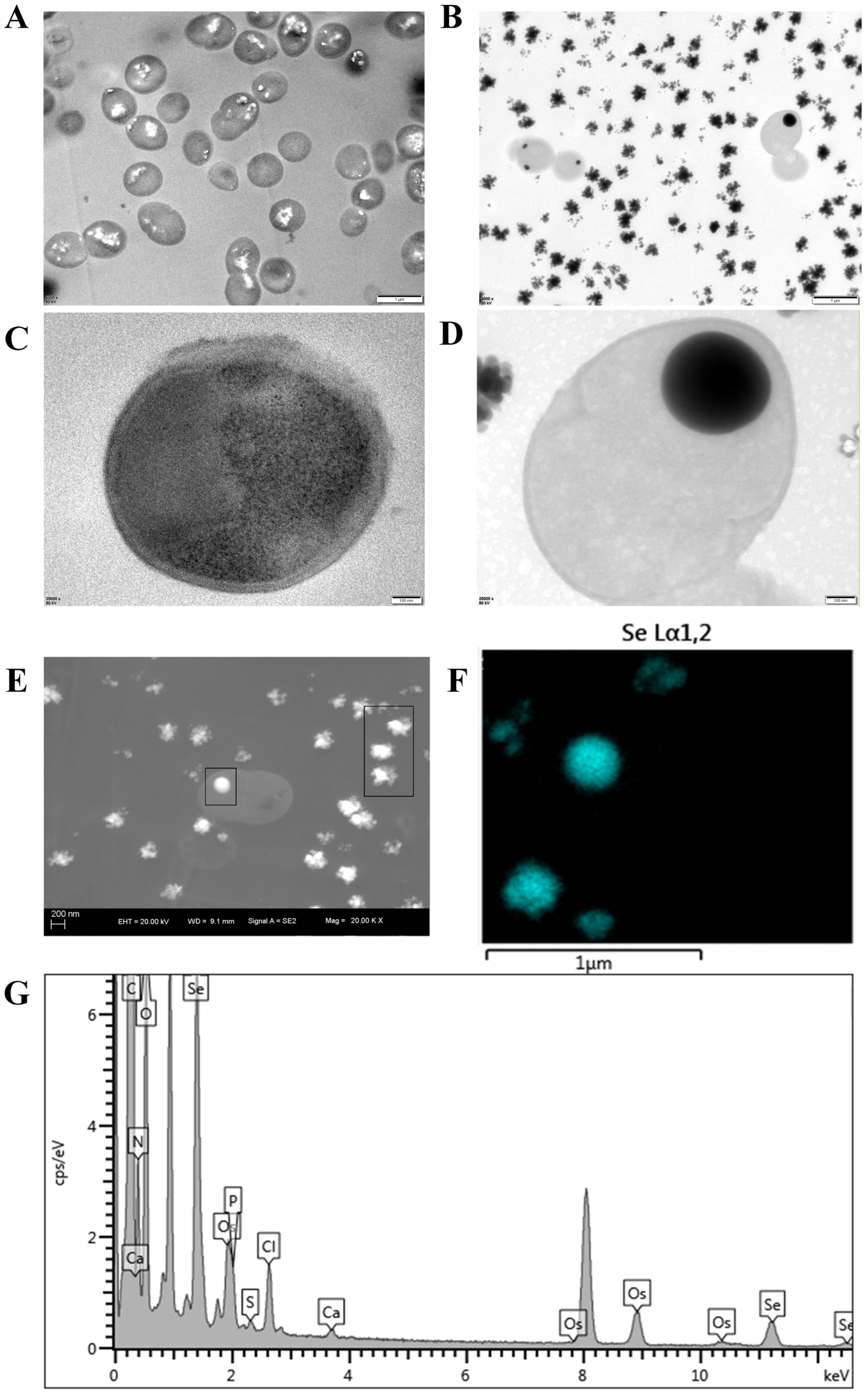
For inorganic selenium enrichment, a sodium selenite stock solution (100 μg/mL, Qingdao Hope Biotech, Qingdao, China) was prepared by dissolving 1 g of the compound in 10 mL of distilled water. After autoclaving, the sterile stock solution was added to MRS medium to achieve a final sodium selenite concentration of 10 μg/mL. RDN-1 was inoculated into the inorganic selenium-supplemented medium and incubated at 37 °C for 12 h. Inorganic selenium-supplemented bacteria were then collected by centrifugation as described above. To confirm inorganic selenium supplementation, the bacterial preparations were analyzed using transmission electron microscopy (TEM, JEOL Ltd., Tokyo, Japan) coupled with energy-dispersive X-ray spectroscopy (EDS). Images were acquired using a digital image acquisition system (Olympus Corporation, Tokyo, Japan).
2.3. Feed Preparation
Commercial feeds specially formulated for sturgeon (Qingdao Qihao Nutrition Technology, Qingdao, China) were used, and the ingredients are composed of fish meal, soybean meal, cottonseed meal, peanut meal, fish soluble, fish oil, soybean oil, amino acids, vitamins, and mineral premixes, and a proximate composition of 44% crude protein, 14% crude fat, 6% crude fiber, 16% crude ash, and 12% moisture. For the probiotic-treated groups, the prepared suspensions of L. lactis and inorganic selenium-supplemented L. lactis were evenly sprayed onto the feed to reach a final bacterial concentration of 1.0 × 10⁷ CFU/g, which was quantified via the plate counting method. For the antibiotic-treated group, feed was coated with a solution of enrofloxacin powder (Jiangsu Xinghai Biotechnology, Dongtai, China) dissolved in PBS to achieve an active enrofloxacin dose of 20 mg/kg fish body weight daily, a routine prophylactic dosage in fish farming. All coated feed samples were air-dried at ambient temperature (23.0 ± 1.0 °C) using an electric fan, prepared within 24 h prior to feeding and administered fresh without prolonged storage.
2.4. Sampling
Samples were collected on days 0, 7, 14, and 21 of the experimental period. At each sampling point, nine sturgeons were taken from each treatment group, with three fish randomly selected from each replicate tank and pooled to form one biological sample per replicate. The fish were anesthetized using 200 mg/L MS-222 (3-Aminobenzoic acid ethyl ester methanesulfonate, Shanghai Aladdin Biochemical Technology, Shanghai, China) before processing. Growth parameters were measured for individual fish specimens, whereas pooled fish samples were used for gut microbiota, gene expression and amino acid profiles. Body weight was measured using an electronic balance (BSA223S, Sartorius Lab Instruments, Göttingen, Germany) to assess growth performance, including weight gain and specific growth rate. The hindgut of fish per replicate tank was dissected and placed in a centrifuge tube containing 4% paraformaldehyde (PFA) fixative (Biosharp, Hefei, China), then stored at 4 °C. For molecular analyses, the body surface was disinfected with 75% ethanol before dissection. Tissue samples including intestine, liver, spleen and kidney were aseptically collected into sterile cryovials, immediately frozen in liquid nitrogen, and stored at −80 °C until further analysis. These samples were subsequently used for 16S rRNA gene-based gut microbiota sequencing and quantitative real-time PCR assays.
2.5. Intestinal Histomorphology of Sturgeon
Intestinal samples were collected from sturgeon and fixed in 4% formaldehyde solution (Beijing Baisha Reagent, Beijing, China). Subsequently, the samples were dehydrated through a graded ethanol series (70%, 80%, 90%, and 100%), with each step lasting 1–2 h to ensure complete water replacement. Following dehydration, the tissues were cleared in xylene (Sinopharm Chemical Reagent, Shanghai, China) to remove residual ethanol and adjust the refractive index of the tissue to match that of paraffin. The samples were then infiltrated with paraffin in a constant-temperature oven (Shanghai Yiheng Scientific Instrument, Shanghai, China) at 60 °C to allow uniform penetration into the tissue. The infiltrated tissues were embedded in paraffin blocks, sectioned at 4–6 μm thickness using a Leica RM 2135 rotary microtome (Leica Biosystems, Wetzlar, Germany), and stained with haematoxylin and eosin (H&E). The sections were mounted on glass slides, dried, and subsequently stained for histological observation. All slides were observed under a ZEISS Lab.A1 microscope (Zeiss, Jena, Germany), and images were captured by a Zeiss Axiocam 305 color microscope camera (Zeiss, Jena, Germany).
2.6. Analysis of Gut Microbiota
Intestinal samples underwent 16S rRNA gene sequence analysis at Novogene China Ltd. (Beijing, China) with the company’s standard laboratory reagents. Genomic DNA was extracted from intestinal microbiome samples using the CTAB method. Samples were lysed with CTAB buffer containing lysozyme, incubated at 65 °C, and centrifuged, and the supernatant was sequentially extracted with phenol-chloroform-isoamyl alcohol (25:24:1) and chloroform-isoamyl alcohol (24:1). DNA was precipitated with isopropanol at −20 °C, washed with 75% ethanol, re-dissolved in ddH
2O, and RNA contamination was removed via treatment with 1 μL RNase A at 37 °C. The 16S rRNA V3-V4 region was amplified using primers (F: CCTAYGGGRBGCASCAG; R: GGACTACNNGGGTATCTAAT). Genomic libraries were constructed with the TruSeq
® DNA PCR-Free Kit; library quality was assessed via Qubit 3.0 and Q-PCR prior to NovaSeq 6000 sequencing. On average, each sample yielded 102,000 raw paired-end reads, and 92,500 high-quality non-chimeric effective tags were retained after strict quality filtering. Filtering criteria: minimum read length ≥ 50 bp, base quality Q ≥ 20, paired-end overlapping length ≥ 10 bp for merging. All samples achieved high sequencing reliability with Q20 > 98% and Q30 > 95%. Raw sequencing reads were assembled with Flash v1.2.7 [
19], quality-controlled with Fastp v0.23.1 [
20], and chimeric sequences were removed using VSEARCH v2.16.0. Clean reads were clustered into operational taxonomic units (OTUs) at a 97% sequence similarity threshold using Uparse v7.0.1001 [
21]. This 97% cutoff is a well-recognized standard for aquatic prokaryotic species classification, ensuring accurate and reliable microbial taxonomic identification. OTU taxonomic annotation was performed against the SILVA138 SSUrRNA database [
22]. Prior to diversity calculation, OTU tables were rarefied to the minimum sequencing depth across all samples for uniform normalization. All samples were processed in a single experimental batch, so no batch-effect correction was required. QIIME v1.9.1 was used for sequence filtering, species annotation, and calculation of alpha diversity indices (Chao1, ACE, Shannon, Simpson, and Goods_coverage) to assess microbial community richness, evenness and sequencing adequacy. One-way ANOVA combined with Duncan’s multiple-range test was used for statistical analysis of alpha diversity among groups. For microbial relative abundance comparison, Bray-Curtis distance-based beta diversity was calculated, and ANOSIM and Adonis analyses (999 permutations) were performed to verify intergroup differences in microbial community structure. PCoA analysis and visualization were conducted in R v2.15.3 with WGCNA, stats, and ggplot2 packages. Differential microbial biomarkers were identified via LEfSe analysis with a threshold of LDA = 3.0 and
p < 0.05. Microbial community functions were predicted using PICRUSt v1.1.4 with KEGG database annotation.
2.7. Expression of Immune-Related Genes
Total RNA was extracted from liver, spleen, and kidney tissues of sturgeon using the SPARKeasy Tissue RNA Extraction Kit (Shandong Sparkjade Biotechnology Co., Ltd., Jinan, China). Subsequently, the extracted RNA was reverse-transcribed into cDNA using the SPARKscript II RT Plus Kit (With gDNA Eraser) (Shandong Sparkjade Biotechnology Co., Ltd., Jinan, China). A quantitative PCR reaction system with a volume of 10 μL was prepared using SYBR Green-based reagents. The primer sequences for
NF-κB,
IL-6,
TLR1 and the internal reference gene
β-actin were presented in
Table 1. RT-qPCR was performed using the 2 × SYBR Green qPCR Mix (Shandong Sparkjade Biotechnology Co., Ltd. Jinan, China) on a LightCycler
® 96 System (Roche, Basel, Switzerland). The thermal cycling conditions were set as follows: initial denaturation at 95.0 °C for 30 s, followed by 40 cycles of denaturation at 95.0 °C for 5 s and annealing/extension at 60.0 °C for 30 s. All reactions were conducted in triplicate to ensure reproducibility. The relative gene expression levels were calculated using the 2
−ΔΔCT method.
2.8. Data Analysis
Data analysis was performed using IBM SPSS software (Version 26). Prior to analysis, the Shapiro-Wilk test was used for normality testing and Levene’s test was applied to verify the homogeneity of variances. Comparisons among different groups were conducted by one-way ANOVA, followed by Duncan’s test for multiple comparison correction. Differences were considered statistically significant at p < 0.05. Data are presented as mean ± standard deviation (SD, n = 3).
3. Results
3.1. Inorganic Selenium Supplementation in RDN-1
TEM revealed the intracellular accumulation of inorganic selenium (Se) in strain RDN-1 (
Figure 1). Compared to the control, RDN-1 grown in sodium selenite-supplemented medium exhibited an markedly reduced growth rate and impaired cellular integrity. EDS analysis revealed substantial extracellular Se deposition, yet it also confirmed that RDN-1 was capable of biotransforming sodium selenite and sequestering Se intracellularly, with 2.62 wt% Se detected in the dry biomass (
Table 2).
3.2. Growth Performance
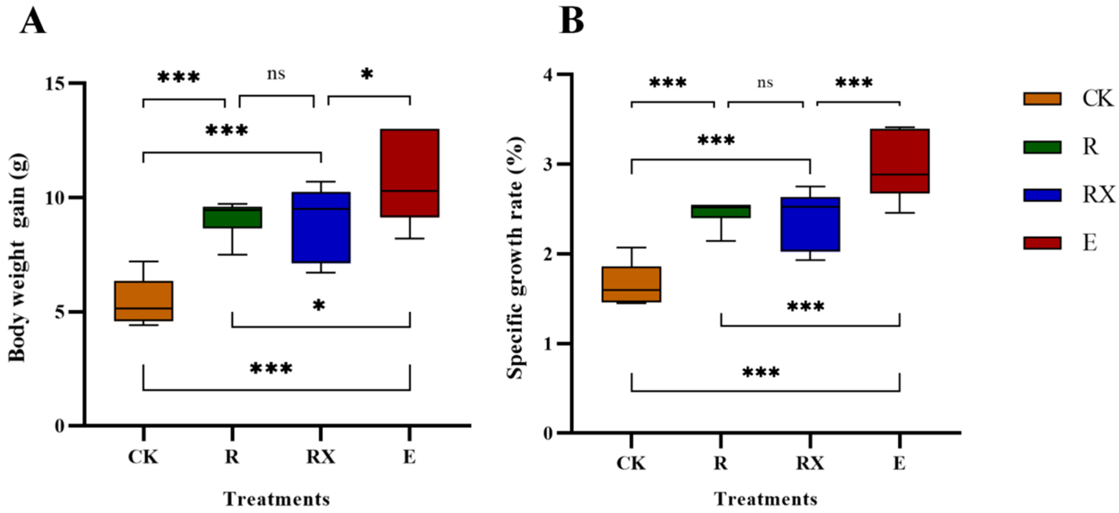
The weight gain and specific growth rate of sturgeons in all treatment groups were significantly higher than those in the control group (
p < 0.05) (
Figure 2). Specifically, both the R and RX groups exhibited increased weight gain and specific growth rates compared to the CK group (
p < 0.05), although no significant difference was observed between the R and RX groups. Notably, the E group demonstrated the highest values in both weight gain and specific growth rate among all groups. The increases were significantly greater than those in the control group (
p < 0.05), and the specific growth rate in the E group showed highly significant differences (
p < 0.05) compared to the CK, R, and RX groups.
3.3. Intestinal Histological Observation
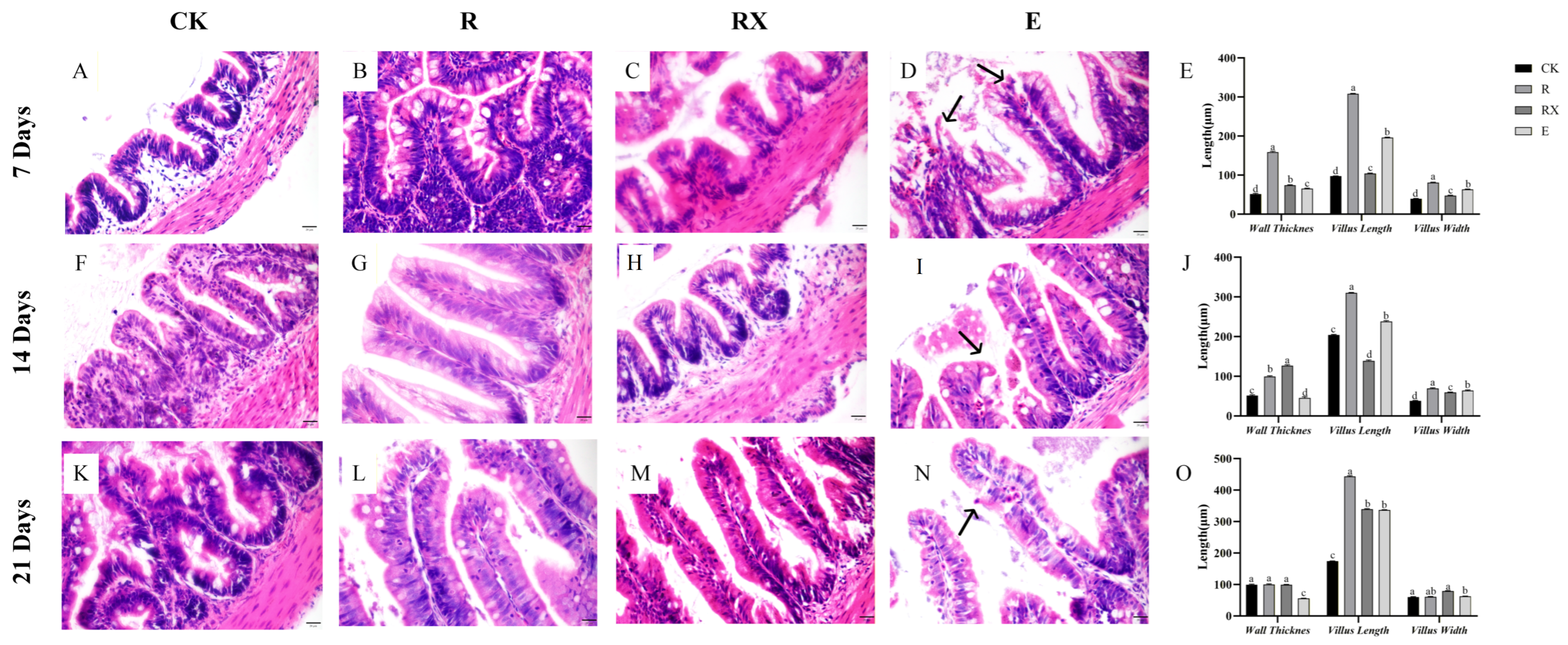
The overall integrity of the intestinal wall and villus structure in the RX group was superior to that of the E group but inferior to the R group (
Figure 3). Compared to the CK group, the R group showed a significant increase in intestinal villus height (
p < 0.05), and exhibited a thickened mucosal layer, well-defined and tightly arranged crypt structures, and intact villus surfaces without evident inflammation or sloughing (
Figure 3B,G,L). The RX group showed increased intestinal wall thickness relative to the CK group, with enhanced villus length and width at later stages (
p < 0.05) (
Figure 3C,H,M). Focal mild vacuolation of epithelial cells was observed in some regions. Notably, in comparison with the CK group, the E group had significantly reduced intestinal wall thickness, villus width, and thickness (
p < 0.05), along with obvious villus atrophy, thinning of the mucosal layer, disorganized crypt architecture, and localized inflammatory cell infiltration (
Figure 3E,I,N).
3.4. Amino Acid Content in Sturgeon Muscle
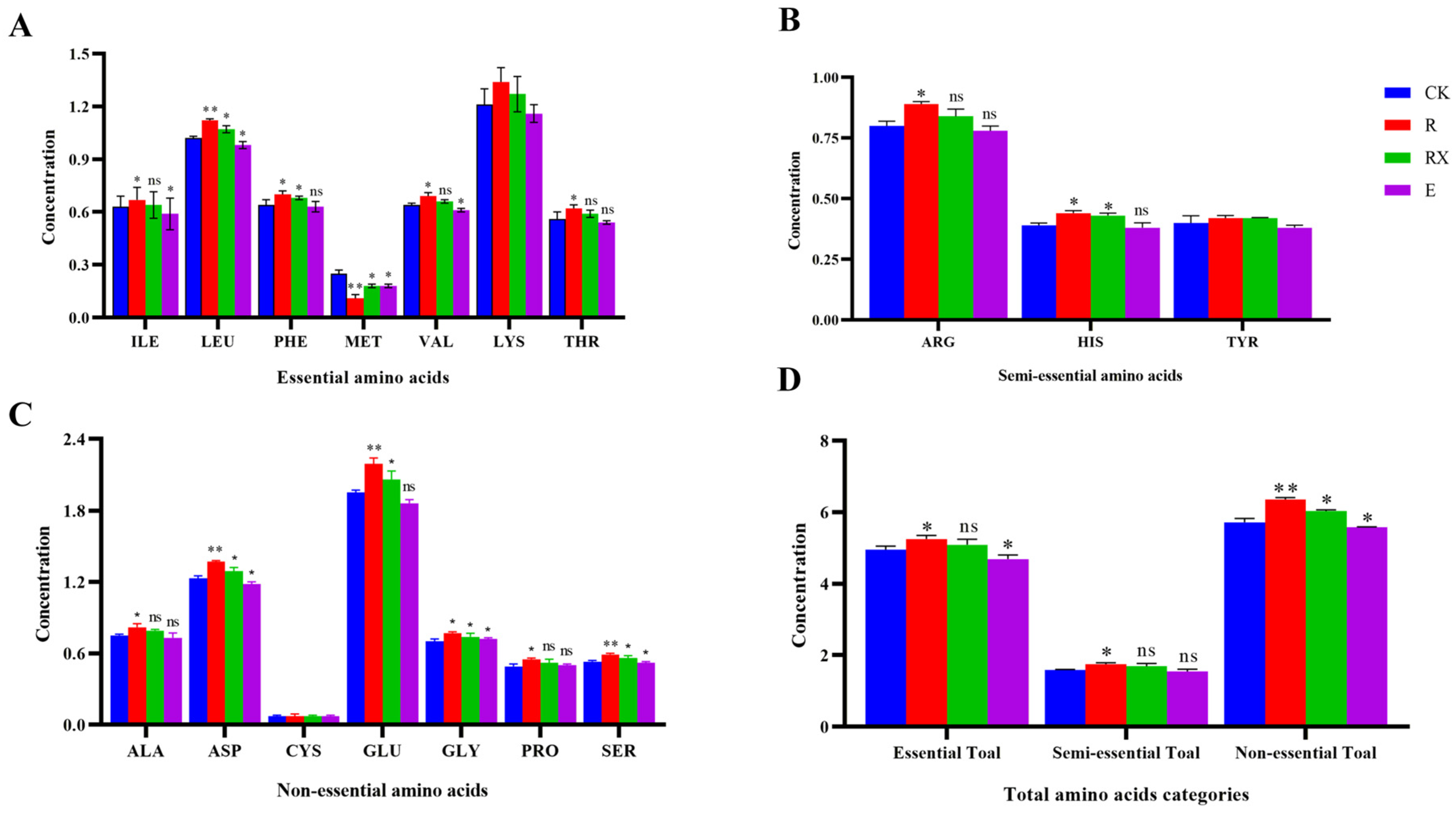
After 21 days of feeding, the majority of amino acids levels in sturgeon muscle from group R were significantly higher than those in the other groups (
Figure 4). In group RX, the levels of leucine, phenylalanine, lysine, histidine, aspartic acid, glutamic acid, glycine, and serine were significantly higher than those in groups CK and E (
p < 0.05), but generally lower than those in group R. Notably, the total content of essential amino acids, semi-essential amino acids, and total amino acids in group E were significantly lower than those in groups R and RX (
p < 0.05). Furthermore, the contents of certain amino acids in group E, such as isoleucine, leucine, methionine, valine, and aspartic acid, were even lower than those in the control group (
p < 0.05).
3.5. Diversity and Structure of Intestinal Microbiota
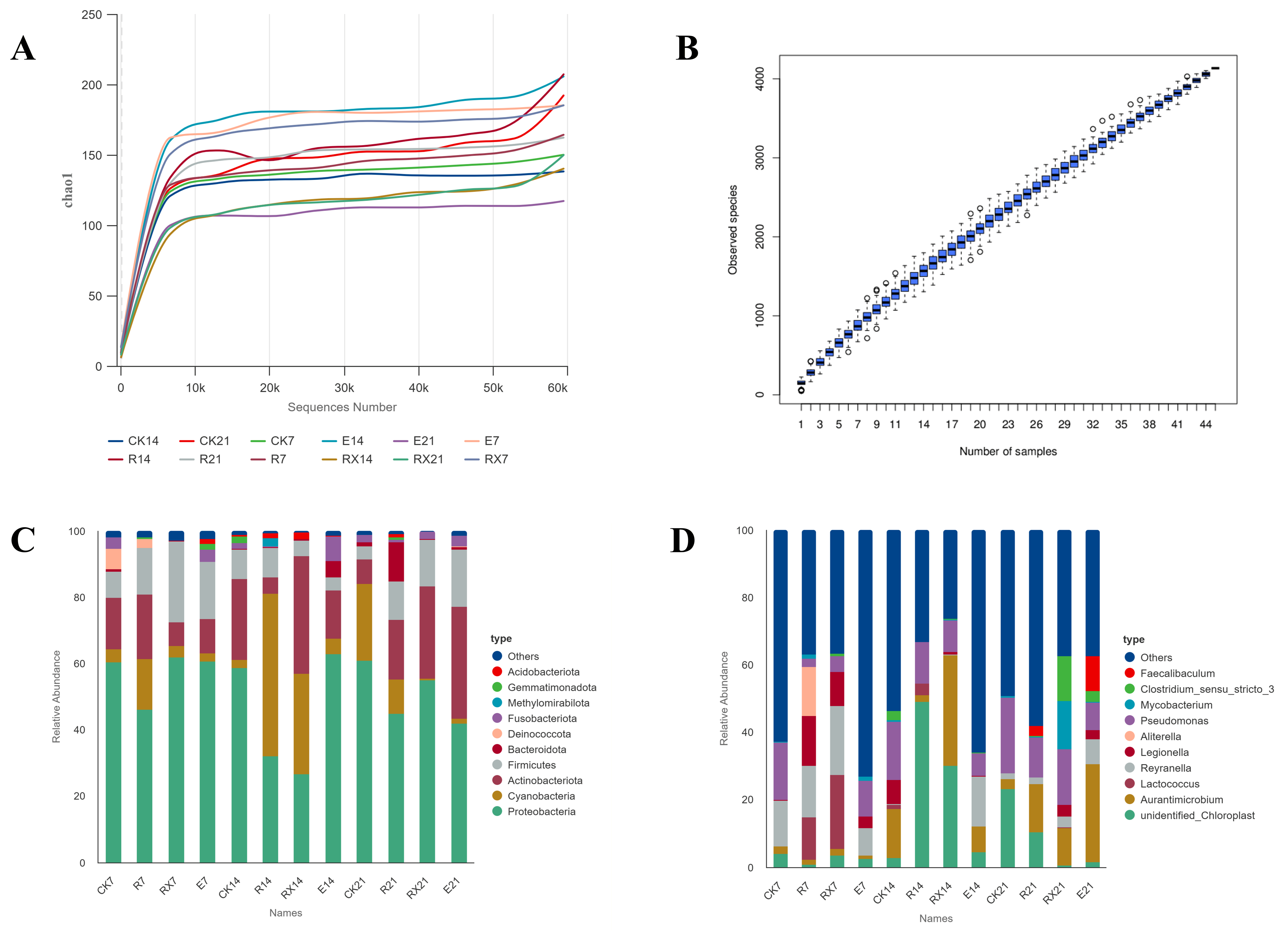
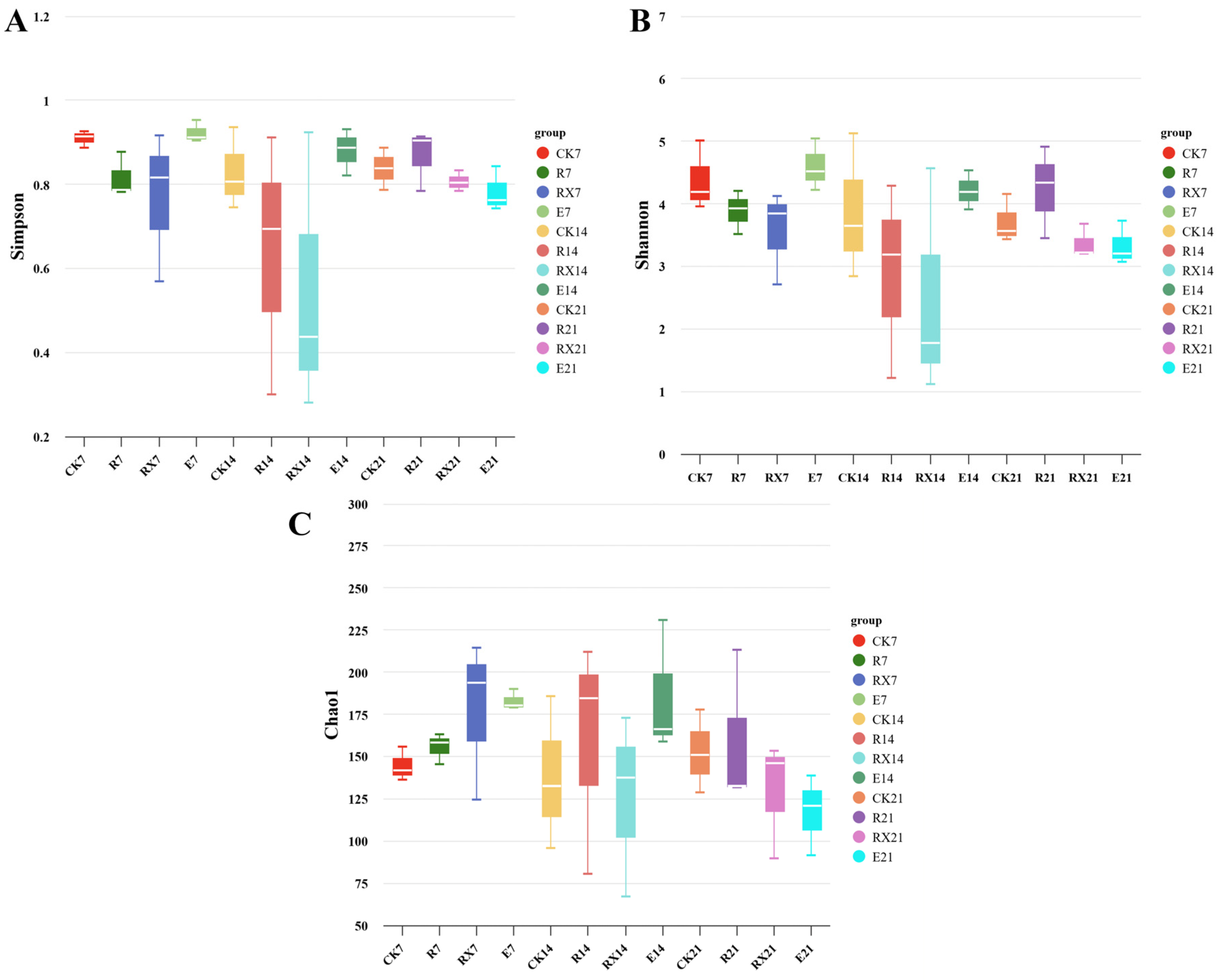
Regarding alpha diversity (Simpson and Shannon indices), group R exhibited an initial (7 day) decrease followed by recovery at the later time point (21 day) (
Figure 5A,B). The RX group displayed a consistent decreasing trend over time. Conversely, group E maintained relatively stable diversity early on but decreased significantly afterward. By day 21, group R maintained stable diversity and restored richness, whereas the E group exhibited an obvious decline in both richness and evenness. The microbial richness (Chao1 index) of the R group increased at the early stage (7 days) and then stabilized in the later phase (21 days) (
Figure 5C). In contrast, both the RX and E groups exhibited an initial increase followed by a decline in richness at the later time point. Notably, outliers were observed in the E group, suggesting considerable variation in richness measurements.
Dilution curves (
Figure 6A) and species accumulation curves (
Figure 6B) indicated satisfactory sequencing depth and sample adequacy, supporting the reliability of the data in representing the natural structure of the intestinal microbial community. At the phylum level, the dominant taxa in sturgeon intestine included Proteobacteria, Cyanobacteria, Actinobacteria, Firmicutes, and Bacteroidetes. The intestinal microbiota underwent significant successional changes when subjected to external interference. Specifically, in the R group, the relative abundance of Proteobacteria was consistently lower, while that of Firmicutes was higher than CK throughout the experiment. Bacteroidetes showed a decrease on day 7, but increased on days 14 and 21. Cyanobacteria increased on days 7 and 14, but returned to near baseline by day 21. In RX group, the relative abundance of Proteobacteria remained lower than that of the CK group across all time points, whereas Firmicutes was significantly higher on days 7 and 21, and Bacteroidetes was lower on these days. Actinobacteria decreased initially on day 7 and increased on days 14 and 21. In E group, the abundance of Proteobacteria was elevated on days 7 and 14 compared to CK. Firmicutes increased on days 7 and 21, while Bacteroidetes decreased on the same days. Cyanobacteria remained lower than CK throughout the sampling period, and Actinobacteria was obviously reduced on days 7 and 14.
3.6. LEfSe Analysis of Differential Microbiota
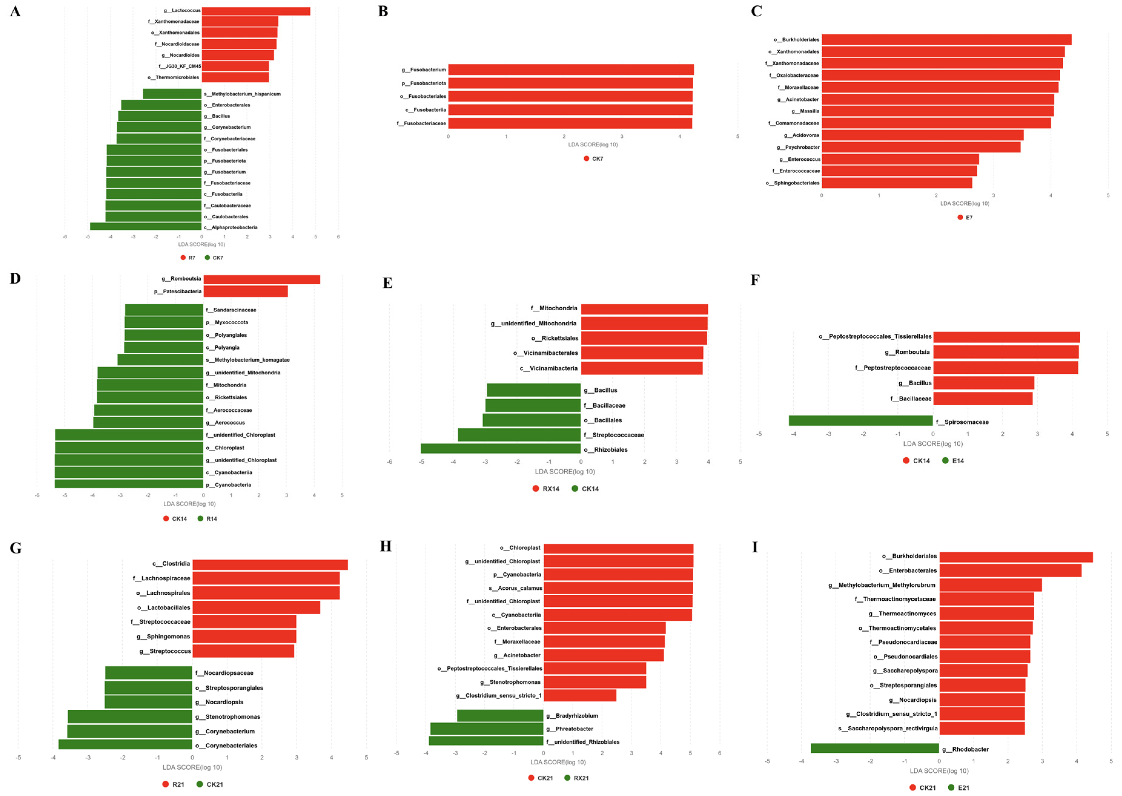
LEfSe analysis revealed significant alterations in the intestinal microbiota composition across treatment groups compared to the control (CK) (
Figure 7). In R group, on day 7, the relative abundances of
Lactococcus,
Xanthomonadales, and
Nocardioides were significantly increased, whereas
Methylobacterium,
Enterobacterales,
Bacillus,
Corynebacterium,
Alphaproteobacteria,
Caulobacteraceae, and
Fusobacterium were markedly reduced (
p < 0.05). On day 14, enrichment was observed in
Cyanobacteria,
Aerococcaceae,
Rickettsiales,
Myxococcota,
Polyangiales, and
Methylobacterium, while
Patescibacteria and
Romboutsia were significantly decreased (
p < 0.05). On day 21,
Clostridia,
Lachnospirales,
Lactobacillales,
Streptococcus, and
Sphingomonas showed notable increases, whereas
Nocardiopsis,
Streptosporangiales,
Stenotrophomonas, and
Corynebacterium were significantly reduced. In RX group, on day 7, a significant decrease was detected in
Fusobacterium (
p < 0.05). On day 14,
Vicinamibacteria and
Rickettsiales were enriched, while
Rhizobiales,
Streptococcaceae, and
Bacillus were significantly decreased (
p < 0.05). On day 21,
Bradyrhizobium and
Phreatobacter were significantly increased, whereas
Cyanobacteria,
Enterobacterales,
Moraxellaceae,
Acinetobacter,
Peptostreptococcales, and
Stenotrophomonas were notably reduced (
p < 0.05). In E group, on day 7, multiple taxa were significantly enriched (
p < 0.05), including
Burkholderiales,
Xanthomonadaceae,
Sphingobacteriales,
Oxalobacteraceae,
Moraxellaceae,
Comamonadaceae,
Enterococcus,
Acinetobacter,
Massilia,
Acidovorax, and
Psychrobacter. On day 14,
Spirosomaceae was significantly increased (
p < 0.05), while
Peptostreptococcaceae,
Romboutsia, and
Bacillus were decreased. On day 21,
Rhodobacter was significantly enriched (
p < 0.05), whereas
Burkholderiales,
Enterobacterales,
Thermoactinomyces,
Pseudonocardiales,
Streptosporangiales,
Methylobacterium,
Saccharopolyspora,
Nocardiopsis, and
Clostridium showed significant reductions (
p < 0.05).
3.7. Intestinal Microbiota Metabolic Pathways
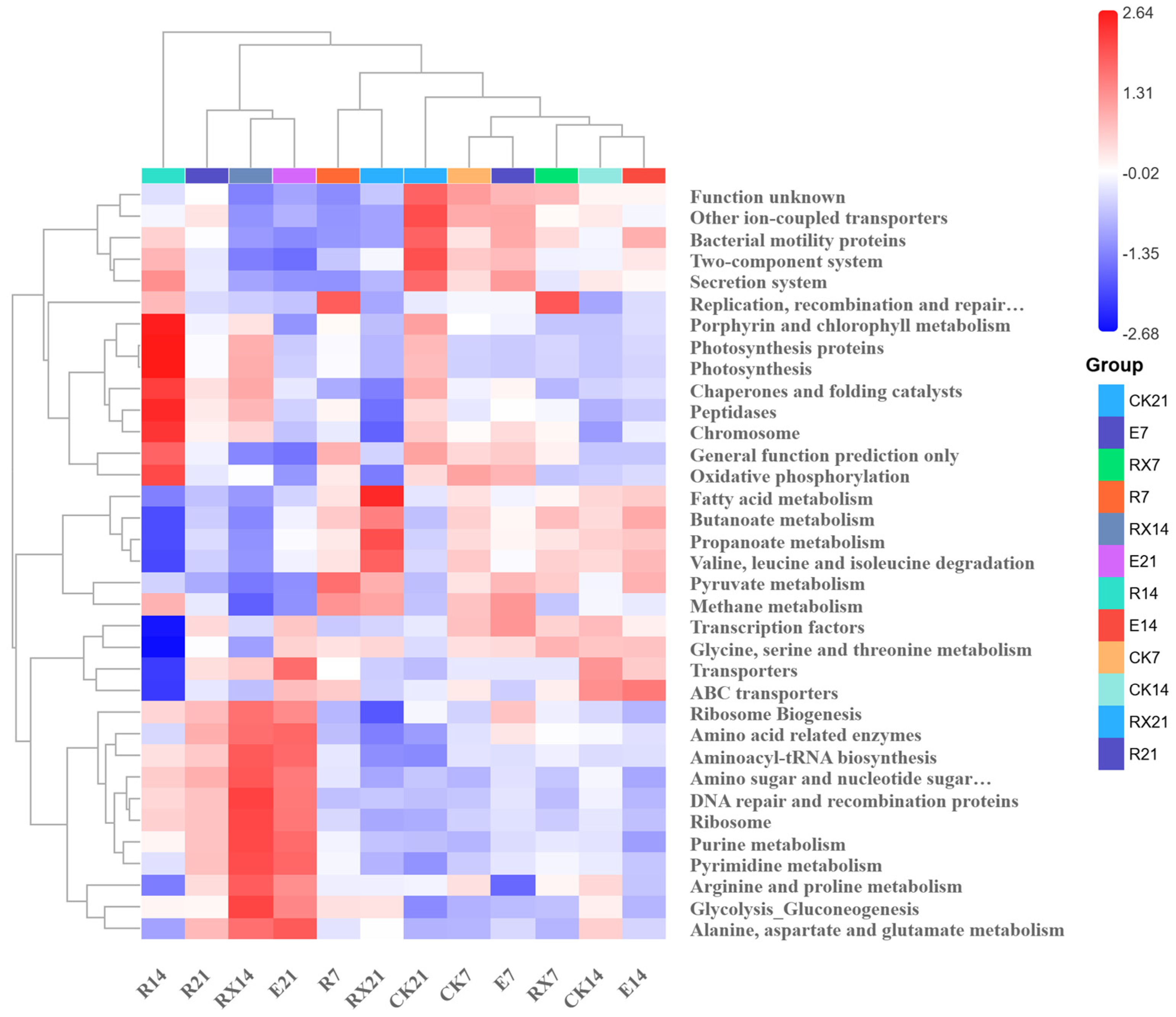
Based on the KEGG pathways, metabolic pathway functions of the gut microbiota were predicted via PICRUSt analysis (
Figure 8). After 7 days of feeding, the R group exhibited significantly enhanced pyruvate metabolism pathways, while bacterial motility protein pathways were remarkably down-regulated (
p < 0.05); the RX group showed significantly up-regulated pathways associated with replication, recombination, and repair proteins; the E group displayed significant down-regulation in the arginine and proline metabolic pathways (
p < 0.05).
After 14 days of feeding, the R group presented significant up-regulation in pathways of chaperone and folding catalysts, peptidases, and oxidative phosphorylation (p < 0.05). Concomitantly, pathways involved in fatty acid metabolism, butyrate metabolism, propionate metabolism, glycine-serine-threonine metabolism, ABC transporters, and valine-leucine-isoleucine degradation were notably down-regulated (p < 0.05). For the RX group, pathways related to ribosomal biosynthesis, amino acid-related enzymes, aminoacyl-tRNA synthesis, amino sugar and nucleotide sugar metabolism, as well as arginine, proline, tryptophan, aspartic acid, and glutamic acid metabolism, and DNA repair and recombination proteins (along with glycolysis/gluconeogenesis pathways) were upregulated (p < 0.05). Conversely, pathways of fatty acid metabolism, butyrate metabolism, propionate metabolism, pyruvate metabolism, valine–leucine–isoleucine degradation, bacterial motility proteins, and the two-component signaling system were down-regulated (p < 0.05). In the E group, the bacterial motility protein pathway was up-regulated, whereas pathways of amino sugar and nucleotide sugar metabolism, purine metabolism, and glycolysis/gluconeogenesis were down-regulated (p < 0.05).
Following 21 days of feeding, the R group showed up-regulation in pathways including alanine–aspartate–glutamate metabolism, glycolysis/gluconeogenesis, ribosomal biosynthesis, aminoacyl-tRNA synthesis, amino sugar and nucleotide sugar metabolism, and DNA repair and recombination proteins. In contrast, ion-coupled transporters, bacterial motility proteins, and two-component secretion system pathways were down-regulated (p < 0.05). In the RX group, glycolysis/gluconeogenesis, fatty acid metabolism, butyrate metabolism, propionate metabolism, and valine–leucine–isoleucine degradation pathways were significantly up-regulated. However, pathways related to ribosomal biosynthesis, oxidative phosphorylation, ion-coupled transporters, bacterial motility proteins, and two-component secretion systems were down-regulated (p < 0.05). For the E group, pathways such as ABC transporters, ribosomal biosynthesis, amino acid-related enzymes, aminoacyl-tRNA synthesis, DNA repair and recombination proteins, ribosomes, amino sugar and nucleoside sugar metabolism, purine metabolism, pyrimidine metabolism, arginine and proline metabolism, glycolysis/gluconeogenesis, and alanine-aspartate-glutamate metabolism were up-regulated. Meanwhile, ion-coupled transporters, bacterial motor proteins, and two-component secretion system pathways were down-regulated (p < 0.05).
3.8. Correlation Analysis Between Intestinal Microbiota and Amino Acids
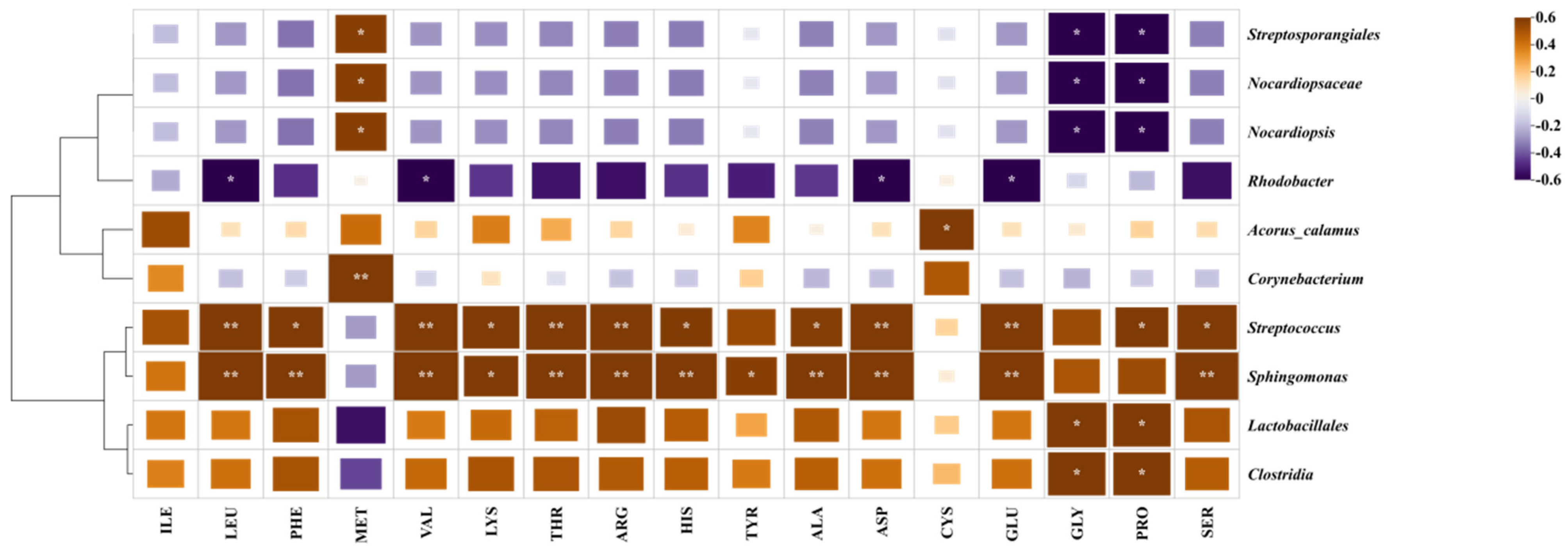
Analysis of gut microbiota-muscle amino acid correlations identified key bacterial contributors (
Figure 9). Streptococcus and Sphingomonas showed a broad-spectrum significant positive association with most amino acids (
p < 0.05), whereas Lactobacillales and Clostridia were specifically significantly correlated with glycine and proline (
p < 0.05). Methionine accumulation was significantly positively associated with Streptosporangiales, Nocardiopsaceae, and Nocardiopsis, genera which were significantly negatively correlated with glycine and proline (
p < 0.05).
3.9. The Expression of Immune-Related Genes
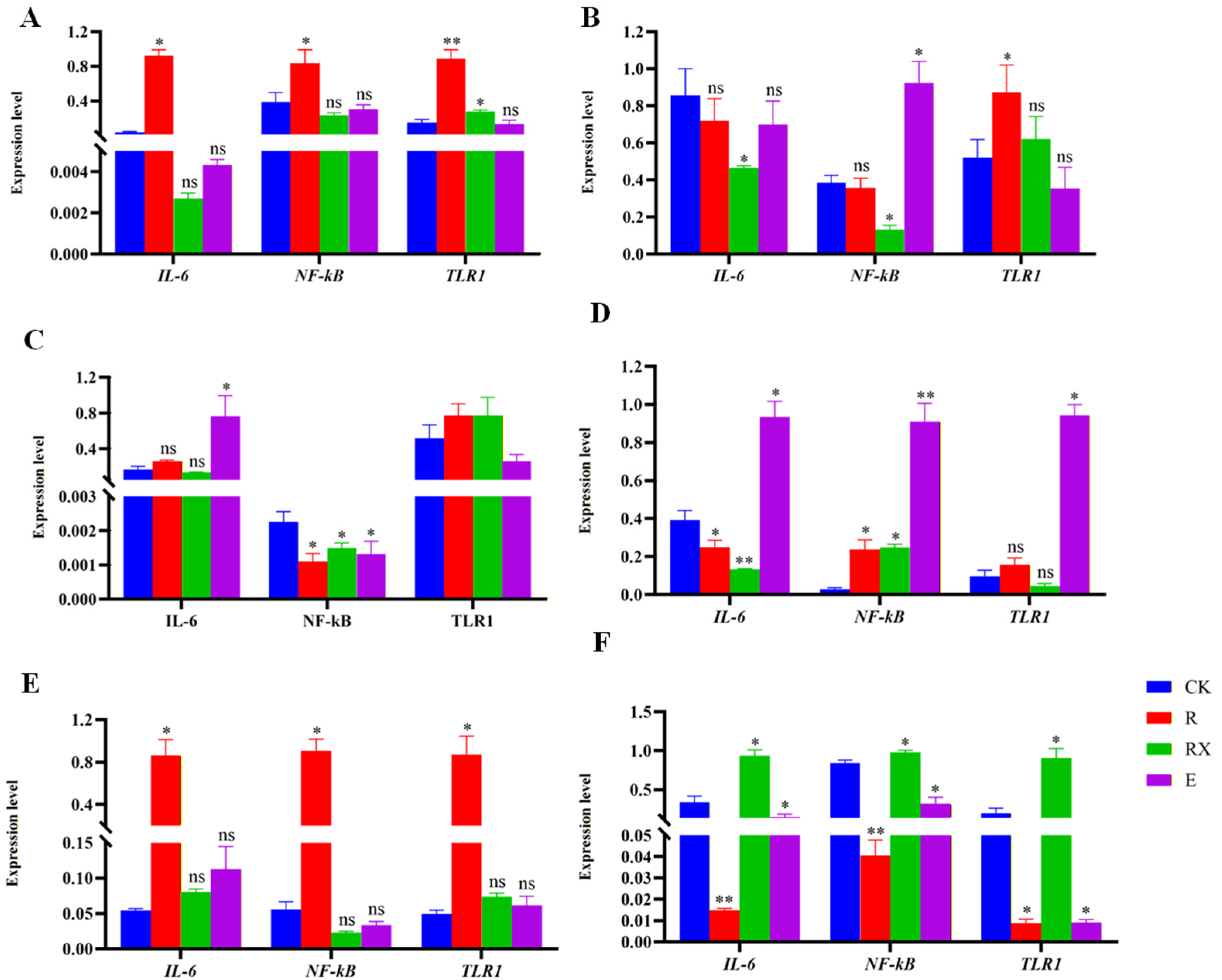
The relative expression levels of immune-related genes (
TLR1,
NF-κB, and
IL-6) in the liver, spleen, and kidney are shown in
Figure 10. After 14 days of feeding, in the liver,
IL-6 and
NF-κB expression were significantly up-regulated in the R group (
p < 0.05), whereas no significant change in
IL-6 was observed in the RX or E groups.
TLR1 expression was significantly elevated in the R and RX groups, but unchanged in the E group. In the spleen,
IL-6 and
NF-κB expression were significantly lower in the RX group than in the CK group (
p < 0.05), while the E group showed significantly higher
NF-κB expression compared to all other groups.
TLR1 expression was significantly increased in the R group; it did not differ significantly between the RX and CK groups but was higher than in the E group. In the kidney,
IL-6 expression was significantly increased in the E group (
p < 0.05), while no significant differences were detected in the R and RX groups compared with the CK group.
NF-κB expression was lower in all treatment groups than in the control, and
TLR1 expression did not differ significantly among groups.
After 21 days of feeding, the liver showed particular sensitivity to E treatment, displaying marked up-regulation of inflammatory genes. In contrast, renal responses were relatively moderate, with an earlier down-regulation trend observed (e.g., NF-κB was generally down-regulated by day 14). In the spleen, the R group showed highly significant elevation in the expression of IL-6, NF-κB, and TLR1 (p < 0.05), with no significant changes in the other groups. Overall, the spleen exhibited the most pronounced and consistent response to R treatment, with all three genes being significantly up-regulated after 21 days. In the kidney, expression of IL-6, NF-κB, and TLR1 was significantly decreased in the R and E groups (p < 0.05), but significantly increased in the RX group. In the liver, IL-6 expression was significantly reduced in the R and RX groups, whereas NF-κB was significantly up-regulated (p < 0.05); TLR1 remained unchanged. In contrast, the E group exhibited highly significant up-regulation of all three genes (p < 0.05).
4. Discussion
In this study, dietary
L. lactis, inorganic selenium enriched
L. lactis, and enrofloxacin all significantly improved the growth performance of sturgeon compared to the control. The significant growth enhancement in the R and RX groups indicates that
L. lactis can improve feed utilization. Notably, the superior growth promotion observed in the E group highlights the immediate effectiveness of antibiotics. However, this approach raises concerns regarding sustainable aquaculture practices, including antibiotic residue, microbial resistance, and long-term health impacts on fish [
23,
24,
25].
In this study, sturgeon in the R group exhibited notably higher levels of most amino acids compared to other groups, suggesting that probiotics may enhance amino acid accumulation by modulating the gut microbiota and improving nutrient absorption efficiency. In the RX group, although most amino acid levels were intermediate between the R and CK groups, methionine content was significantly higher than that in the R group. Notably, RDN-1 also exhibits the ability to biotransform selenium, while electron microscopy analysis revealed a significant decrease in the concentration of
L. lactis. It should be noted that high selenium concentrations may also exert cytotoxic effects on lactic acid bacteria, compromising cell viability and ultimately reducing their probiotic efficacy. Further research is warranted to optimize the selenium concentration and the cultivation duration. Thus, RDN-1 likely directly biotransformed sodium selenite into selenomethionine, which was subsequently absorbed by the intestine, specifically modulating methionine metabolism in sturgeon and thereby altering muscle methionine composition [
26]. In contrast, the E group showed significantly lower levels of total essential amino acids, semi-essential amino acids, and total amino acids compared to the R and RX groups, with some values even falling below those of the CK group. These results imply that although antibiotic use may lead to short-term growth improvements, it could negatively affect amino acid deposition, likely through disruption of intestinal microbial balance and impairment of amino acid absorption and metabolism.
Significant differences in intestinal morphology were observed among the treatment groups. Sturgeon in the R group exhibited pronounced structural improvements, including increased intestinal villus height and width. These findings align with previous studies on
L. lactis, which reported similar enhancements in villus architecture in rainbow trout, thereby improving digestive efficiency [
27,
28]. The RX group showed moderate morphological improvements, with overall structural integrity superior to that of the E group. However, focal mild vacuolization of epithelial cells was detected in some regions, possibly due to the cytotoxic effects of inorganic selenium [
29,
30]. In contrast, the E group showed marked pathological alterations, characterized by significant reductions in intestinal wall thickness, villus width and height, severe villus atrophy, thinning of the mucosal layer, disorganized crypt structures, and localized inflammatory cell infiltration. Although substantial weight gain was recorded in this group, the intestinal degeneration indicates that the growth-promoting effect of enrofloxacin may stem from short-term metabolic stimulation rather than sustainable health benefits.
Gut microbiota serves as a crucial biological barrier for maintaining intestinal health in fish [
31,
32]. In this study, microbial analysis revealed that all treatment groups exhibited temporal succession of gut microbiota, with rapid changes at the early stage (7 days), followed by distinct microbial responses at the mid (14 days) and late (21 days) stages. Under probiotic and antibiotic interventions, the metabolic response of the gut microbiota in sturgeon was pronounced initially and gradually stabilized over time, indicating a strong correlation between metabolic functional shifts and microbial structural succession.
L. lactis has been reported to inhibit harmful bacteria by secreting bacteriocins such as Nisin and suppressing Proteobacteria via competitive exclusion [
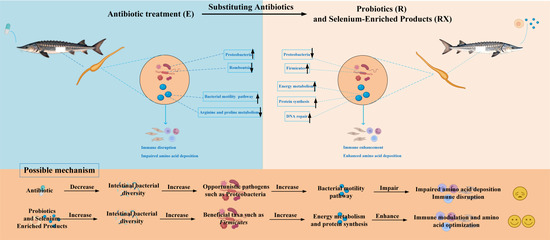
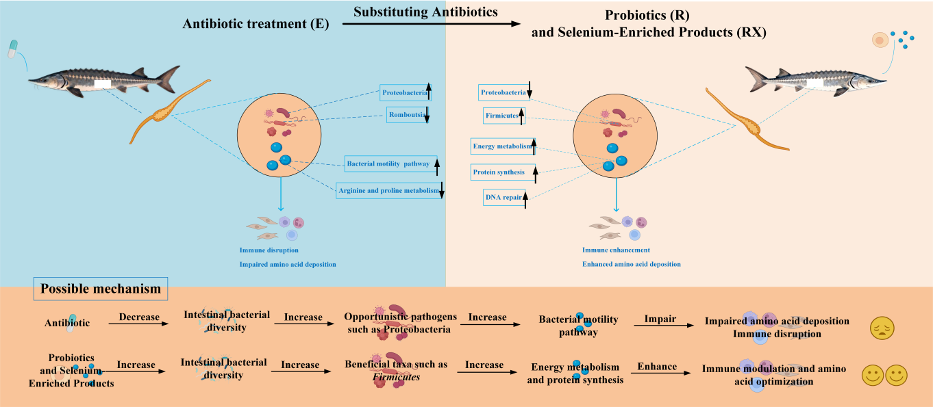
33]. In this study, both the R and RX groups established a microbial structure dominated by Firmicutes, which was positively correlated with host amino acid content. Consistent with this, genomic prediction revealed significant up-regulation of energy metabolism and protein synthesis pathways in these groups. Firmicutes are known to enhance carbohydrate degradation capacity by enriching glycolysis and citrate cycle pathways, thereby producing more volatile fatty acids (e.g., propionate) as energy substrates for the host [
34]. In the R group,
Lactococcus acted as a “pioneer species” at the early stage, inhibiting pathogens such as
Fusobacterium. By the mid-stage,
Cyanobacteria and
Rickettsiales emerged, and the community eventually succeeded to a functional fermentation-type microbiome dominated by
Sphingomonas,
Streptococcus, and
Lactobacillales. This dominant microbial community showed a positive correlation with most amino acids in sturgeon muscle, suggesting that RDN-1 may promote amino acid accumulation by increasing the abundances of
Sphingomonas and
Streptococcus. The RX group developed a more resilient symbiotic micro-ecology, with increased abundance of
Vicinamibacteria and
Rickettsiales at the mid-stage, and
Bradyrhizobium and
Phreatobacter at the late stage. Among these,
Bradyrhizobium was associated with citrate production [
35], which suggested that selenium supplementation might promote the growth of microbial taxa with specialized metabolic capabilities. Previous studies showed that dietary selenium supplementation, including selenomethionine (Se-Met) and selenium nanoparticles (SeNP), significantly promoted growth performance and antioxidant capacity in grass carp [
36], as well as amino acid metabolism (e.g., methionine metabolism) in zebrafish [
37]. In contrast, the E group exhibited the potential for proliferation of opportunistic pathogens. Previous studies showed that antibiotic treatment directly eliminated susceptible microbial communities, created ecological niche vacancies, and thereby promoted the proliferation of opportunistic pathogens such as Proteobacteria [
38]. This observation was consistent with the abnormal proliferation of Proteobacteria and other negatively correlated taxa observed in the early stage. These proliferating taxa may have consumed dietary amino acids to support their own growth or produce harmful metabolites, rather than facilitating host absorption [
39]. Concurrently, amino acid metabolic pathways were suppressed in the early phase. By the mid-stage, beneficial bacteria (e.g.,
Romboutsia) declined, leading to structural degradation and functional impairment of the microbiota, ultimately failing to support effective host amino acid deposition. Previous studies in tilapia have also documented the adverse effects of long-term antibiotic use on intestinal health, including reduced microbial diversity, enrichment of conditionally pathogenic bacteria, and dysregulation of carbohydrate and lipid metabolism pathways [
4]. Moreover, prolonged antibiotic exposure disrupts short-chain fatty acid-mediated cellular signaling, leading to sustained impairment of intestinal mucosal homeostasis [
40].
In this study, groups R and RX led to consistent up-regulation of key metabolic pathways across multiple time points, including energy metabolism (e.g., pyruvate metabolism, oxidative phosphorylation, glycolysis), protein synthesis (ribosome biogenesis, aminoacyl-tRNA biosynthesis), and DNA repair. Specifically, energy metabolic pathways such as pyruvate metabolism and oxidative phosphorylation could increase ATP production and biosynthetic flux, which significantly promoted the enhancement of microbial growth [
41], while the enhanced DNA repair capacity improved metabolic stability [
42]. Notably, the RX group showed a more pronounced enhancement in antioxidant capacity, reflected by the sustained up-regulation of DNA repair pathways at 14 and 21 days, which might be attributed to dietary selenium supplementation boosting the host’s antioxidant defenses. In contrast, the E group displayed a pattern of “metabolic disruption and stress”. Early suppression of arginine and proline metabolism coincided with inhibited microbial growth, while opportunistic pathogens such as
Enterococcus proliferated markedly, accompanied by up-regulation of bacterial motility-related pathways, suggesting increased invasive potential. By the later stage, the E group showed broad compensatory up-regulation of pathways such as ABC transporters and amino acid metabolism. This shift represents a stress response under antibiotic pressure, suggesting a potential risk of resistance selection within the altered intestinal microbiota. Similarly, enrofloxacin reduces intestinal lipopolysaccharide (LPS) activity and enhances the competitive advantage of pathogenic bacterial genera, including
Aeromonas,
Shewanella, and
Shigella, in grass carp (
Ctenopharyngodon idellus) [
43]. Notably, all groups eventually down-regulated bacterial motility and virulence-related pathways by day 21, indicating a general transition toward a more metabolically stable community state. Despite the uniform trend, distinct group differences existed: the R and RX groups ultimately reconstructed a beneficial bacteria-driven metabolic network, whereas the E group might drive persistent compositional and functional alterations shaped by antibiotic selection for resistant taxa. Given that functional profiles were bioinformatically predicted via 16S rRNA, subsequent multi-omics analyses are essential for experimental verification.
The results of immune response indicated that the R group exhibited comprehensive immune-stimulatory effects, particularly at the later stage (21 days), with significant up-regulation of multiple immune genes in the spleen and liver, and such tissue-specific mild upregulation may represent adaptive immune enhancement. Notably, the expression of
TLR1 in the R group was 2.3-fold higher than that in the control group (
p < 0.05), which was consistent with the findings reported in Siberian sturgeon [
44]. Furthermore, similar studies have also demonstrated that feeding the probiotic
Chromobacterium aquaticum can activate the expression of innate immune-related genes such as
NF-κB and
IL-6 in zebrafish [
45];
Bacillus siamensis LF4 can significantly activate the
TLR2,
TLR5, and
MyD88 signaling pathways in
Lateolabrax maculatus [
46]. This indicates that
L. lactis may enhance the host’s pathogen recognition ability and defense capacity through the Toll-like receptor signaling pathway. The RX group demonstrated a more complex and tissue-specific immunomodulatory profile. While it promoted gene expression in certain tissues (e.g., kidney at 21 days), it suppressed expression in others (e.g.,
IL-6 in the liver). This tissue specificity likely stemmed from differences in selenium metabolic pathways: selenium is metabolized by the gut microbiota into various forms (e.g., γ-Glu-SeMet and SeNPs) and then transported via the circulatory system to exert effects different tissues [
47], suggesting a fine-tuned regulatory mechanism that may prevent excessive inflammation and contribute to immune balance. Of particular concern, the E group exhibited marked tissue-specific immune disruption. It induced extremely significant up-regulation of
IL-6,
NF-κB, and
TLR1 in the liver at the later stage, where the dramatic upregulation of inflammatory genes indicated an obvious inflammatory stress response. These results align with those of Qiu et al. (2022), who reported that long-term antibiotic use suppressed immune responses in zebrafish, reducing key immune biomarkers such as macrophage and neutrophil counts and increasing susceptibility to infection [
48].