Article

Preparation and Application of Monoclonal Antibodies Targeting IgM in Pearl Gentian Grouper (Epinephelus fuscoguttatus× E. lanceolatus ♂)

1
Tianjin Key Lab of Aqua-Ecology and Aquaculture, Fisheries College, Tianjin Agricultural University, Tianjin 300384, China
2
College of Marine and Bioengineering, Yancheng Institute of Technology, Yancheng 224051, China
*
Authors to whom correspondence should be addressed.
These authors contributed equally to this work.
Fishes 2026, 11(3), 160; https://doi.org/10.3390/fishes11030160
Submission received: 2 February 2026 / Revised: 6 March 2026 / Accepted: 6 March 2026 / Published: 11 March 2026
(This article belongs to the Special Issue Advances in the Immunology of Aquatic Animals)

Abstract

Viral and bacterial pathogen pathogens cause disease outbreaks that challenge the pearl gentian grouper (Epinephelus fuscoguttatus× E. lanceolatus ♂) industry’s sustainable development. The lack of monoclonal antibodies (mAbs) targeting serum immunoglobulin M (IgM) in this hybrid grouper impedes the development of non-lethal immunoassays for detecting pathogen infections, as well as research on immune responses following vaccination. We purified serum IgM from hybrid pearl gentian grouper and generated two mAbs—designated 41-H2-E1 and 62-E8-G9—against the purified IgM, finding that mAb 62-E8-G9 specifically recognized the IgM heavy chain, whereas mAb 41-H2-E1 specifically recognized the light chain. In indirect immunofluorescence assays, both mAbs reacted with surface Ig-positive (sIg+) lymphocytes. A double-antibody sandwich ELISA was subsequently established using mAb 62-E8-G9 as the capture antibody and HRP-conjugated mAb 41-H2-E1 as the detection antibody, enabling accurate quantification of serum IgM levels. Significant differences in IgM concentrations were observed between larger and smaller individuals (9.11 μg/mL vs. 3.84 μg/mL, p < 0.05). In immunostimulant administration experiments, both low-and high-dose groups exhibited approximately 2.0-fold higher IgM levels than the control group (p < 0.05). In contrast, vaccination with inactivated vaccines did not result in statistically significant differences in total IgM levels. mAb 41-H2-E1 was further applied to detect Vibrio parahaemolyticus- and Vibrio harveyi-specific immunoglobulins in serum under different vaccination regimens. Collectively, these findings demonstrated that the mAbs developed in this study served as reliable immunological tools for investigating immune function in hybrid pearl gentian grouper.
Key Contribution: In this study, IgM-specific monoclonal antibodies against hybrid pearl gentian grouper were generated. A stable and sensitive sandwich ELISA was established for the accurate quantification of total IgM, and an indirect ELISA was developed for the specific detection of Vibrio-induced IgM. These assays provided reliable immunological tools for aquatic immunological research and disease prevention in this species.

1. Introduction

The hybrid pearl gentian grouper (Epinephelus fuscoguttatus× E. lanceolatus ♀) is a hybrid species valued in aquaculture for its strong disease resistance, rapid growth rate, and superior culinary quality [1]. However, under intensive farming conditions, bacterial infections lead to substantial economic losses. Pathogens such as Vibrio alginolyticus [2], Vibrio harveyi [3] and Vibrio parahaemolyticus [4] can suppress immune function and increase host susceptibility to disease [5]. V. parahaemolyticus, a halophilic Gram-negative bacterium, is a major causative agent of Vibriosis and has been reported to induce high mortality and severe histopathological damage in species such as Branchiostoma belcheri tsingtaunese [6]. Similarly, V. harveyi infection in fish often manifests as fin rot, skin lesions, abdominal ascites, hepatosplenomegaly, and abnormal swimming behavior [7]. The distinct pathogenic mechanisms of these bacteria—such as hemolysin-mediated tissue damage and immune barrier disruption by V. harveyivia [8], as well as the inflammatory response triggered by V. parahaemolyticus—complicate the systematic evaluation of humoral immune responses following infection.
Immunoglobulins (Igs) first emerged in jawed vertebrates and played pivotal roles in adaptive immunity by mediating host defense against pathogens following infection [9]. Teleost fish possessed three principal immunoglobulin isotypes—IgM, IgD, and IgZ/IgT [10]—among which IgM served as the predominant mediator of the systemic adaptive immune response. MAbs against serum IgM are valuable tools for studying immune system mechanisms and facilitating pathogen detection, which are central to disease management in aquaculture [11]. These mAbs are widely applied to quantify total IgM [10], detect pathogen-specific IgM [12], and enumerate surface Ig-positive (sIg+) lymphocytes [13]. To date, IgM-specific mAbs have been developed for several teleosts, including Larimichthys crocea [14], Cynoglossus semilaevis [15], Ctenopharyngodon idella [16], Oreochromis niloticus [17], Paralichthys olivaceus [18], and Cyprinus carpio [19]. However, no IgM-specific mAbs have been reported for hybrid pearl gentian grouper, limiting the assessment of immunoglobulin responses after vaccination.
The primary objective of this study was to generate mAbs targeting the IgM of hybrid pearl gentian grouper and to establish a reliable detection platform for quantifying both total and pathogen-specific IgM responses. To this end, we report: [i] the production and characterization of mAbs against the purified IgM; [ii] quantification of serum IgM via double-antibody sandwich ELISA; and [iii] the measurement of antigen-specific IgM responses in fish subjected to different treatments by indirect ELISA.

2. Materials and Methods

2.1. Breeding Environment of Experimental Animals

A total of 300 healthy hybrid pearl gentian grouper (Epinephelus fuscoguttatus× E. lanceolatus ♀) were obtained from a Haifa fish farm in Tianjin, China. The fish were acclimated for 7 days in rearing tanks with continuously aerated water maintained at 25 °C and were fed a commercial dry-pellet diet. Water quality parameters were monitored daily as follows: dissolved oxygen, 5–6 mg/L; pH, 7–8; and ammonia–nitrogen (NH3-N) concentration, ≤0.05 mg/L.

2.2. Purification of Serum Immunoglobulin

Blood was collected from the caudal vein of 20 hybrid pearl gentian groupers (average body weight, 700 ± 70 g), allowed to coagulate at room temperature for 1 h, and then stored at 4 °C overnight. Serum was obtained by centrifugation at 1500 rpm for 30 min at 4 °C, pooled together, and then stored at −20 °C. Crude IgM was precipitated from 50 mL of serum using 50% saturated ammonium sulfate. Subsequently, IgM was further purified from the crude extract using a Protein A column (Meijin Biological Company, Guangzhou, China). The concentration of the purified IgM was determined using the Bradford method, and the molecular weight of the purified IgM was checked by 12% sodium dodecyl sulfate–polyacrylamide gel electrophoresis (SDS-PAGE). Purified lgM was aliquoted into sterile cryovials and stored at −80 °C until further use.

2.3. Production of mAbs Against Hybrid Pearl Gentian Grouper IgM

MAbs against hybrid pearl gentian grouper IgM were produced as previously described [20]. Purified IgM was emulsified with an equal volume of complete Freund’s adjuvant (Sigma, St. Louis, MO, USA), and administered via intraperitoneal injection to five BALB/c mice. A total of three booster immunizations were administered at 1-week intervals, with the same dose of immunogen emulsified in incomplete Freund’s adjuvant (lFA). Following the immunization schedule, cell fusion was performed using the polyethylene glycol (PEG)-mediated method. Splenocytes and SP2/0 myeloma cells were mixed at a 5:1 ratio, and hybridomas were initially screened by indirect ELISA. Then the hybridomas giving positive results were cloned three times by limited dilution. Finally, the mAbs were characterized by indirect ELISA, Western blotting, indirect immunofluorescence assay and subtype detection.

2.4. Screening of mAbs Against Hybrid Pearl Gentian Grouper IgM

2.4.1. Indirect ELISA

Microplates (Naisi, Lishui, China) were coated with purified hybrid pearl gentian grouper IgM (2.5 µg/mL, 100 µL/well) and incubated overnight at 4 °C. Plates were washed with PBST and blocked with 3% BSA at 37 °C for 1 h. Hybridoma supernatants (SP2/0 supernatant as negative control) were added (100 µL/well) and incubated at 37 °C for 1 h, with each sample assayed in triplicate. After washing with PBST, 100 µL/well of HRP-conjugated goat anti-mouse IgG (1:10,000 in PBST; Solarbio, Beijing, China) was added and incubated at 37 °C for 1 h. TMB substrate (100 µL/well) was then added for 30 min at 37 °C, and the reaction was stopped with 50 µL/well of termination buffer (Biosharp, Beijing, China). Absorbance was measured at 450 nm using a microplate reader (Ossence, Shenzhen, China).

2.4.2. Western Blotting Analysis

For Western blot analysis, proteins separated by SDS-PAGE were transferred onto PVDF membranes. Membranes were blocked with 3% BSA in PBS for 1 h at 37 °C, followed by incubation with hybridoma supernatants (SP2/0 supernatant as a negative control) for 1 h at 37 °C. After washing with PBST, they were probed with HRP-conjugated goat anti-mouse IgG (1:2000) at 37 °C for 1 h. Bands were visualized using an ECL (Biosharp, Beijing, China) system.

2.4.3. Monoclonal Antibody Isotype Identification

The isotypes of the monoclonal antibodies were identified using a monoclonal antibody isotype identification kit (Sigma, St. Louis, MO, USA). Hybridoma supernatants, diluted 1:50 in PBST, were added to microplates coated with antibodies specific for different isotypes. After incubation with the enzyme-conjugated secondary antibody for 1 h, the chromogenic substrate was added for color development. Absorbance was measured at 450 nm using a microplate reader, and the isotype corresponding to the well with the highest OD450 nm was assigned as the isotype of the target monoclonal antibody.

2.4.4. Indirect Immunofluorescence

The head kidney tissue was excised and gently pressed through a nylon mesh filter into L-15 medium to prepare a cell suspension. The suspension was layered onto a discontinuous Percoll gradient (1.070/1.020; Solarbio, Beijing, China) and centrifuged at 800× g for 20 min. Isolated leukocytes were resuspended in phosphate-buffered saline (PBS) supplemented with 5% (v/v) newborn calf serum. An appropriate volume of the leukocyte suspension was spread onto glass slides and fixed with 4% paraformaldehyde (Biosharp, Beijing, China) for 10 min. After washing with PBST, the slides were incubated with mAbs 41-H2-E1 and 62-E8-G9 at 37 °C for 1 h, followed by incubation with FITC-conjugated goat anti-mouse IgG (1:250 in PBST; Solarbio, Beijing, China) at 37 °C for 1 h. Slides were then examined using a fluorescence microscope (Leica, Wetzlar, Germany).

2.5. Quantification of Serum IgM via Double-Antibody Sandwich ELISA

2.5.1. Assay Development

For the accurate quantitative detection of IgM content, a double-antibody sandwich ELISA was established using mAb 62-E8-G9 as the capture antibody and HRP-labeled mAb 41-H2-E1 for detection. Microplates were coated with capture antibody (2.5 µg/mL, 100 µL/well) overnight at 4 °C. After blocking, serially diluted hybrid pearl gentian grouper IgM was added to generate a standard curve. Fish serum samples (1:20 dilution in PBST, 100 µL/well) were then applied, followed by incubation with HRP-labeled mAb (1:20,000 in PBST, 100 µL/well). Absorbance was measured at 450 nm using a microplate reader, and serum IgM concentrations were calculated based on the standard curve.

2.5.2. Assay Applicability

The specificity of the sandwich ELISA was assessed by cross-reactivity assays using serum samples from related fish species. Serum IgM was measured in the following species: Epinephelus fuscoguttatus× E. lanceolatus ♂, Epinephelus akaara, Epinephelus moara, Epinephelus fuscoguttatus× E. tukula, Plectropomus leopardus, Scophthalmus maximus, Tongue Sole, and Paralichthys olivaceus, with three individuals analyzed per species (n = 3). All sera were tested following the established ELISA protocol.

2.5.3. Serum IgM Measurement Under Experimental Conditions

To assess serum IgM concentrations in hybrid pearl gentian groupers under different experimental conditions, the established double-antibody sandwich ELISA was employed. Juvenile fish were assigned to the following experimental groups:
(1)
Size-Based Comparison: Serum was collected from large (mean weight, 700 ± 50 g; n = 10) and small (50 ± 5 g; n = 10) hybrid pearl gentian groupers to quantify serum IgM. Experimental fish were from the same batch but different age groups.
(2)
Feed-Additive Ecotechangjia powder (BIOVET, Constantí, Spain) Trial: Thirty hybrid pearl gentian grouper (50 ± 5 g) were randomly assigned to three groups (n = 10/group): high-dose (2 kg/t Ecotechangjia powder-supplemented feed), low-dose (1 kg/t), and control (basal diet). After 90 days, serum IgM levels were assessed. All groups were maintained under identical farming conditions.
(3)
Vaccination Trial: An inactivated vaccine was prepared by emulsifying V. parahaemolyticus with an equal volume of incomplete Freund’s adjuvant. Hybrid pearl gentian groupers (50 ± 5 g) were immunized with this vaccine, while phosphate-buffered saline (PBS) served as the control (n = 6 per group). A single immunization was administered, and serum samples were collected one month post-vaccination for analysis. All groups were maintained under identical farming conditions.

2.6. Evaluation of Antigen-Specific IgM in Vaccination Strategies

2.6.1. Inactivated Vaccine Preparation

V. parahaemolyticus and V. harveyi strains preserved in our laboratory were inoculated onto thiosulfate-citrate-bile salts-sucrose (TCBS) agar medium and cultured at 37 °C with shaking at 180 rpm for 18 h. Bacterial suspensions were adjusted to 1 × 10⁹ CFU/mL in sterile phosphate-buffered saline (PBS), and inactivated with 0.3% formaldehyde at 4 °C for 24 h. Complete inactivation was confirmed using the agar plate method. The inactivated bacterial suspensions were then emulsified with an equal volume of incomplete Freund’s adjuvant to prepare the vaccine at a final concentration of 1 × 10⁸ CFU/mL.

2.6.2. Vaccination Regimen and Sampling

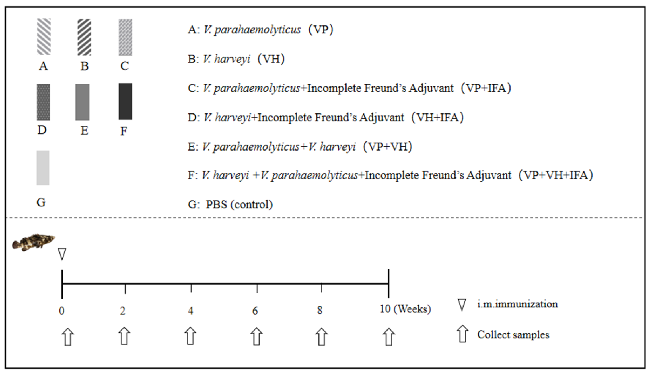
A total of 210 juvenile hybrid pearl gentian grouper (50–60 g, 7–10 cm) from the same batch were randomly assigned to seven vaccination groups (n = 30 per group): inactivated V. parahaemolyticus vaccine (VP), inactivated V. harveyi vaccine (VH), inactivated V. parahaemolyticus with incomplete Freund’s adjuvant (VP + IFA), inactivated V. harveyi with incomplete Freund’s adjuvant (VH + IFA), inactivated V. parahaemolyticus and V. harveyi combined vaccine (VP + VH), inactivated V. parahaemolyticus and V. harveyi combined vaccine with incomplete Freund’s adjuvant (VP + VH + IFA), and PBS as the control. All fish were maintained under identical rearing conditions. Vaccines were administered once via intraperitoneal injection at a dose of 2 × 10⁸ CFU per fish. No booster immunization was performed, as the aim was to evaluate the applicability of the constructed indirect ELISA for monitoring the kinetics of the primary humoral immune response. Serum samples were collected at 2, 4, 6, 8, and 10 weeks post-vaccination, with five fish sampled per time point (Figure 1).

2.6.3. Detection for Pathogen-Specific IgM

Microplates were coated with V. parahaemolyticus or V. harveyi (1 × 10⁸ CFU/mL, 100 µL/well) and incubated overnight at 4 °C. After washing with PBST and blocking with 3% BSA for 1 h at 37 °C, serially diluted sera from vaccinated groupers (1:40 to 1:10,240) were added and incubated for 1 h at 37 °C. Bound antigen-specific IgM was detected using mAb 41-H2-E1 as the primary antibody, followed by HRP-conjugated goat anti-mouse IgG (1:20,000 in PBST). Absorbance was measured at 450 nm. Antibody titers were defined as the highest serum dilution yielding a P/N ratio ≥ 2.1, where P represents the OD450 nm of the vaccinated serum and N represents the OD450 nm of the PBS control at the corresponding dilution. All samples were assayed in triplicate.

2.7. Statistical Analysis

All data were analyzed using GraphPad Prism version 6.0 and SPSS version 20.0. Statistical comparisons were performed using one-way ANOVA, and differences between two groups were assessed with the independent samples t-test. Results were presented as mean ± SD, and differences were considered statistically significant at p < 0.05.

3. Results

3.1. Purification of Serum IgM

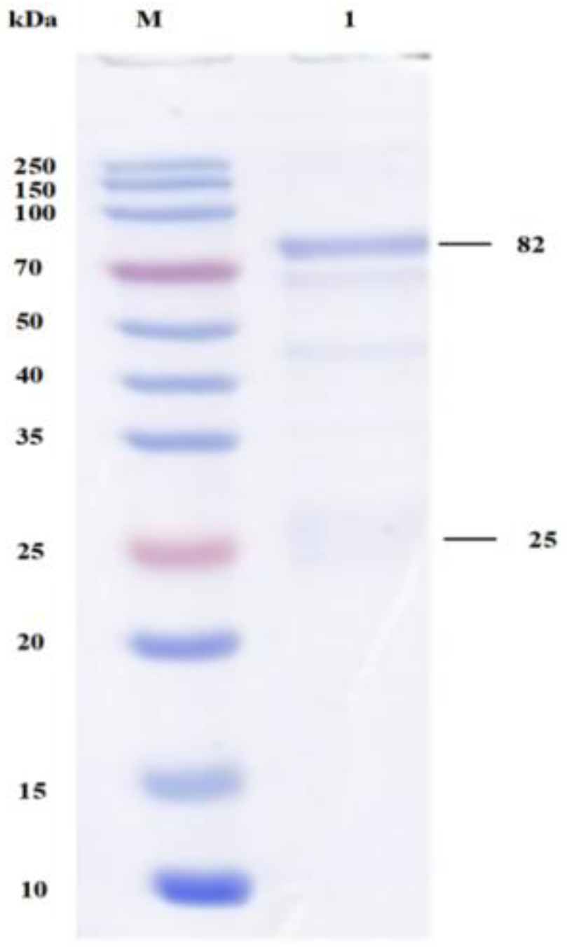
SDS-PAGE analysis revealed that purified IgM from hybrid pearl gentian grouper exhibited two major bands (Figure 2) with molecular weights of 82 kDa and 25 kDa, which corresponding to the heavy and light chains of IgM, respectively. The concentration of purified IgM was quantified to be 1.62 mg/mL using Bradford assay, which fully met the criteria for antigen immunization.

3.2. Production of MAbs

3.2.1. Production and Characterization of MAbs

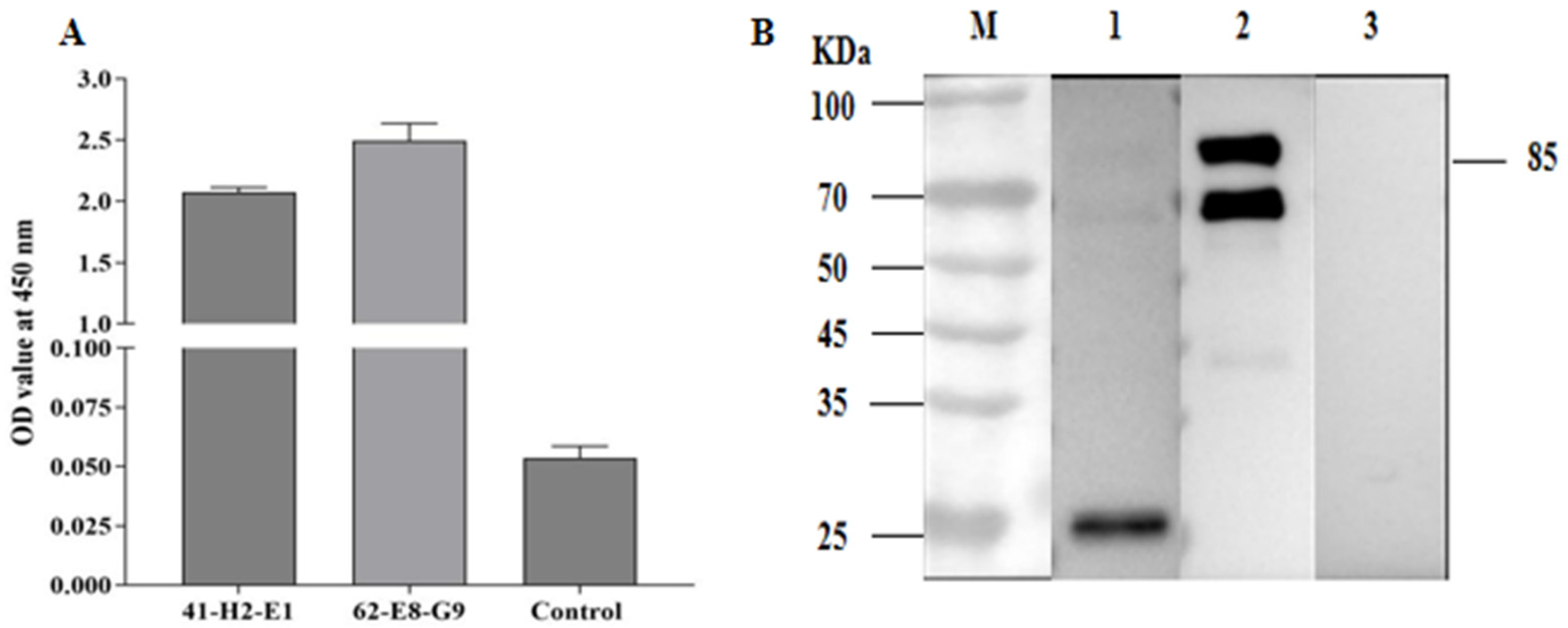
After cell fusion, eight hybridomas tested positive by indirect ELISA, whereas no reactivity was observed in SP2/0 myeloma supernatants. Among these, hybridomas 41-H2-E1 and 62-E8-G9 exhibited strong positive signals (Figure 3A). Western blot analysis confirmed the binding specificity of the mAbs (Figure 3B): mAb 62-E8-G9 recognized two bands at approximately 70 kDa and 85 kDa, corresponding to the IgM heavy chain, while mAb 41-H2-E1 specifically recognized a 24 kDa protein, corresponding to the IgM light chain. No nonspecific binding was observed in the negative controls. MAbs 41-H2-E1 and 62-E8-G9 were identified as IgG2b subtypes.

3.2.2. Indirect Immunofluorescence Results

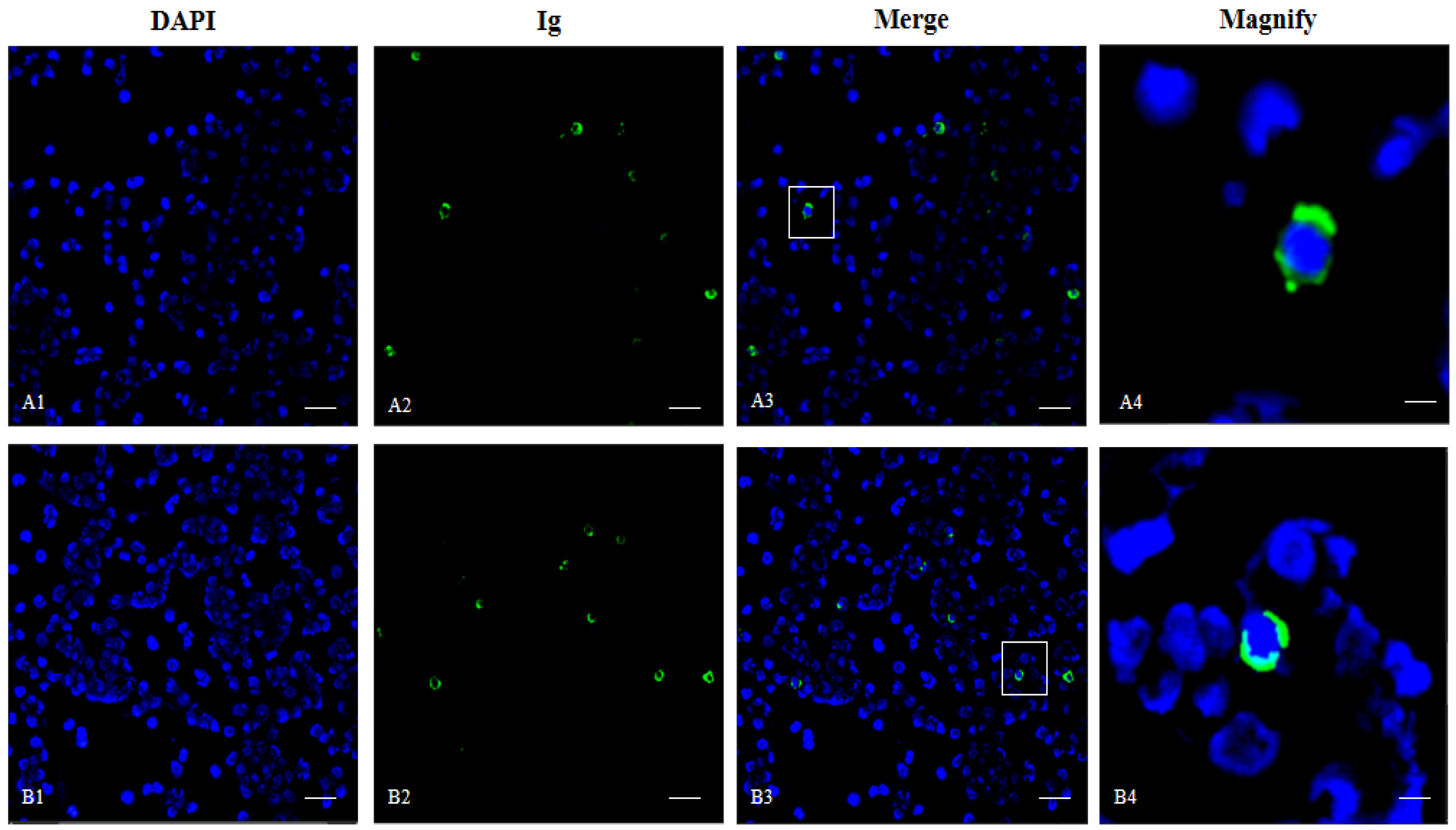
Indirect immunofluorescence detection performed on leukocyte smears from the head kidney revealed that both mAbs specifically bound to surface immunoglobulin-positive (sIg+) lymphocytes, appearing as discrete green punctate fluorescence signals on the cell membrane (Figure 4).

3.3. Development and Application of a Double-Antibody Sandwich ELISA

3.3.1. Establishment of a Standard Curve

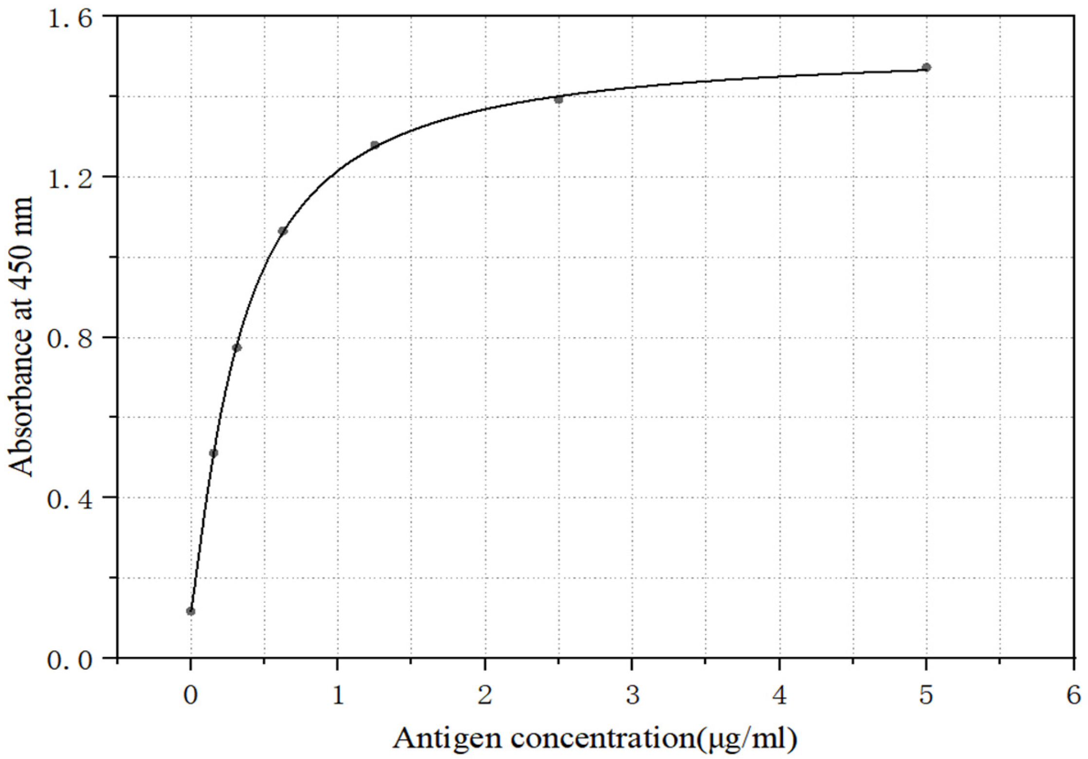
A double-antibody sandwich ELISA was established using the mAb 62-E8-G9 (2.5 µg/mL) as the capture antibody and HRP-labeled mAb 41-H2-E1 as the detection antibody. The assay demonstrated a working range of 0.15625–5 µg/mL, with a standard curve equation of
y = 0.118   +   1.508     0.118 1   +   ( x 0.338 ) 1.202
and a coefficient of determination (R2) of 0.999 (Figure 5).

3.3.2. The Cross-Reactivity Analysis of Double Antibody Sandwich ELISA

The double-antibody sandwich ELISA effectively detected IgM in the sera of groupers, including Epinephelus fuscoguttatus× E. lanceolatus ♂, Epinephelus akaara, Epinephelus moara, and Epinephelus fuscoguttatus× E. tukula (P/N > 2.1). In contrast, the assay was not suitable for non-grouper species such as Plectropomus leopardus, Paralichthys olivaceus, tongue sole, and Scophthalmus maximus (P/N < 2.1) (Table 1).

3.3.3. Performance of Double-Antibody Sandwich ELISA in IgM Quantification

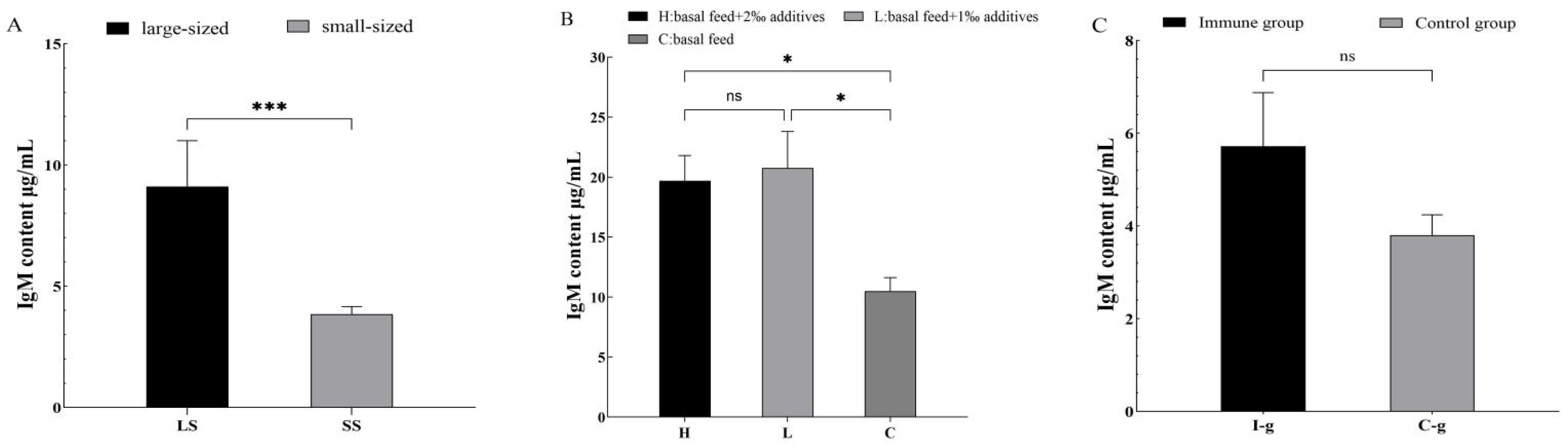
Serum IgM concentrations were significantly higher in larger fish (mean weight, 700 g; 9.11 µg/mL) than in smaller fish (50 g; 3.84 µg/mL). In the larger-fish group, IgM levels ranged from 7.02 to 11.91 µg/mL, whereas in the smaller-fish group, levels ranged from 3.29 to 4.11 µg/mL (Figure 6A). Fish fed with Ecotechangjia powder-supplemented diets showed elevated IgM levels compared to the control group. Both the high-dose group and low-dose additive groups exhibited significantly increased IgM concentrations (p < 0.05), though no significant difference was observed between the two dosage groups (Figure 6B). Following immunization with inactivated VP + IFA, the average serum IgM level in the immunized group was 5.72 µg/mL, compared to 3.8 µg/mL in the control group; this difference was not statistically significant (p ≥ 0.05) (Figure 6C).

3.4. Antigen-Specific IgM Responses Under Different Vaccination Strategies

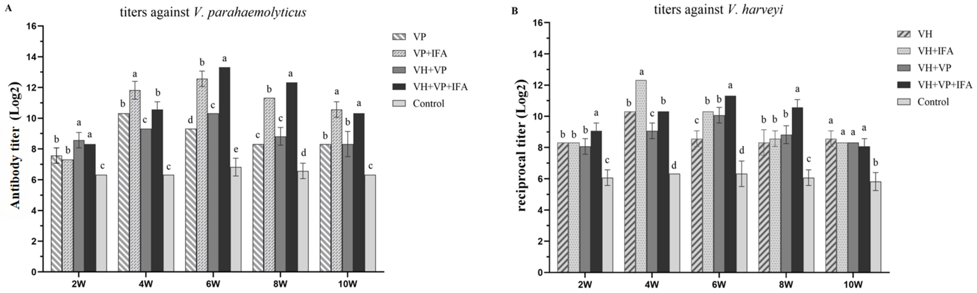
Indirect ELISA was used to monitor pathogen-specific IgM following vaccination with V. parahaemolyticus (VP) and V. harveyi (VH) antigens. Detection of VP-specific IgM antibodies indicated that the VP group reached its peak titer at the fourth week post-immunization. The VP + IFA, VH + VP, and VH + VP + IFA groups all reached their peaks at the sixth week post-immunization. The VH + VP + IFA group had the highest peak titer, followed by the VP + IFA group (Figure 7A). For VP-specific IgM, the VP group reached its peak antibody titer at 4 weeks post-immunization, whereas the VP + IFA, VH + VP, and VH + VP + IFA groups exhibited peak titers at 6 weeks post-immunization. Among these groups, the VH + VP + IFA group demonstrated the highest peak titer, followed by the VP + IFA group (Figure 7A). For VH-specific IgM, both the VH and VH + IFA groups achieved peak titers at 4 weeks post-immunization, while the VH + VP and VH + VP + IFA groups reached peak titers at 6 weeks post-immunization. The VH + IFA group showed the highest peak titer, followed by the VH + VP + IFA group (Figure 7B). Overall, antibody titers displayed a characteristic rise-and-decline kinetic pattern over time. At all sampling points, immunized groups exhibited significantly higher IgM titers than the control group (p < 0.05).

4. Discussion

MAbs targeting fish IgM were successfully developed in various teleost species [14,19]. In the present study, SDS-PAGE analysis showed that the purified IgM from hybrid pearl gentian grouper exhibited two bands with molecular weights of 82 kDa and 25 kDa. MAbs served as valuable tools for pathogen detection and immunochemical characterization in aquatic species [21]. In recent years, mAb-based assays significantly advanced disease diagnosis in aquaculture [22]. In this study, we developed two mAbs against hybrid pearl gentian grouper IgM. Western blot analysis confirmed its ability to bind to antigens, with mAb 62-E8-G9 recognizing two heavy-chain fragments (~70 kDa and 85 kDa); offspring may simultaneously express the heavy chains of both paternal and maternal IgM, similar to findings in Paralichthys olivaceus [23], while mAb 41-H2-E1 specifically detected the light chain at 24 kDa. Such specificity for both native and denatured immunoglobulin forms had been previously documented [23,24]. Indirect immunofluorescence assays further demonstrated that both mAbs specifically recognized surface IgM-positive B lymphocytes in head kidney tissue, supporting their applicability in cellular immunological analyses. Isotyping analysis identified clones 41-H2-E1 and 62-E8-G9 as belonging to the IgG2b subclass. These subclass assignments were consistent with those reported for mAbs, generated by Carassius auratus gibelio [20] and Oreochromis niloticus [25]. The IgG2b subtype was often preferred in immunoassays for its high specificity and broad reactivity [26], consistent with the performance characteristics of the mAbs described in the present study.
MAbs 62-E8-G9 and 41-H2-E1 exhibited strong and specific immunodetection activity. Based on these properties, a double-antibody sandwich ELISA was successfully established, employing mAb 62-E8-G9 as the capture antibody and HRP-conjugated mAb 41-H2-E1 as the detection antibody. This assay demonstrated high sensitivity, specificity, and stability for the detection of hybrid pearl gentian grouper IgM and was suitable for the quantitative determination of serum IgM levels under both physiological and pathological conditions. Notably, the ELISA was specific to grouper IgM and exhibited limited cross-reactivity with IgM from other teleost species, representing its primary limitation. The assay displayed a broad linear detection range (0.15625–5 μg/mL) and high reproducibility. The use of two mAbs targeting distinct epitopes improved both specificity and sensitivity, as previously demonstrated in teleost immunoglobulin assays [27,28]. IgMs from different fish species may have similar antigenic determinants and can lead to cross-reactions among antibodies against IgM of target fish and another teleost [29]. The established double-antibody sandwich ELISA was applicable to IgM detection in other species within the genus Epinephelus, but showed limited reactivity with non-Epinephelus fish. In the process of humoral immunity, when the fish is stimulated by external antigens, it will synthesize and secrete a large number of antibodies of IgM, and resist the invasion of foreign substances through IgM [30]. In the present study, larger individuals exhibited significantly higher serum IgM concentrations than smaller fish. A similar size- and age-associated increase in immunoglobulin levels was reported in Dicentrarchus labrax [31]. Dietary supplementation with Ecotechangjia powder significantly elevated IgM levels, consistent with studies showing immunostimulatory effects of prebiotics in fish [32]. Although vaccination with inactivated V. parahaemolyticus induced a moderate increase in total IgM, the difference was not statistically significant. Gadus morhua was immersed in Vibrio anguillarum for immunization; no significant elevation in total IgM-like protein levels was observed [33].
Antibody titers serve as reliable biomarkers for evaluating vaccine efficacy [34]. Notably, mAb 41-H2-E1 proved effective in an indirect ELISA for detecting pathogen-specific IgM following vaccination. The study showed that the bivalent vaccine increased acquired immunity and promoted protective immune responses against infection with two bacterial pathogens, thereby broadening immune coverage while reducing handling frequency, stress, and production costs in aquaculture systems [35,36]. Bivalent (VP + VH) and adjuvant-supplemented (VP + VH + IFA) vaccine formulations elicited stronger and more-sustained antigen-specific antibody responses than the corresponding monovalent vaccine, with peak titers observed at 6 weeks post-vaccination. The success of a polyvalent vaccine was often regulated by the concentration of individual antigens, cross-reactivity and competition among different antigens. These results were consistent with reports that polyvalent vaccines often induced broader and higher antibody titers than monovalent formulations [37], as observed in Oncorhynchus mykiss [38] and Salmo salar [39]. Furthermore, the incorporation of oil-based adjuvants prolonged the duration of protective immunity, underscoring their importance in the design of long-lasting vaccines for aquaculture applications [35].

5. Conclusions

In conclusion, monoclonal antibodies targeting the IgM of hybrid pearl gentian grouper were successfully generated and characterized. Among the screened clones, mAbs 62-E8-G9 and 41-H2-E1 exhibited high specificity and strong immunoreactivity. A sensitive double-antibody sandwich ELISA was established for the quantitative determination of total serum IgM, and an indirect ELISA was applied to assess pathogen-specific IgM responses. The developed assays demonstrated good stability and reproducibility, providing reliable immunological tools for evaluating vaccine efficacy and monitoring humoral immune responses in hybrid pearl gentian grouper. Nevertheless, the relatively limited sample size may constrain broader generalization of the findings. Future studies should expand sample coverage and further explore the application of these antibodies in field-scale disease surveillance and vaccine optimization strategies.

Author Contributions

Conceptualization, Y.W. and Q.L.; Data curation, X.Q., J.W., J.Q. and X.X.; Formal analysis, X.Q., J.W., J.Q. and X.X.; Investigation, X.Q., J.W., J.Q., Y.L., J.Z. and X.X.; Methodology, Q.L. and Y.W.; Writing—original draft preparation, X.Q.; Writing—review and editing, X.Q., J.W., J.Q., Y.L., J.Z.; X.X.; Y.W. and Q.L.; Funding acquisition, Y.W. and Q.L.; Supervision, Y.W. and Q.L. All authors have read and agreed to the published version of the manuscript.

Funding

This work was supported by the National Natural Science Foundation of China (32473132), the Fund of Key Laboratory of Efficient Utilization and Processing of Marine Fishery Resources of Hainan Province (KLEU-2023-2), and the Fund of National Key Research and Development Program of China (2024YFD2401802).

Institutional Review Board Statement

The study was conducted in accordance with the animal ethics protocols of the Animal Ethical and Welfare Committee (AEWC), Tianjin Agriculture and Forestry University (Approval code: 2025LLSC67; Approval date: 1 June 2025).

Data Availability Statement

The original contributions presented in this study are included in the article. Further inquiries can be directed to the corresponding author.

Acknowledgments

During the preparation of this manuscript, the authors used ChatGPT-5.2 for language optimization. The authors have reviewed and revised all relevant output and take full responsibility for the content of this publication.

Conflicts of Interest

The authors declare that they have no conflicts of interest with the contents of this article.

Abbreviations

The following abbreviations are used in this manuscript:
mAbsmonoclonal antibodies
sIg+secretory Immunoglobulin positive
V. harveyiVibrio harveyi
V. parahaemolyticusVibrio parahaemolyticus
IgMimmunoglobulin M
IgDimmunoglobulin D
IgZimmunoglobulin Z
IgTimmunoglobulin T
SDS-PAGESodium Dodecyl Sulfate–Polyacrylamide
PBSPhosphate-Buffered Saline
PBSTPhosphate-Buffered Saline with Tween-20
ECLChemiluminescence Reagent
HRPHorseradish Peroxidase
FITCFluorescein Isothiocyanate
ELISAEnzyme-Linked Immunosorbent Assay
VPVibrio parahaemolyticus
VHVibrio harveyi
VP + IFAVibrio parahaemolyticus + Incomplete Freund’s Adjuvant
VH + IFAVibrio harveyi + Incomplete Freund’s Adjuvant
VP + VHVibrio parahaemolyticus + Vibrio harveyi
VP + VH + IFAVibrio parahaemolyticus + Vibrio harveyi + Incomplete Freund’s Adjuvant

References

  1. Pang, A.; Wang, T.; Xie, R.; Wang, Z.; Xin, Y.; Tan, B.; Zhang, W. Effects of soybean meal on immunity and transcriptomics of liver in pearl gentian grouper (Epinephelus fuscoguttatus ♀ × Epinephelus lanceolatus ♂). Aquac. Rep. 2004, 35, 101969. [Google Scholar] [CrossRef]
  2. Yuan, S.; Jin, X.; Hu, Y.; Zhou, X.; Zhao, Z. Transcriptomic responses to RGNNV and Vibrio alginolyticus infection in the spleen of hybrid grouper (Epinephelus lanceolatus ♂ × Epinephelus fuscoguttatus ♀). Comp. Immunol. Rep. 2004, 6, 200125. [Google Scholar] [CrossRef]
  3. Liu, Y.; Lu, S.; Guo, M.; Wang, Z.; Hu, B.; Zhou, B. The dynamic immune response of the liver and spleen in leopard coral grouper (Plectropomus leopardus) to Vibrio harveyi infection based on transcriptome analysis. Front. Immunol. 2004, 15, 1457745. [Google Scholar] [CrossRef] [PubMed]
  4. He, L.; Liang, Y.; Yu, X.; Peng, W.; He, J.; Fu, L.; Lin, H.; Zhang, Y.; Lu, D. Vibrio parahaemolyticus flagellin induces cytokines expression via toll-like receptor 5 pathway in orange-spotted grouper, Epinephelus coioides. Fish Shellfish Immunol. 2019, 87, 573–581. [Google Scholar] [CrossRef] [PubMed]
  5. Xu, L.-W.; Liu, X.-H.; Zhang, J.-Y.; Liu, G.-F.; Feng, J. Outbreak of enteric microsporidiosis of hatchery-bred juvenile groupers, Epinephelus spp., associated with a new intranuclear microporidian in China. J. Fish Dis. 2017, 40, 183–189. [Google Scholar] [CrossRef]
  6. Huang, G.; Liu, H.; Han, Y.; Fan, L.; Zhang, Q.; Liu, J.; Yu, X.; Zhang, L.; Chen, S.; Dong, M.; et al. Profile of acute immune response in Chinese amphioxus upon Staphylococcus aureus and Vibrio parahaemolyticus infection. Dev. Comp. Immunol. 2007, 31, 1013–1023. [Google Scholar] [CrossRef]
  7. Nguyen, H.T.; Nguyen, T.T.; Tsai, M.-A.; Ya-Zhen, E.; Wang, P.-C.; Chen, S.-C. A formalin-inactivated vaccine provides good protection against Vibrio harveyi infection in orange-spotted grouper (Epinephelus coioides). Fish Shellfish Immunol. 2017, 65, 118–126. [Google Scholar] [CrossRef]
  8. Yuan, B.; Zhuang, Z.; Wang, X.; Huang, H.; Yan, Q. Isolation and characterization of a highly pathogenic strain of Vibrio harveyi EFL-2201 from pearl gentian grouper. Aquac. Rep. 2025, 41, 102668. [Google Scholar] [CrossRef]
  9. Schroeder, H.W.; Cavacini, L. Structure and function of immunoglobulins. J. Allergy Clin. Immunol. 2010, 125, S41–S52. [Google Scholar] [CrossRef]
  10. Fillatreau, S.; Six, A.; Magadan, S.; Castro, R.; Sunyer, J.O.; Boudinot, P. The astonishing diversity of Ig classes and B cell repertoires in teleost fish. Front. Immunol. 2013, 4, 28. [Google Scholar] [CrossRef]
  11. Hikima, J.; Jung, T.-S.; Aoki, T. Immunoglobulin genes and their transcriptional control in teleosts. Dev. Comp. Immunol. 2011, 35, 924–936. [Google Scholar] [CrossRef] [PubMed]
  12. Sánchez, C.; López-Fierro, P.; Zapata, A.; Dominguez, J. Characterisation of monoclonal antibodies against heavy and light chains of trout immunoglobulin. Fish Shellfish Immunol. 1993, 3, 237–251. [Google Scholar] [CrossRef]
  13. Dooley, H.; Flajnik, M.F. Antibody repertoire development in cartilaginous fish. Dev. Comp. Immunol. 2006, 30, 43–56. [Google Scholar] [CrossRef] [PubMed]
  14. Huang, Y.; Yuan, X.; Mu, P.; Li, Q.; Ao, J.; Chen, X. Development of monoclonal antibody against IgM of large yellow croaker (Larimichthys crocea) and characterization of IgM(+) B cells. Fish Shellfish Immunol. 2019, 91, 216–222. [Google Scholar] [CrossRef] [PubMed]
  15. Yang, S.; Tang, X.; Sheng, X.; Xing, J.; Zhan, W. Development of monoclonal antibodies against IgM of half-smooth tongue sole (Cynoglossus semilaevis) and analysis of phagocytosis of fluorescence microspheres by mIgM+ lymphocytes. Fish Shellfish Immunol. 2017, 66, 280–288. [Google Scholar] [CrossRef]
  16. He, Y.; Jiang, Y.; Lu, L. Serodiagnosis of grass carp reovirus infection in grass carp Ctenopharyngodon idella by a novel Western blot technique. J. Virol. Methods 2013, 194, 14–20. [Google Scholar] [CrossRef]
  17. Al-Harbi, A.H.; Truax, R.; Thune, R.L. Production and characterization of monoclonal antibodies against tilapia Oreochromis niloticus immunoglobulin. Aquaculture 2000, 188, 219–227. [Google Scholar] [CrossRef]
  18. Sheng, X.Z.; Guo, Y.; Zhu, H.; Chai, B.; Tang, X.; Xing, J.; Chi, H.; Zhan, W. Transepithelial Secretion of Mucosal Immunoglobulin Receptor of Flounder (Paralichthys olivaceus): In-Vivo and In-Vitro Evidence. Front. Immunol. 2022, 13, 868753. [Google Scholar] [CrossRef]
  19. Li, Y.; Zheng, S.; Wang, Q.; Bergmann, S.M.; Zeng, W.; Wang, Y.; Liu, C.; Shi, C. Detection of koi herpesvirus (KHV) using a monoclonal antibody against Cyprinus carpio IgM. Arch. Virol. 2017, 162, 2381–2385. [Google Scholar] [CrossRef]
  20. Guo, B.; Wei, C.; Wang, Y.; Li, Q. Preparation and application of monoclonal antibody against IgM in gibel carp (Carassius gibelio). J. Dalian Fish. Univ. 2022, 37, 450–456, (In Chinese with English abstract). [Google Scholar]
  21. Zhang, Y.; Wu, S.; Wang, J.; Wernike, K.; Lv, J.; Feng, C.; Zhang, J.; Wang, C.; Deng, J.; Yuan, X.; et al. Expression and purification of the nucleocapsid protein of Schmallenberg virus, and preparation and characterization of a monoclonal antibody against this protein. Protein Expr. Purif. 2013, 92, 1–8. [Google Scholar] [CrossRef]
  22. Aamelfot, M.; Dale, O.B.; Falk, K. Infectious salmon anaemia-pathogenesis and tropism. J. Fish Dis. 2014, 37, 291–307. [Google Scholar] [CrossRef]
  23. Jang, H.N.; Woo, J.-K.; Cho, Y.-H.; Kyong, S.-B.; Choi, S.-H. Characterization of monoclonal antibodies against heavy and light chains of flounder (Paralichthys olivaceus) immunoglobulin. J. Biochem. Mol. Biol. 2004, 37, 314–319. [Google Scholar] [CrossRef]
  24. Wu, R.; Shen, J.; Lai, X.; He, T.; Li, Y. Development of monoclonal antibodies against serum immunoglobulins from gibel carp (Carassius auratus gibelio) and their applications in serodiagnosis of inapparent infection and evaluation of vaccination strategies. Fish Shellfish Immunol. 2020, 96, 69–77. [Google Scholar] [CrossRef] [PubMed]
  25. Velázquez, J.; Rodríguez, A.; Aragón, H.; Haidar, A.; González, M.; Valdés, R.; Garay, H.E.; Abreu, D.D.; Ramos, Y.; Cabrales, A.; et al. Monoclonal antibody against Nile tilapia (Oreochromis niloticus) IgM heavy chain: A valuable tool for detection and quantification of IgM and IgM+ cells. Fish Shellfish Immunol. 2020, 11, 44–54. [Google Scholar] [CrossRef] [PubMed]
  26. Lin, L.; Zhao, C.; Zhang, W.; Zhang, Y.; Yong, L.; Tang, J.; LYU, D.; Wei, P.; MO, M. Preparation and identification of broad-spectrum monoclonal antibodies against N protein of avian infectious bronchitis virus. J. South. Agric. 2022, 53, 2674–2682, (In Chinese with English abstract). [Google Scholar]
  27. Ding, X.-X.; Li, X.-F.; Deng, Y.Q.; Guo, Y.H.; Hao, W.; Che, X.-Y.; Qin, C.-F.; Fu, N. Development of a double antibody sandwich ELISA for West Nile virus detection using monoclonal antibodies against non-structural protein 1. PLoS ONE 2014, 9, e108623. [Google Scholar] [CrossRef]
  28. Krebs, T.; Kilic, I.; Mütze, K.; Kleinhans, S.; Lücking, D.; Hennies, M.; Tetens, J. Establishment of a Sandwich-ELISA for simultaneous quantification of bovine pregnancy-associated glycoprotein in serum and milk. PLoS ONE 2021, 16, e0251414. [Google Scholar] [CrossRef]
  29. Gong, H.; Cao, J.; Liu, C.; Zhu, C.; Bergmann, S.M.; Zheng, Y.; Wang, Q. Development and application of monoclonal antibody against IgM of hybrid snakehead (Channa maculate ♀ × Channa argus ♂). Aquac. Res. 2021, 52, 4371–4379. [Google Scholar] [CrossRef]
  30. Gao, Y.-L.; Tang, X.-Q.; Sheng, X.-Z.; Xing, J.; Zhan, W.-B. Immune responses of flounder Paralichthys olivaceus vaccinated by immersion of formalin-inactivated Edwardsiella tarda following hyperosmotic treatment. Dis. Aquat. Org. 2015, 116, 111–120. [Google Scholar] [CrossRef]
  31. Scapigliati, G.; Scalia, D.; Marras, A.; Meloni, S.; Mazzini, M. Immunoglobulin levels in the teleost sea bass Dicentrarchus labrax (L.) in relation to age, season, and water oxygenation. Aquaculture 1999, 174, 207–212. [Google Scholar] [CrossRef]
  32. Reyes-Becerril, M.; Ascencio, F.; Gracia-Lopez, V.; Macias, M.E.; Roa, M.C.; Esteban, M.Á. Single or combined effects of Lactobacillus sakei and inulin on growth, non-specific immunity and IgM expression in leopard grouper (Mycteroperca rosacea). Fish Physiol. Biochem. 2014, 40, 1169–1180. [Google Scholar] [CrossRef] [PubMed]
  33. Jonsson, A.; López-Porras, A.; Nørstebø, S.F.; Guslund, N.C.; Sørum, H.; Qiao, S.W.; Johansen, F.E. Protective IgM-mediated immunity against Vibrio anguillarum in Atlantic cod with evolutionary losses of mhc class II and cd4. Front. Immunol. 2025, 16, 1579541. [Google Scholar] [CrossRef] [PubMed]
  34. Xu, G.; Sheng, X.; Xing, J.; Zhan, W. Effect of temperature on immune response of Japanese flounder (Paralichthys olivaceus) to inactivated lymphocystis disease virus (LCDV). Fish Shellfish Immunol. 2011, 30, 525–531. [Google Scholar] [CrossRef]
  35. Tafalla, C.; Bøgwald, J.; Dalmo, R.A. Adjuvants and immunostimulants in fish vaccines: Current knowledge and future perspectives. Fish Shellfish Immunol. 2013, 35, 1740–1750. [Google Scholar] [CrossRef]
  36. Liu, S.; Guo, H.; Yi, J.; Yang, Z.; Zhou, S.; Xiu, Y. Efficacy of bivalent vaccine against Aeromonas salmonicida and Edwardsiella tarda infections in turbot. Fish Shellfish Immunol. 2023, 139, 108837. [Google Scholar]
  37. Abotaleb, M.M.; Soliman, H.M.; Tawfik, R.G.; Mourad, A.; Khalil, R.H.; Abdel-Latif, H.M.R. Efficacy of combined inactivated vaccines against Vibrio alginolyticus and Streptococcus agalactiae infections in Nile tilapia in Egypt. Aquac. Int. 2024, 32, 1317–1334. [Google Scholar] [CrossRef]
  38. Nikoskelainen, S.; Verho, S.; Järvinen, S.; Madetoja, J.; Wiklund, T.; Lilius, E. Multiple whole bacterial antigens in polyvalent vaccine may result in inhibition of specific responses in rainbow trout (Oncorhynchus mykiss). Fish Shellfish Immunol. 2007, 22, 206–217. [Google Scholar] [CrossRef]
  39. Hoel, K.; Salonius, K.; Lillehaug, A. Vibrio antigens of polyvalent vaccines enhance the humoral immune response to Aeromonas salmonicida antigens in Atlantic salmon (Salmo salar L.). Fish Shellfish Immunol. 1997, 7, 71–80. [Google Scholar] [CrossRef]
Figure 1. Schematic of the experimental vaccination and sampling schedule.
Fishes 11 00160 g001
Figure 2. SDS-PAGE analysis of serum IgM of hybrid pearl gentian grouper. Lane M: Molecular; lane 1: purified IgM of hybrid pearl gentian grouper by Protein A.
Fishes 11 00160 g002
Figure 3. ELISA and Western blotting analysis of the MAbs against the IgM of hybrid pearl gentian grouper. (A) ELISA of MAbs 41-H2-E1 and 62-E8-G9 reacting with IgM of hybrid pearl gentian grouper (mean ± S.D., n = 3); (B) Western blotting analysis of mAb specificity. Lane: Protein marker; Lane 1: mAb 41-H2-E1; Lane 2: mAb 62-E8-G9; Lane 3: negative control.
Fishes 11 00160 g003
Figure 4. Immunofluorescence staining of IgM+ leukocytes from hybrid pearl gentian grouper head kidney. MAb labeling was visualized by FITC-labeled goat-anti-mouse IgG (green). Cells were stained with DAPI (blue). (A1A4) mAb 41-H2-E1, (B1B4) mAb 62-E8-G9. Scale bar = 20 μm.
Fishes 11 00160 g004
Figure 5. Standard curve for the quantification of hybrid pearl gentian grouper IgM by double-antibody sandwich ELISA. IgM was detected at concentrations of 0.15625, 0.3125, 0.625, 1.25, 2.5, and 5 µg/mL.
Fishes 11 00160 g005
Figure 6. (A) Comparison of serum IgM levels between large and small hybrid pearl gentian grouper. Serum IgM concentrations were quantified by a double-antibody sandwich ELISA. LS: Large-sized fish group; SS: small-sized fish group. Data were presented as mean ± SD (*** p < 0.001); (B) Effect of dietary additive supplementation on serum IgM levels in hybrid pearl gentian grouper. Serum IgM concentrations were measured by double-antibody sandwich ELISA after a 90-day feeding trial with diets containing a high or low dose of additive (Ecotechangjia powder), compared to a control diet. Data were presented as mean ± SD (* p < 0.05, ns, p ≥ 0.05); (C) serum IgM levels in hybrid pearl gentian grouper following immunization with an inactivated vaccine. Serum IgM concentrations were quantified by double-antibody sandwich ELISA one month after intraperitoneal injection with inactivated VP + IFA or PBS (control). Data are presented as mean ± SD. No significant difference (ns; p ≥ 0.05) was observed between the immunized and control groups.
Fishes 11 00160 g006
Figure 7. Dynamics of pathogen-specific IgM titers in hybrid pearl gentian grouper following vaccination. (A) Pathogen-specific IgM titers against V. parahaemolyticus were determined by indirect ELISA in fish immunized with different vaccine formulations. (B) Pathogen-specific IgM titers against V. harveyi were determined by indirect ELISA in fish immunized with different vaccine formulations. At each corresponding time point, groups labeled with different letters differed significantly from the control group (p < 0.05).
Fishes 11 00160 g007
Table 1. Cross-reactivity analysis results.
FishOD (450)Results
Epinephelus fuscoguttatus× E. lanceolatus1.296+
Epinephelus akaara1.349+
Epinephelus moara1.056+
Epinephelus fuscoguttatus× E. tukula1.889+
Plectropomus leopardus0.086-
Tongue Sole0.168-
Paralichthys olivaceus0.074-
Scophthalmus maximus0.065-
PBST0.128
“+” Positive result; “-” Positive result.
Disclaimer/Publisher’s Note: The statements, opinions and data contained in all publications are solely those of the individual author(s) and contributor(s) and not of MDPI and/or the editor(s). MDPI and/or the editor(s) disclaim responsibility for any injury to people or property resulting from any ideas, methods, instructions or products referred to in the content.