Article

First Morphological and Molecular Identification of Intestinal Helminths in Wild Turbot Scophthalmus maximus (Linnaeus, 1758) Along the Bulgarian Black Sea Coast

by
Alexander Atanasoff
1,
Cigdem Urku
2,*,
Elitsa Petrova-Pavlova
3 and
Feriha Tserkova
3
1
Faculty of Veterinary Medicine, Trakia University, 6014 Stara Zagora, Bulgaria
2
Faculty of Aquatic Sciences, Istanbul University, 34134 Istanbul, Türkiye
3
Institute of Fish Resources, Agriculture Academy, 9000 Varna, Bulgaria
*
Author to whom correspondence should be addressed.
Fishes 2025, 10(8), 395; https://doi.org/10.3390/fishes10080395
Submission received: 23 June 2025 / Revised: 2 August 2025 / Accepted: 4 August 2025 / Published: 7 August 2025
(This article belongs to the Special Issue Advances in Fish Pathology and Parasitology)

Abstract

Turbot Scophthalmus maximus (Linnaeus, 1758) is one of the most valuable and economically important species for the Black Sea countries. In Bulgaria, their numbers are limited and stocks are depleted; therefore, monitoring development and health status is extremely important. Internal helminths are widespread among turbots on the Bulgarian Black Sea coast. However, description of this infection is relatively limited, and they have not been reported in scientific papers. For this purpose, a total of 36 hauls were made at depths from 15 to 90 m, and 65 turbots were examined for intestinal parasites. The present study represents the first report of internal helminths in turbot from Bulgarian marine waters through the spawning season, characterized morphologically based on a microscope observation and molecular identification. Evaluation of laboratory analyses revealed that two different parasites were determined: Bothriocephalus sp. (Müller, 1776) and Hysterothylacium aduncum (Rudolphi, 1802) and that 73.85% of the turbot were infected with one or more parasites. Based on the results, control measures and treatment for the wild population are unrealistic but should be considered for the containment and spread of diseases in aquaculture facilities.
Key Contribution: This study presents detailed findings on the molecular identification and characterization of parasitic species detected in turbot (Scophthalmus maximus), which is considered the most economically significant and commercially valuable fish species in Bulgaria.

1. Introduction

The scientific literature refers to two generic names for turbot: Scophthalmus maximus and Psetta maxima (Linnaeus, 1758). Nevertheless, the scientific community has strongly recommended the utilisation Scophthalmus as the valid generic name [1]. The Black Sea turbot (Scophthalmus maximus) is a demersal species that inhabits the continental shelf on sedimentary sandy-silty mixed bottoms up to 60–70 m and plays an important role in the fisheries [2]. The turbot is considered to be one of the most valuable and economically significant species in the Black Sea countries [3]. The current status of the wild turbot population in the Black Sea has been characterised as “overexploited” and at risk of significant depletion [4]. The biological monitoring of turbot catches along the Bulgarian coast of the Black Sea has revealed that the population is limited (Endangered EN [A1b, d; B1b (v), c (iv)]) and that the stocks are depleted, estimated to be approximately 2000 tons [5]. From an ecological perspective, the turbot is considered a pivotal species for the assessment of ecological health and the impact of human activities in the context of monitoring and analysing the processes that occur within the marine environment of the Black Sea. This is attributable to the fact that, during its lifecycle, the turbot inhabits a wide range of habitats [2]. Despite the economic and ecological importance of this species in aquatic ecosystems, there is a dearth of research on the health status of turbot, particularly with regard to parasitic infections.
In recent years, there has been an increasing interest in the study of marine parasite fauna. Gastrointestinal helminthic infections are prevalent in wild fish, and their omnivorous nature and feeding habits are likely the main reason for this. Consequently, the monitoring of health status is of paramount importance. Notably, parasites are now being used as biological indicators to trace host population dynamics, including fish migration patterns and stock structure assessments. The hypothesis that parasitism may have played a role in the decline of wild fish populations has been postulated, with the potential to impact ecological stability and fisheries productivity [6,7,8]. The potential threat posed to humans by turbot parasites and their significance within European fishing industries render them a subject of particular interest [9].
In the field of ichthyology, the identification of protozoan and metazoan parasites in fish is conventionally undertaken through the use of stained tissue sections and light microscopy. This process is complemented by the utilisation of classical keys, which are based on the morphology of the entire organism. The molecular PCR method has been recommended as a diagnostic technique for identifying parasite species in turbot. It is considered to be both sensitive and rapid, and to complement microscopy-based identification methods [10].
The prevalence of internal helminths among turbot fish along the Bulgarian Black Sea coast has been well documented in the mass media, including on the official Bulgarian national television channel. However, the extant literature on this infection is scant, with no reports in the scientific papers [11].
The present study was conducted with the objective of investigating the infection levels of internal helminths in turbot (Scophthalmus maximus) from the Bulgarian Black Sea coast throughout the spawning season. The study provides the first detailed morphologically characterised observations, based on microscope analysis and molecular identification.

2. Materials and Methods

2.1. Data Collection

The scientific demersal trawl survey in the Bulgarian Black Sea, conducted between 9 and 19 May 2021, was undertaken as part of the National Fisheries Data Collection Programme, in accordance with the terms of the contract with the National Agency for Fisheries and Aquaculture (NAFA) in Burgas. This survey was conducted in compliance with Council Regulation No. 199/2008 and Commission Decision 2010/93/EU. The areas in which trawling was performed are illustrated in Figure 1.
The survey was conducted using the 19.5 m long fishing vessel EGEO2, which has an engine power of 367.75 kW, and equipment with 32/27-34 bottom trawl with a vertical opening of 2 m, an effective head rope of 13 m, and an effective footrope of 15 m, respectively, with a mesh size of 200 mm. On board the vessel, the length and individual body weight of each specimen were measured and recorded. The fish were euthanised by concussion in accordance with Council Regulation (EU) 1099/2009. They were then transferred within a separate bag (to prevent leakage) and surrounded by ice packs in the shipment box to maintain their freshness.

2.2. Data Analyses

The fish were then transported to the Ichthyology Laboratory at the Institute of Fish Resources in Varna for autopsy.
The age of the turbot was determined through the examination of otoliths under a binocular microscope.
The abdominal cavity of the turbot was opened, and the main digestive organs, such as the stomach and gut, were meticulously dissected after ligaturing the contents to prevent leakage. The mesenterium, peritoneum, and associated organs (e.g., pancreas, spleen) were also examined during the dissection. The contents of the digestive tract of the fish were examined. The stomach content analysis included identification of the composition and total number of food components and the weight and frequency of occurrence of each food component. The index of relative importance (IRI) was used to determine the significance of each food component in the trophic spectrum [12]:
IRI = ( C N + C W )   ×   F , IRI = ( C N + C W )   ×   F
CN—percentage share of the food item i in total number; CW—percentage share of the food item i in total weight; F—frequency of occurrence.
IRI, expressed as a percentage, was calculated by the following equation [13].
% I R I i = 100 × I R I i i n I R I i
n—total number of the taxonomic categories at a given taxonomic level
Spearman’s rank correlation coefficient (rs) was used to determine the strength of the relationship between host body length, body weight, age, depth, and the prevalence of specific parasites.

2.3. Parasitical Examination

The gut contents were washed into cups with lower amounts of water. The gut was then cut lengthways and examined under an Accu-Scope 3078 binocular stereo microscope (Accu-Scope Inc., New York, NY, USA). Once the parasites had been located, the final specimens were removed from the gut and transferred to tubes containing 70% ethanol for further identification [14]. Permanent slides were prepared without staining, following the methods outlined by Moravec [14] and Yoshinaga et al. [15] for nematodes and by Yamaguti [16] for cestodes. These methods are based on morphological criteria.

2.4. Real-Time PCR Analysis

2.4.1. DNA Isolation

The procedure for the direct extraction and purification of deoxyribonucleic acid (DNA) from the fresh samples was carried out in accordance with the instructions provided by the manufacturer (QIAmp DNA Mini Kit, Qiagen, Hilden, Germany).

2.4.2. Primer Optimization and Synthesis

Primer synthesis was performed for Bothriocephalus sp. using the 18S rRNA gene region with the accession number AJ228776.1. Primer synthesis was also performed for Hysterothylacium aduncum of the Anisakidae family using the 18S rRNA gene region with the accession number MF072693.1. The designed primer sequences are presented in Table 1.

2.4.3. PCR Analysis

PCR amplification was performed using 100 ng of template DNA, along with 125 pmol of each primer. The reaction mixture included 2 µL of a 0.8 µM dNTP mix, 10 µL of 10 × PCR buffer supplemented with 20 mM MgCl2, and 0.8 µL of Taq DNA polymerase (5 U/µL; Bioasia, Shenzhen, China). Additionally, 0.1 µL of SYBR Green dye was added per sample to enable real-time detection.
The thermal cycling conditions were as follows: an initial denaturation at 94 °C for 5 min, followed by 30 amplification cycles consisting of denaturation at 94 °C for 30 s, annealing at 58 °C for 30 s, and extension at 72 °C for 1 min. A final extension step was carried out at 72 °C for 5 min. All reactions were conducted using a Rotor-Gene Q real-time PCR system (Qiagen, Hilden, Germany).

2.5. Histopathological Examination

The histological method was used to detect tissue damage caused by the identified parasites. To this end, tissue samples were taken from the internal organs (such as the intestine, stomach, liver, kidneys and spleen) and fixed in 10% buffered formalin. All samples were dehydrated using a graded series of ethanol, cleared in xylene and embedded in paraffin wax according to standard histological procedures. The paraffin blocks were then cut into serial longitudinal sections (4–5 µm) using a Leica RM2125 microtome (Leica Biosystems, Richmond, IL, USA), and stained with Mayer’s haematoxylin (Sigma-Aldrich—HHS16, Saint Louis, MA, USA) and eosin (Merck—109844, Merck KGaA, Darmstadt, Germany) (H&E). The slides were then examined under an Olympus BX-51 (Olympus Europe GmbH, Hamburg, Germany) light microscope equipped with an Olympus DP72 (Olympus Europe GmbH, Hamburg, Germany) digital camera [17].

3. Results

A total of 65 turbots, comprising 34 females (mean length: 52.53 cm; mean body weight: 3056 g) and 31 males (mean length: 49.1 cm; mean body weight: 2279 g), were collected from 36 hauls (duration: 60 min; speed: 2.2–2.6 knots) at depths ranging from 15 to 90 m.
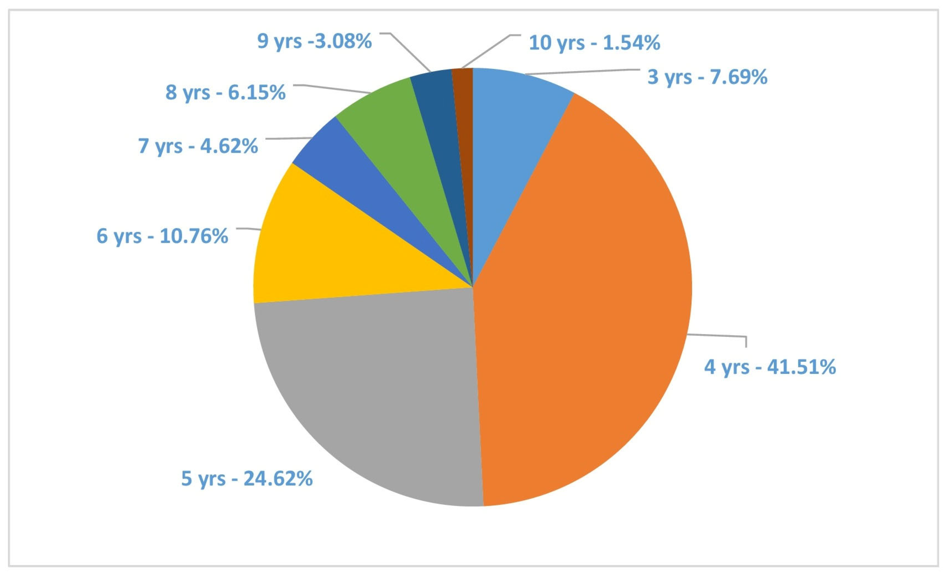
The age composition of the turbot population was ascertained through a meticulous analysis of 65 pairs of otoliths. The age structure of the population under scrutiny comprised classes ranging from 3 to 11 years of age. The 4- (41.54%) and 5 (24.62%)-year classes exhibited a combined prevalence of 66.16%, with the 6-year class accounting for 10.76% of the total (Figure 2).
The average stomach fullness index was 0.13% BW ± 0.04 SE. The analysis of spatial distribution of the stomach fullness index indicated high values in the southern zones along the Bulgarian Black Sea coast. In the current study, the turbot food spectrum was mainly formed by Merlangius merlangus (Linnaeus, 1758)—IRI = 6250.95 (79.12% IRI), Gobiidae c IRI = 664.36 (8.41%) and Engraulis encrasicolus (Linnaeus, 1758)—IRI = 409.70 (5.19%).
Evaluation of findings from intestinal parasite examinations revealed that 73.85% of the turbot were infected with one or more parasites. Two parasites were identified: one nematode and one cestode.
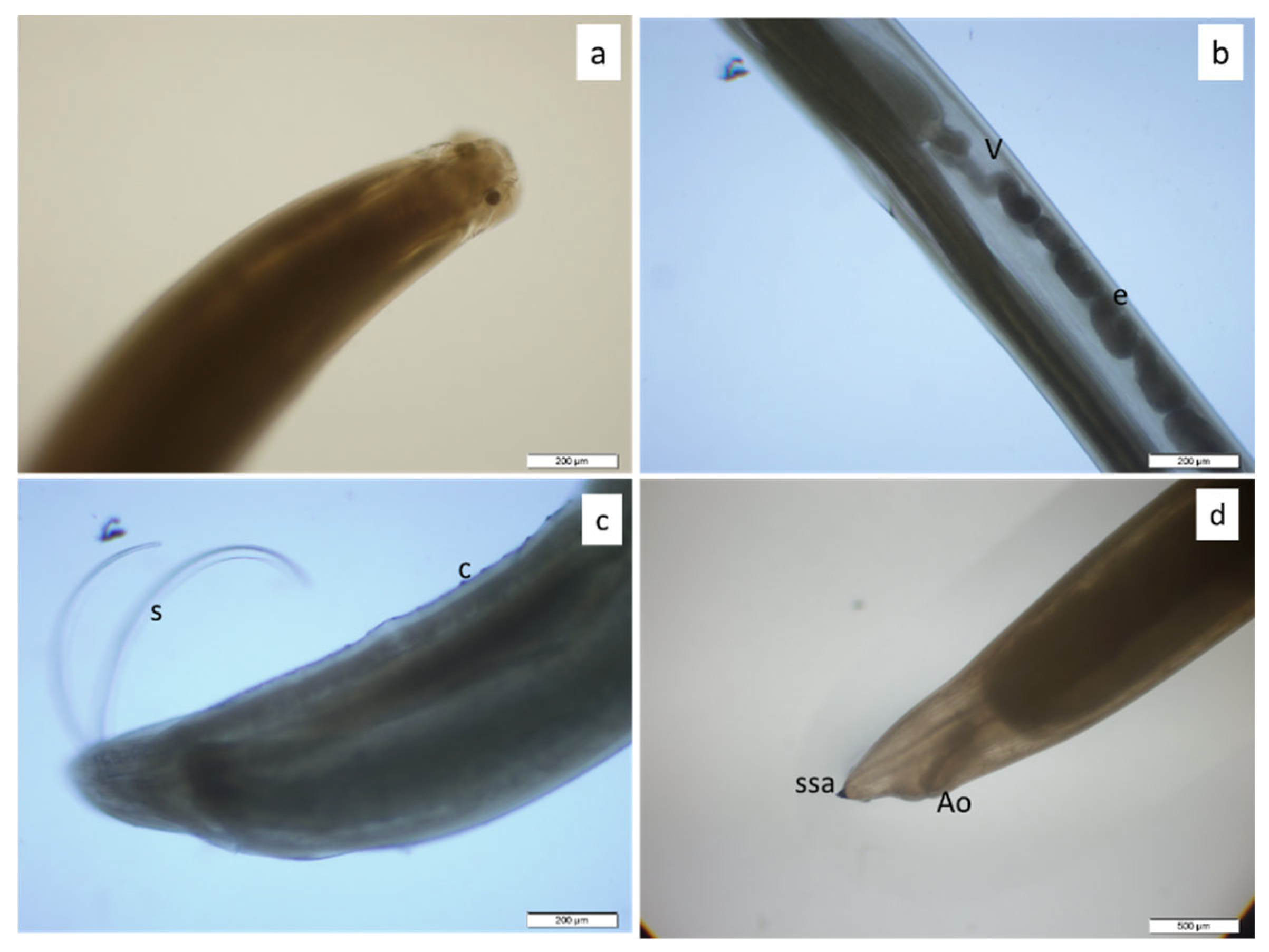
The nematode parasite extracted from the fish was whitish and elongated, generally measuring between 5 and 15 mm in length and 0.10–0.15 mm in width. Microscopic examination of the anterior extremity revealed a small protuberance, or tooth, and three poorly differentiated incipient labia. A small spine was observed at the posterior extremity. This morphological structure is similar to that reported by Yoshinaga et al. [15] for H. aduncum (Figure 3). However, it is easy to differentiate these from other ascaridoid nematodes using external morphology alone and proper taxonomic keys [14]. In this study, molecular identification was also used to confirm the results.
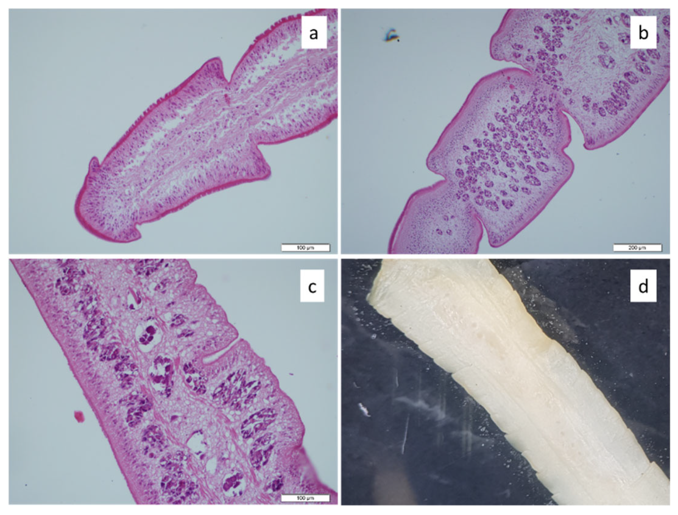
The morphological features of the cestode parasite, such as the shape of the proglotids and the scolex with an apical disc and shallow bothria, as well as the presence of two sets of reproductive organs per proglottid, were consistent with the morphological structure of Bothriocephalus sp. reported by Yamaguti [16] (Figure 4d). Therefore, the parasite was identified as Bothriocephalus sp. This morphological identification was confirmed by real-time PCR. Additionally, the parasite’s histological structure was examined (Figure 4a–c).
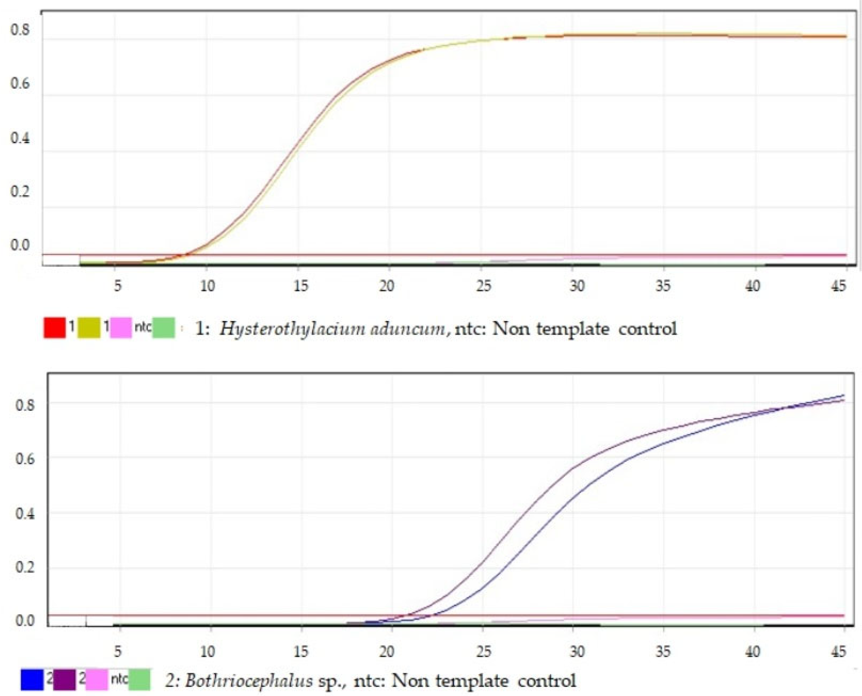
As part of the study, each parasite sample was analysed in two replicates using real-time PCR. One negative control sample was used for each parasite identification analysis. The real-time PCR results for each parasite species are given separately below (Figure 5). High-resolution melting (HRM) analysis, performed after real-time PCR analysis, showed that the amplicons displayed a single peak.
The most prevalent parasite identified among the female subjects was Bothriocephalus sp., constituting 70.58% of the cases, followed by H. aduncum, which accounted for 64.70%. The prevalence of Bothriocephalus sp. in male turbot was found to be 77.41%. The least detected parasite was H. aduncum, with a rate of 58.06%, being identified in 18 male fish. The prevalence of Bothriocephalus sp. was expressed by the number of parasitized individuals out of the total number analysed. The highest extent of parasitism was observed in the 60–70 m depth range, where 13 out of 16 individuals were infested (see Table 2 for further details). In the 70–80 m depth range, the prevalence of parasitized fish reached 75% of the total fish analysed. With regard to the quantity of H. aduncum in turbot, parasitism was most prevalent in the 70–80 m station, where 7 out of 8 fish analysed (87.50%) were infested by cestodes. In the other deep stations (50–60 m and 60–70 m), the percentage of cestode parasite-infested turbot individuals ranged between 81.25 and 84.62% of the total number of individuals analysed.
The present study examined the correlation between the prevalence of infection with parasites and the following variables: body length, body weight, age, and depth. The analysis revealed no statistically significant correlation between the mean host length and prevalence (rs = 0.0508, p = 0.69), mean host weight and prevalence (rs = 0.0334, p = 0.79), host age and prevalence (rs = 0.0323, p = 0.80), and different depths and prevalence (rs = 0.0955, p = 0.45).
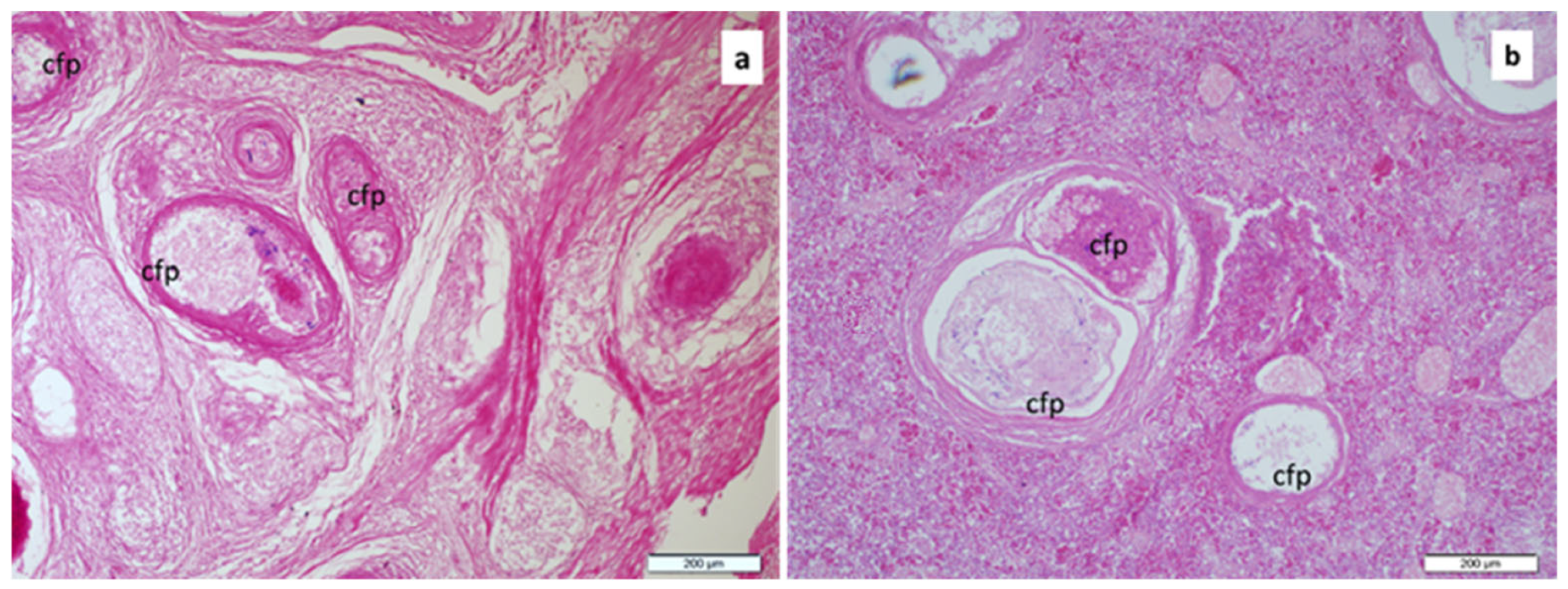
Histopathological analysis revealed that the most affected tissues were the intestine and spleen. In the intestinal tissue of infected fish, necrosis and shedding of epithelial cells, structures formed by the parasite surrounded by fibrous connective tissue in the lamina propria and intense haemorrhage and hyperaemia in this region were detected (Figure 6a and Figure 7a). The cystic structure was detected in the pancreas and spleen tissue (see Figure 6b).
Degeneration and haemorrhage in the gastric glands were another histopathological finding that attracted attention in other stomach tissues. It was determined that was Bothriocephalus sp., especially detected in the intestine, caused blockage in the intestinal tissue (Figure 7b).

4. Discussion

In consideration of the results obtained from the present study, it is evident that the observed aggregation patterns do not appear to be directly associated with variations in parasite load, irrespective of factors such as gender, habitat and dietary habits. This observation aligns with the conclusions reported by Yavuzcan et al. [18]. As posited by Amarante et al. [19], the extent of parasite aggregation, particularly in the context of endoparasites, is contingent on the feeding habits of the host. An increase in the proportion of fish in the turbot diet has been demonstrated to affect its infection with parasites. H. aduncum is one of the most prevalent nematodes identified in marine fish species, including Trachurus trachurus (Linnaeus, 1758) [18], Merlangius merlangus [20], Boops boops (Linnaeus, 1758) [21], Alosa immaculata (Bennet, 1835) and Spicara smaris (Linnaeus, 1758) [22]. Despite the existence of reports indicating a high level of infection in numerous host fish species and geographical regions of the Black Sea, the present study documents the first occurrence of H. aduncum in turbot along the Bulgarian Black Sea coast.
There can be no doubt that the parasite fauna is related to the host’s diet. Black Sea turbot represents the fish feeding on small-sized prey: fish and crustaceans such as copepods, amphipods, decapods, and shrimps which are the intermediate hosts of different parasite species. Giragosov and Khanaychenko [23] noted that turbot are more susceptible to infection with the cestode Bothriocephalus sp. than other fish species. Our findings are consistent with previous research that observed high infection prevalence levels from 33% to 70% by this parasite in the digestive tract of turbot in the Black Sea [2,17,21,22,23,24]. Maximov [25] reported Bothriocephalus sp. infection prevalence levels ranging between 35 and 50% of the analysed turbot individuals from the Romanian Black Sea coast. A similar infection rate (33.3%) for caught turbot has also been noted along the Sinop coasts of the Black Sea [22]. The highest infection rate, at a level of a few tens of parasites in the individual turbot and bleeding lesions on the intestinal wall, was reported by Țoțoiu et al. [24].
It is interesting to note that, despite the lack of correlations between mean host length, mean host weight, host age and different depths and parasite distributions, the highest prevalence was found at the greatest depths. It is possible that this phenomenon is due to the relief of the Black Sea, which is characterised by the presence of pronounced vertical depressions, canyons and unique conditions. It is thought that these conditions are maintained by strong stratification, river runoff and inflows, which have the potential to increase the number of nematodes and other meiobenthic organisms in the marine environment [26]. Meanwhile, the dominance of nematodes has been observed to increase with depth by up to 90% [27,28].
The effects of parasitic infections on host organisms vary widely, ranging from mechanical damage and non-lethal physiological stress (manifested as growth retardation and loss of appetite) to more serious consequences such as organ and tissue destruction, systemic intoxication, and the facilitation of secondary disease agents [29,30]. The effects on fish tissue often depend on the intensity of the pathogen’s infection. Branson et al. [31] reported severe enteritis and high mortality in turbot (S. maximus) with a high level of intestinal infection.
Many cestode species have a shallow attachment to the intestinal mucosa and do not cause significant tissue damage. In contrast, the haemorrhage and necrosis caused by nematode parasites generally result from their attachment to the intestines with the scoleces in turbot and induce an inflammatory response [32,33]. A rarely reported consequence of Bothriocephalus sp. infection is to penetrate the body cavity, extending as far as some internal organs, as in the current case affecting the spleen and pancreas [21,34].
Stressors exerting influence on the ecosystem, such as parasitic infection, have the capacity to exert a significant effect on the overall health of the ecosystem and its components, including human beings [35] Therefore, from a public health perspective, the assessment of parasite contamination in fish is a critical component of any comprehensive food safety monitoring program. For instance, it is imperative to ensure that the fish is cooked to the appropriate temperature and duration. Specifically, a temperature of approximately 74 °C is required for optimal cooking, while the duration must also be taken into consideration. Furthermore, the consumption of under-well-cooked turbot by humans has been identified as an important risk factor for parasitic infections [36]. The primary symptoms of this zoonotic disease are contingent on the location of the parasite within the digestive tract or the manifestation of severe allergic reactions [37]. These symptoms typically emerge within 12 h post-ingestion of infected fish. In some cases, the method of freezing plays a crucial role in maintaining the quality of the fish. It is essential to ensure that the fish is frozen at a temperature below −20 °C to prevent the onset of disease.

5. Conclusions

To the best of our knowledge, this study constitutes the first documented report on nematode and cestode parasites infecting turbot through the spawning season along the Bulgarian Black Sea coast.
The objective of the research was twofold: firstly, to ascertain the presence of the parasites in question and to identify them; secondly, to assess the histopathological damage that the parasites inflict on the tissues of the host. The present study hypothesises that the results thereof may facilitate more straightforward identification of helminth parasites that pose a threat to human health, due to the potential for parasite contamination in turbot (zoonosis). Furthermore, it is possible that the results may provide arguments that could be used in favour of conducting a future study to achieve the much-needed genomic characterisation of gastrointestinal helminths in turbot in the Black Sea.

Author Contributions

Conceptualization, A.A. and C.U.; methodology, A.A. and C.U.; software, A.A. and C.U.; validation, A.A. and C.U.; formal analysis, E.P.-P.; investigation, E.P.-P. and F.T.; resources, E.P.-P. and F.T.; data curation, E.P.-P. and F.T.; writing—original draft preparation, A.A. and C.U.; writing—review and editing, A.A. and C.U.; visualization, C.U.; supervision, A.A.; project administration, A.A.; funding acquisition, E.P.-P. and F.T. All authors have read and agreed to the published version of the manuscript.

Funding

This study was supported by the Agriculture Academy, Bulgaria, under the framework of project number: Ж167/2021.

Institutional Review Board Statement

No specific ethical approval was required: (i) the research does not describe laboratory experiments with fishes; (ii) the described fish species, turbot (Scophthalmus maximus), is a typical fishery subject in the Black Sea, studied within the framework of the National Fisheries Data Collection Program in Bulgaria, with financial support from the EU; (iii) as a data collection methodology, a widely applied fishery technique, namely bottom trawling, was used; (iv) the research methods were approved by the National Agency of Fisheries in Bulgaria; (v) specific procedures for animal treatment are not required.

Informed Consent Statement

Not applicable.

Data Availability Statement

The data presented in this study are available upon request from the corresponding author. The data are not publicly available due to institutional restrictions.

Conflicts of Interest

The authors declare no conflicts of interest.

References

  1. Nikolov, V.; Ivanova, P.; Dzembekova, N.; Panayotova, M.; Raykov, V.; Dobrovolov, I. Application of allozyme markers for screening of turbot populations along Western Black Sea coast. ZooNotes 2015, 79, 1–15. [Google Scholar]
  2. Țoțoiu, A.; Patriche, N.; Niță, V.; Sîrbu, E.; Dima, F.M.; Nenciu, M.I.; Nistor, V. Epidemiology of Turbot (Scophthalmus maeoticus) bacterial contamination, a fishery limiting factor on the Romanian Black Sea. Fishes 2023, 8, 418. [Google Scholar] [CrossRef]
  3. Hulak, B.; Leonchyk, Y.; Maximov, V.; Tiganov, G.; Shlyakhov, V.; Pyatnitsky, M. The current state of the turbot, Scophthalmus maximus (Linnaeus, 1758), population in the northwestern part of the Black Sea. Fish. Aquat. Life 2021, 9, 164–175. [Google Scholar] [CrossRef]
  4. GFCM. Working Group on the Black Sea (WGBS). In Proceedings of the Report of the 2nd Meeting of the Subregional Group on Stock Assessment in the Black Sea (SGSABS), Constanta, Romania, 10–12 November 2014; p. 237. [Google Scholar]
  5. Petrova, E.; Stoykov, S.; Mihneva, V.; Valchev, S.; Penchev, P.; Tserkova, F. Bottom Trawl Surveys in the Bulgarian Black Sea Area, Autumn 2022; Report under contract with the Executive Agency Fisheries and Aquacultures, program for data collection for fishing in 2021; Agricultural Academy Institute of Fish Resources: Varna, Bulgaria, 2022; p. 52. [Google Scholar]
  6. MacKenzie, K.; Abaunza, P. Parasites as biological tags for stock discrimination of marine fish: A guide to procedures and methods. Fish. Res. 1998, 38, 45–56. [Google Scholar] [CrossRef]
  7. Lester, R.J.G.; MacKenzie, K. The use and abuse of parasites as stock markers for fish. Fish. Res. 2009, 97, 1–2. [Google Scholar] [CrossRef]
  8. Güven, A.; Öztürk, T. Balık parazitlerinin biyoizlemdeki (Biyomonitoring) önemi. Süleyman Demirel Üniversitesi Eğirdir Su Ürünleri Fakültesi Derg. 2018, 14, 59–73. [Google Scholar] [CrossRef]
  9. Skrzypczak, M.; Rolbiecki, L. Helmintofauna of turbot Scophthalmus maximus (Linnaeus, 1758) from the southern Baltic Sea including new data. Pol. J. Vet. Sci. 2015, 18, 599–605. [Google Scholar] [CrossRef]
  10. Türe, M. Molecular identification of Uronema marinum (Protozoa, Ciliophora, Scuticociliatia) in cultured turbot Psetta maxima larvae. Vet. Res. Forum 2021, 12, 121–124. [Google Scholar]
  11. Ürkü, C.; Atanasoff, A.; Petrova-Pavlova, E.; Tserkova, F. Morphological identification of intestinal parasites from turbot (Psetta maxima) along the Bulgarian Black sea coast. In Proceedings of the of 2nd Aquatic Biotechnology Symposium, Istanbul, Turkiye, 13–15 October 2021; p. 53. Available online: https://aquabiotech.istanbul.edu.tr/en/content/past-events/2021 (accessed on 2 August 2025).
  12. Pinkas, L.; Oliphant, M.S.; Iverson, I.L.K. Food habits study of albacore, bluefin tuna, and bonito in California waters. In Fish Bulletin; USA and California Department of Fish and Game: Sacramento, CA, USA, 1971; Volume 152, pp. 1–105. [Google Scholar]
  13. Cortes, E. A critical review of methods of studying fish feeding based on analysis of stomach contents: Application to elasmobranch fishes. Can. J. Fish. Aquat. Sci. 1997, 54, 726–738. [Google Scholar] [CrossRef]
  14. Moravec, F. Parasitic Nematodes of Freshwater Fishes of Europe; Kluwer Academic Publishers: Dordrecht, The Netherlands, 1994; p. 473. [Google Scholar]
  15. Yoshinaga, T.; Ogawa, K.; Wakabayashi, H. Experimental life cycle of Hysterothylacium aduncum (Nematoda: Anisakidae) in fresh water. Fish Pathol. 1987, 22, 243–251. [Google Scholar] [CrossRef]
  16. Yamaguti, S. The cestodes of vertebrates. In Systema Helminthum; Interscience Publıshers: New York, NY, USA, 1959; Volume II, p. 271. [Google Scholar]
  17. Roberts, R.; Smail, D.; Munro, E. Laboratory methods. In Fish Pathology; WB Saunders: London, UK, 2001; pp. 380–412. [Google Scholar]
  18. Yavuzcan Yildiz, H.; Genç, E.; Kaya, D.; Yilmaz, B. Predictive model for the nematode (Hysterothylacium aduncum) in horse mackerel, Trachurus trachurus from the Black Sea. Mar. Life Sci. 2024, 6, 10–16. [Google Scholar] [CrossRef]
  19. Amarante, C.F.D.; Tassinari, W.D.S.; Luque, J.L.; Pereira, M.J.S. Factors associated with parasite aggregation levels in fishes from Brazil. Rev. Bras. Parasitol. Veterinária 2015, 24, 174–182. [Google Scholar] [CrossRef]
  20. Pekmezci, G.Z. Molecular characterization of Hysterothylacium aduncum (Nematoda: Raphidascarididae) larvae infecting Merlangius merlangus euxinus (Linnaeus, 1758) from the Turkish Black Sea coast based on mitochondrial small subunit ribosomal RNA gene analysis. Etlik Vet. Mikrobiyoloji Derg. 2019, 30, 64–69. [Google Scholar] [CrossRef]
  21. Aldık, R.; Cakır, F.; Tonguç-Yayintas, O.; Altinagac, U. Endoparacite investigation in some fish species with high economic value obtained from Canakkale, Turkey. J. Adv. VetBio Sci. Tech. 2021, 6, 202–210. [Google Scholar] [CrossRef]
  22. Özer, A.; Olguner, A.M. Metazoan parasites of some marine fish species collected at the Sinop coasts of the Black Sea. Ege J. Fish. Aquat. Sci. 2013, 30, 93–97. [Google Scholar] [CrossRef]
  23. Giragosov, V.E.; Khanaychenko, A.N. Macroscopic characteristics of the epizootic situation in the Black Sea turbot Scophthalmus maeoticus. In Contemporary Problems of Theoretical and Marine Parasitology; Bondarenko Publishing: Moscow, Russia, 2016; pp. 219–221. [Google Scholar]
  24. Totoiu, A.; Tiganov, G.; Galatchi, M.; Nenciu, M. Food array analysis in turbot (Psetta maeotica) at the Romanian Black Sea coast in 2013. Cercet. Mar. 2014, 44, 164–172. [Google Scholar]
  25. Maximov, V. Managementul Durabil al Calcanului la Litoralul Românesc; Editura Boldaş: Constanţa, Romania, 2012; p. 199. [Google Scholar]
  26. Stanev, E. Understanding Black Sea dynamics an overview of recent numerical modeling. Oceanography 2005, 18, 56–75. [Google Scholar] [CrossRef]
  27. Leduc, D.; Rowden, A.A.; Nodder, S.D.; Berkenbusch, K.; Probert, P.K.; Hadfield, M.G. Unusually high food availability in Kaikoura Canyon linked to distinct deep-sea nematode community. Deep Sea Res. Part II: Topical Stud. Oceanography 2014, 104, 310–318. [Google Scholar]
  28. Sapir, A. Why are nematodes so successful extremophiles? Commun. Integr. Biol. 2021, 14, 24–26. [Google Scholar] [CrossRef]
  29. Țoțoiu, A.; Radu, G.; Nenciu, M.I.; Patriche, N. Overview of health status of the main Romanian Black Sea coast fish. J. Environ. Prot. Ecol. 2018, 4, 1591–1602. [Google Scholar]
  30. Stoyanov, B.; Mutafchiev, Y.; Georgiev, B.B. Helminth parasites of the three-spined stickleback Gasterosteus aculeatus L., 1758 (Actinopterygii: Gasterosteidae) from a Black Sea coastal wetland, Bulgaria. Acta Zool. Bulg. 2023, 75, 285–300. [Google Scholar]
  31. Branson, E.; Riaza, A.; Alvarez-Pellitero, P. Myxosporean infection causing intestinal disease in farmed turbot, Scophthalmus maximus (L.), (Teleostei: Scophthalmidae). J. Fish Dis. 1999, 22, 395–399. [Google Scholar] [CrossRef]
  32. Britton, J.R.; Pegg, J.; Williams, C.F. Pathological and ecological host consequences of infection by an introduced fish parasite. PLoS ONE 2011, 6, e26365. [Google Scholar] [CrossRef] [PubMed]
  33. Davey, J.T.; Peachey, J.E. Bothriocephalus scorpii [Cestoda: Pseudophyllidea] in turbot and brill from british coastal waters. J. Mar. Biol. Assoc. U.K. 1968, 48, 335–340. [Google Scholar] [CrossRef]
  34. Ahmad, F.; Fazili, K.M.; Sofi, O.M.; Sheikh, B.A.; Sofi, T.A. Distribution and pathologycaused by Bothriocephalus acheilognathi, Yamaguti 1934 (Cestoda: Bothriocephalidae): A review. Rev. Vet. 2018, 29, 142–149. [Google Scholar] [CrossRef]
  35. Țoțoiu, A.; Stoica, E.; Ciucă, A.M.; Harcotă, G.E.; Nită, V.; Patriche, N. Parasites and microplastics in the gastrointestinal tract of Alosa immaculata from the Black Sea—Implications for health and condition. J. Mar. Sci. Eng. 2025, 13, 1316. [Google Scholar] [CrossRef]
  36. Audicana, M.T. Anisakis, something is moving inside the fish. Pathogens 2022, 11, 6. [Google Scholar] [CrossRef]
  37. Baron, L.; Branca, G.; Trombetta, C.; Punzo, E.; Quarto, F.; Speciale, G.; Barresi, V. Intestinal anisakidosis: Histopathological findings and differential diagnosis Pathology. Res. Pract. 2014, 210, 746–750. [Google Scholar] [CrossRef]
Figure 1. Map of turbot sample collection stations.
Fishes 10 00395 g001
Figure 2. Age structure of turbot.
Fishes 10 00395 g002
Figure 3. Photomicrograph of H. aduncum (Rudolphi, 1802), (a) anterior part, (b) female parasite, (c) male posterior part, (d) female posterior end (S: spicules, Ao: anal opening, V: vulva, E e: egg, ssa: single spine-like appendix, c: caudal papillae).
Fishes 10 00395 g003
Figure 4. Photomicrograph of Bothriocephalus sp. (a) scolex; (b) segments; (c,d) detail of mature segment (ac)—Histological section of parasite, H&E staining.
Fishes 10 00395 g004
Figure 5. Real-time PCR analysis results for H. aduncum and Bothriocephalus sp. (all samples gave positive results).
Fishes 10 00395 g005
Figure 6. The presence of cystic-forming parasites (cfp) in the lamina propria of the intestinal tissue (a) and spleen (b) (H&E).
Fishes 10 00395 g006
Figure 7. Hyperemia in the lamina propria (a), degeneration and necrosis of epithelial tissue of intestinal villi and blockage in the intestinal tissue (b), (hi: hyperemia, d: degeneration, n: necrosis and Bs: Bothriocephalus sp.)
Fishes 10 00395 g007
Table 1. Primers used in the present study for Bothriocephalus sp. and H. aduncum.
Primers and ProbeSequence 5′-3′TmAmp Self Comp
Bothriocephalus sp. 18S-FCGTTTTCCGACTCCGCTCTA59.831450.00
Bothriocephalus sp. 18S-RCTCGGAAGCAGACACCACTT59.970.00
H. aduncum 18S-FAAAGCGGGGACTGCTGTTTC61.17701.00
H. aduncum 18S-RACTGCGATTAAGGCGGTTTC58.920.00
Table 2. Prevalence of intestinal parasites at different depths.
Species of Parasites10–20
(n = 2)
30–40
(n = 3)
40–50
(n = 18)
50–60
(n = 13)
60–70
(n = 16)
70–80
(n = 8)
80–90
(n = 5)
Bothriocephalus sp.1
(50.00%)
2
(66.67%)
10
(55.56%)
7
(53.84%)
13
(81.25%)
6
(75.00%)
3
(60.0%)
Hysterothylacium aduncum1
(50.00%)
2
(66.67%)
11
(61.11%)
11
(84.62%)
13
(81.25%)
7
(87.50%)
3
(60.0%)
Disclaimer/Publisher’s Note: The statements, opinions and data contained in all publications are solely those of the individual author(s) and contributor(s) and not of MDPI and/or the editor(s). MDPI and/or the editor(s) disclaim responsibility for any injury to people or property resulting from any ideas, methods, instructions or products referred to in the content.