Article

Morphomolecular Characterization of Rhadinorhynchus niloticus (Acanthocephala: Rhadinorhynchidae) from Nile Perch (Lates niloticus, Perciformes: Latidae)

by
Ebtsam Sayed Hassan Abdallah
1,
Marco Albano
2,* and
Hasnaa Thabit
3
1
Aquatic Animal Medicine and Management Department, Faculty of Veterinary Medicine, Assiut University, Assiut 71529, Egypt
2
Department of Veterinary Sciences, University of Messina, Polo Universitario Dell’ Annunziata, 98168 Messina, Italy
3
Department of Zoology and Entomology, Faculty of Science, Assiut University, Assiut 71526, Egypt
*
Author to whom correspondence should be addressed.
Fishes 2025, 10(8), 397; https://doi.org/10.3390/fishes10080397
Submission received: 5 June 2025 / Revised: 25 July 2025 / Accepted: 6 August 2025 / Published: 8 August 2025
(This article belongs to the Special Issue Advances in Fish Pathology and Parasitology)

Abstract

Adults of Rhadinorhynchus niloticus, a member of the Rhadinorhynchidae family, were isolated from the intestines of wild Nile perch (Lates niloticus (Linnaeus, 1758); Perciformes: Latidae) caught from the River Nile and its tributaries in Assiut City, Egypt. The parasite was found freely in the intestinal lumen with a prevalence of 10.71%, and the burden varied from one to five parasites per fish. The mean intensity and abundance were 2.16 ± 0.47 (95% CI: 1.33 to 3.17) and 0.23 ± 0.08 (95% CI: 0.11 to 0.43), respectively. The parasite was described using light and scanning electron microscopy. Molecular species identification as well as phylogenetic relationship analysis of the isolated parasite were achieved by sequencing and comparisons of the mitochondrial cytochrome oxidase C subunit I (COI) and nuclear 18S rRNA genes. The sequences were deposited in GenBank under the accession numbers PP859185 and MZ727194. Furthermore, phylogenetic analysis demonstrated that the parasites emerged from a separate branch belonging to the Rhadinorhynchidae family, which was clearly distinguished from other genospecies.
Key Contribution: Adults of Rhadinorhynchus niloticus were isolated from the intestines of wild Nile perch, investigating their mean intensity and abundance. Through microscopy and molecular sequencing, the parasite was characterized, shedding light on its morphology and phylogeny.

1. Introduction

The Nile perch, Lates niloticus (Linnaeus, 1758), a member of the Latidae family, is a tropical freshwater piscivorous potamodromous predator. It is highly valued commercially and recreationally in Africa and can be found in various habitats, such as rivers, lakes, and irrigation channels. It has the potential to grow up to 2 m in length, weigh as much as 200 kg, and live for up to 16 years [1]. It is an ideal fish for aquaculture because of its bone-free white flesh, which is high in protein and vitamins, including omega-3 fatty acids. These nutrients are crucial for human nutrition and overall well-being [2].
Parasitism has a critical impact on fish production, industrial productivity, and economic value [3]. For example, Neoechinorhynchus buttnerae causes economic losses of up to 100% in tambaqui (Colossoma macropomum) production in northern Brazil [4]. In aquatic ecosystems, fish serve as key hosts for parasites, which encompass a wide variety of adult and immature forms. For these parasites, fish may function as primary hosts in parasitic life cycles or as one of multiple hosts in a series. Although most individual fish in wild or cultivated populations are parasite-infested, in most cases, no significant harm occurs to the fish. The number of parasites (burden) needed to harm a fish varies considerably depending on the species, the size of the host, and its health status [5]. Fish parasites contribute to fish disease and can negatively impact the appearance of fish, potentially leading to consumer rejection [6]. Some parasites can also cause economically significant disease outbreaks by exploiting fish or reducing productivity through nutritional effects. Other factors may be responsible for long-term changes in population structure. Some fish parasites are transmissible to humans, whereas others reduce the market value of fish products by spoiling host tissue or decreasing the demand for fish as food [7].
Acanthocephalans are a group of endoparasitic helminths that are commonly found in both marine and freshwater fish worldwide. The phylum Acanthocephala includes at least 1289 species of relatively small, vermiform, heterosexual endoparasites [8] with low specificity for intermediate, definitive, and transport hosts. The genus Rhadinorhynchus (Lühe, 1911) belongs to the family Rhadinorhynchidae [9]. These worms are commonly referred to as spiny-headed or thorny-headed worms. In this study, we assessed the morphometric and morphological characteristics of the parasite found in the intestines of L. niloticus inhabiting the Nile River in Assiut City, Egypt. Our objective was to determine the taxonomic classification of the parasite. We also calculated the prevalence of the parasite, worm load, mean intensity, and abundance.

2. Materials and Methods

2.1. Ethical Statement

Fish were handled according to the standard protocol approved by Assiut University, Faculty of Science Research Ethics Committee for Animal Use and Care (Number-01-2024-0018).

2.2. Study Area and Fish Sampling

A total of one hundred and twelve L. niloticus were caught by fishermen from September 2020 to August 2021 from the River Nile in Assiut City, southern Egypt (latitude 27°10′51.46″ N, Longitude 31°11′1.25″ E; Figure 1). The number of fish was determined using a method published by Shvydka et al. [10] and Reiczigel et al. [11]. The fish were promptly transported to the Parasitology Laboratory (Faculty of Science, Assiut University) for examination. The fish were humanely euthanized using 200 µL of clove oil per liter of tank water for tissue sampling, as described by Kildea et al. [12], and identified using a technique published by Paugy et al. [13]. The total length measured from the tip of the snout to the tip of the longer lobe of the caudal fin, the standard length measured from the tip of the snout to the posterior end of the last vertebra, and the body weight measurements were recorded for each fish. The total length of the examined fish varied from 22 to 39 cm, with an average of 31.05 ± 3.48 cm. The standard length ranged from 19 to 33 cm, with an average of 26.1 ± 2.98 cm, and the body weight ranged from 112 to 674 g, with an average of 325.9 ± 115.67 g. They were directly evaluated for any visible clinical signs and postmortem lesions, following the methods outlined by Eissa [14] and Noga [15]. Fish were examined as previously described by Abdallah and Hamouda [16,17], Hassan et al. [18], Mahmoud et al. [19], and Thabit and Abdallah [20].

2.3. Parasitological and Epidemiological Examinations

The fish were dissected as described by Noga [15]. The intestines were then placed in a Petri dish containing 0.85% physiological saline solution, and R. niloticus was detected. After carefully cleaning the parasites in physiological saline, the total number of R. niloticus was determined per fish. The method described by Bush et al. [21] was utilized to calculate the parasite’s prevalence (number of infected fish/total number of examined fish ×100), mean intensity (total number of parasites collected/total number of infected fish), and mean abundance (total number of parasites collected/total number of examined fish) throughout the study period (one year; from September 2020 to August 2021).

2.4. Morphological Examination

The worms were fixed in 70% ethanol after being submerged in cold water for 2–5 h or until completely expanded. Following a fine-needle puncture, the worms were stained with Mayer’s acid carmine, detained in 70% ethanol with 4% hydrochloric acid, dehydrated in increasing ethanol concentrations for 24 h each, and cleared in graduated concentrations of terpineol in 100% ethanol to 100% terpineol, followed by 50% terpineol in 50% Canada balsam for 24 h each. After that, whole worms were mounted in DPX. The worms were examined using a light microscope (Ernst, Leitz, Wetzlar, Gmbh, Germany) with a digital camera (Mu1803-HS; AmScope, Irvine, CA, USA).
For scanning electron microscopy examination, male and female specimens of R. niloticus were preserved for 1 h at 4 °C in 0.1 M phosphate buffer (pH 7.2) containing 3% glutaraldehyde. The samples were then post-fixed in 1% osmium tetroxide for 1 h at 4 °C in the same buffer, after being rinsed in the buffer. Subsequently, the samples were dried at the critical point using an alcohol series. Following the method described by Hassan, Mahmoud, Metwally and Moktar [18], they were sputter-coated in gold, and SEM images were captured at the electron microscope facility at Assiut University using a JEOL 5400 LV electron microscope (JEOL, Tokyo, Japan). Morphometric characteristics of different body regions were identified using 10 parasites.

2.5. Molecular Characterization of the Parasite

DNA was extracted using the CTAB method described by Abdallah et al. [22]. The concentration and purity of the DNA were evaluated using a nanophotometer (Implen GmbH, Munchen, Germany) at an optical density (OD) of 260 nm and relative OD of 260/280 nm, respectively. DNA samples were stored at −20 °C until needed. PCR amplification was performed using a Veriti® model 9902 thermal cycler (Applied Biosystems, Waltham, MA, USA). The PCR reactions were carried out in a 50 µL volume, including 25 µL of MyTaq red mix (Bioline, London, UK), 2 µL of each primer, 4 µL of template DNA (containing 100 ng of parasite DNA), and 17 µL of H2O (RNase/DNase free). A 776-base pair (bp) segment of the mitochondrial cytochrome oxidase c subunit I (COI) was amplified using the primer pair LCO1490 (5′-GGTCAACAAATCATAAAGATATTGG-3′) and HCO2198 (5′-TAAACTTCAGGGTGACCAAAAAATCA-3′) as outlined by Folmer et al. [23]. The PCR amplification process began with an initial denaturation step at 95 °C for 5 min, followed by 35 cycles of denaturation at 95 °C for 1 min, annealing at 40 °C for 1 min, extension at 72 °C for 1.5 min, and a final extension step at 72 °C for 7 min. Additionally, the nuclear 18S rRNA gene was amplified using the primer pair 18S F (5′-AGATTAAGCCATGCATGCGTAAG-3′) and 18S R (5′-TGATCCTTCTGCAGGTTCACCTAC-3′) following the method performed by Garey et al. [24]. PCR was performed using the following cycling conditions: initial denaturation at 94 °C for 4 min, followed by 30 s at 94 °C, 30 s at 60 °C, and 120 s at 72 °C for 35 cycles, with a final extension at 72 °C for 5 min. The amplified products were separated on 1.5% agarose gels in Tris-acetate EDTA (TAE) buffer, stained with 0.05 μg/mL ethidium bromide (Serva, Germany), and visualized using a UV transilluminator (MultiDoc-It, UVP, UK). The sizes of the PCR products were determined using a 100-bp DNA ladder H3 RTU (GeneDireX, Taiwan). The PCR products from the gels were purified using a Zymoclean Gel DNA Recovery Kit (Zymo Research, USA).
The purified PCR products were sequenced using the same amplification primers (SolGent Company, Daejeon, Republic of Korea). A BLAST search was conducted through the NCBI website and analyzed using the Basic Local Alignment Search Tool (BLAST; http://www.ncbi.nlm.nih.gov/BLAST, accessed on 5 August 2025).
The Hasegawa–Kishino–Yano model [25] and maximum likelihood techniques were used to infer the evolutionary history of the COI gene. The tree with the highest log likelihood (−7242.42) is displayed. By employing the Maximum Composite Likelihood (MCL) technique to create a matrix of pairwise distances, the Neighbor-Join and BioNJ algorithms automatically generated the initial trees for the heuristic search, and the topology with the highest log likelihood value was selected. A discrete Gamma distribution was used to model evolutionary rate differences among sites (5 categories; +G, parameter = 0.3409). The tree is drawn to scale, with branch lengths indicating the number of substitutions per site (next to the branches). The present analysis involved 15 nucleotide sequences, resulting in 1045 positions in the final dataset. Evolutionary analyses were performed using MEGA11 (ver. 11.0.13) by Tamura et al. [26]. Furthermore, we calculated the number of base substitutions per site to estimate the evolutionary divergence between sequences. The analyses were conducted using the maximum composite likelihood model and included 15 nucleotide sequences (Table 1), with all ambiguous positions removed for each sequence pair through the pairwise deletion option. The final dataset contained 1045 positions. Evolutionary analyses were performed using MEGA11 [26].
The evolutionary history of the 18S rRNA gene was inferred using the maximum-likelihood method and the Tamura 3-parameter model [27]. The tree with the highest log likelihood (−7701.65) is displayed. The initial trees for the heuristic search were automatically obtained by applying the Neighbor-Join and BioNJ algorithms to a matrix of pairwise distances estimated using the Tamura 3 parameter model and then selecting the topology with the highest log likelihood value. A discrete Gamma distribution was used to model evolutionary rate differences among sites (5 categories; +G, parameter = 0.4240). The tree is drawn to scale, with branch lengths measured as the number of substitutions per site (next to the branches). This analysis involved 18 nucleotide sequences (Table 1), resulting in 1867 positions in the final dataset. Evolutionary analyses were performed using MEGA11 software [26]. The number of base substitutions per site between sequences is shown. The analyses were performed using the maximum composite likelihood model. This study included 18 nucleotide sequences. Any ambiguous positions in each sequence pair were eliminated using the pairwise deletion option. The final dataset comprised 1867 positions. Evolutionary analyses were performed using MEGA11 software [26].
Table 1. GenBank accession numbers for the taxa used in the phylogenetic analysis.
Parasite NameHostCountryGenBank Accession NumbersReference
COI18S rRNA
Rhadinorhynchidae Lühe, 1912
RhadinorhynchusniloticusNile perch (Lates niloticus)EgyptPP859185MZ727194This study
Rhadinorhynchus gerberi (Lisitsyna, Kudlai, Cribb and Smit, 2019)Trachinotus botlaSouth AfricaMN104897MN105739[28]
Rhadinorhynchus hiansi (Soota and Bhattacharya, 1981)Sarda orientalisVietnamMN203137MN203133[29]
Rhadinorhynchus sp.marine fishWestern Pacific coast of Mexico -AY062433[30]
HNA (Family Scianidae)USADQ089712˗[31]
Rhadinorhynchus dorsoventrospinosus (Amin, Heckmann and Nguyen Van Ha 2011) DecapteruskurroidesVietnamMN267179MH384435[32]
Rhadinorhynchus laterospinosus (Amin, Heckmann & Nguyen Van Ha 2011)AuxisrockeiVietnamMK572743MK457183[33]
Rhadinorhynchus carangis (Yamaguti 1939)Australian marine teleostsAustralia MN705830[34]
Rhadinorhynchus biformis (Smales 2014) MN705829
Rhadinorhynchus johnstoni (Golvan 1969) MN705827
Rhadinorhynchus pristis (Rudolphi 1802)Nyctiphanes couchii (Bell)SpainJQ061132JQ061133[35]
Leptorhynchoididae Witenberg, 1932
Leptorhynchoides thecatus (Linton 1891, Kostylev 1924) ˗USAAY690577˗[36]
Pomphorhynchidae Monticelli, 1905
Tenuiproboscis sp.Lutjanus argentimaculatusIndiaJF694276˗NPY
Longicollum pagrosomi (Yamaguti 1935) OplegnathusfasciatusChinaKY490048-[37]
cultured red sea breamKorea-KX641270NPY
Echinorhynchidae Cobbold, 1879
Echinorhynchus sasakiae (Kita & Kajihara 2023)Hexagrammos lagocephalus (Pallas)JapanLC757487LC757488[38]
Echinorhynchus gadi (Zoega in Müller 1776)Salvelinus malmaRussiaKF156892-[39]
LimnognathiamaerskiUSA-AY218123[40]
Neoechinorhynchidae Ward, 1917
NeoechinorhynchussalmonisSalvelinus malmaRussiaKF156889-[39]
Neoechinorhynchus sp.Mugil cephalusIndia-MN992025NPY
Tenuisentidae Van Cleave, 1936
Tenuisentis niloticus (Meyer 1932)Heterotis niloticus (Cuvier)Burkina FasoKT970469KT970471[41]
Brachionidae
Brachionus plicatilis (Müller 1786)Arthrospira platensisChina-KY886363NPY
ScirpophagaincertulasIndiaAY218090-
NPY: Not published yet.

3. Results

3.1. Parasitological and Epidemiological Examinations

Out of the 112 fish analyzed, 12 were found to be infected with the adult stage of the parasite, resulting in a prevalence of 10.71%. The parasite was found freely present in the intestinal lumen, with no particular clinical signs observed externally or internally in the infected fish. The parasitic burden ranged from one to five parasites per fish, with a mean intensity and mean abundance of 2.16 ± 0.47 (95% CI: 1.33 to 3.17) and 0.23 ± 0.08 (95% CI: 0.11 to 0.43), respectively.

3.2. Morphological Examination

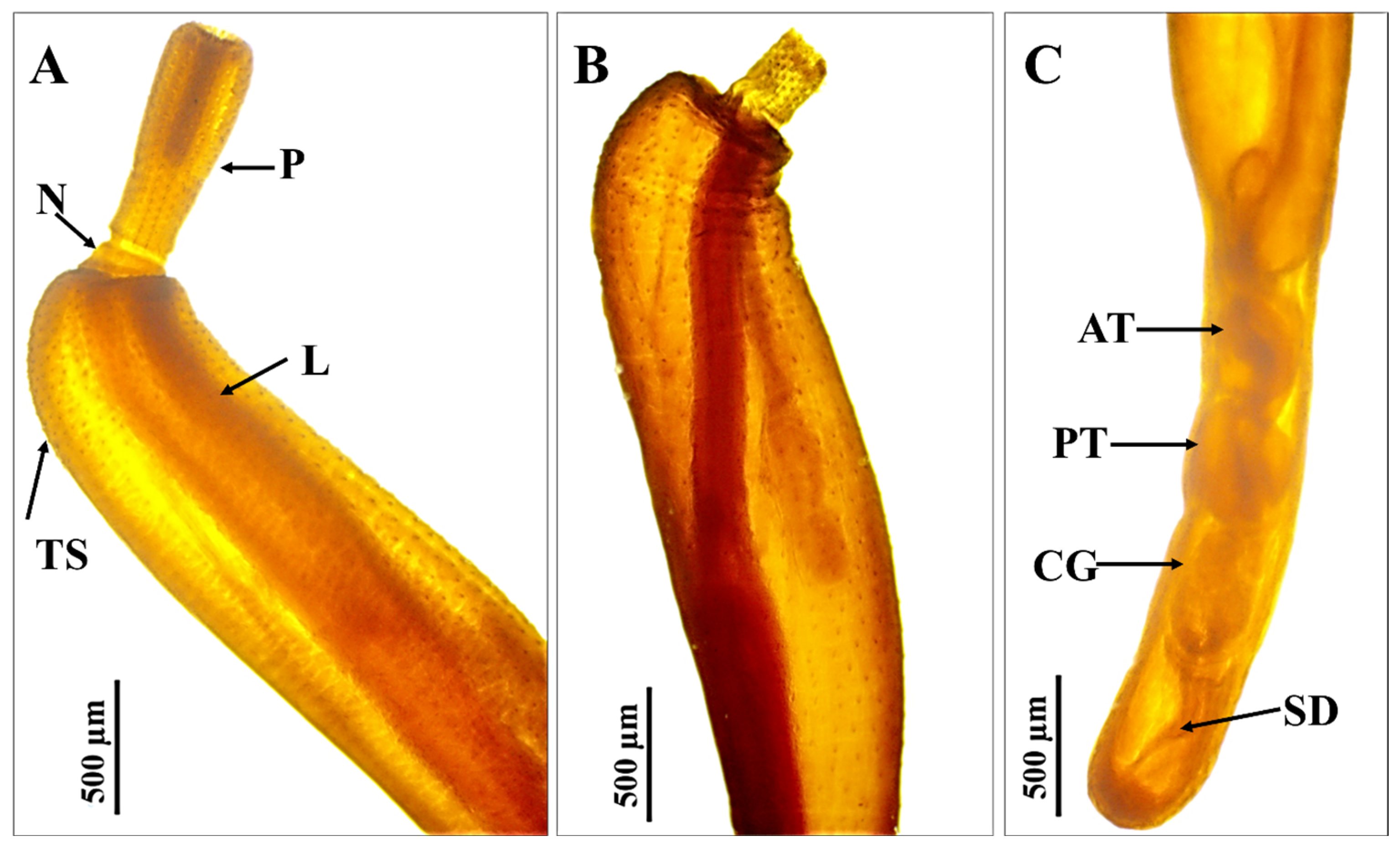
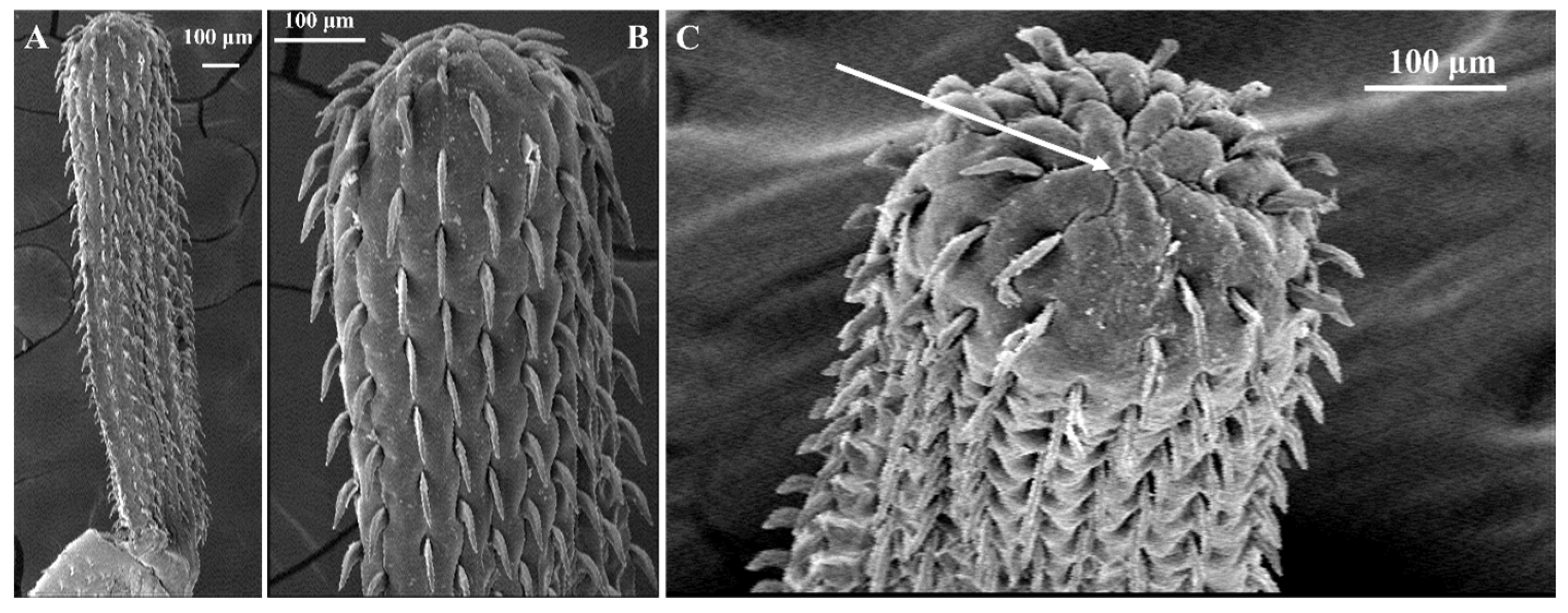
The parasite’s elongated, cylindrical body with a spiny proboscis was discovered through light and scanning electron microscopy (Figure 2A and Figure 3A). The lemnisci or proboscis sheath is long, cylindrical, and attached to the base of the proboscis. The proboscis is either fully everted (Figure 2A and Figure 3A) or partially retracted into the proboscis receptacle (Figure 2B).
The anterior end has small-sized apical hooks compared to the subapical hooks (Figure 3B,C). The apical end of the proboscis displays a centrally located apical epidermal cone (Figure 3C).
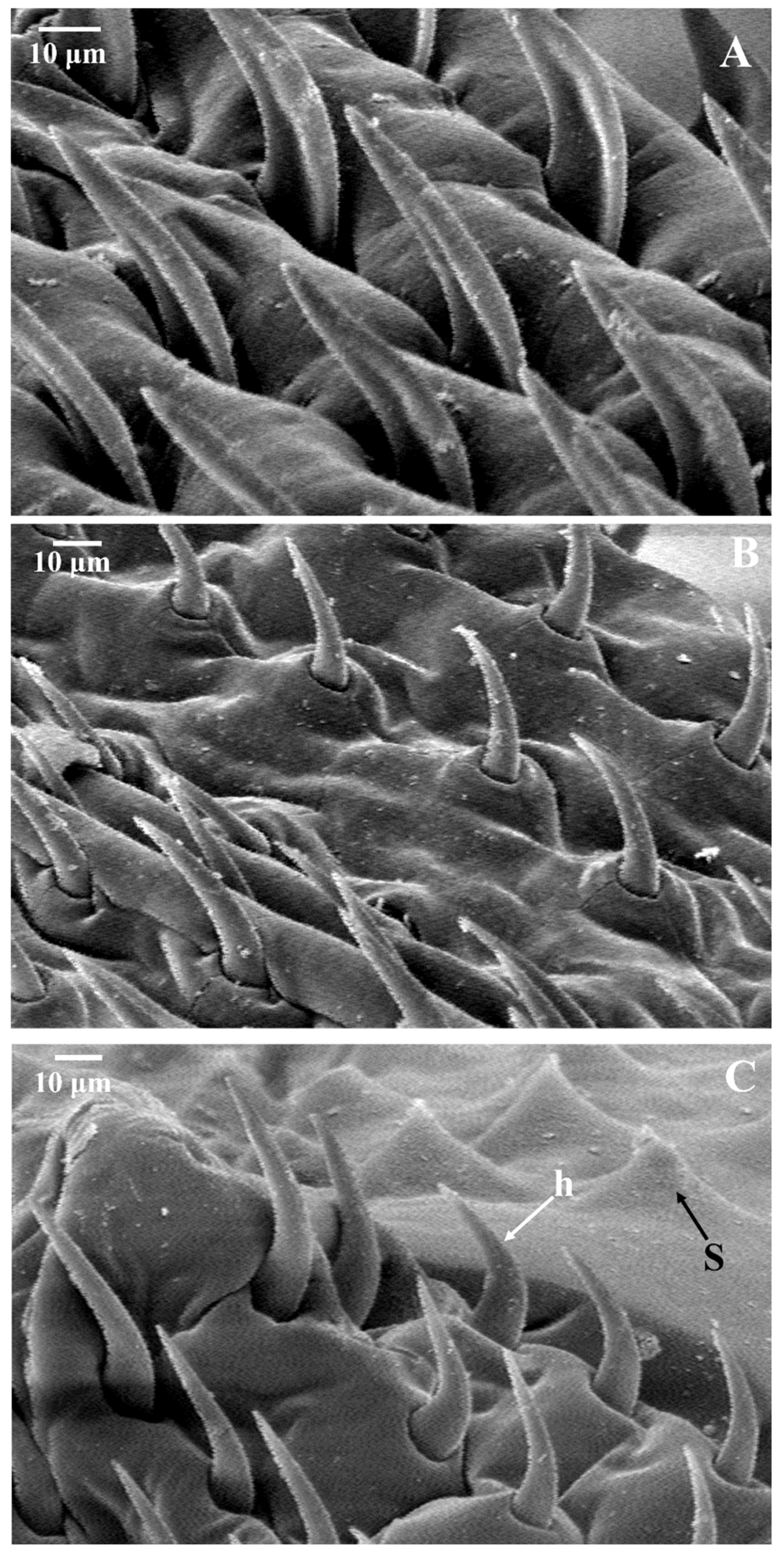
The proboscis is completely covered with several hooks that are evenly distributed in rows and pointed ventrally (Figure 4). The anteriorly located hooks (Figure 4A) are wider and more flattened than the intermediate hooks (Figure 4B) or the basal hook (Figure 4C). All the proboscis hooks at different parts are larger than the tegumental spines (Figure 4C).
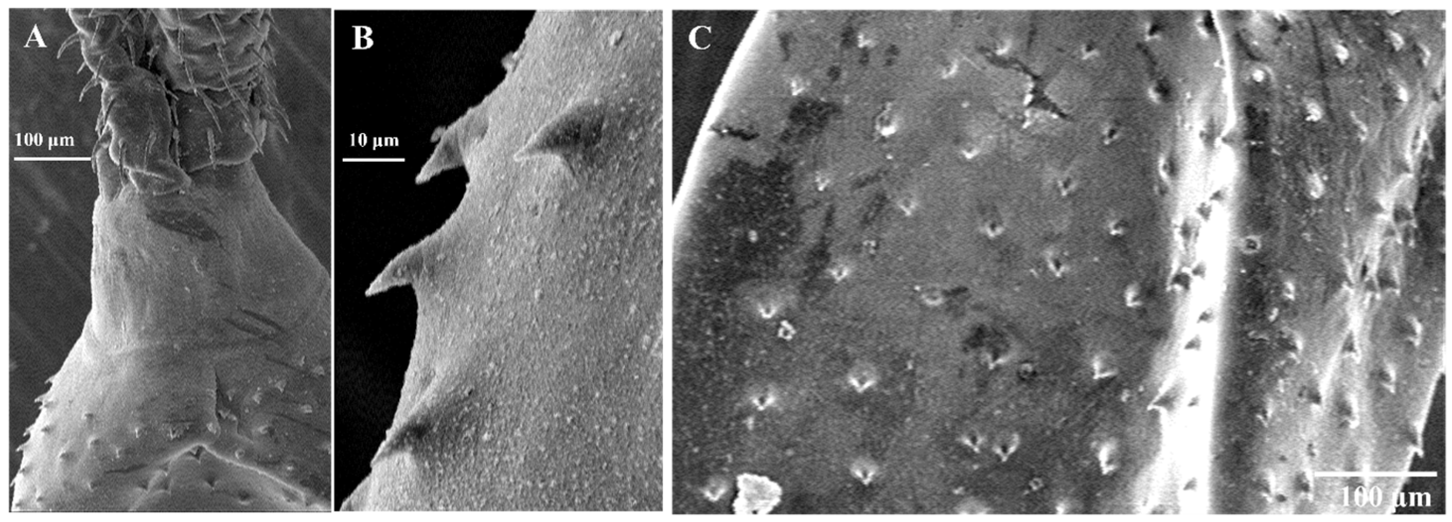
Following the proboscis, a short, smooth conical neck that is free from spines is observed (Figure 5A). The tegumental spines are directed ventrally (Figure 5B,C), originate from the trunk cone, and cover the anterior half of the parasite’s body (Figure 5C).
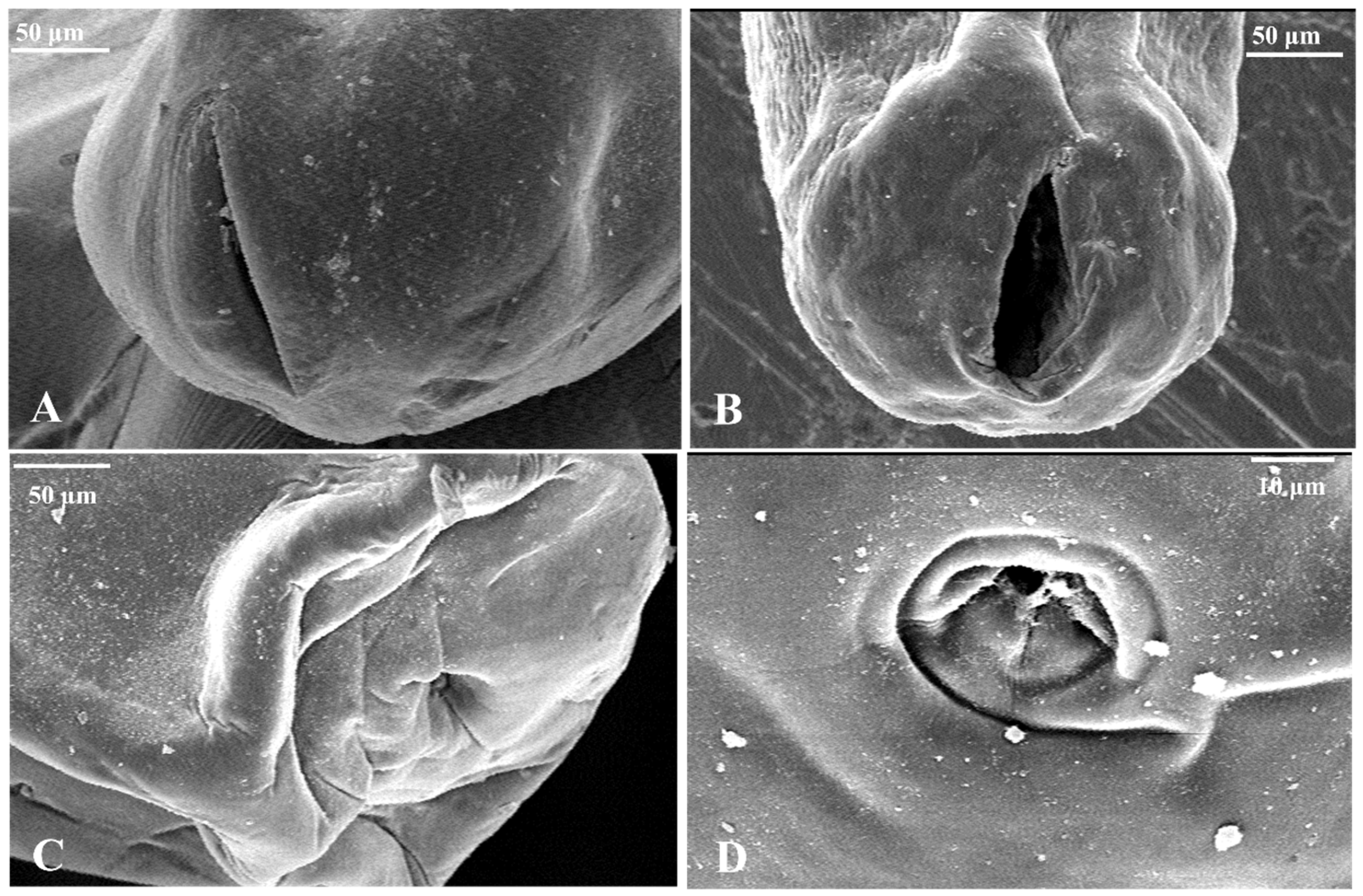
The male genitalia occupy the posterior half of the trunk: two ovoid testes, one behind the other, followed by a cement gland and a clear male bursa at the posterior end (Figure 2C). The gonopore is located terminally in both sexes. In females, a genital opening is found at the end of the worm, varying in size and shape; it can be round or a vertical slit that occupies nearly the entire diameter of the trunk, with noticeable lips (Figure 6A,B). The male bursa has a thick muscular margin and a terminal gonopore (Figure 6C), which is equipped with a mildly serrated, thick muscular rim and an internal ring of sensory papillae (Figure 6D).

3.3. Molecular Characterization

Both the mitochondrial COI, with an amplicon of approximately 776 bp (accession numbers PP859185: https://www.ncbi.nlm.nih.gov/nuccore/PP859185; deposited on 4 March 2025), and the nuclear 18S rRNA gene sequence (amplicon = approximately 1700 bp; accession numbers MZ727194: https://www.ncbi.nlm.nih.gov/nuccore/MZ727194; deposited on 20 August 2021), were used for species identification.
Phylogenetic analysis using MEGA11 revealed the relationship between R. niloticus isolated in this study and other species of Rhadinorhynchidae.
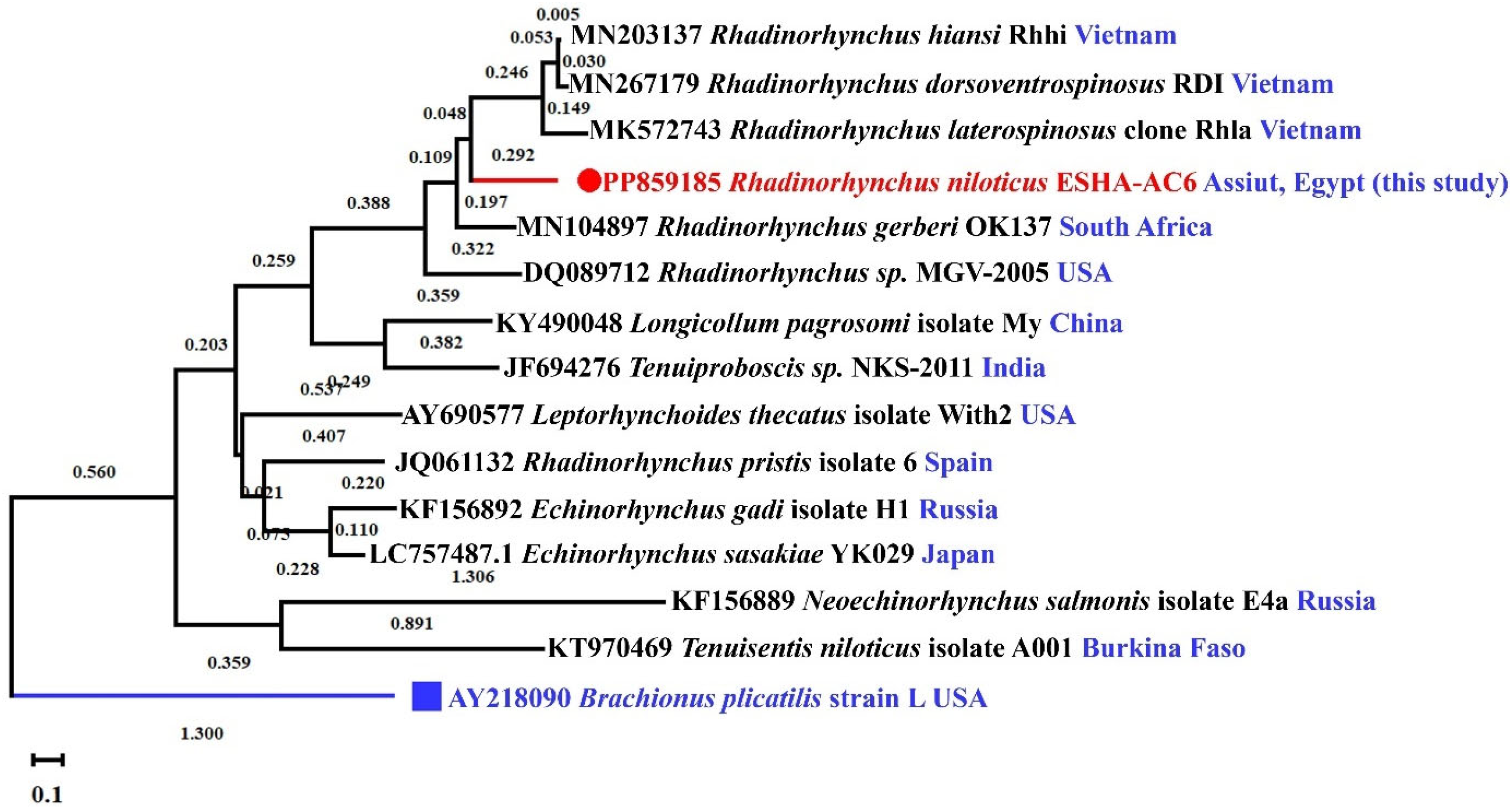
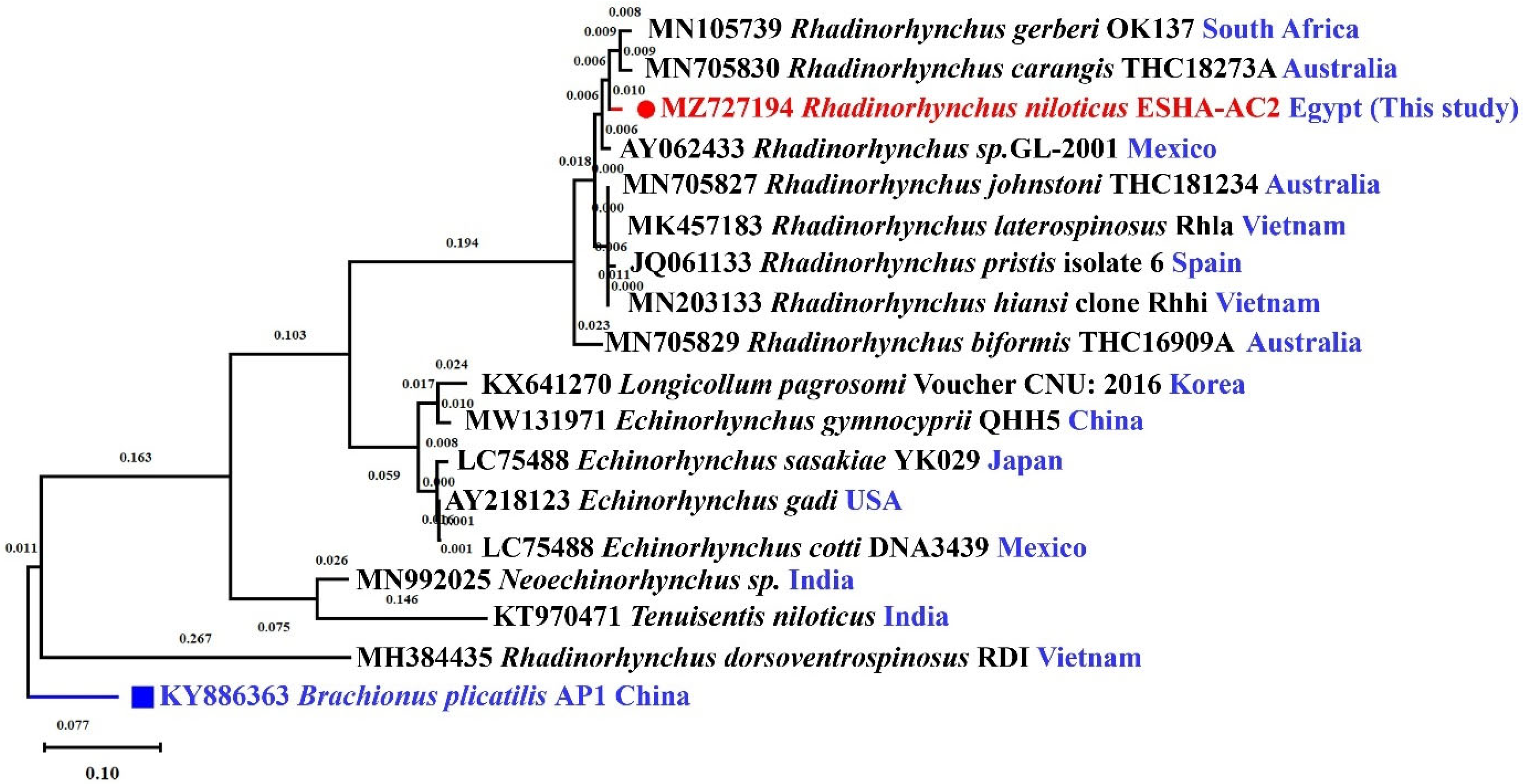
In this study, R. niloticus isolates displayed a monophyletic group that distinguished them from other genospecies using the maximum-likelihood method (Figure 7 and Figure 8). Both sequences of the COI and nuclear 18S rRNA genes from the isolated parasites were deposited in GenBank under accession numbers PP859185 and MZ727194, respectively.
The alignment of the mitochondrial COI gene contained 15 taxa and 1045 bp. Out of the Rhadinorhynchus species used in maximum-likelihood analysis, nine of ten Rhadinorhynchus exhibited high phylogenetic signaling and were recovered as a monophyletic clade. This clade includes R. gerberi Lisitsyna, Kudlai, Cribb and Smit, 2019 (MN105739; Lisitsyna, Kudlai, Cribb and Smit [28]), R. carangis Yamaguti, 1939 (MN705830; Huston, Cribb and Smales [34]), this study isolate (MZ727194), Rhadinorhynchus sp. isolated from a marine fish caught on the western Pacific coast of Mexico (AY062433; García-Varela, Cummings, Pérez-Ponce de León, Gardner and Laclette [30]), R. laterospinosus Amin, Heckmann and Nguyen Van Ha, 2011 (MK457183; Amin, Heckmann, Dallarés, Constenla and Ha [33]), R. johnstoni Golvan, 1969 (MN705827, Huston, Cribb and Smales [34]), R. hiansi Soota and Bhattacharya, 1981 (MN203133; Amin, Heckmann, Dallarés, Constenla and Van Ha [29]), R. pristis Rudolphi, 1802 (JQ061133; Gregori, Aznar, Abollo, Roura, González and Pascual [35]), and R. biformis Smales, 2014 (MN705829; Huston, Cribb and Smales [34]). However, R. dorsoventrospinosus (MH384435, Chaudhary, Amin, Heckmann and Singh [32]) arose as a subclade separate from other Rhadinorhynchus.
The alignment of the 18S rRNA gene of the small subunit (SSU) consisted of 18 taxa and 1867 bp, exhibited high phylogenetic signaling, and recovered nine out of ten Rhadinorhynchus species as a monophyletic clade. This clade includes R. gerberi Lisitsyna, Kudlai, Cribb and Smit, 2019 (MN105739; Lisitsyna, Kudlai, Cribb and Smit [28]), R. carangis Yamaguti, 1939 (MN705830; Huston, Cribb and Smales [34]), this study isolate (MZ727194), Rhadinorhynchus sp. isolated from a marine fish caught on the western Pacific coast of Mexico (AY062433; García-Varela, Cummings, Pérez-Ponce de León, Gardner and Laclette [30]), R. laterospinosus Amin, Heckmann and Nguyen Van Ha, 2011 (MK457183; Amin, Heckmann, Dallarés, Constenla and Ha [33]), R. johnstoni Golvan, 1969 (MN705827, Huston, Cribb and Smales [34]), R. hiansi Soota and Bhattacharya, 1981 (MN203133; Amin, Heckmann, Dallarés, Constenla and Van Ha [29]), R. pristis Rudolphi, 1802 (JQ061133; Gregori, Aznar, Abollo, Roura, González and Pascual [35]), and R. biformis Smales, 2014 (MN705829; Huston, Cribb and Smales [34]). Again, R. dorsoventrospinosus (MH384435, Chaudhary, Amin, Heckmann and Singh [32]) arose as a subclade separate from other Rhadinorhynchus.
The genetic divergence estimated among the COI gene showed high divergence distances, ranging from 0.27 to 0.44 among species of the Rhadinorhynchidae family. However, this range increased to 0.45–0.60 among other tested Acanthocephala members from different families (Table 2). In contrast, the sequences of 18S rRNA gene of the study isolate and the most closely related species within the same family had closer divergence distances, ranging from 0.01 to 0.05, except for R. dorsoventrospinosus, which had 0.34 divergence distances with this study isolate. However, it ranged from 0.19 to 0.34 among other tested Acanthocephala species from different families (Table 3).

4. Discussion

Lates niloticus is a large piscivorous fish belonging to the Latidae family, with high value in commercial, artisanal, and recreational fisheries [42]. It can be infected with numerous parasitic infections, such as the myxosporean Henneguya ghaffari, the monogeneans Diplectanum lacustris, D. simile, and Tylodelphys spp., the nematodes Philometra ovata and the third larval stage of Contracaecum quadripapillatum, and the crustacean parasites Ergasilus kandti and E. latus [20,43,44,45,46,47,48,49]. This study represents the first comprehensive morphological and molecular identification of R. niloticus.
Acanthocephalans are thorny-headed helminths that complete their life cycles using arthropods and vertebrates as intermediate hosts. They are sexually separated and commonly found in the intestines of freshwater and marine fish, as well as in other vertebrates [37]. The presence of a proboscis equipped with hooks, a syncytial epidermis, and a lacuna system with circulatory channels is characteristic of these helminths. They lack an alimentary tract and facilitate direct nutrient absorption through the body wall [50]. Rhadinorhynchidae, as described by Travassos (1923) [51], is a family of parasitic worms belonging to the order Echinorhynchidae. The genus Rhadinorhynchus, first identified by Lühe in 1911 [52], has been found in more than 47 host individuals in both freshwater and marine habitats [53]. These worms parasitize the intestines of teleosts as adults. The anterior body region is equipped with small, scattered, pointed, backwards-directed cuticular spines covered by cuticular folds. The proboscis and proboscis receptacles are very long. The lemnisci are long and finger-like. The proboscis is armed with hooks arranged in separate longitudinal rows, with the ventral proboscis hooks being stronger and pointed with arched ends compared to the dorsal hooks. Similar studies have documented the presence of this parasite in Egyptian Nile perch in many localities, such as in Aswan (Upper Egypt) by Hamouda et al. [54] and Bazh and Hamouda [55], Qena Governorate by El-Shahawy et al. [49], and, in a recent manuscript, Al Minya by El-Siefy, Ibraheem and Abd El-Kareem [47]. The isolated parasite was identified as R. niloticus based on morphological traits previously described by Mohamadain [56], Mazen and Thabit [46], and Bazh and Hamouda [55].
The sample size was selected following Shvydka, Sarabeev, Estruch and Cadarso-Suárez [10] to ensure accurate calculation of the prevalence, mean intensity, and mean abundance of a specific fish parasite. The present study encountered several challenges due to the limited literature describing the parasite and its prevalence in natural habitats. The prevalence rate was found to be 10.7%, which was lower than that reported by Bazh and Hamouda [55], who found that 51.5% of the examined fish were infected with R. niloticus. This difference could be attributed to variations in time and location, as well as biotic and abiotic factors.
In the current study, the 18S rRNA gene, a commonly used marker for deeper phylogenetic relationships, was used for molecular identification. Based on a total of 1656 bases of the 18S rRNA sequence, the currently isolated R. niloticus (MZ727194) is closely related to Rhadinorhynchidae members R. laterospinosus (MK457183) and R. hiansi (MN203133) isolated from Vietnamese marine teleosts, sharing identities of 98.70% and 98.68%, respectively. Additionally, both R. gerberi (MN105739), isolated from a South African marine fish, and R. pristis (JQ061133), isolated from Spain, share a 98.28% identity with the 18S rRNA sequence of the current study. Furthermore, Rhadinorhynchus sp. (AY062433) was collected from the intestines of fish belonging to the family Scianidae, displaying a 98.19% identity. However, the 18S rRNA sequence of the current study showed significant differences from other Rhadinorhynchidae members isolated from Australian marine teleosts. For example, R. carangis (MN705830) had a 99% coverage and 98.04% identity, R. johnstoni (MN705827) showed a 97.59% identity, and the lowest identity of 95.31% with 98% coverage was seen with R. biformis (MN705829). This close relationship to members of Rhadinorhynchidae was supported by narrow divergence distances ranging from 0.01 to 0.05 between the current study isolate (MZ727194) and other members in the Rhadinorhynchidae. However, this distance increased significantly when the current study isolate was compared with acanthocephalan parasites from other families and orders, ranging between 0.19 and 0.34. Like the 18S rRNA sequence, the COI gene sequence of R. niloticus in the current study (PP859185) is closely related to members of Rhadinorhynchidae. Specifically, it shares 100% coverage and 77.26% similarity with R. gerberi (MN104897) isolated from South Africa, 98% coverage and 77.04% similarity with R. hiansi (MN203137) isolated from Vietnamese marine teleosts, 98% coverage and 76.23% similarity with Rhadinorhynchus sp. (DQ089712) isolated in the USA, R. dorsoventrospinosus (MN267179) with 89% coverage and 75.80% similarity, 100% coverage and 79.92% similarity with R. laterospinosus (MK572743) isolated from Vietnamese marine teleosts, and 71% coverage and 71.36% similarity with R. pristis (JQ061132) isolated from Spain. These results were supported by narrow divergence distance ranges between 0.27 and 0.44 among the Rhadinorhynchidae members and the current study isolate (PP859185). There was a wider divergence distance ranging between 0.45 and 0.60 between this study’s parasite and other tested acanthocephalan COI gene sequences. However, the parasite studied in the current research has been documented in many locations along the Egyptian Nile in L. niloticus; molecular study encountered a challenge due to a lack of sequences in GenBank. Both gene sequences will contribute to future studies on this parasite in L. niloticus.

5. Conclusions

Adults of R. niloticus, belonging to the Rhadinorhynchidae family, were identified in Assiut City, Egypt, using an integrated morphological and molecular approach. They were found living freely in the intestines of wild L. niloticus. Their prevalence was 10.7%, with a mean intensity of 2.16 ± 0.47 (95% CI: 1.33 to 3.17) and 0.23 ± 0.08 (95% CI: 0.11 to 0.43). Morphological identification was conducted using both light and scanning electron microscopy. Molecular identification was also performed using both the universal metazoan cytochrome oxidase 1 gene (often used for species-level identification and resolving more recent evolutionary divergences) and the 18S rRNA gene, one of the commonly used ribosomal markers for deeper phylogenetic relationships. This confirmed their identification, and the sequences were deposited in GenBank under accession numbers PP859185 and MZ727194. The current research is the first study in Egypt to identify R. niloticus in wild L. niloticus using both morphological and molecular techniques, with the assistance of two universal gene sequences. However, additional studies should be conducted to clarify its impact on the fish’s immune system.

Author Contributions

Conceptualization, E.S.H.A. and H.T.; methodology, E.S.H.A. and H.T.; validation, E.S.H.A., M.A., H.T.; formal analysis, E.S.H.A.; investigation, E.S.H.A., M.A., H.T.; data curation, E.S.H.A., M.A., H.T.; writing—original draft preparation, E.S.H.A.; writing—review and editing, E.S.H.A., M.A., H.T.; visualization, E.S.H.A. and M.A.; supervision, E.S.H.A.; funding acquisition, M.A. All authors have read and agreed to the published version of the manuscript.

Funding

This research received no external funding.

Institutional Review Board Statement

All methods used in this study were carried out in accordance with relevant guidelines and regulations. Ethical approval for this study was obtained from Assiut University, Faculty of Science Ethics Committee for Animal Use and Care (Number 01/2024/0018). The study was carried out in compliance with the ARRIVE guidelines.

Data Availability Statement

Data and materials are available upon reasonable request from the corresponding author. The datasets generated during the current study are available in GenBank under accession numbers PP859185, accessed on 4 March 2025 (https://www.ncbi.nlm.nih.gov/nuccore/PP859185) and MZ727194, accessed on 20 August 2021 (https://www.ncbi.nlm.nih.gov/nuccore/MZ727194).

Acknowledgments

We would like to thank Mahmoud Mostafa Mahmoud Mohamed, of the Department of Aquatic Animal Medicine and Management at Assiut University, for proofreading and English editing the manuscript.

Conflicts of Interest

The authors declare no conflicts of interest.

References

  1. Aloo, P.A.; Njiru, J.; Balirwa, J.S.; Nyamweya, C.S. Impacts of Nile Perch, Lates niloticus, introduction on the ecology, economy and conservation of Lake Victoria, East Africa. Lakes Reserv. Sci. Policy Manag. Sustain. Use 2017, 22, 320–333. [Google Scholar] [CrossRef]
  2. Asnake, W. Nile Perch (Lates niloticus): The Promising White Meat of the World. J. Nutr. Food Sci. 2018, 8, 680–683. [Google Scholar] [CrossRef]
  3. Başusta, N.; Mutlu, E.; Deval, M.C. Parasitic isopods (Anilocra frontalis H. Milne Edwards, 1830 and Ceratothoa capri (Trilles, 1964)) from the Antalya Bay (Turkey) with new host records. Turk. J. Sci. Technol. 2017, 12, 11–15. [Google Scholar]
  4. Sebastião, F.d.A.; Braga de Oliveira, M.I.; Rocha, M.J.S.; Souza, D.C.d.M.; Ribeiro, P.; Majolo, C.; Crescêncio, R.; Chagas, E.C. Effect of a food additive in the control of the acanthocephalan Neoechinorhynchus buttnerae in Colossoma macropomum. Aquac. Res. 2021, 52, 635–642. [Google Scholar] [CrossRef]
  5. Roy, P.E. Nematode (Round Worm) Infection in Fish, 2nd ed.; IFAS: Gainesville, FL, USA, 2002. [Google Scholar]
  6. Paperna, I. Fish Disease and Disorders; CABI Publishing: Kelowna, BC, Canada, 2001; Volume 1. [Google Scholar]
  7. Ismen, A.; Bingel, F. Nematode infection in the whiting Marsangius euxinus off Turkish coast of the black sea. Fish. Res. 1999, 42, 183–189. [Google Scholar] [CrossRef]
  8. Amin, O.M. Classification of the acanthocephala. Folia Parasitol. 2013, 60, 273–305. [Google Scholar] [CrossRef]
  9. Gibson, D.; Wayland, M. World List of Marine Acanthocephala. Rhadinorhynchus Lühe, 1911. World Register of Marine Species. Available online: https://www.marinespecies.org/aphia.php?p=taxdetails&id=20399 (accessed on 28 June 2025).
  10. Shvydka, S.; Sarabeev, V.; Estruch, V.D.; Cadarso-Suárez, C. Optimum Sample Size to Estimate Mean Parasite Abundance in Fish Parasite Surveys. Helminthologia 2018, 55, 52–59. [Google Scholar] [CrossRef] [PubMed]
  11. Reiczigel, J.; Marozzi, M.; Fábián, I.; Rózsa, L. Biostatistics for parasitologists–A primer to quantitative parasitology. Trends Parasitol. 2019, 35, 277–281. [Google Scholar] [CrossRef] [PubMed]
  12. Kildea, M.A.; Allan, G.L.; Kearney, R.E. Accumulation and clearance of the anaesthetics clove oil and AQUI-S™ from the edible tissue of silver perch (Bidyanus bidyanus). Aquaculture 2004, 232, 265–277. [Google Scholar] [CrossRef]
  13. Paugy, D.; Lévêque, C.; Teugels, G. The Fresh and Brackish Water Fishes of West Africa; Faune et Flore Tropicales: Paris, France, 2003; Volume 1. [Google Scholar]
  14. Eissa, A.E. Clinical and Laboratory Manual of Fish Diseases; LAP LAMBERT Academic Publishing: Saarbrücken, Germany, 2016. [Google Scholar]
  15. Noga, E.J. Fish Disease: Diagnosis and Treatment, 2nd ed.; Iowa State University Press: Ames, IA, USA, 2010. [Google Scholar]
  16. Abdallah, E.S.H.; Hamouda, A.H. Livoneca redmanii Leach, 1818 (Cymothoidae) a parasitic isopod infesting the gills of the European seabass, Dicentrarchus labrax (Linnaeus, 1758): Morphological and molecular characterization study. BMC Vet. Res. 2022, 18, 330. [Google Scholar] [CrossRef]
  17. Abdallah, E.S.H.; Hamouda, A.H. Morphological and molecular characterization of Lernanthropus kroyeri, a copepod infesting the gills of European seabass Dicentrarchus labrax. Egypt. J. Aquat. Res. 2023, 49, 49–55. [Google Scholar] [CrossRef]
  18. Hassan, E.S.; Mahmoud, M.M.; Metwally, A.M.; Moktar, D.M. Lamproglena monodi (Copepoda: Lernaeidae), infesting gills of Oreochromis niloticus and Tilapia zillii. Glob. J. Fish. Aquac. Res. 2013, 6, 1–16. [Google Scholar]
  19. Mahmoud, M.M.; Hassan, E.S.; Haridy, M.; Nour El Deen, E.A.; Kuraa, H.M.M.; Hanna, H.N.S. Parasitic infections of the gills of wild African Sharptooth Catfish (Clarias gariepinus). Assiut Vet. Med. J. 2018, 64, 31–39. [Google Scholar] [CrossRef]
  20. Thabit, H.; Abdallah, E.S.H. Morphological and molecular identification of third-stage Contracaecum larvae (Nematoda: Anisakidae) parasitizing Nile perch Lates niloticus in Egypt. Aquac. Res. 2022, 53, 4869–4881. [Google Scholar] [CrossRef]
  21. Bush, A.O.; Lafferty, K.D.; Lotz, J.M.; Shostak, A.W. Parasitology meets ecology on its own terms: Margolis et al. revisited. J. Parasitol. 1997, 83, 575–583. [Google Scholar] [CrossRef]
  22. Abdallah, E.S.H.; Mahmoud, M.M.; Abdel-Rahim, I.R. Trichosporon jirovecii infection of red swamp crayfish (Procambarus clarkii). J. Fish. Dis. 2018, 41, 1719–1732. [Google Scholar] [CrossRef]
  23. Folmer, O.; Black, M.; Hoeh, W.; Lutz, R.; Vrijenhoek, R. DNA primers for amplification of mitochonrial cytochrome c oxidase subunit I from diverse metazoan invertebrates. Mol. Mar. Biol. Biotechnol. 1994, 3, 294–299. [Google Scholar]
  24. Garey, J.R.; Near, T.J.; Nonnemacher, M.R.; Nadler, S.A. Molecular evidence for Acanthocephala as a subtaxon of Rotifera. J. Mol. Evol. 1996, 43, 287–292. [Google Scholar] [CrossRef]
  25. Hasegawa, M.; Kishino, H.; Yano, T. Dating the human-ape split by a molecular clock of mitochondrial DNA. J. Mol. Evol. 1985, 22, 160–174. [Google Scholar] [CrossRef]
  26. Tamura, K.; Stecher, G.; Kumar, S. MEGA11: Molecular Evolutionary Genetics Analysis version 11. Mol. Biol. Evol. 2021, 38, 3022–3027. [Google Scholar] [CrossRef]
  27. Tamura, K. Estimation of the number of nucleotide substitutions when there are strong transition-transversion and G + C-content biases. Mol. Biol. Evol. 1992, 9, 678–687. [Google Scholar] [CrossRef] [PubMed]
  28. Lisitsyna, I.O.; Kudlai, O.; Cribb, H.T.; Smit, J.N. Three new species of acanthocephalans (Palaeacanthocephala) from marine fishes collected off the East Coast of South Africa. Folia Parasitol. 2019, 66, 1–20. [Google Scholar] [CrossRef] [PubMed]
  29. Amin, O.M.; Heckmann, R.A.; Dallarés, S.; Constenla, M.; Van Ha, N. Morphological and Molecular Description of Rhadinorhynchus hiansi Soota and Bhattacharya, 1981 (Acanthocephala: Rhadinorhynchidae) from Marine Fish off the Pacific Coast of Vietnam. J. Parasitol. 2020, 106, 56–70, 15. [Google Scholar] [CrossRef] [PubMed]
  30. García-Varela, M.; Cummings, M.P.; Pérez-Ponce de León, G.; Gardner, S.L.; Laclette, J.P. Phylogenetic analysis based on 18S ribosomal RNA gene sequences supports the existence of class polyacanthocephala (acanthocephala). Mol. Phylogenet Evol. 2002, 23, 288–292. [Google Scholar] [CrossRef]
  31. García-Varela, M.; Nadler, S.A. Phylogenetic relationships among Syndermata inferred from nuclear and mitochondrial gene sequences. Mol. Phylogenet. Evol. 2006, 40, 61–72. [Google Scholar] [CrossRef]
  32. Chaudhary, A.; Amin, O.M.; Heckmann, R.; Singh, H.S. The molecular profile of Rhadinorhynchus dorsoventrospinosus Amin, Heckmann, and ha 2011 (Acanthocephala: Rhadinorhynchidae) from Vietnam. J. Parasitol. 2020, 106, 418–427. [Google Scholar] [CrossRef]
  33. Amin, O.M.; Heckmann, R.A.; Dallarés, S.; Constenla, M.; Ha, N.V. Morphological and molecular description of Rhadinorhynchus laterospinosus Amin, Heckmann & Ha, 2011 (Acanthocephala, Rhadinorhynchidae) from marine fish off the Pacific coast of Vietnam. Parasite 2019, 26, 14. [Google Scholar] [CrossRef]
  34. Huston, D.C.; Cribb, T.H.; Smales, L.R. Molecular characterisation of acanthocephalans from Australian marine teleosts: Proposal of a new family, synonymy of another and transfer of taxa between orders. Syst. Parasitol. 2020, 97, 859–861. [Google Scholar] [CrossRef]
  35. Gregori, M.; Aznar, F.; Abollo, E.; Roura, A.; González, A.; Pascual, S. Nyctiphanes couchii as intermediate host for Rhadinorhynchus sp. (Acanthocephala, Echinorhynchidae) from NW Iberian Peninsula waters. Dis. Aquat. Org. 2013, 105, 9–20. [Google Scholar] [CrossRef]
  36. Steinauer, M.L.; Nickol, B.B.; Ortí, G. Cryptic speciation and patterns of phenotypic variation of a highly variable acanthocephalan parasite. Mol. Ecol. 2007, 16, 4097–4109. [Google Scholar] [CrossRef]
  37. Li, L.; Chen, H.-X.; Amin, O.M.; Yang, Y. Morphological variability and molecular characterization of Pomphorhynchus zhoushanensis sp. nov. (Acanthocephala: Pomphorhynchidae), with comments on the systematic status of Pomphorhynchus Monticelli, 1905. Parasitol. Int. 2017, 66, 693–698. [Google Scholar] [CrossRef] [PubMed]
  38. Kita, Y.; Kajihara, H. Morphological and molecular characterization of a new species of the genus Echinorhynchus Zoega in Müller, 1776 (Acanthocephala: Echinorhynchidae) parasitizing the rock greenling Hexagrammos lagocephalus (Pallas) (Scorpaeniformes: Hexagrammidae) from eastern Hokkaido, Japan. Syst. Parasitol. 2023, 100, 735–743. [Google Scholar] [CrossRef] [PubMed]
  39. Malyarchuk, B.; Derenko, M.; Mikhailova, E.; Denisova, G. Phylogenetic relationships among Neoechinorhynchus species (Acanthocephala: Neoechinorhynchidae) from North-East Asia based on molecular data. Parasitol. Int. 2014, 63, 100–107. [Google Scholar] [CrossRef] [PubMed]
  40. Giribet, G.; Sørensen, M.V.; Funch, P.; Kristensen, R.M.; Sterrer, W. Investigations into the phylogenetic position of Micrognathozoa using four molecular loci. Cladistics 2004, 20, 1–13. [Google Scholar] [CrossRef]
  41. Amin, O.M.; Evans, R.P.; Boungou, M.; Heckmann, R. Morphological and molecular description of Tenuisentis niloticus (Meyer, 1932) (Acanthocephala: Tenuisentidae) from Heterotis niloticus (Cuvier) (Actinopterygii: Arapaimidae), in Burkina Faso, with emendation of the family diagnosis and notes on new features, cryptic genetic diversity and histopathology. Syst. Parasitol. 2016, 93, 173–191. [Google Scholar] [CrossRef]
  42. Koblmüller, S.; Schöggl, C.A.; Lorber, C.J.; Van Steenberge, M.; Kmentová, N.; Vanhove, M.P.M.; Zangl, L. African lates perches (Teleostei, Latidae, Lates): Paraphyly of Nile perch and recent colonization of Lake Tanganyika. Mol. Phylogenet Evol. 2021, 160, 107141. [Google Scholar] [CrossRef]
  43. Elhawary, N.M.; Hamouda, A.H.; Bazh, E.K.; El-Bahy, N.M.; Sorour, S.S. Phenotypic and genomic characterization of Tylodelphys sp. metacercaria (Diesing 1850)(Trematoda: Diplostomidae) recovered from Lates niloticus (Linnaeus, 1758) Egypt. Vet. Med. Soc. Parasitol. J. EVMSPJ 2022, 18, 15–34. [Google Scholar]
  44. Thon, C.; Otachi, O.; Oldewage, A. Endo-helminths infestation in Nile perch, Lates niloticus, (L.,) and Nile tilapia, Oreochromis niloticus (L.,) in Winam Gulf of Lake Victoria, Kenya. Egerton J. Sci. Technol. 2019, 16, 1–139. [Google Scholar]
  45. Outa, J.O.; Dos Santos, Q.M.; Avenant-Oldewage, A.; Jirsa, F. Parasite diversity of introduced fish Lates niloticus, Oreochromis niloticus and endemic Haplochromis spp. of Lake Victoria, Kenya. Parasitol. Res. 2021, 120, 1583–1592. [Google Scholar] [CrossRef]
  46. Mazen, N.; Thabit, H. Light and scanning electron microscopy of three parasitic helminths from freshwater fishes in Assiut, Egypt. Assiut Vet. Med. J. 2005, 51, 1–12. [Google Scholar]
  47. El-Siefy, A.M.; Ibraheem, M.H.; Abd El-Kareem, S.G. Ovarian balls (Floating ovaries) of Rhadinorhynchus niloticus Mohamadain, 1989 from the Nile perch Lates niloticus Linnaeus, 1758; an electron microscope study. Helminthologia 2024, 61, 194–200. [Google Scholar] [CrossRef] [PubMed]
  48. Ebraheem, M.E. Studies on Some of the Parasites in Some of Nile Fishes in Sohag Governorate; Assiut University: Asyut, Egypt, 1992. [Google Scholar]
  49. El-Shahawy, I.; El-Seify, M.; Metwally, A.; Fwaz, M. Survey on endoparasitic fauna of some commercially important fishes of the River Nile, southern of Egypt (Egypt). Revue de Médecine Vétérinaire 2017, 168, 126–134. [Google Scholar]
  50. Smales, L.R. The genus Rhadinorhynchus (Acanthocephala: Rhadinorhynchidae) from marine fish in Australia with the description of four new species. Acta Parasitol. 2014, 59, 721–736. [Google Scholar] [CrossRef] [PubMed]
  51. Travassos, L. Informações sobre a fauna helrninthologica de Matto Grosso (II Nota). Folha Medica 1923, 4, 12–16. [Google Scholar]
  52. Lühe, M. Acanthocephalen: Register der Acanthocephalen und Parasitischen Plattwürmer, Geordnet nach ihren Wirten; Brauer, A., Ed.; Gustav Fischer Verlag: Jena, Germany, 1911. (In German) [Google Scholar]
  53. Barton, D.P.; Smales, L.R. Acanthocephalan cystacanths from flatfish (Order pleuronectiformes) in Tropical Australian waters. J. Parasitol. 2015, 101, 429–435. [Google Scholar] [CrossRef]
  54. Hamouda, A.H.; Sorour, S.S.; El-Habashi, N.M.; Adam, E.-H.A. Parasitic infection with emphasis on Tylodelphys spp. as new host and locality records in Nile perch; Lates niloticus from Lake Nasser, Egypt. World’s Vet. J. 2018, 8, 19–33. [Google Scholar]
  55. Bazh, E.; Hamouda, A. Scanning morphology, prevalence and histopathology of some acanthocephalans infecting some River Nile fish. Bulg. J. Vet. Med. (Online First) 2019, 87, 239–250. [Google Scholar] [CrossRef]
  56. Mohamadain, H.S. Studies on Helminth Parasites of the Nile Fishes in Quena Province, A.R, Egypt; Faculty of Science Quena Branch, Assiut University: Qena, Egypt, 1989. [Google Scholar]
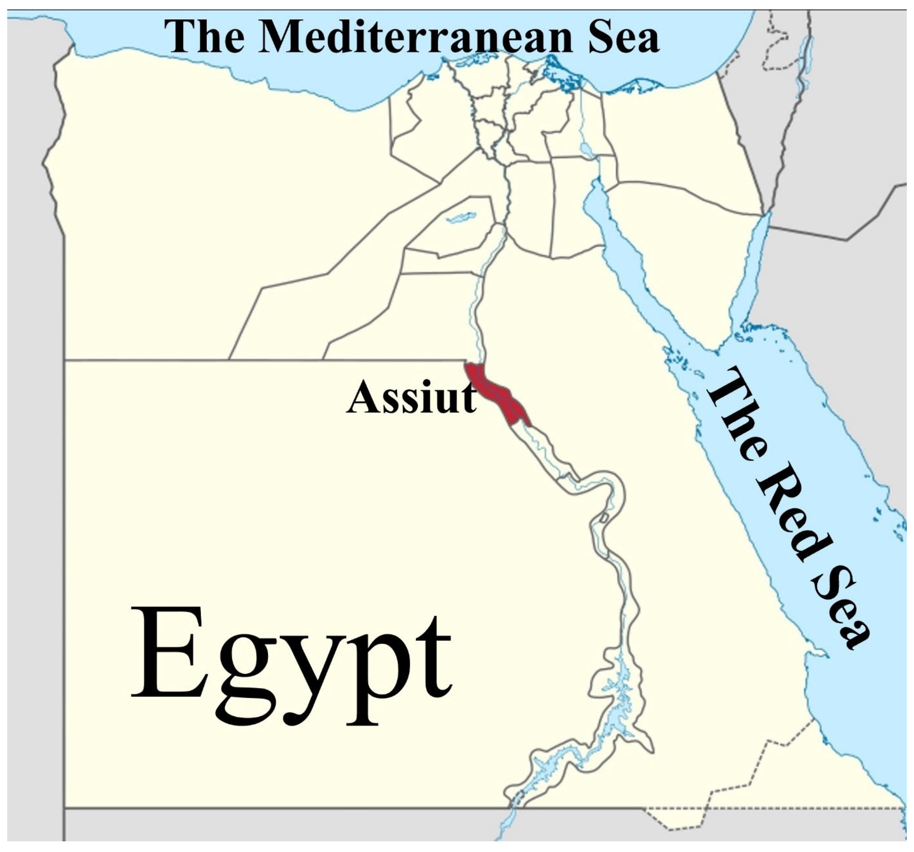
Figure 1. Map of Assiut governorate (highlighted in red) in Egypt.
Fishes 10 00397 g001
Figure 2. Light micrographs of Rhadinorhynchus niloticus (Acanthocephala: Rhadinorhynchidae) from Nile perch (Lates niloticus; Perciformes: Latidae) reveal the following features: (A) a completely everted proboscis and the anterior part of the trunk; (B) a partially retracted proboscis in the proboscis receptacle; (C) the posterior end of a male parasite. Abbreviations: P, proboscis; N, neck; L, Lemnisci; TS, trunk spines; AT, anterior testis; PT, posterior testis; CG, cement gland; SD, seminal duct.
Fishes 10 00397 g002
Figure 3. Scanning electron micrographs of Rhadinorhynchus niloticus (Acanthocephala: Rhadinorhynchidae) from Nile perch (Lates niloticus; Perciformes: Latidae) reveal the following: (A) a fully everted curved proboscis, neck, and anterior part of the trunk; (B) the anterior end of the proboscis, highlighting the small size of the apical hooks compared to the subapical hooks; (C) the apical end of the proboscis displaying the apical epidermal cone (arrow).
Fishes 10 00397 g003
Figure 4. Scanning electron micrographs of Rhadinorhynchus niloticus (Acanthocephala: Rhadinorhynchidae) from Nile perch (Lates niloticus; Perciformes: Latidae) reveal the shapes of the proboscis hooks in three regions: (A) the anterior region of the proboscis; (B) the middle region of the proboscis; and (C) the basal hooks of the proboscis. It is important to note the differences in shape and size between the proboscis hooks (h) and the tegumental spines (S).
Fishes 10 00397 g004
Figure 5. Scanning electron micrographs of Rhadinorhynchus niloticus (Acanthocephala: Rhadinorhynchidae) from Nile perch (Lates niloticus; Perciformes: Latidae) reveal: (A) the spineless smooth conical neck located between the posterior end of an everted proboscis and the anterior trunk cone; (B) a closer look at the tegumental spines on the anterior trunk cone; (C) tegumental spines in the middle region of the trunk.
Fishes 10 00397 g005
Figure 6. Scanning electron micrographs of Rhadinorhynchus niloticus (Acanthocephala: Rhadinorhynchidae) from Nile perch (Lates niloticus; Perciformes: Latidae) reveal the following: (A) and (B) show a partially and fully opened female gonopore, respectively, occupying almost the entire trunk diameter with prominent lips; (C,D) display a male bursa with a thick muscular margin and a terminal gonopore, of which (D) provides a ventral view of the male bursa in (C) showing a mildly serrated thick muscular rim and an internal ring of sensory papillae.
Fishes 10 00397 g006
Figure 7. The maximum likelihood method was used to estimate phylogenetic relationships among acanthocephala and the study isolate of Rhadinorhynchus niloticus (PP859185, red color) isolated from Nile perch (Lates niloticus) intestine based on mitochondrial cytochrome C (COI) gene sequences using the MEGA11 program. Brachionus plicatilis was used as the outgroup (blue color). The bar indicates the genetic distance resulting from the sequence variation.
Fishes 10 00397 g007
Figure 8. The maximum likelihood method was used to estimate the phylogenetic relationships among acanthocephala. The study focused on Rhadinorhynchus niloticus (MZ727194; red color) isolated from Nile perch (Lates niloticus) based on nuclear 18S rRNA gene sequences using MEGA11. Brachionus plicatilis was used as the outgroup (blue color). The bar indicates the genetic distance resulting from the sequence variation.
Fishes 10 00397 g008
Table 2. Estimates of evolutionary divergence of COI gene sequence pairs between Rhadinorhynchus niloticus (this study isolate) and other acanthocephalans.
123456789101112131415
1-PP859185 Rhadinorhynchus niloticus
2-MN104897 R. gerberi0.27
3-MN203137 R. hiansi0.290.27
4-DQ089712 Rhadinorhynchus sp.0.300.310.30
5-MN267179 R. dorsoventrospinosus0.310.280.030.30
6-MK572743 R. laterospinosus0.320.300.150.320.17
7-JQ061132 R. pristis0.440.420.410.430.440.45
8-JF694276 Tenuiproboscis sp.0.450.440.470.420.440.510.47
9-LC757487 Echinorhynchus sasakiae0.480.430.480.480.470.500.350.42
10-KF156892 E. gadi0.490.440.470.490.480.490.320.430.22
11-KY490048 Longicollum pagrosomi0.490.420.440.410.420.490.480.330.430.47
12-AY690577 Leptorhynchoides thecatus0.490.390.460.490.470.490.390.450.360.380.47
13-KF156889 Neoechinorhynchus salmonis0.550.570.440.580.600.600.500.550.560.530.630.60
14-KT970469 Tenuisentis niloticus0.600.600.620.590.650.650.490.550.490.490.630.530.53
15-AY218090 Brachionus plicatilis0.730.670.640.620.650.650.630.700.590.610.670.590.760.66
Table 3. Estimates of evolutionary divergence of 18S rRNA gene sequence pairs between Rhadinorhynchus niloticus (this study isolate) and other acanthocephalans.
123456789101112131415161718
1-MZ727194 Rhadinorhynchus niloticus
2-MN203133 R. hiansi0.01
3-AY062433 Rhadinorhynchus sp.0.010.01
4-MK457183 R. laterospinosus0.010.000.01
5-MN105739 R. gerberi0.020.020.020.02
6-MN705827 R. johnstoni0.020.000.020.000.02
7-JQ061133 R. pristis0.020.000.020.000.020.00
8-MN705830 R. carangis0.020.010.020.010.010.030.02
9-MN705829 R. biformis0.050.050.050.050.050.050.050.06
10-KX641270 Longicollum pagrosomi0.190.200.200.200.200.200.200.200.19
11-LC757488 Echinorhynchus sasakiae0.200.210.210.210.200.210.210.210.210.04
12-AY218123 E. gadi0.200.210.210.210.210.220.210.210.210.040.00
13-MW172280 E. cotti0.200.210.210.210.210.220.210.220.210.040.000.00
14-KT970471 Tenuisentis niloticus0.200.240.210.250.220.210.230.210.210.120.120.120.12
15-MW131971 E. gymnocyprii0.210.210.220.210.210.220.210.220.220.020.040.040.040.11
16-MH384435 R. dorsoventrospinosus0.340.290.340.290.320.340.280.350.350.260.300.300.300.220.31
17-MN992025 Neoechinorhynchus sp.0.340.350.340.350.350.350.360.350.330.260.270.270.270.070.260.21
18-KY886363 Brachionus plicatilis0.330.320.330.330.330.340.310.340.340.240.260.260.260.170.250.190.28
Disclaimer/Publisher’s Note: The statements, opinions and data contained in all publications are solely those of the individual author(s) and contributor(s) and not of MDPI and/or the editor(s). MDPI and/or the editor(s) disclaim responsibility for any injury to people or property resulting from any ideas, methods, instructions or products referred to in the content.