Review

Molecular Mechanisms and Biomarker-Based Early-Warning Indicators of Heavy Metal Toxicity in Marine Fish

Chemical Oceanography and Marine Pollution Department, National Institute for Marine Research and Development (NIMRD) “Grigore Antipa”, 300 Mamaia Blvd., 900581 Constanta, Romania
*
Authors to whom correspondence should be addressed.
Fishes 2025, 10(7), 339; https://doi.org/10.3390/fishes10070339
Submission received: 30 May 2025 / Revised: 30 June 2025 / Accepted: 8 July 2025 / Published: 10 July 2025
(This article belongs to the Section Environment and Climate Change)

Abstract

Heavy metals are among the most persistent and bioaccumulative pollutants in marine ecosystems, posing significant toxicological threats to fish via complex molecular and cellular disruptions. This review synthesizes current knowledge on the cascade of mechanistic responses in marine fish following HM exposure, which includes oxidative stress, modulation of antioxidant responses, activation of detoxification systems, DNA damage, inflammation, apoptosis, neuroendocrine disruption, and ultimately, cellular energy imbalance. In addition to established pathways, the review highlights recent advances in mechanistic understanding and biomarker development, including cellular stress responses, epigenetic regulation, metal homeostasis mechanisms, and novel molecular indicators. These mechanisms support the development of an integrated biomarker framework that combines classical indicators (e.g., antioxidant enzymes, metallothionein) with next-generation endpoints (e.g., miRNA profiles, gene-level responses of metal transporters or stress chaperones, epigenetic alterations). The interpretation of biomarker responses requires consideration of the exposure context, environmental variables, and physiological status to ensure accurate assessment of sublethal toxicity in field settings. By bridging mechanistic understanding with biomonitoring relevance, this review provides a comprehensive foundation for advancing molecular tools in pollution monitoring and risk assessment. Special emphasis is placed on biomarkers specific to heavy metal exposure, enhancing their diagnostic value relative to general stress indicators.
Key Contribution: By integrating mechanistic data on oxidative stress, detoxification, genotoxicity, transcriptional and epigenetic regulation, and lysosomal dysfunction from recent studies, this review constructs a comprehensive adverse-outcome framework for heavy metal toxicity in marine fish. It proposes a tiered biomarker panel, ranging from classical enzymatic markers to emerging indicators of subcellular stress, to support the development of early-warning tools for environmental assessment, with particular emphasis on responses specifically associated with metal exposure.

Graphical Abstract

1. Introduction

Heavy metals (HMs) are persistent and pervasive pollutants in marine environments, originating from both natural processes (e.g., erosion, volcanic activity) and anthropogenic activities such as mining, industrial discharge, and agricultural runoff [1,2,3]. Once introduced, HMs do not degrade but instead accumulate in sediments and organisms, potentially biomagnifying through food webs and posing significant risks to marine life and human health [4,5]. Fish are particularly vulnerable due to their ecological importance, commercial value, and physiological sensitivity to metal exposure [6,7].
The most encountered toxic metals in marine ecosystems include cadmium (Cd), lead (Pb), mercury (Hg), arsenic (As), copper (Cu), zinc (Zn), chromium (Cr), and nickel (Ni) [8]. These elements vary in biological function, with essential metals such as Cu, Zn, Fe, Mn, and Se required in trace amounts for enzymatic and metabolic functions [9]. For example, Cu is a cofactor for cytochrome c oxidase in mitochondrial respiration, Zn contributes to antioxidant defense via superoxide dismutase (SOD), and Se is integral to glutathione peroxidase (GPx) activity [10]. However, excess accumulation disrupts homeostasis, leading to oxidative stress, enzyme inhibition, and metabolic dysregulation [11]. In contrast, non-essential metals such as Cd, Pb, Hg, and As lack known physiological roles and are toxic even at low concentrations [12]. Cd tends to accumulate in the kidneys and liver, impairing calcium signaling and antioxidant capacity. Methylmercury, the most toxic form of Hg, crosses the blood–brain barrier, inhibits acetylcholinesterase (AChE), and induces neurotoxicity [13].
Due to their high bioaccumulation potential and susceptibility to environmental change, fish serve as valuable bioindicators of HM pollution. Metals enter fish through gill uptake, ingestion, and dermal absorption, subsequently accumulating in target organs such as the liver, kidneys, and brain [14,15]. Once internalized, HMs interfere with macromolecular functions, disrupt enzymatic activity, and promote reactive oxygen species (ROS) generation, triggering oxidative damage and inflammation [16,17].
In natural marine environments HMs rarely act alone. They co-occur with various pollutants including microplastics, pesticides, and pharmaceuticals, often resulting in synergistic or antagonistic toxic effects [18]. For instance, metals in combination with microplastics may exacerbate redox imbalance and inflammatory responses beyond individual exposures [19,20], while combined exposure to Cd and Hg has been shown to potentiate organ damage [21]. Conversely, trace elements like Zn and Se may mitigate cadmium or mercury toxicity through competitive uptake or enhanced antioxidant enzyme activity [22]. These interactions complicate environmental impact assessments and highlight the importance of mechanistic understanding [23].
Metal speciation also influences toxicity. Methylmercury is more bioavailable and neurotoxic than inorganic Hg [24], while free ionic forms of Cu and Cd readily penetrate gill epithelia and cause cellular injury [25]. Uptake pathways, accumulation patterns, and cellular defense capacity vary by species, life stage, and environmental conditions [26]. Some metals are sequestered into inert complexes, while others bind to proteins and nucleic acids, impairing physiological functions [27].
Although considerable research has characterized bioaccumulation patterns and ecological risks, there remains a critical need to elucidate the molecular mechanisms driving HM toxicity at sub-organismal levels [28]. These include oxidative stress, DNA disruption, altered gene expression, immune activation, and activation of detoxification pathways [29,30]. Molecular and biochemical markers often precede visible signs of pathology, providing a sensitive foundation for early detection and biomonitoring [31].
This review synthesizes current knowledge on the molecular and biochemical mechanisms of HM toxicity in marine teleosts, with particular emphasis on oxidative stress, antioxidant disruption, genotoxicity, programmed cell death, and cellular defense systems such as metallothioneins and glutathione. These responses disrupt cellular homeostasis and contribute to tissue damage, immune suppression, and impaired physiological performance. By integrating data from laboratory and field studies, this review highlights how these mechanistic molecular markers can inform environmental monitoring and risk assessment. Special attention is given to biomarkers that are specific to HM exposure, distinguishing them from generalized stress responses, thereby enhancing their utility in diagnostic, regulatory, and conservation contexts. The review is structured around major mechanistic domains, followed by sections on tissue-level outcomes, biomonitoring applications, and future directions aimed at increasing ecological and regulatory relevance.

2. Methodology

This study was conducted as a narrative review, structured to identify, select, and synthesize recent scientific literature on molecular, biochemical, and cellular biomarkers of HM toxicity in marine fish. Narrative reviews allow for a more flexible and integrative analysis of complex scientific fields. This approach is particularly suitable for evaluating diverse mechanistic responses and biomarker endpoints related to metal-related toxic effects in fish, where experimental designs, species, and biomarker types vary widely. The review focuses on mechanistic responses linked to early-warning indicators of contamination, including oxidative stress, detoxification, apoptosis, endocrine disruption, neurotoxicity, and energy metabolism disturbances. A structured search and data extraction strategy was applied to enhance transparency and ensure thematic consistency throughout the synthesis.
A comprehensive literature search was carried out using major indexing databases (Scopus, Web of Science, PubMed, and Google Scholar). The search strategy combined keywords using Boolean operators such as (“heavy metals” AND “marine fish”) AND (“biomarkers” OR “oxidative stress” OR “metallothionein” OR “heat shock protein” OR “transcriptomics” OR “miRNA” OR “DNA damage” OR “apoptosis” OR “neurotoxicity” OR “environmental monitoring”). The search was limited to English-language peer-reviewed publications between 2000 and 2024, with a focus on the last 5 years, and included only open-access articles or those available through institutional subscriptions. The inclusion criteria were as follows: (i) studies on marine or euryhaline fish, (ii) reporting molecular and biochemical responses to individual or mixed HMs (e.g., Cd, Pb, Hg, As, Cu, Zn), (iii) providing original experimental or field data. Excluded were studies on non-metal contaminants, non-fish aquatic species, terrestrial species, or non-peer-reviewed or modeling-only publications. A small number of recent review articles were retained to support synthesis and contextual interpretation.
Although this review was conducted as a narrative synthesis rather than a formal systematic review, elements of the PRISMA framework [32] were applied during the literature screening and data extraction phases to enhance methodological transparency and consistency. Studies were prioritized based on the following: (i) clarity and validation of biomarker endpoints, (ii) tissue and species specificity, (iii) reported exposure concentrations and durations. Biomarkers were further prioritized based on their mechanistic relevance to known toxicological pathways (e.g., oxidative stress, apoptosis, detoxification), their recurrence across multiple studies and species, and their application in environmental monitoring frameworks. From each eligible study, we extracted species name, HM(s) tested, biomarker(s) assessed, target tissue(s), exposure conditions, and observed effects (e.g., gene/protein upregulation, enzymatic inhibition, oxidative damage). Preference was given to biomarkers validated in marine or euryhaline fish under realistic exposure conditions, and those supported by dose–response or tissue-specific evidence. Data were grouped by biological mechanisms (oxidative stress, detoxification, genotoxicity, etc.) to support a mechanistic interpretation of biomarker responses across taxa.
Findings were synthesized narratively, focusing on early-warning potential, pollutant specificity, and application in contamination monitoring. Biomarkers were classified as indicators of exposure, effect, or susceptibility, and their diagnostic utility evaluated in ecological risk contexts. To complement the thematic synthesis, Table A1 (Appendix A) provides a curated overview of representative laboratory and field studies, including species, metal types, biomarker endpoints, tissue specificity, exposure conditions, and main findings.

3. Molecular and Biochemical Mechanisms of Toxicity

HMs exert toxic effects through a cascade of interrelated biochemical and molecular disruptions. These begin with the overproduction of reactive oxygen species (ROS), leading to oxidative stress, and extending to genotoxicity, pro-inflammatory response, programmed cell death, and disrupted cellular energetics [31]. Understanding these mechanisms provides a foundation for developing biomarker-based monitoring strategies.
As depicted in Figure 1, interaction with HMs initiates several inter-connected molecular events, such as (i) ROS-mediated oxidative stress, (ii) induction of detoxification pathways, (iii) DNA damage with attendant epigenetic change, (iv) inflammation and apoptosis, (v) neuro- and endocrine disruption, (vi) energy-metabolism collapse, that converge on tissue- and organ-level dysfunctions.
This approach supports a comprehensive understanding of how ichthyofauna respond to metal pollution at multiple biological levels, from molecular damage to ecological implications. This chapter is organized to reflect the mechanistic cascade triggered by HM exposure, progressing from early molecular interactions to downstream physiological outcomes. It begins with the core biochemical and molecular mechanisms of toxicity, highlighting how HMs induce redox imbalance and disrupt antioxidant defenses. It then examines cellular defense strategies involving metallothioneins, glutathione systems, and metal transport proteins, which constitute the organism’s primary defense. When these mechanisms are overwhelmed, metals and ROS may interact directly with DNA, leading to genotoxic damage and epigenetic alterations. This cellular injury often triggers a broader molecular reprogramming and stress adaptation response, including transcriptional activation of defense pathways, modulation of energy metabolism, and altered microRNA expression. As the perturbation response escalates, fish may exhibit immune activation and programmed cell death, followed by neurotoxicity and endocrine disruption, ultimately culminating in cellular dysfunction and energy failure.

3.1. Oxidative Stress and Antioxidant Disruption

HMs induce stress through redox cycling and binding to sulfur-, nitrogen-, and oxygen-containing biomolecules, thereby impairing proteins, enzymes, and other cellular components [33,34]. A primary mechanism involves excessive production of reactive oxygen species (ROS) such as superoxide anions, hydroxyl radicals, and hydrogen peroxide, originating from mitochondrial dysfunction, Fenton/Haber–Weiss reactions, nicotinamide adenine dinucleotide phosphate (NADPH) oxidase activity, and impaired antioxidant systems [35,36,37,38,39]. This imbalance between ROS generation and antioxidant defenses causes oxidative damage to lipids, proteins, and nucleic acids, ultimately disrupting cellular and organ-level function [40,41].
To mitigate ROS-induced damage, organisms activate the following key protective enzymes: superoxide dismutase (SOD), catalase (CAT), and glutathione peroxidase (GPx). These enzymes constitute the first line of defense by catalyzing the dismutation of superoxide radicals, decomposition of hydrogen peroxide, and reduction in lipid hydroperoxides, respectively (Figure 2). Acute metal interaction often results in their upregulation as a compensatory mechanism. However, under persistent HM contamination, antioxidant systems may become overwhelmed or inhibited, resulting in sustained oxidative damage. Lipid peroxidation biomarkers, such as malondialdehyde (MDA), and oxidative protein damage markers like protein carbonyls are frequently elevated under HM-induced oxidative stress. These indicators are widely validated in laboratory and field studies as sensitive molecular markers of early toxic effects in fish [42,43,44].
The response of antioxidant systems varies with interaction conditions. For instance, manganese (Mn) exposure in marine medaka embryos (Oryzias melastigma) led to increased MDA and enzyme activity (SOD, CAT, and GPx), demonstrating an acute compensatory response [43]. Conversely, prolonged exposure to arsenic or cadmium can deplete glutathione (GSH), suppress SOD/CAT/GPx activity, and elevate MDA levels, as observed in juvenile rainbow trout (Oncorhynchus mykiss) [42,44]. Thus, in some cases, antioxidant activity increases initially as a compensatory response, but declines under prolonged stress, indicating exhaustion of the defense system [45,46]. Adaptive adjustments are evident in some species. In fat snook (Centropomus parallelus) exposed to metal-laden particulate matter, CAT, GST, and GSH levels increased in the kidney, effectively preventing lipid peroxidation or protein carbonylation, although tissue lesions still developed in gills and kidney [45]. Similarly, transient antioxidant spikes in lab studies suggest the potential for physiological acclimation, but long-term compensation often carries energetic trade-offs [46].
Wild fish from long-term contaminated sites exhibit elevated baseline antioxidant enzyme levels compared to reference populations [47], which, although protective, may impair growth, immunity, or reproduction over time due to energy diversion [48,49]. For instance, a recent field study found that Nile tilapia (Oreochromis niloticus) from metal-contaminated environments showed reduced expression of growth-related genes (myogenic differentiation 1, MyoD, and insulin-like growth factor 1, IGF-1) and elevated inflammatory markers, highlighting the physiological stress and impaired growth linked to sustained metal exposure [49].
Another critical antioxidant response involves heme oxygenase-1 (HO-1), also known as heat shock protein 32 (Hsp32), which is strongly upregulated during HM-induced oxidative stress. Induction of HO-1 is not merely a stress signal: it actively contributes to cytoprotection. HO-1 catalyzes heme breakdown into biliverdin (subsequently reduced to the antioxidant bilirubin), free iron (sequestered by ferritin), and carbon monoxide (CO), a gaseous messenger with anti-inflammatory properties. HO-1 induction is regulated by the NF-E2-related nuclear factor2 (Nrf2)/Kelch-like-ECH-associated protein1 (keap1) (Nrf2/Keap1) signaling pathway, which is activated under oxidative conditions [50,51,52]. Although HO-1 responds robustly to HMs such as cadmium, mercury, and arsenic, it also reacts to other oxidative agents, making it a broad oxidative stress biomarker rather than a metal-specific indicator [53]. For instance, in juvenile golden pompano (Trachinotus ovatus) copper triggered increased expression of Nrf2 and its downstream targets HO-1, SOD, CAT, NAD(P)H quinone oxidoreductase 1 (NQO1), and GPx [51]. Similar activation patterns occur with cadmium and mercury, which bind to thiol groups and deplete GSH, triggering Nrf2-mediated antioxidant gene expression [52,54].
HO-1 plays a vital cytoprotective role. Its induction, typically regulated via the Nrf2 pathway, contributes to oxidative damage mitigation, immune modulation, and enhanced cell viability. For example, in a mammalian mercury exposure model, inhibition of HO-1 nullified antioxidant protection against HgCl2 toxicity, underscoring its essential role [55]. Comparable effects occur in fish, for instance in swamp eel (Monopterus albus), HO-1 knockdown increased ROS-induced damage and apoptosis, while its induction improved antioxidant defense and survival [56]. In cadmium-exposed carp (Cyprinus carpio), HO-1 suppressed inflammatory cytokines through Nrf2 activation [57].
HO-1 meets the following key biomarker criteria: low baseline expression, strong stress responsiveness, physiological relevance, and compatibility with non-lethal tissue sampling (e.g., gill or liver biopsies for mRNA/protein analysis) [58]. Elevated HO-1 expression in fish from polluted estuaries aligns with increased metallothionein and antioxidant enzyme levels, indicating metal-induced oxidative stress [59]. It is sensitive to low contaminant doses and responds earlier than overt toxicity markers, making it a powerful early-warning biomarker. HO-1’s function also enables health status interpretation as moderate induction suggests adaptation, while excessive or prolonged elevation signals severe stress. As a result, HO-1 is now increasingly included in multi-biomarker ecotoxicological panels, complementing markers such as ROS, lipid peroxidation, and DNA impairment to enhance the assessment of HM impacts in marine ecosystems [59].
To sum up, numerous transcriptomic studies confirm that oxidative stress response genes are among the earliest molecular indicators of HM toxicity. Upregulation of sod1, cat, gpx1, and nrf2 has been observed in Sparus aurata and Oncorhynchus mykiss under cadmium and mercury exposure [44,60]. In Danio rerio, cadmium induces gclc, gpx1a, and prdx1, suggesting activation of the glutathione system [61]. Proteomic profiles further support this response, revealing the overexpression of antioxidant enzymes and redox-sensitive chaperones in the liver and gill tissues of Danio rerio upon copper exposure [62]. These molecular endpoints offer dose-responsive and metal-specific insights into redox homeostasis and complement biochemical assays of oxidative damage. Overall, oxidative stress constitutes a central mechanism of HM toxicity. The dynamic responses of redox defense proteins and cytoprotective systems such as HO-1 offer valuable insight into early toxic effects, while their modulation under prolonged conditions reflects both organismal adaptation and potential physiological cost. Together, these responses underscore the utility of oxidative indicators in pollution monitoring frameworks.

3.2. Detoxification Pathways: Metallothioneins, Glutathione Systems, Metallochaperones, and Metal Transporters

Marine teleosts inhabiting polluted environments often accumulate toxic HMs through both waterborne and dietary sources. To cope with this stress, fish employ specialized molecular pathways that neutralize and eliminate excess metals. Cellular defenses are broadly categorized into the following two main strategies: (1) membrane transport systems: pumps and transporters that regulate metal uptake, efflux, and compartmentalization; (2) intracellular chelation via metal-binding proteins [63].
Among the primary intracellular defense components are metallothioneins (MTs) and the reduced glutathione (GSH) antioxidant system. These systems function alongside other molecular players, such as metallochaperones and metal-sequestering proteins, forming an integrated response to HM toxicity. Their upregulation is often coordinated with antioxidant enzyme activity, underscoring the inter-connectedness of detoxification and redox imbalance responses (Figure 3). These pathways not only safeguard cellular functions but are also widely used as indicators in ecotoxicological studies to assess environmental metal burden [47].
Redox-active metals like iron and copper can catalyze the production of ROS, while non-redox-active metals such as cadmium and mercury disrupt antioxidant defenses and bind to protein sulfhydryl groups [17]. As discussed in the previous section, fish mount an antioxidant response to counteract ROS, including the induction of metal-binding and redox-buffering proteins. MT and GSH represent two key lines of intracellular defense; MTs sequester toxic metal ions through their cysteine-rich domains, reducing their bioavailability and mitigating oxidative damage [63], while GSH, a tripeptide composed of γ-L-glutamyl-L-cysteinyl-glycine, acts both as a direct antioxidant and as a cofactor in enzymatic metal neutralization processes [64,65]. The cysteine thiol (-SH) group of GSH is central to its metal-binding and redox-regulating functions [66].
In addition to these, fish possess metallochaperones (specialized carrier proteins) that escort metal ions to target sites or to cellular compartments for storage or excretion, maintaining metal homeostasis and minimizing free ion toxicity [67,68]. For instance, the copper chaperone antioxidant protein 1 (ATOX1) delivers Cu+ to ATPase transporters in the trans-Golgi network for incorporation into cuproenzymes and regulation of cellular copper export, while the copper chaperone for superoxide dismutase (CCS) ensures the correct delivery of Cu+ to SOD, safeguarding enzymatic function and preventing oxidative damage. Similar chaperoning and transporter systems exist for other metals, highlighting an integrated network of metal homeostasis [69]. In essence, fish living in polluted waters mount a complex physiological response of increasing metal-binding proteins, activating redox defense enzymes, and pumping excess metals out or into inert storage, all of which can be measured as indicators of metal contamination.

3.2.1. Metallothioneins: Metal-Binding Proteins

Metallothioneins (MTs) are small cysteine-rich proteins that contribute significantly to HM neutralization and metal homeostasis in fish. Their induction and detoxification efficacy are known to vary across species, tissues, and exposure contexts, so it is important to interpret MT responses within species-specific physiological and ecological frameworks. This variability is due to a combination of factors, including the specific metal involved, the concentration and duration of exposure, and the inherent characteristics of the fish species and its tissues. For example, red mullet (Mullus barbatus) exhibited elevated hepatic and intestinal MT levels in response to copper in Mediterranean harbors [70], while coho salmon (Oncorhynchus kisutch) showed pronounced MT mRNA responses in olfactory tissues rather than the liver during cadmium exposure [71]. Different species have evolved with varying capacities for MT induction and detoxification. Some might exhibit a stronger MT response to certain metals, while others may show a more robust response in specific tissues. A study investigating the induction of MT in common carp (Cyprinus carpio), gibel carp (Carassius gibelio), and rainbow trout (Oncorhynchus mykiss) following Cu exposure revealed significant differences in tissue-specific responses among these species. Gibel carp (Carassius gibelio) demonstrated superior adaptive responses compared to the other species, highlighting species-specific mechanisms of Cu detoxification and MT regulation [72].
Another study examined the role of MT in the accumulation and elimination of dietary cadmium (Cd) in the European sea bass (Dicentrarchus labrax) and Senegalese sole (Solea senegalensis). While MT concentrations were not found to directly reflect Cd exposure in either species, suggesting their limitation as a real-time biomarker for acute contamination, species-specific basal MT pools emerged as a critical physiological factor. Notably, the higher basal MT concentrations observed in the liver of Dicentrarchus labrax are hypothesized to have enhanced its capacity for Cd biliary elimination and subsequent relocation to other tissues like muscle, likely through the increased formation of Cd/MT complexes. This highlights that intrinsic, species-specific MT levels play a significant role in determining the variability of metal concentrations in marine fish, rather than inducible MT responses [73].
MT numerous thiol (-SH) groups confer high affinity for a broad range of divalent and thiophilic metal ions, including Cd2+, Hg2+, Pb2+, Ag+, and As5+, as well as essential metals like Cu+ and Zn2+ [74]. By binding these metals, MTs effectively sequester them in non-reactive forms, reducing their cellular toxicity. This process primarily occurs in the liver, but also in the kidneys, gills, and intestine depending on the route and duration of contact [75]. As such, MTs not only immobilize toxic ions, preventing them from catalyzing oxidative reactions or inhibiting enzymes, but also contribute to antioxidant defense by directly scavenging free radicals [63].
A key characteristic of MTs is their inducibility by HMs; upon acute or chronic exposure, MT gene expression and protein synthesis rise significantly once internal contamination surpasses a regulatory threshold [76]. This response is mediated by metal-sensitive transcription factors, particularly metal-responsive transcription factor-1 (MTF-1), which detects elevated intracellular metal levels and activates MT gene transcription. Induced MTs bind surplus metals, supporting cellular defense mechanisms. However, when interaction is excessive or prolonged, MT binding capacity may be saturated, leading to unbound metals accumulating in tissues, a condition often observed as elevated liver metal concentrations when MT neutralization is insufficient [75].
Rapid upregulation of MT in organs such as the liver, kidney, and gills is a well-established defensive mechanism. Field studies confirm this adaptive response. For instance, demersal fish inhabiting the Rio Doce estuary in Brazil, two years after a mining tailings disaster, exhibited persistently elevated MT and GSH levels in the liver and muscle. These parameters showed strong correlations with metal burdens, indicating sustained physiological adaptation to persistent contamination [47]. In apex predators like blue sharks (Prionace glauca) from mercury- and cadmium-contaminated North Atlantic waters, a substantial fraction of tissue metal loads was found bound to MTs. The study documented MT-associated neutralization of diverse elements (As, Cd, Cs, Cu, Hg, Pb, Se, Ti, and Zn) in the liver, and several in muscle. Notably, it provided the first evidence of MT-binding of titanium (Ti), cobalt (Co), and vanadium (V) in sharks (Prionace glauca), expanding our understanding of metal handling in elasmobranchs [77]. In coastal flatfish and benthic feeders, MT levels closely reflect environmental metal levels. Red mullet (Mullus barbatus), for example, show increased hepatic MT concentrations in metal-polluted harbors and bays across the Mediterranean, making them valuable bioindicators of sustained HM interaction [78]. Additionally, MT levels in intestinal tissue have been reported to increase in red mullet (Mullus barbatus) from the Adriatic Sea, demonstrating that intestinal MT expression may serve as an early-warning marker of dietary metal uptake [70]. Laboratory studies corroborate these patterns. Fish exposed to cadmium or mercury consistently show dose-dependent MT induction in the liver, indicating a threshold-driven gene expression response [57]. Recent studies confirm species- and tissue-specific MT responses to HM exposure in marine fish. For example, Trematomus hansoni (Antarctic teleost) exhibited significant upregulation of mt1 mRNA in gill and liver tissue following Cu and Cd exposure [79]. As thiol groups in MTs bind and neutralize metal ions, the proteins help facilitate their storage or excretion. Nevertheless, under high concentrations MTs may become saturated, leading to a “spillover” of metals into non-detoxifying tissues like muscle. This phenomenon was observed in fish from the Rio Doce estuary, where liver MT capacity was exceeded, and excess metals accumulated in muscle [47].
Overall, MT induction represents a hallmark adaptive mechanism against HM harmful effects in fish. Through metal binding and radical scavenging, MTs serve dual roles in metal neutralization and ROS-induced damage mitigation. Their inducibility, tissue specificity, and correlation with contamination make MTs widely adopted biomarkers in ecotoxicological studies. It is important to emphasize that while MTs are widely used biomarkers of metal exposure, their baseline expression levels and inducibility are influenced by taxonomic differences, environmental parameters (e.g., salinity, temperature), and tissue-specific metabolism [80]. Thus, MT responses should be validated for each target species and integrated with supporting biomarkers and environmental data to enhance diagnostic accuracy.

3.2.2. Glutathione and Antioxidant Systems in Metal Detoxification

The glutathione (GSH) system represents a central component of the cellular defense, functioning in both direct detoxification and support of broader oxidative stress responses. Glutathione is a tripeptide (γ-L-glutamyl-L-cysteinyl-glycine) present in cells, with its cysteine thiol group enabling non-enzymatic binding to metals like cadmium, lead, and mercury. These interactions yield GSH–metal conjugates, which are less reactive and more readily excretable [66]. A key route involves the active export of these conjugates via ATP-dependent transporters, especially members of the multidrug resistance-associated protein (MRP) family. Studies in fish cell lines demonstrate that prolonged cadmium exposure induces MRP pumps, enhancing the efflux of Cd2+-GSH complexes and conferring increased metal tolerance [81,82].
In addition to direct metal conjugation, glutathione underpins several enzymatic pathways that mitigate ROS-induced damage. While the previous section discussed general antioxidant enzyme responses, this section emphasizes the GSH-dependent enzymatic system of glutathione peroxidase (GPx), which uses GSH to reduce hydrogen and lipid peroxides, thereby protecting cellular membranes from oxidative injury; glutathione S-transferase (GST) that catalyzes the conjugation of GSH to electrophilic metal species and secondary toxicants, tagging them for removal; and glutathione reductase (GR) that regenerates GSH from oxidized glutathione (GSSG), maintaining intracellular redox balance [83,84].
In contaminated marine habitats, elevated GSH levels and associated enzyme activities are frequently observed. For instance, estuarine fish from Brazil’s Rio Doce displayed increased GSH and MT levels in correlation with tissue metal loads, indicating a dual antioxidant–detoxification response [47]. In laboratory settings, zebrafish (Danio rerio) larvae exposed to cadmium exhibited upregulated GSH and GST activity, affirming activation of the glutathione-mediated detox pathway [84]. Similarly, wild gobies (Acanthogobius ommaturus) from metal-impacted estuaries showed elevated GPx and GR activities in liver tissues, correlating with sediment contamination [85]. These enzyme activities were identified as sensitive indicators of metal presence, rising in tandem with tissue metal burdens and oxidative damage markers. Beyond metal neutralization, the cysteine thiol in GSH plays a role in conjugating and neutralizing not only free metal ions but also byproducts of HM-induced redox imbalance. This includes the detoxification of reactive metabolites and lipid peroxides. Depletion of GSH under high contaminant burdens leaves cells vulnerable to oxidative injury, making GSH content and enzyme activity sensitive molecular markers of sublethal toxicity [64,83].
Some HMs directly impair components of the antioxidant system. Mercury, for instance, binds selenol groups and can inhibit selenoenzymes such as GPx and thioredoxin reductase (TrxR). A study on Hawaiian reef fish found that TrxR (thioredoxin reductase) transcript levels were significantly lower in individuals with high hepatic Hg burdens. Conversely, MT gene expression increased in the kidneys of the same fish, indicating parallel activation of cellular defense mechanisms via MTs [86]. This illustrates the interplay between GSH- and MT-mediated responses, forming a network of compensatory defenses during metal stress. Under severe or prolonged contamination, when detoxification systems are overwhelmed, unbound metal ions may accumulate in sensitive tissues, increasing the risk of systemic damage. Although GSH and MT pathways are efficient under moderate contamination, their protective effects may fail in high-exposure scenarios, leading to spillover effects and toxicity [17,87].
In addition to these systems, ethoxyresorufin-O-deethylase (EROD) activity serves as a Phase I biotransformation marker and reflects cytochrome P450 1A (CYP1A) enzyme induction. While primarily associated with aryl hydrocarbon receptor (AhR)-mediated responses to organic pollutants such as PAHs, EROD can also be induced by HM–organic complexes, particularly those involving Cu, Ni, and Cd, which enhance their capacity to act as AhR agonists [88,89]. Elevated EROD activity signals upregulation of CYP1A enzymes, facilitating the oxidative metabolism of xenobiotics into more hydrophilic, excretable forms. This response is often followed by Phase II conjugation reactions, including those mediated by GSTs, which collectively enhance cellular defense mechanisms.
Co-induction of EROD and GST activity has been observed in HM-exposed fish, indicating overlapping cellular responses to metal–organic toxicants. For instance, Espinoza et al. [71] reported that GST upregulation coincided with MT expression in fish, highlighting coordinated defense mechanisms. Although EROD is best known as a marker of organic contaminant pollution, its responsiveness to mixed pollutant loads, especially metal–ligand complexes, makes it a useful tool for assessing combined stressor effects in ecotoxicological studies. Given this, EROD measurement in liver or gill tissue is commonly included in biomonitoring programs. When interpreted alongside MT and GST, EROD activity provides insight into both the nature and complexity of contamination. Its use is particularly valuable in polluted marine systems characterized by overlapping sources of metals and organics, offering a more comprehensive assessment of cellular defense capacity and contaminant effects.

3.2.3. Other Mechanisms: Metallochaperones and Metal Transporters

In addition to MTs and the GSH system, organisms rely on a broader suite of molecular strategies to mitigate HM toxicity. These include metallochaperones, metal transport proteins, and intracellular sequestration processes that ensure metals are safely distributed, stored, or eliminated, thereby reducing their reactivity and potential for cellular damage.
Metallochaperones are proteins that bind metal ions and deliver them to specific intracellular destinations, such as enzymes, organelles, or storage vesicles. While initially described for essential metals like copper and zinc, these proteins contribute to HM neutralization by minimizing the pool of free, unbound metal ions in the cytosol, thus preventing unintended interactions or oxidative damage. In fish, well-characterized copper chaperones include antioxidant protein 1 (ATOX1), which transports Cu+ to ATPase transporters (ATP7A/B) in the Golgi apparatus for incorporation into cuproenzymes or for excretion; copper chaperone for superoxide dismutase (CCS), which delivers Cu+ to SOD; and cytochrome c oxidase copper chaperone (COX17), which supplies copper to mitochondrial cytochrome c oxidase [69]. These systems ensure that essential metals are used safely and efficiently, and that redox-active ions like copper do not catalyze harmful Fenton reactions. Similar mechanisms exist for zinc and other trace elements, involving heat shock proteins or zinc-finger regulatory proteins that buffer excess ions and modulate metal bioavailability [69].
Although chaperones are not specialized for toxic metals, HMs such as cadmium and lead can hijack these pathways due to chemical similarity with essential metals. For example, cadmium can bind to zinc-dependent proteins or chaperones [57], and lead can mimic calcium ions, following Ca2+ transport routes [21]. These incidental interactions may reduce the immediate toxicity of HMs by transiently sequestering them within cellular compartments.
There is also a diverse array of membrane transporters that regulate the uptake and removal of metals. On the influx side, proteins such as the zinc-regulated transporter (ZIP), iron-regulated transporter (IRT), and divalent metal transporters (DMT1) facilitate the cellular import of essential ions but may also inadvertently transport toxic metals such as Cd2+ or Pb2+ under polluted conditions [90]. On the efflux side, P-type ATPases (ATP-powered ion pumps) like copper-transporting ATPases (ATP7A/B) and Zn transporters (ZnT) help export excess copper and zinc from the cytoplasm or sequester them into vesicles for storage [91]. Their upregulation under HM stress represents a protective cellular response to limit cytosolic metal overload. Another critical transporter group is the ATP-binding cassette (ABC) family, particularly the multidrug resistance-associated proteins (MRPs). These transporters actively extrude metal conjugates, such as GSH–Cd complexes, from the cytosol [92]. For example, MRP2 in fish liver facilitates the biliary excretion of cadmium bound to glutathione [93,94]. Studies in zebrafish (Danio rerio) and trout (Oncorhynchus mykiss) demonstrate that MRP expressions increase in response to cadmium and arsenic, contributing to enhanced HM tolerance [95,96].
When cellular defense systems like MT and GSH become saturated, organisms may convert excess metals into insoluble, biologically inert mineralized deposits within lysosomes from liver or kidney tissues. These granules often contain metal–sulfur or metal–selenium complexes, such as CdS (cadmium sulfide) and HgSe (mercury selenide), that prevent further metal reactivity [27,97]. The formation of these granules involves MTs (which transport metals to storage sites) and enzymes or proteins that provide sulfide or selenide for precipitation. Selenium is particularly important in mercury neutralization as it complexes with Hg to form insoluble granules in the liver, often visible under microscopy as dark inclusions [98]. This sequestration mechanism is critical for long-term survival under persistent conditions.
These alternative defense mechanisms complement MT and GSH-based pathways and reflect a multi-tiered defense network in fish [63]. They are regulated by metal-sensitive transcription factors such as MTF-1, which induces MT expression, and others that govern metal transporter genes [99,100]. The efficiency and coordination of these pathways determine the resilience of fish inhabiting contaminated environments.
Recent studies suggest that expression levels of metallochaperones or transporter genes may serve as emerging biomarkers of sublethal or chronic metal exposure [76,101]. While still under development, these markers could offer a more nuanced insight into metal handling at the cellular level [102]. Field and laboratory data reinforce the ecological relevance of this integrated response. From blue sharks (Prionace glauca) in the Atlantic [69] to estuarine fish in South America [46] and reef fish in the Pacific [77], MT and GSH system upregulation is consistently observed in response to environmental metal burden [47,77,86]. These responses are now routinely measured in monitoring programs as indicators of ecosystem health [85]. Moreover, transcriptional indicators such as MT isoforms, TrxR, GST, and heat shock proteins are enhancing our ability to detect HM stress early. For example, Espinoza et al. (2012) [71] showed that MT mRNA in olfactory tissues was a more reliable indicator of cadmium presence than GST due to its stronger and more consistent transcriptional response.
The combined activity of metallothioneins, glutathione systems, metallochaperones, metal transporters, and selenium-dependent sequestration constitutes a robust cellular defense architecture [103]. These mechanisms neutralize metal ions, prevent ROS generation, and protect critical biomolecules from damage. Understanding these molecular pathways enhances our knowledge of fish resilience to HM interaction and supports the development of reliable molecular markers for environmental monitoring. Advancing research on less-characterized mechanisms, such as metallochaperones and gene regulatory networks, will further strengthen marine pollution assessment [104].
Nevertheless, under sustained HM exposure, even these systems can be overwhelmed. This leads to uncontrolled metal accumulation, ROS formation, and genotoxic effects such as DNA strand breaks and chromosomal aberrations [105,106]. In such cases, fish activate broader adaptive responses, including transcriptional reprogramming, energy redistribution, and the activation of repair pathways to re-establish cellular homeostasis [107].

3.3. DNA Damage and Genotoxicity

HMs can produce significant genotoxic effects through direct interaction with DNA or indirectly via the overproduction of ROS. These mechanisms lead to DNA strand breaks, base modifications (such as 8-hydroxy-2′-deoxyguanosine, 8-oxodG), and chromosomal aberrations [5,11] (Figure 4). Among these, 8-oxodG is one of the most widely recognized biomarkers of oxidative DNA disruption, formed when ROS oxidize guanine bases in DNA. Elevated 8-oxodG levels have been observed in the liver, kidney, and brain tissues of fish exposed to cadmium (Cd) and arsenic (As) [42,44]. However, 8-oxodG is not exclusive to metal pollution. Increased levels have also been reported following pesticide exposure (e.g., dieldrin, paraquat), indicating that this lesion reflects a general oxidative insult across contaminant classes [108,109]. Its measurement, therefore, serves as a broad indicator of oxidative genotoxic stress.
DNA strand breaks are commonly assessed using the single-cell gel electrophoresis (Comet) assay, a sensitive method capable of detecting early DNA disruptions at the individual cell level. The assay derives its name from the comet-like appearance of DNA fragments migrating from the nucleus under an electric field and visualized by fluorescence microscopy. Numerous studies have demonstrated correlations between increased DNA damage and tissue metal concentrations in both field and laboratory settings. For instance, tilapia (Oreochromis niloticus) and catfish (Clarias gariepinus) inhabiting metal-contaminated sites showed increased DNA strand breakage that was positively associated with Pb and Cd levels in tissues, suggesting a dose-dependent genotoxic response [110,111].
To complement Comet analysis, the micronucleus (MN) assay is widely used to evaluate structural and numerical chromosomal alterations. Micronuclei are extranuclear bodies resulting from chromosome fragments or whole chromosomes that fail to incorporate into the daughter nuclei during mitosis. Elevated MN frequencies have been reported in fish exposed to HMs in both experimental and field studies [112,113]. The MN test is particularly valuable as it detects chromosomal instability that may have long-term genetic and reproductive consequences.
Importantly, these genotoxic effects occur even under sublethal and environmentally relevant metal concentrations, raising concerns about population-level impacts such as reduced reproductive success, increased mutation rates, and carcinogenesis. The convergence of findings across multiple assays, including increased 8-oxodG formation, longer Comet assay tail moments [44,110], elevated micronucleus frequencies [112,113], and disrupted expression of DNA repair genes [114], underscores the central role of DNA impairments in HM toxicity mechanisms. In response to genotoxic stress, fish may activate DNA repair pathways or initiate programmed cell death, depending on the extent of damage [111,115]. These responses, while protective, can also divert energy from growth and reproduction, with potential fitness costs in chronically exposed populations.
Beyond direct genotoxicity, HMs are increasingly recognized for inducing epigenetic alterations, which modify gene expression without altering the DNA sequence itself. These changes include shifts in DNA methylation and histone modification (e.g., acetylation/deacetylation), which can aberrantly activate or silence genes [116,117]. Unlike DNA damage, epigenetic modifications are often reversible, yet heritable, with the potential to persist across cell divisions and, in some cases, be transmitted to future generations. This raises additional concerns about the long-term and even transgenerational effects of HM harmful effects on marine organisms.

3.4. Molecular Reprogramming and Stress Adaptation

HMs induce broad transcriptional reprogramming, altering gene expression profiles that regulate detoxification, oxidative defense, metabolism, cytokine-mediated reactions, and survival [118,119]. These coordinated molecular responses represent core adaptive mechanisms by which cells attempt to restore homeostasis under toxic stress (Figure 5).
Gene expression changes are central to the cellular response to HMs. Although responses vary depending on metal type and environmental level, common targets include metal transporters (e.g., ZIP, DMT1), redox regulators (e.g., Nrf2), immune and inflammatory mediators (e.g., nuclear factor kappa-light-chain-enhancer of activated B cells, NF-κB, and cytokines), and apoptotic regulators (tumor protein 53, p53, Bcl-2-associated X protein, BAX, and cysteine-aspartic protease 3, caspase-3) [119,120]. These pathways work in concert to manage metal influx, neutralize ROS, control immune signaling, and eliminate damaged cells.
Heat shock proteins (HSPs) act as molecular chaperones that stabilize and refold damaged proteins, while hypoxia-inducible factor 1α (HIF-1α) supports cell survival under oxygen stress. HSPs induction in response to HMs is often interpreted as a general stress response. However, the magnitude and pattern of HSP upregulation differ across marine taxa [121]. In gilthead seabream (Sparus aurata), exposure to cadmium and mercury significantly increased hepatic HSP70 and HIF-1α transcript levels [60], indicating metabolic adaptation. Conversely, estuarine gobies (Acanthogobius ommaturus) from highly polluted areas exhibited no significant HSP activation despite elevated tissue metal loads [85], possibly reflecting chronic acclimation or species-specific stress tolerance. African catfish (Clarias gariepinus) embryos are highly susceptible to lead exposure. Their HSP70 gene expression, a key stress indicator, actually decreased at high lead concentrations (500 µg/L) rather than increasing. This suggests that these early developmental stages lack the typical cellular coping mechanisms [122]. These observations highlight the necessity of validating HSP biomarkers in ecologically relevant contexts.
Additionally, AMP-activated protein kinase (AMPK) functions as an energy-sensing enzyme activated by mitochondrial dysfunction or ROS-induced damage. AMPK shifts cellular priorities by downregulating anabolic pathways and stimulating ATP-generating catabolic processes, helping to restore energy balance during HM exposure [30,60,123]. Its activation reflects a trade-off from growth and reproduction to survival, positioning AMPK as a valuable biomarker of metabolic disruption in environmental toxicology. While initially protective, sustained activation may impair tissue function and contribute to chronic physiological dysfunction [124].
The Nrf2 pathway plays a pivotal role in orchestrating antioxidant and detoxification gene expression. In Sparus aurata (gilthead seabream), sublethal cadmium and mercury significantly upregulated Nrf2, HIF-1α, and AMPK transcripts in the liver, reflecting a shift toward perturbation response and cellular maintenance [60]. Interestingly, these changes were accompanied by the downregulation of lipogenic genes such as fatty acid synthase (FAS), indicating reduced lipid synthesis in favor of energy conservation under HM stress [60].
In addition to direct gene regulation, HMs can induce epigenetic modifications, which are heritable, reversible changes in gene expression without altering the DNA sequence. These include DNA methylation and histone acetylation, which may result in gene silencing or inappropriate gene activation [116,117]. Metals such as cadmium and lead are known to disrupt methylation patterns in fish, causing either global hypomethylation or gene-specific hypermethylation, with potential long-term effects on cellular function [125]. Although epigenetic changes are also triggered by other pollutants like PAHs and endocrine disruptors [117], their integration into biomarker panels enhances the detection of sublethal and potentially transgenerational impacts of HMs.
HMs also alter the expression and function of metal transporter proteins, including DMT1 (Divalent Metal Transporter-1), ZIP family transporters, and copper-specific ATPases (ATP7A/B) [82,126]. These proteins maintain intracellular metal homeostasis by regulating uptake, compartmentalization, and excretion. For instance, cadmium can upregulate DMT1, disturbing the balance of essential metals like iron and zinc and potentially causing systemic effects such as anemia or metabolic dysfunction [82,127].
Small non-coding RNAs (microRNAs or miRNAs) are emerging as critical post-transcriptional regulators in fish exposed to metal stress [128,129]. High metals levels including Cd, Cu, Pb, Hg, and As can alter miRNA expression profiles, affecting pathways related to oxidative stress, pro-inflammatory response, apoptosis, and repair [107,130]. For example, miR-155 and miR-132 are linked to immune modulation, while liver-specific miR-122 is sensitive to hepatotoxic damage [131]. These molecules are being explored as early-warning indicators, although their specificity for HMs is limited as they may also respond to organic pollutants, microplastics, or pharmaceuticals [128]. Consequently, miRNA-based interpretations should be integrated with other biomarker responses to improve diagnostic accuracy.
In summary, HMs cause pervasive gene expression reprogramming in marine fish, engaging cellular defense mechanisms, repair, and stress-response pathways at the molecular level. The progression from stress adaptation to cell damage is often marked by the activation of pro-inflammatory and programmed cell death pathways, which are discussed in the following section.
Integrated transcriptomic and proteomic analyses have deepened the understanding of HM toxicity in fish by linking specific gene and protein expression changes to cellular stress responses. For instance, nrf2, ho-1, gpx1, and mt1 are consistently upregulated under cadmium, mercury, and arsenic exposure in species like Sparus aurata, Oncorhynchus mykiss, and Gobiocypris rarus [44,60,132]. The metallothionein isoform mt1 shows strong, dose-dependent induction under cadmium and copper exposure in species such as Prionace glauca, Acanthogobius ommaturus, and Oncorhynchus kisutch [71,77,85]. Similarly, apoptosis-related genes such as p53, bax, and caspase-3 are elevated in response to arsenic and chromium in Oncorhynchus mykiss and Danio rerio [42,133]. Antioxidant enzymes (e.g., CAT, GST, SOD), heat shock proteins (hsp70 and hsp90), and apoptosis regulators (BAX and caspase-3) are differentially expressed in the liver and gill tissues of Gambusia affinis exposed to copper and zinc [134]. Moreover, omics studies on Fugu rubripes and Pelteobagrus fulvidraco have identified transcription factors (mtf-1 and nrf2) and epigenetic regulators as upstream modulators of HM stress responses [135,136]. Together, these findings reinforce the value of omics-based markers for mechanistic diagnosis and early warning in marine biomonitoring frameworks.

3.5. Inflammation and Apoptosis

HMs activate several molecular pathways involved in both pro-inflammatory responses and programmed cell death (apoptosis) (Figure 6). A central mediator of the inflammatory response is nuclear factor kappa-light-chain-enhancer of activated B cells (NF-κB), a transcription factor that regulates numerous immune-related genes [137]. HMs frequently induce NF-κB activation in fish, signaling the initiation of immune surveillance and defense mechanisms [138]. Once activated, NF-κB promotes the transcription of pro-inflammatory cytokines, such as tumor necrosis factor-alpha (TNF-α) and interleukin-6 (IL-6), which play critical roles in early immune responses. These cytokines facilitate leukocyte recruitment, enhance phagocytic activity, and amplify inflammatory signaling, reflecting the organism’s attempt to control HM-induced cellular injury [42,60].
Under conditions of prolonged or high-dose exposure, inflammatory signals can converge with ROS-induced damage and DNA disruption pathways, shifting the cellular response from repair to apoptosis [139]. Apoptotic signaling in HM-exposed fish is governed by several well-characterized genes [140]. The p53 transcription factor serves as an early molecular sensor of genotoxic and redox imbalance. Upon activation, p53 induces the expression of pro-apoptotic genes such as bax, which disrupts the mitochondrial membrane, allowing the release of internal signals that initiate the cell death process [141,142]. This triggers the activation of caspase-3, a key executioner protease responsible for degrading structural and regulatory proteins, culminating in the controlled dismantling of the cell [142]. Evidence from toxicological studies supports this cascade. In medaka (Oryzias melastigma) embryos exposed to elevated manganese (Mn) levels, increased expression of p53 and tert (telomerase reverse transcriptase, TERT) was observed, suggesting a linkage between DNA damage sensing and apoptotic regulation [43]. Similarly, in trout (Oncorhynchus mykiss) exposed to arsenic, simultaneous upregulation of inflammatory cytokines and caspase-3 in brain tissue highlighted a combined neuroinflammatory and apoptotic response [42].
A study by Shaw et al. (2022) [133] further demonstrated that environmentally relevant concentrations of hexavalent chromium (Cr(VI)) induced significant DNA disruptions and activated the intrinsic apoptotic pathway. This was characterized by the upregulation of p53, BAX, caspase-9, and caspase-3, alongside the downregulation of the anti-apoptotic gene Bcl-2. The proposed mechanism involves the intracellular reduction of Cr(VI) to Cr(III), generating oxidative stress and triggering hepatocellular programmed cell death, ultimately disrupting liver homeostasis [133]. These findings underscore the sensitivity of the liver as a primary target of HM toxicity and validate apoptosis-related gene markers as relevant tools in aquatic toxicology.
Overall, transcriptomic studies have identified a conserved apoptotic response cascade in fish exposed to HMs, including cadmium, arsenic, and chromium. Genes such as p53, bax, and caspase-3 are consistently upregulated in response to genotoxic or oxidative stress, as shown in Danio rerio, Oncorhynchus mykiss, and Sparus aurata [42,60,133]. Notably, Cr(VI) exposure in Danio rerio induced the simultaneous upregulation of p53, bax, caspase-9, and caspase-3 with the concurrent suppression of anti-apoptotic bcl-2, confirming activation of the intrinsic apoptotic pathway [133]. These molecular endpoints complement histological or enzymatic biomarkers and serve as sensitive indicators of cellular injury due to HM stress.
Inflammation and programmed cell death pathways are often interlinked with oxidative and genotoxic stress. The severity and duration of exposure determine whether these molecular responses remain adaptive, aimed at damage control, or become dysfunctional, culminating in cell death and tissue injury [143]. While transient activation may help restore cellular homeostasis, sustained or excessive activation can impair organ function and contribute to long-term physiological dysfunction in marine species.

3.6. Neurotoxicity and Cholinergic Disruption

HMs can induce significant neurotoxic effects in fish, primarily through oxidative damage to neural tissue, disruption of neurotransmitter regulation, and interference with neurodevelopmental processes. A key biomarker in neurotoxicity studies is acetylcholinesterase (AChE), an enzyme responsible for hydrolyzing acetylcholine in synaptic clefts, thus terminating synaptic transmission [144] (Figure 7). Although AChE inhibition is classically associated with organophosphate and carbamate pesticide [144,145], several metals, including mercury (Hg), cadmium (Cd), lead (Pb), and arsenic (As), also reduce AChE activity, typically through oxidative stress-mediated mechanisms [146].
Inhibition of AChE by these metals leads to the accumulation of acetylcholine at synapses, resulting in prolonged neuronal stimulation and impaired signal transmission. This can manifest as behavioral abnormalities, including impaired locomotion, reduced feeding efficiency, altered predator avoidance, and compromised coordination [146,147].
Richetti et al. (2011) [146] demonstrated that adult zebrafish (Danio rerio) exposed to concentrations of Cd, Pb, Hg, and Zn showed differential effects on brain AChE activity. Mercury and lead significantly inhibited AChE, while cadmium and zinc did not. Notably, the study found no changes in AChE gene expression, suggesting a post-transcriptional mechanism of inhibition. Mercury also markedly reduced antioxidant capacity in brain tissue, implicating ROS-induced damage as a key driver of neurotoxicity [146]. Similarly, in juvenile trout (Oncorhynchus mykiss) arsenic caused decreased AChE activity in brain tissue accompanied by increased oxidative stress and caspase-3 expression, indicating the involvement of apoptotic pathways in neural damage [42]. In another study, medaka (Oryzias melastigma) embryos exposed to manganese exhibited early neurodevelopmental toxicity, marked by a disrupted expression of cardiac and inflammatory genes, suggesting central nervous system impairment at early life stages [43].
Beyond cholinergic disruption neurotoxic effects include oxidative damage to brain lipids and DNA, the upregulation of pro-inflammatory cytokines (e.g., TNF-α, IL-6), and mitochondrial dysfunction, all of which compromise neuronal integrity and function [3].
Moreover, metals such as lead and mercury inhibit critical ion-regulatory enzymes such as sodium–potassium adenosine triphosphatase (Na+/K+-ATPase) and calcium adenosine triphosphatase (Ca2+-ATPase), which are essential for maintaining electrochemical gradients across neuronal membranes [148]. Inhibition of these enzymes disrupts nerve impulse transmission and ion homeostasis. For instance, lead has been shown to suppress Na+/K+-ATPase activity in the brain, gills, and kidneys, impairing neuronal excitability and contributing to neurotoxicity. These effects are commonly accompanied by increased lipid peroxidation and ROS generation, linking enzyme inhibition to redox imbalance [149]. Mercury exerts similar effects by binding to thiol (-SH) groups in Na+/K+-ATPase, altering its conformation and reducing its function [150]. Mercury also impairs Ca2+-ATPase activity, disrupting calcium homeostasis which is crucial for neurotransmitter release and muscle function, thereby compounding its neurotoxic potential [151].
The inhibition of AChE and ion-regulatory enzymes underscores the neurological vulnerability of fish to environmental contamination. Given the ecological importance of behavior for foraging, predator avoidance, and reproduction, indicators such as AChE, Na+/K+-ATPase, and Ca2+-ATPase serve as valuable tools for detecting sublethal neurotoxic effects that may not be captured by traditional biomarkers. Incorporating these endpoints into biomonitoring frameworks enhances the early detection of metal-induced neural dysfunction and helps to assess ecological risk in polluted environments.
In essence, the molecular evidence for neurotoxicity under HM exposure is increasingly supported by transcriptomic profiling. In arsenic-exposed Oncorhynchus mykiss, brain tissues exhibited significant upregulation of inflammatory cytokines (nf-κb, tnf-α, il-6) and oxidative stress regulators (nrf2, 8-oxodG) alongside inhibition of ache mRNA and increased caspase-3, linking transcriptional dysregulation to cholinergic and apoptotic neural damage [42]. Similarly, Mn exposure in Oryzias melastigma induced the differential expression of tert and pro-inflammatory genes, suggesting a molecular basis for early neurodevelopmental toxicity [43]. These gene-level changes are consistent with observed behavioral and enzymatic alterations and reinforce the role of molecular markers in neurotoxicological assessment.

3.7. Endocrine Disruption and Hormonal Dysregulation

HMs are increasingly recognized as endocrine-disrupting chemicals (EDCs), capable of interfering with hormonal signaling pathways in marine fish even at low, environmentally relevant concentrations [152]. These disruptions impact key physiological functions, including growth, metabolism, reproduction, and development.
A primary target of HM-induced endocrine disruption is the hypothalamic–pituitary–gonadal (HPG) axis, which regulates reproductive function. HMs such as cadmium (Cd), mercury (Hg), arsenic (As), and lead (Pb) can impair gonadotropin-releasing hormone (GnRH) signaling from the hypothalamus, which in turn suppresses the release of luteinizing hormone (LH) and follicle-stimulating hormone (FSH) from the pituitary gland [153,154]. These hormones are essential for steroidogenesis and gametogenesis in the gonads (Figure 8). Disruption of this axis can lead to altered sex hormone production, impaired gamete development, and reduced fertility. In addition, several metals exhibit estrogenic activity, either by mimicking endogenous estrogens or by modulating estrogen receptor activity. Such effects result in the induction of vitellogenin (VTG), a female-specific egg yolk precursor protein, in male or juvenile fish, which serves as a sensitive biomarker of estrogenic endocrine disruption [155,156].
In addition to VTG induction, the plasma 17β-estradiol/testosterone (E2/T) ratio serves as a complementary endocrine biomarker [157]. This ratio reflects the balance between estrogenic and androgenic hormone signaling, which is critical for proper gonadal development and function. Deviations in the E2/T ratio, such as elevated estradiol or suppressed testosterone levels, may indicate HM-induced disruption of steroidogenesis. Such imbalances are frequently observed alongside VTG induction in male fish and provide mechanistic insight into hormonally mediated reproductive toxicity [158].
Metals such as Cd, Pb, Hg, and As have been shown to alter the levels of sex steroids (e.g., estradiol, testosterone) and reproductive markers such as vitellogenin (VTG) [159]. For instance, Cd can inhibit aromatase (CYP19A1) activity, leading to reduced estradiol synthesis [153], while various metals may mimic estrogenic effects, resulting in VTG induction in male fish [160]. Altered expressions of genes related to steroidogenesis, like 17β-hydroxysteroid dehydrogenase (17β-HSD) and cytochrome P450 family 19 subfamily A member 1, commonly known as aromatase (CYP19A1), and hormone receptors have been documented in fish exposed to HMs, indicating direct molecular interference with endocrine function [161,162].
Thyroid hormone pathways are also sensitive to metal and organic pollutants. Disruption of triiodothyronine (T3) and thyroxine (T4) homeostasis impairs metabolic regulation, larval development, and metamorphosis, especially critical processes during early life stages [163,164].
Additionally, HMs inhibit enzymes critical to hormonal synthesis and metabolism [52], such as δ-aminolevulinic acid dehydratase (ALAD), which catalyzes the second step of heme biosynthesis. ALAD is particularly sensitive to Pb, which can displace zinc from the enzyme’s active site, thus hindering its activity [165]. ALAD inhibition disrupts the synthesis of heme, a cofactor essential for various enzymes involved in steroidogenesis and thyroid hormone metabolism [166]. These include cytochrome P450 enzymes (e.g., CYP11A1, CYP19A1), which catalyze key reactions in steroid hormone biosynthesis. Impaired heme production can thus indirectly suppress steroidogenic activity, leading to altered levels of sex hormones, with downstream effects on gametogenesis and reproductive success [115]. Furthermore, reduced heme availability can impair thyroid peroxidase (TPO) activity, which is essential for thyroid hormone synthesis, thereby contributing to T3/T4 imbalance. These mechanisms highlight how metal-induced disruption of a fundamental metabolic pathway can cascade into broader endocrine and reproductive dysfunctions in saltwater fish [111].
In addition to ALAD inhibition, Pb can also interfere with the final step of heme biosynthesis by inhibiting ferrochelatase, the enzyme responsible for inserting iron into protoporphyrin IX. When this step is impaired, zinc protoporphyrin (ZPP) is formed instead. ZPP accumulates in erythrocytes and serves as a specific biomarker of lead-induced disruption in heme synthesis. Field studies, such as Schmitt et al. (2007) [167], found ZPP detectable only in fish from contaminated sites, underscoring its value as a sensitive and mechanistically specific biomarker of Pb exposure.
Endocrine effects of HMs are reflected not only in hormonal disruption but also in the expression of genes regulating steroidogenesis and hormonal pathways. Studies have shown that cadmium, mercury, and lead modulate the expression of vtg, cyp19a1 (aromatase), and 17β-hsd, particularly in the liver and gonad tissues of Oreochromis niloticus and Gambusia affinis [153,154,160]. Cadmium downregulates cyp19a1, impairing estrogen synthesis, while arsenic and lead induce vtg in male fish, transcriptional effects that are measurable well before reproductive failure occurs. These biomarkers, used in conjunction with plasma E2/T ratios, provide mechanistic insight into HM-induced endocrine disruption. Although endocrine disruption is less frequently studied in marine fish than oxidative or genotoxic stress, its implications for reproductive fitness and population sustainability make it a priority for future research. Incorporating endocrine markers such as VTG, plasma sex hormones, and steroidogenic gene expression into biomarker panels will enhance the sensitivity and ecological relevance of metal toxicity assessments [168].

3.8. Cellular Dysfunction and Energy Disruption

At the cellular level, HMs disrupt essential biochemical and structural processes, leading to energy imbalance, organelle damage, and ultimately, physiological dysfunction [21] (Figure 9). Among cellular targets, mitochondria are especially vulnerable. Documented effects include impaired ATP production, altered mitochondrial membrane potential, and the activation of apoptosis pathways [169,170]. Concurrently, lipid peroxidation of cellular membranes compromises structural integrity, resulting in the leakage of enzymes such as alanine aminotransferase (ALT) and aspartate aminotransferase (AST) and the disruption of ion regulation [43,45].
To mitigate damage, fish activate repair mechanisms, including chaperone proteins and DNA repair enzymes [121]. However, these responses demand substantial energetic resources, often diverting energy from critical processes such as growth, reproduction, and immune function [48,49]. In long-term polluted environments, fish populations may develop tolerance to HMs, yet this often occurs at a cost of reduced fitness or increased sensitivity to secondary stressors [171,172]. Thus, adaptation may delay overt toxicity but does not eliminate underlying biochemical disruption.
Persistent cellular dysfunction commonly manifests at the tissue level. Histopathological findings such as hepatocyte vacuolization, liver necrosis, glomerular degeneration, and gill epithelial lifting have been reported in fish exposed to metals like Cd, Hg, Pb, and As [173]. These tissue lesions are often associated with upstream molecular events, including ROS-induced damage, immune activation, and programmed cell death, and correlate with elevated leakage of cytosolic enzymes and disrupted ion balance [174]. Although histology is not the main focus of this review, these findings validate molecular markers and emphasize the ecological relevance of sublethal HM toxicity.
A critical cellular defense process against HM-induced stress is autophagy, a lysosome-dependent pathway that degrades and recycles damaged organelles and misfolded proteins [175]. Under conditions of oxidative or proteotoxic stress, autophagy supplements proteasomal degradation and helps to maintain protein homeostasis [176]. However, prolonged metal exposure can impair lysosomal function, reducing autophagic flux and causing the accumulation of cellular debris. This contributes to metabolic imbalance, prolonged stress, and, in severe cases, tissue degeneration or apoptosis [177].
HMs disrupt lysosomal membrane stability (LMS) through multiple mechanisms. ROS generated under metal stress peroxidize lysosomal membrane lipids, increasing permeability and allowing the release of lysosomal hydrolases (e.g., cathepsins) into the cytosol, an event that promotes apoptotic or necrotic cell death [178,179]. Additionally, metals can impair vacuolar-type H+-ATPases, which maintain the lysosomal acidity required for enzymatic degradation, further compromising lysosomal function [180]. When proteasome activity is also inhibited, oxidized proteins accumulate, overwhelming lysosomes and increasing the risk of membrane rupture [181].
LMS has emerged as a sensitive subcellular biomarker for detecting contaminant-induced cellular damage and bioenergetic stress. Initially used in sentinel species like Mytilus [182], LMS analysis is increasingly applied in marine fish, particularly in hepatocytes and renal cells, as an early-warning indicator of sublethal HM toxicity [183]. Reduced LMS reflects compromised lysosomal integrity and impaired cellular homeostasis, particularly when combined with other stress indicators [178].
HMs also interfere with key enzymes essential for metabolic and physiological homeostasis. For example, carbonic anhydrase (CA), which regulates acid–base balance and gas exchange in gill epithelia is inhibited by Cd, Hg, and Cu, leading to reduced respiratory efficiency and ion imbalance [184,185,186]. Additionally, HMs such as cadmium and arsenic inhibit proteasomal enzymes, impairing protein turnover and causing accumulation of damaged proteins, contributing to sustained cellular stress [187].
Collectively, HMs cause mitochondrial dysfunction, lysosomal destabilization, and the inhibition of vital enzymes such as CA, proteasomes, and ion-transporting ATPases. These disruptions compromise ATP synthesis, protein and ion homeostasis, and respiratory function, culminating in cellular damage and tissue injury [142,178]. While organisms exhibit adaptive responses, such as the upregulation of antioxidants, MTs, and stress response pathways, that can buffer acute toxicity, these mechanisms may not fully prevent sublethal physiological impacts [188]. The balance between adaptation and sustained cellular stress is central to understanding HM toxicodynamics in marine fish.
Overall, proteomic and gene expression studies in fish have highlighted mitochondrial and lysosomal vulnerabilities under metal stress. Genes involved in energy regulation, such as ampk, hif-1α, and fas, are consistently modulated by Cd and Hg exposure in Sparus aurata, indicating energy reallocation under oxidative burden [60]. Transcriptomic suppression of carbonic anhydrase (ca) and ATPase genes has also been linked to impaired ion regulation and acid–base balance in species like Danio rerio, Oncorhynchus mykiss, and Carassius carassius [148,185]. These transcriptional disruptions align with observed mitochondrial dysfunction and lysosomal membrane instability, confirming multi-level cellular energy compromise during HM toxicity.

4. Applications in Biomonitoring and Risk Assessment

The mechanistic disruptions caused by HMs, including oxidative stress, genotoxicity, neurotoxicity, and endocrine dysregulation, form the biological foundation for a wide range of sensitive and specific biomarkers used in marine environmental monitoring. Translating these cellular and molecular effects into quantifiable endpoints allows for the early detection of contaminant stress in marine fish populations, often prior to the emergence of overt physiological or ecological symptoms [189].
Molecular markers are pivotal tools in modern ecotoxicology, enabling the detection of sublethal and mechanistically informative effects of HMs at molecular, biochemical, and physiological levels [189,190]. HMs initiate complex cascades of biological responses, such as ROS generation, detoxification, and immune activation, that are reflected in established and emerging biomarker systems. These include classic indicators like metallothioneins (MTs), glutathione-dependent enzymes, and acetylcholinesterase (AChE) inhibition, as well as advanced endpoints such as lysosomal membrane stability (LMS) and microRNA (miRNA) profiles [183,191,192].
A tiered classification, encompassing biomarkers of exposure, effect, and susceptibility, helps to contextualize biomarker responses within ecological and health risk assessments [191]. Exposure reveals initial contaminant contact, effect captures resultant physiological disturbances, and susceptibility provides insight into organismal condition and vulnerability [190]. Differentiating metal-specific responses from general pollutant effects remains a key challenge but is essential for accurate source attribution [193].
As described in previous sections, HMs activate tightly linked toxicodynamic pathways involving redox imbalance, antioxidant depletion, genotoxic damage, cytokine-mediated reactions, apoptotic signaling, endocrine disruption, and energetic failure [7,23]. These responses form the basis for mechanistic biomarker selection. For instance, indicators such as MTs and GSH-dependent enzymes are upregulated in response to the cellular defense demands imposed by metal accumulation, while indicators like MDA and DNA strand breaks reflect oxidative membrane damage and genotoxic stress, respectively. Neurotoxicity, endocrine disruption, and impaired cellular energetics are likewise represented by established biomarkers such as AChE inhibition, VTG induction, and altered LMS. Fish exhibit adaptive mechanisms to mitigate HM toxicity, including species- and stage-specific upregulation of MTs and antioxidant enzymes [171,194]. These compensatory responses are commonly reflected in biomarker modulation and serve as key indicators of redox regulation under environmental stress [195,196]. However, prolonged activation of these systems may not fully restore homeostasis and can mask underlying damage, reinforcing the value of biomarker panels that integrate multiple endpoints across various responses pathways. Biomarkers such as MTs and HSPs, although frequently applied in pollution monitoring, exhibit variable sensitivity across taxa and tissues [72,122]. Their interpretation must therefore consider baseline expression, physiological status, and exposure context. Without species-specific calibration or threshold validation, these biomarkers may yield ambiguous results, especially under complex environmental stressor regimes. Integrating them within a multi-biomarker panel improves ecological relevance and mitigates the overinterpretation of individual endpoints.
The utility of these molecular markers in marine monitoring is increasingly evident. For example, MTs, SOD, CAT, MDA, and DNA strand breaks are employed as early-warning signals in wild fish exposed to polluted environments. These endpoints are compatible with multilevel assessment frameworks that link contaminant levels to biological effect and ecological relevance (Figure 10; Table 1). To support biomarker selection for environmental monitoring, Table 1 presents a ranked synthesis of molecular, biochemical, and physiological indicators of HMs toxicity in marine fish. Biomarkers were categorized by mechanistic function (e.g., oxidative stress, detoxification, genotoxicity), and evaluated based on their specificity to metal exposure, diagnostic role (exposure, effect, or susceptibility), and early-warning potential. This structured comparison highlights the most sensitive and specific responses for the early detection of HM stress, whilst also acknowledging broader condition biomarkers relevant to chronic or sublethal effects. The table integrates established and emerging indicators, offering a comparative framework to support monitoring design and interpretation in marine ecotoxicology.
Biological indicators also have predictive value in ecological risk assessments, helping to estimate safe environmental levels and identify at-risk species or habitats [197]. Their use supports national and international environmental assessment programs, including the Marine Strategy Framework Directive (MSFD), particularly Descriptor 8 that addresses concentrations of contaminants in the environment and associated biological effects, while Descriptor 9 targets contaminant levels in seafood intended for human consumption [198].
To support the selection and classification of molecular biomarkers presented below, Table A1 (Appendix A) compiles field and laboratory studies documenting exposure–response relationships across a range of fish species and HMs.
The practical relevance of biomarker-based monitoring is demonstrated in programs such as the OSPAR Joint Assessment and Monitoring Programme (JAMP), which employs a suite of validated biomarkers, LMS, EROD activity, AChE inhibition, and DNA damage, to assess early biological responses in marine fish and bivalves [199,200,201]. Furthermore, the International Council for the Exploration of the Sea (ICES) has standardized biomarker protocols under the Techniques in Marine Environmental Sciences (TIMES) series. Widely applied endpoints include MTs, GST, VTG, and micronucleus assays in fish species such as flounder (Platichthys flesus) and dab (Limanda limanda). These standardized procedures ensure inter-laboratory comparability and facilitate the integration of mechanistic molecular markers into MSFD and OSPAR frameworks [202,203].
In North America, the NOAA Mussel Watch Program serves as a leading example of biomarker integration into national-scale contaminant monitoring. In addition to chemical residue analysis, this program includes biological effect markers such as MT expression, AChE inhibition, and redox defense enzymes (e.g., CAT, GPx) in mussels and oysters to assess the cumulative stress from both metals and organic contaminants. This multi-endpoint approach has proven effective in detecting pollution hotspots and evaluating temporal trends across U.S. coastlines [204,205]. Similarly, in Europe, the HELCOM monitoring program in the Baltic Sea employs the core biological indicators of EROD/CYP1A activity, oxidative stress indicators, and histopathological endpoints in fish and bivalves, feeding directly into the Holistic Assessments (HOLAS) and supporting MSFD Descriptor 8 compliance [206]. These cases highlight the transition of biomarkers from research tools to operational monitoring instruments. The biomarker framework presented in this review, encompassing exposure, effect, and susceptibility indicators, aligns with these regulatory strategies and is well-suited for regional adaptation in under-monitored systems like the Black Sea. Implementing mechanistically grounded bioindicators into existing networks enhances early detection, source attribution, and management decisions for coastal ecosystem protection [207].
To enhance the interpretability and utility of molecular markers in ecotoxicology, they are commonly categorized into the following three functional groups: exposure, effect, and susceptibility or condition [190]. This classification framework reflects both the biological level of response and the ecological relevance of the endpoint, enabling the targeted selection of indicators for the specific monitoring and evaluation of contamination impact.
  • Biomarkers of exposure
These indicators detect the early interaction between contaminants and biological systems, typically before the onset of overt pathology. They reflect molecular or enzymatic responses to toxicant contact and are especially valuable for early-warning monitoring. Classic examples include the following:
Metallothioneins (MTs): Cysteine-rich proteins induced in response to metals (e.g., Cd, Zn, Hg), functioning in sequestration and neutralization. Field and laboratory studies confirm that MT mRNA and protein levels increase significantly in exposed fish, supporting its reliability as a biomarker of metal stress [45,47]. Consequently, MTs are increasingly used in environmental monitoring programs employing resident fish as sentinel species [80,208,209].
The GSH system reflects cellular defense mechanisms and redox homeostasis. Elevated activities of glutathione S-transferase (GST) and glutathione peroxidase (GPx) in fish from polluted harbors, for example, indicate active phase II detoxification processes [210]. These enzymes are highly responsive to both metals and organic pollutants.
Acetylcholinesterase (AChE) inhibition: Although widely used in pesticide biomonitoring, AChE activity is also suppressed by neurotoxic metals such as Pb and Hg. In zebrafish (Danio rerio) and trout (Oncorhynchus mykiss), reduced brain AChE activity has been directly linked to cadmium and arsenic [42,146].
Carbonic anhydrase (CA) activity: A respiratory enzyme inhibited by Cu, Hg, and other metals, CA disruption compromises acid–base balance and gill ion exchange. Fish inhabiting Cu-contaminated sites show decreased CA activity in gill tissues, indicating early respiratory stress [186].
Notably, enzymatic antioxidants occupy a transitional role between exposure and effect indicators. Their early modulation signals contact with contaminants and the activation of defensive pathways [210], while prolonged alterations, such as enzyme inhibition or GSH depletion, indicate a failure to maintain redox balance and the onset of oxidative damage [211].
  • Biomarkers of effect
These parameters capture biological damage or physiological disruption resulting from toxicant interactions. They are critical for assessing sublethal stress, identifying thresholds for harm, and interpreting ecological significance. Key examples include the following:
Lipid peroxidation (LPO): Measured as malondialdehyde (MDA) accumulation, LPO is a hallmark of oxidative membrane injury. For instance, increased MDA levels in fish from metal-polluted estuaries reflect cumulative ROS burden [212].
Lysosomal membrane stability (LMS): A sensitive indicator of autophagic and cytosolic stress, LMS decline correlates with Cd, Pb, and mixed contaminants in both laboratory and field settings [179,183].
Micronuclei formation: This cytogenetic biomarker indicates chromosomal fragmentation or mitotic disruption. Field studies in tilapia (Oreochromis niloticus) and catfish (Clarias gariepinus) downstream of industrial discharges have shown elevated MN frequencies linked to Pb and As exposure [110,112].
Altered gene or protein expressions: Upregulation of heat shock proteins (HSPs) and CYP1A enzymes reflects cellular defense activation. In Baltic flatfish (Platichthys flesus), EROD/CYP1A activity has been used as an indicator of both PAH and HM presence [206].
  • Biomarkers of susceptibility or condition
These indicators reflect organismal health, resilience, and vulnerability, offering insights into the long-term impacts of pollutants such as reproductive disruption and population viability.
Representative examples include the following:
Vitellogenin (VTG) induction: A key marker of endocrine disruption, particularly sensitive to estrogenic HMs like Cd and As. VTG upregulation in male or juvenile fish has been widely documented in contaminated estuaries [156,157,158].
Histopathological alterations: Structural damage in the liver (e.g., necrosis), gills (e.g., epithelial lifting), or kidney (e.g., glomerular degeneration) confirm biochemical disruptions. These lesions validate molecular responses and correlate with MT, GST, or LMS [174,175].
Condition indices like the gonadosomatic index (GSI) and hepatosomatic index (HSI): These morphological metrics track energy allocation and reproductive investment. Reduced GSI and altered HSI have been reported in metal-exposed populations of European flounder (Platichthys flesus), highlighting metabolic strain [195].
Growth and reproductive output: Long-term endpoints integrating cumulative stressors. For instance, metal-exposed medaka (Oryzias melastigma) and zebrafish (Danio rerio) show reduced fecundity and delayed development, which are linked mechanistically to MT induction and antioxidant enzyme imbalance [197].
Organizing biological indicators in this functional hierarchy facilitates rational biomarker selection tailored to different monitoring contexts such as detecting early exposure, diagnosing physiological impact, or assessing population-level risk.
The application of integrated multi-biomarker approaches has become a powerful tool for evaluating pollutant effects in marine organisms, especially in environments affected by complex mixtures of contaminants [213]. By combining indicators that reflect exposure, physiological effect, and susceptibility, such strategies allow for a more mechanistically informed and holistic evaluation of organismal health than any single endpoint alone [212]. These approaches capture cumulative biological responses across molecular, cellular, and tissue levels, offering early-warning signals of environmental stress before ecological consequences such as population declines or reproductive failures become evident [214].
This integrated strategy has proven highly effective in field-based studies. For example, a biomonitoring study on gobies (Acanthogobius ommaturus) used hepatic MT levels, GPx and GR activities, along with MDA concentrations, to assess site-specific metal levels and ROS-induced damage. These endpoints were synthesized into an Integrated Biomarker Response (IBR) index, which successfully distinguished between highly and moderately contaminated locations with high diagnostic resolution. The strong correlation between biomarker responses and sediment HM concentrations confirmed the approach’s field applicability [85].
Another case, from Holbert et al. (2023) [86], investigated mercury effects in reef-associated fish from Hawaiian coastal ecosystems. Researchers applied gene expression analysis targeting MT and TrxR in kidney and liver tissues. MT mRNA levels positively correlated with tissue Hg burden, confirming its utility as a metal-specific biomarker. In contrast, TrxR expression declined with increasing Hg, reflecting mercury’s oxidative inhibition of selenoenzyme activity. These molecular markers provided early evidence of physiological disruption in fish populations before overt pathology emerged. When combined with traditional enzyme assays, this multi-tiered biomarker approach greatly enhanced sensitivity and specificity. Together with other case studies, these findings reinforce the practical relevance of integrated biomarker frameworks for the ecological risk assessment and long-term surveillance of marine pollution.
Beyond MT and TrxR, gene-level responses involving heat shock proteins (HSPs) and metallochaperone proteins also show promise as early-warning indicators of sub-toxic metal stress [121]. Proteomic investigations further expand this toolkit by identifying shifts in proteins involved in metal transport, mitochondrial function, and stress response in fish from impacted vs. reference areas [215]. In practice, combining established markers (e.g., MT levels, GSH-related enzymes, GSI/HSI indices) with emerging molecular markers (e.g., stress-responsive transcripts or proteomic/metabolomic profiles) provides the most robust ecological insight [216]. However, interpreting biomarker responses requires attention to influencing variables. Species, age, sex, and environmental parameters (e.g., temperature, salinity) can modulate biomarker expression. For instance, a spike in fish MT levels or a decline in GSH does not confirm pollution unless contextualized within environmental and organismal baselines [217]. While biomarker panels provide a functional view of pollutant-induced responses, their interpretation in natural environments must also account for confounding environmental variables. The integration of biomarker data into environmental assessment must consider the influence of multiple stressors upon organisms beyond chemical exposure alone [218]. As demonstrated by a study on freshwater invertebrates [219], bioenergetics modeling can provide mechanistic insight into stressor interactions and improve predictive accuracy. Similar approaches should be explored for marine fish, with the goal of building more ecologically realistic and transferable biomarker frameworks.
Factors such as temperature, salinity, and dissolved oxygen (hypoxia) can independently influence gene and protein expression, potentially masking or mimicking the effects of toxic exposure [220]. For instance, heat shock proteins (e.g., HSP70) and antioxidant enzymes (e.g., CAT, SOD) are known to respond strongly to thermal stress and low oxygen levels [121,221], while metallothionein expression may fluctuate with salinity changes and seasonal variation [222]. For instance, a study on tilapia (Oreochromis niloticus) exposed to Cd, Cu, and Zn under different salinity conditions (0, 10, and 20 ppt) demonstrated that salinity significantly modulates both metal uptake and MT mRNA expression. Increased salinity generally reduced cadmium and zinc accumulation and suppressed MT mRNA induction in most internal organs, while copper-induced MT expression increased in the gills at higher salinities. Despite these variations, MT mRNA levels in the liver and kidney correlated well with tissue metal accumulation, supporting their utility as biomarkers across salinity gradients. These findings highlight that MT expression is salinity-sensitive, and biomarker interpretation must account for such environmental variables [223]. Ignoring these influences may reduce the specificity and reliability of biomarker-based assessments in field conditions. To improve ecological relevance, biomonitoring efforts should incorporate environmental covariates, consider reference baseline variability, and apply multivariate approaches where possible to better distinguish metal-specific responses from other stress-related signals [193,224].
To improve the practical implementation of biomarker-based marine monitoring, it is useful to distinguish biomarkers that are relatively specific to HM and those that are part of a broader cellular stress response triggered by diverse pollutant classes [225]. Figure 11 and Table A2 summarize this functional classification, delineating biomolecular endpoints that are tied to metal toxicity, such as metallothionein induction and metal transporter regulation, from generalized stress parameters like oxidative damage, apoptosis, or neurotoxicity which may also arise from interaction with organic contaminants, microplastics, or pharmaceutical residues [190]. This distinction strengthens the rational selection of endpoints in ecotoxicological studies and enhances the regulatory relevance of biomarker data.
Despite their proven utility, challenges remain. Harmonizing biomarker protocols, ensuring interspecies comparability, and integrating results with ecological outcomes requires standardization and contextual baselining. Environmental factors such as seasonal variability and nutritional status must be accounted for. As a best practice, multi-biomarker assessments should be embedded in comprehensive frameworks that incorporate site conditions, chemical data, and biological baselines [226].
In summary, combining molecular markers of exposure, effect, and susceptibility, and integrating molecular- to organismal-level responses, significantly improves the accuracy, sensitivity, and ecological relevance of marine pollution assessments [190,212]. This approach facilitates early detection, supports contamination impact assessment, and enhances the ability to differentiate between stressor types and their biological impacts. As biomarker science progresses, the integration of omics-based, immunological, and microbiome-derived endpoints is expected to enhance both the predictive accuracy and ecological relevance of marine pollution assessments [227]. Ensuring their effectiveness and regulatory applicability requires continued validation and harmonization across species, regions, and protocols [228].
Biomarkers are most effective when embedded in decision-making frameworks that guide ecological status assessments, pollution control, and regulatory enforcement [229]. Figure 12 presents an illustrative overview to guide the application of biomarker-based tools in environmental assessment. It synthesizes key themes, including exposure-effect interpretation, weight-of-evidence integration, and relevance for management strategies. For instance, elevated MT or GSH enzyme activities in fish can indicate recent HM interactions, prompting targeted investigations [216]. Likewise, a combined signal of increased lipid peroxidation, AChE inhibition, and genotoxicity (e.g., micronuclei, Comet assay) may reveal sublethal stress requiring a managed response [230]. These biomarker signals are valuable tools for prioritizing interventions in vulnerable areas, such as marine protected zones or aquaculture regions, and for supporting actions like seafood advisories or pollution mitigation strategies [228].
In this context, the weight-of-evidence (WoE) framework has gained prominence in regulatory ecotoxicology. WoE integrates chemical concentrations, biomarker responses, ecological indices, and habitat status into a comprehensive environmental status assessment [231]. Biological indicators are key components of WoE by providing mechanistic and early-warning indicators of contaminant stress, often preceding community or population-level changes [232]. Within the MSFD context, WoE approaches may combine metal concentrations in sediments, MT induction in fish, histopathological changes, and reproductive indices to assess GES compliance [233,234]. For comparability and regulatory applicability, it is critical to define biomarker thresholds and reference ranges across species and sites. Baseline values from clean reference locations enable stress indices or deviation scores to be computed. Action thresholds can be based on dose–response relationships from lab studies, long-term field data, and ecosystem modeling [76,235]. Several established programs, such as HELCOM and NOAA Mussel Watch, already use IBR scores and deviation indices to pinpoint pollution hotspots and guide management interventions [236,237].
Despite the growing utility of biomarker-based approaches, several challenges hinder their full implementation in environmental monitoring. Biomarker use is limited by variability across species and environments, and by responses to multiple stressors, which complicates interpretation [229]. Integrating molecular indicators into broader frameworks with multivariate tools and harmonized baselines can improve their reliability and regulatory relevance [76,212].
In this context, the integration of omics technologies such as transcriptomics, proteomics, metabolomics, epigenomics, and microRNAomics is revolutionizing biomarker science in marine ecotoxicology [227,238]. These high-throughput methods offer unprecedented resolution, mechanistic insight, and diagnostic precision in assessing pollutant effects.
Transcriptomic tools (e.g., RNA-seq, microarrays) allow for the gene-level profiling of stress pathways altered by HMs, including those regulating oxidative defense (nrf2, ho-1, and gpx1), programmed cell death (bax, caspase-3, and p53), metal detoxification (mt1 and mt2), and endocrine signaling (vtg and estrogen receptor genes) [239].
Proteomic analyses provide insight into post-transcriptional changes and functional protein networks by identifying differentially expressed proteins which play key roles in detoxification, cellular defense, and energy metabolism, such as heat shock proteins (HSP70, and HSP90), cytochrome P450 enzymes, superoxide dismutase (SOD), and glutathione-related enzymes [240].
Metabolomic profiling detects shifts in small molecules (e.g., amino acids, fatty acids, energy intermediates), providing indicators of metabolic stress and energy disruption, particularly under HM-induced mitochondrial dysfunction [240].
Epigenomic studies, including DNA methylation and histone modification assays, have begun to uncover persistent regulatory effects from chronic or transgenerational exposures, particularly from HMs like Cd or Pb and complex pollutant mixtures, highlighting their potential as long-term effect biomarkers in marine species [116].
microRNA (miRNA) profiling (e.g., miR-122, miR-155, miR-132) regulate gene expression post-transcriptionally and are sensitive to diverse stressors. Altered expression patterns under metal interactions provide non-invasive, early-stage indicators, although tissue-specificity and functional validation remain ongoing challenges [241,242].
Collectively, omics-based approaches enable multi-layered biomarker discovery and enhance the mechanistic understanding of contaminant effects in marine organisms. Their integration with conventional biomarkers supports a systems-level framework, linking molecular responses to organismal health, population resilience, and ecosystem-level impacts relevant to monitoring and management.

5. Knowledge Gaps and Future Perspectives

Despite substantial progress in understanding the molecular and biochemical mechanisms of HM toxicity in fish from marine environments, several critical gaps still limit the ecological relevance, predictive capacity, and regulatory integration of biomarker-based frameworks. Addressing these limitations requires the inclusion of emerging biomarker domains, standardization efforts, and broader environmental contexts to enhance sensitivity, specificity, and interpretability.
A key limitation is the lack of long-term and multigenerational studies assessing the cumulative and potentially transgenerational effects of persistent HM presence [116,117,171]. Most biomarker assessments are based on short-term laboratory experiments or single-time-point field sampling, which may overlook adaptive or delayed effects, particularly during sensitive life stages such as embryonic development or early juvenile growth. This restricts our capacity to predict population-level impacts or evaluate post-remediation recovery [243].
Another major challenge is the limited assessment of mixture toxicity. In natural environments, fish are exposed to complex contaminant mixtures, such as metals co-occurring with PAHs, PCBs, pesticides, pharmaceuticals, or microplastics, that may interact synergistically or antagonistically [18,19,87]. Current biomarker frameworks often fail to distinguish between stressor types. Future studies should employ multivariate designs and develop mechanism-specific biomarker panels capable of discerning contaminant classes.
Environmental variables, including temperature, salinity, oxygen, and food quality, also modulate both HM bioavailability and organismal responses, yet they are rarely integrated into biomarker interpretation frameworks [226]. Experimental designs and monitoring programs must account for these modulators to improve site comparability and seasonal resolution.
Recent advances in omics technologies offer promising avenues for identifying sensitive, mechanistically anchored biomarkers [130,238,239,240]. Transcriptomic, proteomic, metabolomic, and epigenomic tools have revealed early molecular signatures of HM stress, including altered miRNA profiles, DNA methylation, and shifts in metabolic intermediates [244,245]. However, their routine application is still limited by cost, complexity, and lack of standardization.
Among the underrepresented domains in current biomarker frameworks, the following warrant focused developments:
  • Behavioral and neuroendocrine endpoints, such as altered locomotion, predator avoidance, or hormone levels, are ecologically meaningful indicators of fitness and survival but are rarely included in monitoring efforts [42,146,160].
  • Gut microbiome profiling is emerging as a sensitive integrative marker of contaminant-induced dysbiosis, affecting immunity, nutrient absorption, and detoxification capacity [107,187].
  • Cellular immune responses, including phagocytic activity and cytokine profiling, can complement molecular biomarkers by reflecting systemic physiological alterations [214,246].
  • Autophagy and lysosomal integrity, through markers such as LMS or autophagic flux, provide early warning of proteotoxic stress and subcellular dysfunction [247].
  • Metal transport and homeostasis pathways, involving metallochaperones (e.g., ATOX1) and transporters (e.g., ZIP, DMT1, ATP7A/B) [69,82,126], remain poorly integrated into monitoring despite their specificity to HM.
Additionally, inconsistent threshold values and reference ranges across species, life stages, and regions hinder the regulatory use of biomarkers. Establishing standardized baselines, stressor-specific response ranges, and harmonized protocols for tissue sampling, quantification, and data normalization is crucial to ensure interstudy comparability and decision-making applicability [236].
A major step forward will be the integration of traditional biochemical markers with emerging omics-based endpoints into multi-tiered biomarker frameworks. This systems-level approach, linking gene expression, protein function, histological changes, and ecological performance, can significantly improve diagnostic resolution and ecological relevance [85,208]. Complementing biomarker data with ecological indicators (e.g., growth, reproduction, population structure) strengthens the connection between sub-organismal stress and ecosystem-level outcomes [236,248].
While omics platforms have demonstrated considerable promise in elucidating early molecular responses and pathway-level disruptions, their integration into regulatory frameworks remains limited [249]. Future efforts should prioritize the validation of candidate biomarkers across taxa and exposure scenarios, the standardization of omics workflows, and the interpretation of molecular data in conjunction with classical indicators to generate robust, predictive, and policy-relevant biomarker systems [250].
In summary, while considerable progress has been made in biomarker research for HM toxicity, future efforts should prioritize the following: (i) incorporating emerging biomarker domains with demonstrated mechanistic relevance, (ii) evaluating real-world mixture effects and environmental variables, (iii) validating biomarkers across taxa and life stages, (iv) standardizing methodologies for broader application in environmental management [76]. The integration of next-generation molecular tools with traditional endpoints will enable more robust, predictive, and ecologically meaningful assessments of marine pollution [251].

6. Conclusions

Despite notable progress in identifying molecular and biochemical biomarkers of heavy metal toxicity in marine fish, several critical gaps hinder their broad application in environmental monitoring and regulatory assessments. While many biomarkers, such as metallothioneins, antioxidant enzymes, and DNA damage indicators, have been widely used, their variability across species, tissues, and environmental contexts limits comparability. Cross-species standardization, harmonized threshold values, and clear interpretation frameworks remain essential for translating biomarker data into actionable ecological assessments.
Future efforts should prioritize the following:
(i)
The integration of emerging endpoints such as microRNAs, epigenetic markers, and metabolomics for higher diagnostic resolution;
(ii)
Evaluating the effects of real-world pollutant mixtures, which often involve complex interactions between metals and organics;
(iii)
Understanding how environmental variables (e.g., temperature, salinity, hypoxia) influence biomarker responses;
(iv)
Validating biomarker responses across life stages and ecological scenarios.
Additionally, the use of artificial intelligence and machine learning models to analyze multi-biomarker datasets may improve predictive accuracy and enable the development of adaptive monitoring systems [252,253,254,255,256]. These tools could help distill complex molecular responses into robust environmental risk indicators. Ultimately, advancing biomarker-based frameworks will require interdisciplinary collaboration, long-term field validation, and integration into policy-relevant monitoring schemes to support sustainable marine ecosystem management.

Author Contributions

Conceptualization, A.O. and L.L.; methodology, A.O., V.C., N.D., D.D., E.R. and L.L.; software, A.O. and L.L.; literature search and selection, A.O., V.C., N.D., D.D., E.R. and L.L.; data curation, A.O., V.C. and L.L.; writing—original draft preparation, A.O., V.C., N.D., D.D., E.R. and L.L.; writing—review and editing, A.O., V.C., N.D., D.D., E.R. and L.L.; visualization, A.O. and L.L. All authors have read and agreed to the published version of the manuscript.

Funding

This research received no external funding.

Institutional Review Board Statement

Not applicable.

Data Availability Statement

Not applicable.

Acknowledgments

Infographics and schematic figures in this manuscript were created with the assistance of OpenAI tools (ChatGPT-4.0 Plus) (July 2024 release). All visual materials were critically reviewed and edited by the authors, who take full responsibility for their content, accuracy, and scientific interpretation.

Conflicts of Interest

The authors declare no conflicts of interest.

Abbreviations

The following abbreviations are used in this manuscript:
HMsHeavy metals
SODSuperoxide dismutase
GPxGlutathione peroxidase
ROSReactive oxygen species
NADPHNicotinamide adenine dinucleotide phosphate
CATCatalase
MDAMalondialdehyde
GSHReduced glutathione (γ-L-glutamyl-L-cysteinyl-glycine)
MyoDMyogenic differentiation 1
IGF-1Insulin-like growth factor 1
HO-1Heme oxygenase-1
Hsp32Heat shock protein 32
Nrf2/Keap1NF-E2-related nuclear factor2/Kelch-like-ECH-associated protein1
NQO1NAD(P)H quinone oxidoreductase 1
MTsMetallothioneins
ATOX1Antioxidant protein 1
CCSCopper chaperone for superoxide dismutase
MTF-1Metal-responsive transcription factor-1
MRPMultidrug resistance-associated protein
GSTGlutathione S-transferase
GRGlutathione reductase
TrxRThioredoxin reductase
ERODEthoxyresorufin-O-deethylase
CYP1ACytochrome P450 1A
AhRAryl hydrocarbon receptor
COX17Cytochrome c oxidase copper chaperone
ZIPZinc-regulated transporter
IRTIron-regulated transporter
DMT1Divalent metal transporters
ATP7A/BCopper-transporting ATPases
ZnTZn transporters
ABCATP-binding cassette
8-oxodG8-hydroxy-2′-deoxyguanosine
NF-κBNuclear factor kappa-light-chain-enhancer of activated B cells
p53Tumor protein 53
BAXBcl-2-associated X protein
caspase-3Cysteine-aspartic protease 3
HSPsHeat shock proteins
HIF-1αHypoxia-inducible factor-1α
AMPKAMP-activated protein kinase
FASFatty acid synthase
microRNAs or miRNAsSmall non-coding RNAs
TNF-αTumor necrosis factor-alpha
IL-6Interleukin-6
TERTTelomerase reverse transcriptase
AChEAcetylcholinesterase
Na+/K+-ATPaseSodium-potassium adenosine triphosphatase
Ca2+-ATPaseCalcium adenosine triphosphatase
EDCsEndocrine-disrupting chemicals
HPG axisHypothalamic–pituitary–gonadal axis
GnRHGonadotropin-releasing hormone
LHLuteinizing hormone
FSHFollicle-stimulating hormone
VTGVitellogenin
17β-HSD17β-hydroxysteroid dehydrogenase
CYP19A1Cytochrome P450 family 19 subfamily A member 1, commonly known as aromatase
T3Triiodothyronine
T4Thyroxine
ALADδ-aminolevulinic acid dehydratase
TPOThyroid peroxidase
ALTAlanine aminotransferase
ASTAspartate aminotransferase
LMSLysosomal membrane stability
CACarbonic anhydrase
IBRIntegrated biomarker response
qPCRquantitative Polymerase Chain Reaction
E2/T ratio17β-estradiol/testosterone ratio
ZPPZinc protoporphyrin
PUFAPolyunsaturated fatty acids
MnSODManganese superoxide dismutase
AREAntioxidant response element

Appendix A

Table A1. Selected findings from field and laboratory studies on molecular and biochemical effects of heavy metals in marine fish.
StudyMetal (s)Fish SpeciesBiomarkers/
Mechanisms
Major Findings
Field studies
Gabriel et al. (2020) (Atlantic Oc.) [47]Mixed mine tailing metals (As, Cr, Cu, Hg, Zn, etc.)Demersal fish species
(Cathorops spixii,
Genidens genidens,
Eugerres brasilianus,
Diapterus rhombeus, and
Mugil sp.)
MT and GSH in liver and muscle Chronic exposure led to elevated MT and GSH in fish tissues, with levels significantly correlated to tissue metal concentrations;
indicates active metal sequestration and antioxidant compensation in wild fish living in an area affected by a mine tailings disaster.
Hauser et al. (2021) (Atlantic Oc.) [77]Broad range of metals (Ag, Al, As, Cd, Co, Cr, Cs, Cu, Fe, Hg, Mn, Ni, Pb, Sb, Sr, Ti, V, and Zn)Blue shark
(Prionace glauca)
MT and GSH in liver and muscleMT plays a role in detoxifying As, Cd, Cs, Pb, Se, and Zn in both liver and muscle, while for Cu, Hg, and Ti only in liver;
GSH appears to be involved in the physiological response to Co and Zn;
this was the first report of MT-mediated detox for elements like Ti and Co in this species, providing baseline data for biomonitoring.
Filipovic Marijic et al. (2007) (Adriatic Sea) [70]CuRed mullet
(Mullus barbatus)
Intestinal MT and cytosolic CuCu concentrations and MT levels in the intestines are significantly higher in the polluted areas, indicating a direct relationship between copper exposure and MT induction.
Liu et al. (2023) (Pacific Oc.) [85]Hg, Cd, As, and ZnGobies (Acanthogobius ommaturus)Liver MT;
GPx, GR;
MDA; and IBR index
MT, MDA, GPx, and GR in liver were elevated in fish from more polluted sites;
an integrated biomarker response (IBR) analysis clearly distinguished heavily polluted stations by their higher combined biomarker scores.
This study demonstrates a successful application of multiple detoxification-related biomarkers for environmental risk assessment.
Holbert et al. (2023) (Pacific Oc.) [86]Total mercury (THg)Bonefish
(Albula spp.)
Trevally
(Caranx spp.)
THg in muscle, liver, kidney;
MT (MET) gene expression; and
TrxR gene expression
MT gene (MET) expression was elevated in high-Hg fish, supporting MT mRNA as a biomarker for Hg exposure;
in contrast, TrxR gene expression was inversely related to Hg in liver, implying that Hg may suppress this antioxidant enzyme’s expression or activity.
The study suggests that monitoring these molecular biomarkers can help detect the subclinical effects of mercury in fish before overt toxicity occurs.
Moussa et al. (2022) (Mediterranean Sea) [110]Cd, Pb, Cu, Zn, Fe, and AlOreochromis niloticus and Clarias gariepinusMetal levels;
DNA damage (Comet assay, 8-oxodG);
and organ indices
Fish chronically exposed to polluted agricultural drain waters accumulated high levels of metals in liver, kidney, and gills. Genotoxicity was evident as fish from the contaminated site had significantly more DNA damage (Comet tail length, fragmentation) than reference fish, proportional to tissue metal burdens.
Despite signs of metal tolerance, the persistence of genotoxic effects underscores the long-term risk of exposure, even at environmentally relevant concentrations.
Schmitt et al. (2002) (N. America, rivers impacted by smelters) [166] Pb and ZnDemersal fish species: catostomids (Hypentelium nigricans,
Carpiodes carpio,
Catostomus macrocheilus,
C. platyrhynchus) salmonids (Oncorhynchus mykiss, Salvelinus fontinalis), carp (Cyprinus carpio), and channel catfish (Ictalurus punctatus)
Erythrocyte ALAD;
hemoglobin (Hb);
and blood/liver/muscle Pb and Zn;
ALAD activity was significantly inhibited in fish from rivers downstream of smelters compared to upstream reference sites;
catostomids showed greater ALAD sensitivity to Pb than salmonids or carp;
no ALAD activity was detected in channel catfish at some sites, possibly due to species-specific sensitivity;
blood and tissue Pb concentrations correlated with ALAD inhibition;
and Zn concentrations showed less variation and may have modulated Pb effects.
Schmitt et al. (2007) (N. America, rivers impacted by Pb/Zn mining activities) [167]Pb, Zn, Cd, Co, Ni, and FeLargescale stoneroller (Campostoma oligolepis),
Longear sunfish (Lepomis megalotis), and
Northern hog sucker (Hypentelium nigricans)
ALAD activity;
ZPP;
hepatic MT gene expression;
lipid peroxidation;
hemoglobin (Hb);
and HMs in blood.
Blood Pb concentrations were significantly elevated in fish from sites near active and historical mines;
ALAD activity was negatively correlated with blood Pb levels across all species;
ZPP was only detectable in fish from contaminated sites;
MT gene expression showed positive correlation with liver Zn concentrations, although differences between contaminated and reference sites were not statistically significant;
and lipid peroxidation levels and MT differences were not significant among groups of fish classified by Pb exposure levels.
Laboratory experiments
Espinoza et al. (2012) [71]
Acute (8–48 h) exposures to low (3.7 ppb) and high (347 ppb) levels of Cd
CdCoho Salmon (Oncorhynchus kisutch)mRNA expression of GST isoforms and MT in liver, gill, and olfactory tissues, using qPCR While GSTs do respond to cadmium-induced oxidative stress, their inconsistent and transient changes make them less reliable as biomarkers.
In contrast, MT mRNA expression, particularly in the olfactory system, shows a more consistent and pronounced response to cadmium exposure.
Thus, MT mRNA expression may serve as a more reliable biomarker for assessing short-term cadmium exposure in fish compared to GST isoforms.
Taysı et al. (2024) [44]
Exposure to three Cd concentrations (1, 3, and 5 mg/L) over three time-points (24, 48, and 96 h)
CdRainbow trout
(Oncorhynchus mykiss) (juvenile)
SOD, CAT, GSH, and MDA;
8-oxodG;
caspase-3; and histology
Cd exposure induced oxidative stress (SOD increased, GSH, and CATdecreased) and DNA damage (8-oxodG levels rose significantly in liver/kidney);
caspase-3 activity increased (apoptosis);
and histopathology showed liver and kidney tissue injury.
Monteiro et al. (2023) [45]
Exposure to metallurgical particles (SePM) (0.0, 0.01, 0.1 and 1.0 g L−1), for 96 h.
Metallife-rous particulate mixture
(Al, Ti, V, Cr, Mn, Fe, Ni, Cu, Zn, As, Se, Rb, Sr, Y, Zr, Nb, Mo, Ag, Cd, Sn, Ba, La, Ce, W, Hg, Pb, and Bi)
Fat snook (Centropomus parallelus)SOD, CAT, GPx, GST, and GSH in gill, liver, and kidney;
lipid peroxidation; and tissue lesions
Waterborne exposure to metal-rich particulates caused tissue-specific antioxidant responses: CAT, GST, and GSH increased in kidney, while SOD/CAT decreased in liver, whileGPx increased;
no rise in MDA or protein oxidation occurred (effective antioxidant defense), but gill and kidney lesions were observed, indicating partial physiological stress.
Xie et al. (2020) [51]
Exposure to 0.95 mg Cu/L in water for one week.
CuGolden pompano
(Trachinotus ovatus)
mRNA expression of Nrf2 and keap1, SOD, CAT, HO-1, NQO1, and GPXHO-1 upregulated (mRNA increased);
Nrf2–HO-1 pathway activated;
and SOD, CAT, NQO1, and GPx transcripts significantly increased, indicating oxidative stress response.
Dong et al. (2016) [257]
Exposure to 0.001–10 µM MeHg, HgCl2, HgS up to 10 days.
MethylHg (MeHg) and inorganic HgMedaka fish embryos
(Oryzias spp.)
MT and HO-1 gene expression (RT-PCR)HO-1 upregulated (mRNA increased);
HO-1 and MT strongly induced by toxic MeHg and inorganic HgCl2, but not by less toxic HgS forms;
And HO-1 induction correlates with mercury toxicity.
Bonsignore et al. (2022) [60]
Exposure to Hg (4.9 ± 1.2 μg L−1) and Cd (29 ± 5 μg L−1) for 25 days.
Hg and CdGilthead seabream (Sparus aurata) (adults)Liver fatty acids (polyunsaturated fatty acids, PUFA); MDA;
and gene expression (Nrf2, HIF-1α, AMPK, NF-κB, and fatty acid synthase, FAS)
Chronic exposure to Cd and Hg led to bioaccumulation (highest in gill, liver, and kidney) and oxidative stress (MDA increased, liver PUFA decreased);
both metals triggered significant upregulation of Nrf2, HIF-1α, and AMPK genes (adaptive metabolic responses);
Hg specifically increased NF-κB (suggesting inflammation);
and FAS expression dropped, showing altered lipid metabolism under metal stress.
Kamila et al. (2024) [258]
Exposure to environmentally relevant concentrations of As and Cr for 15, 30 and 60 days.
As (III) + Cr (VI) mixtureZebrafish
(Danio rerio)
MDA, GSH, and CAT;
and HO-1 (mRNA), Nrf2, NQO1, and
MnSOD
Chronic exposure;
elevated MDA level, GSH content, altered CAT activity indicated oxidative stress;
upregulation of Nrf2-Keap1-ARE (Antioxidant response element) pathway;
HO-1 upregulated (mRNA increased);
and sustained elevation of HO-1 (and Nrf2, NQO1, MnSOD) at all time-points during exposure, indicating prolonged antioxidant defense activation under metal co-exposure.
Zang et al. (2020) [259]
Exposure to 15 mg/L cadmium chloride 24 h
CdSwamp eel
(Monopterus albus)
HO-1 and Nrf2 mRNA expression in liver.Acute exposure:
HO-1 upregulated (mRNA/protein increased);
HO-1 significantly induced under Cd-induced oxidative stress, being a crucial mediator of antioxidant and anti-apoptotic defenses;
HO-1 knockdown increased Cd toxicity.
Liu et al. (2023) [43]
Acute exposure to different concentrations of MnCl2 (0–152.00 mg/L)
MnMarine medaka (Oryzias melastigma) (embryos)Developmental indices (hatching, malformations);
SOD, CAT, GPx, and MDA;
and gene expression (cardiac genes, p53, TNF-α, and IL-1β)
Mn exposure caused embryotoxicity (delayed hatching, malformations) and oxidative stress (MDA and antioxidant enzyme activities increased dose-dependently);
notably, Mn disrupted expression of cardiac development genes and upregulated stress markers (telomerase, p53) and inflammatory cytokines (TNF-α, IL-1β), thus implicating ROS and inflammation in Mn toxicity.
Shaw et al., (2022) [133]
Exposure to 2 mg L−1 Cr [VI]
Cr(VI)Zebrafish (Danio rerio)DNA damage (comet assay);
and apoptosis-related gene expression (p53, Bax, Caspase-3, Caspase-9, and Bcl2)
Significant increase in DNA damage index;
intrinsic pathway activation (p53, Bax, Caspase-9, Caspase-3); decreased Bcl2.
Mechanism: Cr(VI) reduced to Cr(III) in hepatocytes, causing ROS generation followed by DNA damage and apoptosis.
Zhang et al. (2024) [48]
Exposure to four concentrations: 0, 0.2, 0.4, and 0.6 mg/L Cd2+, for 30 days
CdNile tilapia (Oreochromis niloticus) (juveniles)Growth performance; digestive enzymes; and antioxidant gene expression (CAT, SOD, GST, and GPx)Sub-chronic Cd exposure in a marine aquaculture context led to stunted growth and lower muscle protein/lipid content;
Cd significantly downregulated hepatic antioxidant genes (CAT, SOD, GST, and GPx), indicating an impaired antioxidant system; digestive enzyme activities dropped, suggesting general physiological stress.
This study shows Cd can repress antioxidant defenses and metabolism during prolonged exposure.
Sac et al. (2023) [42]
Exposure at concentrations of 25, 50, and 75 mg/L As for 96 h
AsRainbow trout (Oncorhynchus mykiss) (juveniles)Brain oxidative stress markers (SOD, CAT, GPx, GSH, and MDA); and NF-κB, TNF-α, IL-6, Nrf2, 8-oxodG, AChE, and caspase-3 (using ELISA)Acute As exposure resulted in pronounced oxidative stress and neurotoxicity;
in-brain levels of oxidative stress markers MDA, NF-κB, IL-6, TNF-α, Nrf2, and 8-OHdG were significantly elevated, while antioxidant enzymes (SOD, CAT, and GPx) and GSH were markedly reduced;
a significant increase in caspase-3 expression, accompanied by inhibition of AChE activity, indicated activation of apoptotic pathways and neural dysfunction.
These results demonstrate that As simultaneously induces oxidative damage, inflammation, and apoptosis in fish brain tissue.
Richetti et al. (2011) [146]
Exposure to mercury chloride and lead acetate (20 μg/L), zinc chloride (5 mg/L) and cadmium acetate (0.1 mg/L), 24 h, 96 h and 30 days.
Cd, Pb, Hg, and Zn Adult zebrafish (Danio rerio)AChE activity;
AChE gene expression;
and antioxidant capacity (brain)
The findings suggest that Hg and Pb can impair cholinergic neurotransmission by inhibiting AChE activity and that mercury can also compromise antioxidant defenses, potentially leading to neurotoxic effects.
Ayllon et al., 2000 [112]
Exposure (intraperitoneal injections) to different doses (0.17, 1.7, 2 × 1.7, and 3.4 mg/kg) of cadmium and mercury for 24 h
Cd and HgPhoxinus phoxinus and Poecilia latipinnaMicronuclei induction and
other nuclear abnormalities
P. phoxinus shows limited sensitivity to HM genotoxicity, but is responsive to other clastogens (e.g., colchicine); and
P. latipinna is sensitive to both Cd and Hg and is a valid model for HM genotoxicity testing.
Puar et al. (2020) [260]
Exposure to 4 μM Zn
Zn exposure
HM analyzed: Zn, Ca, Ni, Cu, Mn, Fe, Co, andSe
Danio rerio (zebrafish), early life stagesWhole-body metal content;
mRNA expression levels of ZIP transporters via ddPCR;
and spatial localization of ZIPs via in situ hybridization
High Zn exposure led to increased Zn body burden and transient reductions in Ca, Ni, and Cu;
Zn exposure significantly altered the expression of ZIP transporters: zip1 and zip8 were upregulated while zip4 was downregulated;
ZIPs showed distinct spatial distribution, e.g., zip8 localized in the intestinal tract, zip14 in the pronephric tubules and head regions.
The study highlights Zn-induced disruption of metal homeostasis and differential ZIP transporter regulation in developing zebrafish.

Appendix B

Table A2. Biomarkers and molecular mechanisms in marine fish: specific indicators of HM exposure and general stress responses to environmental pollutants.
Biomarker/MechanismPollutant TypesNotes
Metallothioneins (MTs)Specific to HMsClassic biomarker of metal exposure.
Metal transporters (DMT1, ZIP, and ATP7A/B)Specific to HMsRegulate uptake/homeostasis of metals; altered expression under HM stress.
Metallochaperones (e.g., ATOX1)Specific to HMsMediate intracellular delivery and compartmentalization of metal ions; disrupted by HM exposure.
δ-ALA dehydratase (ALAD) inhibitionSpecific to Pb and other HMsEnzyme activity is used as HM exposure marker.
SOD, CAT, GPx, GSH, and MDAGeneral (HMs, organics, and microplastics)Core oxidative stress markers across pollutant types.
Heme oxygenase-1 (HO-1)HMs, PAHs, organic EDCs, and oxidative agentsCytoprotective enzyme upregulated via Nrf2 pathway; early indicator of oxidative stress but broadly inducible by ROS and not exclusive to metals.
DNA base oxidation (8-oxodG)HM, PAHs, pesticides, and oxidative agentsMarker of oxidative DNA damage; induced by multiple ROS-generating pollutants. Useful for genotoxicity screening, but not HM-specific.
EROD/CYP1A inductionPrimarily organic pollutants (PAHs), but metals in complexes may also trigger it.Indicates activation of the AhR pathway; reflects mixed exposure to organic pollutants and metal–organic complexes.
Comet assay/DNA strand breaksHMs, PAHs, pesticides, pharmaceuticals, and oxidative agentsSensitive genotoxicity assay used across pollutant classes; detects single- and double-strand breaks, alkali-labile sites, and DNA crosslinking in individual cells. Not HM specific.
MicroRNA (miRNA) expression profilesEmerging for HMs, organic pollutants, microplastics, and pharmaceuticalsAltered expression of specific miRNAs (e.g., miR-122, miR-155) regulates oxidative stress, apoptosis, and immune pathways; sensitive to various pollutants but not HM specific.
Epigenetic alterations (DNA methylation patterns)HMs, PAHs, EDCs, and hypoxiaDisruption of global or gene-specific methylation affects gene regulation; indicative of chronic and possibly transgenerational stress, but broadly responsive to multiple stressors.
Lysosomal membrane stability (LMS)Common across pollutantsAffects bivalves, fish, in response to multiple stressors.
Apoptosis genes, caspase-3, p53 General cellular stressActivated in metal and organic toxicity.
AChE inhibitionNeurotoxicants: organophosphates, HMs, and microplasticsCommon neurotoxicity biomarker.
Carbonic anhydrase (CA) inhibitionHM, pesticides, and other pollutantsSensitive to various pollutants; indicative of general environmental stress, not metal specific.
Vitellogenin (VTG) inductionClassical for endocrine disruptorsInduced by both metals (Hg, Cd, and Pb) and EDCs (e.g., BPA, phthalates).

References

  1. El-Sharkawy, M.; Alotaibi, M.O.; Li, J.; Du, D.; Mahmoud, E. Heavy Metal Pollution in Coastal Environments: Ecological Implications and Management Strategies: A Review. Sustainability 2025, 17, 701. [Google Scholar] [CrossRef]
  2. Haidar, Z.; Fatema, K.; Shoily, S.S.; Sajib, A.A. Disease-Associated Metabolic Pathways Affected by Heavy Metals and Metalloid. Toxicol. Rep. 2023, 10, 554–570. [Google Scholar] [CrossRef]
  3. Kumar, M.; Singh, S.; Jain, A.; Yadav, S.; Dubey, A.; Trivedi, S.P. A Review on Heavy Metal-Induced Toxicity in Fishes: Bioaccumulation, Antioxidant Defense System, Histopathological Manifestations, and Transcriptional Profiling of Genes. J. Trace Elem. Med. Biol. 2024, 83, 127377. [Google Scholar] [CrossRef]
  4. Mitra, S.; Chakraborty, A.J.; Tareq, A.M.; Emran, T.B.; Nainu, F.; Khusro, A.; Idris, A.M.; Khandaker, M.U.; Osman, H.; Alhumaydhi, F.A.; et al. Impact of Heavy Metals on the Environment and Human Health: Novel Therapeutic Insights to Counter the Toxicity. J. King Saud Univ.-Sci. 2022, 34, 101865. [Google Scholar] [CrossRef]
  5. Taslima, K.; Al-Emran, M.; Rahman, M.S.; Hasan, J.; Ferdous, Z.; Rohani, M.F.; Shahjahan, M. Impacts of Heavy Metals on Early Development, Growth and Reproduction of Fish—A Review. Toxicol. Rep. 2022, 9, 858–868. [Google Scholar] [CrossRef]
  6. Phaenark, C.; Phankamolsil, Y.; Sawangproh, W. Ecological and Health Implications of Heavy Metal Bioaccumulation in Thai Fauna: A Systematic Review. Ecotoxicol. Environ. Saf. 2024, 285, 117086. [Google Scholar] [CrossRef] [PubMed]
  7. Oros, A. Bioaccumulation and Trophic Transfer of Heavy Metals in Marine Fish: Ecological and Ecosystem-Level Impacts. J. Xenobiotics 2025, 15, 59. [Google Scholar] [CrossRef]
  8. Santhosh, K.; Kamala, K.; Ramasamy, P.; Musthafa, M.S.; Almujri, S.S.; Asdaq, S.M.B.; Sivaperumal, P. Unveiling the Silent Threat: Heavy Metal Toxicity Devastating Impact on Aquatic Organisms and DNA Damage. Mar. Pollut. Bull. 2024, 200, 116139. [Google Scholar] [CrossRef]
  9. Maret, W. Metalloproteomics, Metalloproteomes, and the Annotation of Metalloproteins. Metallomics 2010, 2, 117–125. [Google Scholar] [CrossRef]
  10. Andreini, C.; Bertini, I.; Rosato, A. Metalloproteomes: A Bioinformatic Approach. Acc. Chem. Res. 2009, 42, 1471–1479. [Google Scholar] [CrossRef]
  11. Lall, S.P.; Kaushik, S.J. Nutrition and Metabolism of Minerals in Fish. Animals 2021, 11, 2711. [Google Scholar] [CrossRef] [PubMed]
  12. Tolkou, A.K.; Toubanaki, D.K.; Kyzas, G.Z. Detection of Arsenic, Chromium, Cadmium, Lead, and Mercury in Fish: Effects on the Sustainable and Healthy Development of Aquatic Life and Human Consumers. Sustainability 2023, 15, 16242. [Google Scholar] [CrossRef]
  13. Naz, S.; Chatha, A.M.M.; Téllez-Isaías, G.; Ullah, S.; Ullah, Q.; Khan, M.Z.; Shah, M.K.; Abbas, G.; Kiran, A.; Mushtaq, R.; et al. A Comprehensive Review on Metallic Trace Elements Toxicity in Fishes and Potential Remedial Measures. Water 2023, 15, 3017. [Google Scholar] [CrossRef]
  14. Dhaneesh, K.V.; Gopi, M.; Ganeshamurthy, R.; Kumar, T.T.A.; Balasubramanian, T. Bio-Accumulation of Metals on Reef Associated Organisms of Lakshadweep Archipelago. Food Chem. 2012, 131, 985–991. [Google Scholar] [CrossRef]
  15. Ahmed, I.; Zakiya, A.; Fazio, F. Effects of Aquatic Heavy Metal Intoxication on the Level of Hematocrit and Hemoglobin in Fishes: A Review. Front. Environ. Sci. 2022, 10, 919204. [Google Scholar] [CrossRef]
  16. Shahjahan, M.; Islam, M.J.; Hossain, M.T.; Mishu, M.A.; Hasan, J.; Brown, C. Blood Biomarkers as Diagnostic Tools: An Overview of Climate-Driven Stress Responses in Fish. Sci. Total Environ. 2022, 843, 156910. [Google Scholar] [CrossRef] [PubMed]
  17. Wang, Y.; Noman, A.; Zhang, C.; AL-Bukhaiti, W.Q.; Abed, S.M. Effect of Fish-Heavy Metals Contamination on the Generation of Reactive Oxygen Species and Its Implications on Human Health: A Review. Front. Mar. Sci. 2024, 11, 1500870. [Google Scholar] [CrossRef]
  18. Elgendy, M.Y.; Ali, S.E.; Abbas, W.T.; Algammal, A.M.; Abdelsalam, M. The Role of Marine Pollution on the Emergence of Fish Bacterial Diseases. Chemosphere 2023, 344, 140366. [Google Scholar] [CrossRef]
  19. Zeng, H.; Zhong, Y.; Wei, W.; Luo, M.; Xu, X. Combined Exposure to Microplastics and Copper Elicited Size-Dependent Uptake and Toxicity Responses in Red Swamp Crayfish (Procambarus clarkia). J. Hazard. Mater. 2025, 487, 137263. [Google Scholar] [CrossRef]
  20. Wen, B.; Jin, S.-R.; Chen, Z.-Z.; Gao, J.-Z.; Liu, Y.-N.; Liu, J.-H.; Feng, X.-S. Single and Combined Effects of Microplastics and Cadmium on the Cadmium Accumulation, Antioxidant Defence and Innate Immunity of the Discus Fish (Symphysodon aequifasciatus). Environ. Pollut. 2018, 243, 462–471. [Google Scholar] [CrossRef]
  21. Banaee, M. Perspective Chapter: Exploring the Toxicity Effect of Heavy Metals on Aquatic Organisms—A Comprehensive Analysis. In Heavy Metals in the Environment—Contamination, Risk, and Remediation; IntechOpen: Rijeka, Croatia, 2024. [Google Scholar]
  22. Gochfeld, M.; Burger, J. Mercury Interactions with Selenium and Sulfur and the Relevance of the Se:Hg Molar Ratio to Fish Consumption Advice. Environ. Sci. Pollut. Res. 2021, 28, 18407–18420. [Google Scholar] [CrossRef]
  23. Connon, R.E.; Geist, J.; Werner, I. Effect-Based Tools for Monitoring and Predicting the Ecotoxicological Effects of Chemicals in the Aquatic Environment. Sensors 2012, 12, 12741–12771. [Google Scholar] [CrossRef] [PubMed]
  24. Xiang, Y.; Zhu, A.; Guo, Y.; Liu, G.; Chen, B.; He, B.; Liang, Y.; Yin, Y.; Cai, Y.; Jiang, G. Decreased Bioavailability of Both Inorganic Mercury and Methylmercury in Anaerobic Sediments by Sorption on Iron Sulfide Nanoparticles. J. Hazard. Mater. 2022, 424, 127399. [Google Scholar] [CrossRef] [PubMed]
  25. McGeer, J.C.; Niyogi, S.; Smith, D.S. Cadmium. In Fish Physiology; Wood, C.M., Farrell, A.P., Brauner, C.J., Eds.; Academic Press: New York, NY, USA, 2011; Volume 31, pp. 125–184. ISBN 9780123786340. [Google Scholar]
  26. Ray, S.; Vashishth, R. From Water to Plate: Reviewing the Bioaccumulation of Heavy Metals in Fish and Unraveling Human Health Risks in the Food Chain. Emerg. Contam. 2024, 10, 100358. [Google Scholar] [CrossRef]
  27. Almashhadany, D.A.; Rashid, R.F.; Altaif, K.I.; Mohammed, S.H.; Mohammed, H.I.; Al-Bader, S.M. Heavy Metal (Loid) Bioaccumulation in Fish and Its Implications for Human Health. Ital. J. Food Saf. 2024, 14, 12782. [Google Scholar] [CrossRef]
  28. Khushbu; Gulati, R.; Sushma; Kour, A.; Sharma, P. Ecological Impact of Heavy Metals on Aquatic Environment with Reference to Fish and Human Health. J. Appl. Nat. Sci. 2022, 14, 1471–1484. [Google Scholar] [CrossRef]
  29. Amin Rajizadeh, M.; Pourbabaki, R. Oxidative Stress and Exposure to Metals. In Biochemical and Physiological Response During Oxidative Stress—From Invertebrates to Vertebrates; Cordaro, M., Di Paola, R., Fusco, R., Eds.; IntechOpen: Rijeka, Croatia, 2024. [Google Scholar]
  30. Ju, Z.; Ndandala, C.B.; Lei, Y.; Shija, V.M.; Luo, J.; Wang, P.; Wen, C.; Liang, H. Cadmium-Induced Oxidative Stress, Histopathology, and Transcriptomic Changes in the Hepatopancreas of Fenneropenaeus merguiensis. Aquac. Rep. 2024, 36, 102061. [Google Scholar] [CrossRef]
  31. Schuijt, L.M.; Peng, F.-J.; van den Berg, S.J.P.; Dingemans, M.M.L.; Van den Brink, P.J. (Eco)Toxicological Tests for Assessing Impacts of Chemical Stress to Aquatic Ecosystems: Facts, Challenges, and Future. Sci. Total Environ. 2021, 795, 148776. [Google Scholar] [CrossRef]
  32. Moher, D.; Liberati, A.; Tetzlaff, J.; Altman, D.G. Preferred Reporting Items for Systematic Reviews and Meta-Analyses: The PRISMA Statement. PLoS Med. 2009, 6, e1000097. [Google Scholar] [CrossRef]
  33. Livingstone, D.R. Contaminant-Stimulated Reactive Oxygen Species Production and Oxidative Damage in Aquatic Organisms. Mar. Pollut. Bull. 2001, 42, 656–666. [Google Scholar] [CrossRef]
  34. Shahjahan, M.; Taslima, K.; Rahman, M.S.; Al-Emran, M.; Alam, S.I.; Faggio, C. Effects of Heavy Metals on Fish Physiology—A Review. Chemosphere 2022, 300, 134519. [Google Scholar] [CrossRef]
  35. Lee, J.-W.; Choi, H.; Hwang, U.-K.; Kang, J.-C.; Kang, Y.J.; Kim, K.I.; Kim, J.-H. Toxic Effects of Lead Exposure on Bioaccumulation, Oxidative Stress, Neurotoxicity, and Immune Responses in Fish: A Review. Environ. Toxicol. Pharmacol. 2019, 68, 101–108. [Google Scholar] [CrossRef] [PubMed]
  36. Lee, J.-W.; Jo, A.-H.; Lee, D.-C.; Choi, C.Y.; Kang, J.-C.; Kim, J.-H. Review of Cadmium Toxicity Effects on Fish: Oxidative Stress and Immune Responses. Environ. Res. 2023, 236, 116600. [Google Scholar] [CrossRef]
  37. Lushchak, V.I. Contaminant-Induced Oxidative Stress in Fish: A Mechanistic Approach. Fish Physiol. Biochem. 2016, 42, 711–747. [Google Scholar] [CrossRef]
  38. Collin, F. Chemical Basis of Reactive Oxygen Species Reactivity and Involvement in Neurodegenerative Diseases. Int. J. Mol. Sci. 2019, 20, 2407. [Google Scholar] [CrossRef] [PubMed]
  39. Genchi, G.; Sinicropi, M.S.; Lauria, G.; Carocci, A.; Catalano, A. The Effects of Cadmium Toxicity. Int. J. Environ. Res. Public. Health 2020, 17, 3782. [Google Scholar] [CrossRef]
  40. Checa, J.; Aran, J.M. Reactive Oxygen Species: Drivers of Physiological and Pathological Processes. J. Inflamm. Res. 2020, 13, 1057–1073. [Google Scholar] [CrossRef] [PubMed]
  41. Rauf, A.; Khalil, A.A.; Awadallah, S.; Khan, S.A.; Abu-Izneid, T.; Kamran, M.; Hemeg, H.A.; Mubarak, M.S.; Khalid, A.; Wilairatana, P. Reactive Oxygen Species in Biological Systems: Pathways, Associated Diseases, and Potential Inhibitors—A Review. Food Sci. Nutr. 2024, 12, 675–693. [Google Scholar] [CrossRef]
  42. Saç, H.; Yeltekin, A.Ç. Investigation of Oxidative Stress Status and Apoptotic Markers of Juvenile Trout Exposed to Arsenic Toxicity. Toxicol. Res. 2023, 12, 608–614. [Google Scholar] [CrossRef]
  43. Liu, K.; Yu, D.; Xin, M.; Lü, F.; Zhang, Z.; Zhou, J.; Liu, T.; Liu, X.; Song, J.; Wu, H. Exposure to Manganese (II) Chloride Induces Developmental Toxicity, Oxidative Stress and Inflammatory Response in Marine Medaka (Oryzias melastigma) Embryos. Aquat. Toxicol. 2023, 261, 106622. [Google Scholar] [CrossRef]
  44. Taysı, M.R. Assessing the Effects of Cadmium on Antioxidant Enzymes and Histological Structures in Rainbow Trout Liver and Kidney. Sci. Rep. 2024, 14, 27453. [Google Scholar] [CrossRef]
  45. Monteiro, R.; Souza, I.d.C.; Morozesk, M.; Soares, M.P.; De Angelis, C.F.; Vieira, N.S.; Bendhack, F.; Monferrán, M.V.; Wunderlin, D.A.; Fernandes, M.N. Metalliferous Atmospheric Settleable Particulate Matter Action on the Fat Snook Fish (Centropomus parallelus): Metal Bioaccumulation, Antioxidant Responses and Histological Changes in Gills, Hepatopancreas and Kidneys. Chemosphere 2023, 330, 138715. [Google Scholar] [CrossRef] [PubMed]
  46. Yin, Y.; Ma, Y.; Li, Q.; Chen, N.; Wen, S. Stressful Effects of Individual and Combined Exposure to Low-Concentration Polylactic Acid Microplastics and Chromium on Marine Medaka Larvae (Oryzias melastigma). Toxics 2024, 12, 594. [Google Scholar] [CrossRef] [PubMed]
  47. Gabriel, F.Â.; Hauser-Davis, R.A.; Soares, L.; Mazzuco, A.C.A.; Rocha, R.C.C.; Saint Pierre, T.D.; Saggioro, E.; Correia, F.V.; Ferreira, T.O.; Bernardino, A.F. Contamination and Oxidative Stress Biomarkers in Estuarine Fish Following a Mine Tailing Disaster. PeerJ 2020, 8, e10266. [Google Scholar] [CrossRef]
  48. Zhang, Q.; Xie, Y.; Qin, R.; Huang, E.; Zhang, Z.; Zhou, J.; Liu, D.; Meng, L.; Liu, Y.; Tong, T. Effects of Cadmium on the Growth, Muscle Composition, Digestion, Gene Expression of Antioxidant and Lipid Metabolism in Juvenile Tilapia (Oreochromis niloticus). Front. Mar. Sci. 2024, 11, 1443484. [Google Scholar] [CrossRef]
  49. Shaalan, W.M. Hazardous Effects of Heavy Metal Pollution on Nile Tilapia in the Aquatic Ecosystem of the Eastern Delta in Egypt. BMC Vet. Res. 2024, 20, 585. [Google Scholar] [CrossRef] [PubMed]
  50. Jancsó, Z.; Hermesz, E. Impact of Acute Arsenic and Cadmium Exposure on the Expression of Two Haeme Oxygenase Genes and Other Antioxidant Markers in Common Carp (Cyprinus carpio). J. Appl. Toxicol. 2015, 35, 310–318. [Google Scholar] [CrossRef]
  51. Xie, J.; He, X.; Fang, H.; Liao, S.; Liu, Y.; Tian, L.; Niu, J. Identification of Heme Oxygenase-1 from Golden Pompano (Trachinotus ovatus) and Response of Nrf2/HO-1 Signaling Pathway to Copper-Induced Oxidative Stress. Chemosphere 2020, 253, 126654. [Google Scholar] [CrossRef]
  52. Dey, K.K.; Kamila, S.; Das, T.; Chattopadhyay, A. Lead Induced Genotoxicity and Hepatotoxicity in Zebrafish (Danio rerio) at Environmentally Relevant Concentration: Nrf2-Keap1 Regulated Stress Response and Expression of Biomarker Genes. Environ. Toxicol. Pharmacol. 2024, 107, 104396. [Google Scholar] [CrossRef]
  53. Zheng, F.; Gonçalves, F.M.; Abiko, Y.; Li, H.; Kumagai, Y.; Aschner, M. Redox Toxicology of Environmental Chemicals Causing Oxidative Stress. Redox Biol. 2020, 34, 101475. [Google Scholar] [CrossRef]
  54. Ni, M.; Li, X.; Yin, Z.; Jiang, H.; Sidoryk-Węgrzynowicz, M.; Milatovic, D.; Cai, J.; Aschner, M. Methylmercury Induces Acute Oxidative Stress, Altering Nrf2 Protein Level in Primary Microglial Cells. Toxicol. Sci. 2010, 116, 590–603. [Google Scholar] [CrossRef] [PubMed]
  55. Li, S.; Wang, X.; Xiao, Y.; Wang, Y.; Wan, Y.; Li, X.; Li, Q.; Tang, X.; Cai, D.; Ran, B.; et al. Curcumin Ameliorates Mercuric Chloride-Induced Liver Injury via Modulating Cytochrome P450 Signaling and Nrf2/HO-1 Pathway. Ecotoxicol. Environ. Saf. 2021, 208, 111426. [Google Scholar] [CrossRef] [PubMed]
  56. Sun, X.; Li, J.; Dong, P.; Li, L.; Gu, Z.; Yuan, J. Antioxidant and Anti-Apoptotic Properties of Heme Oxygenase-1 in Red Swamp Crayfish Procambarus Clarkii. Fish Shellfish. Immunol. 2025, 162, 110348. [Google Scholar] [CrossRef]
  57. Lee, J.-W.; Jo, A.-H.; Kang, Y.-J.; Lee, D.; Choi, C.-Y.; Kang, J.-C.; Kim, J.-H. Review of Cadmium Bioaccumulation in Fish Exposed to Cadmium. Toxics 2024, 13, 7. [Google Scholar] [CrossRef]
  58. Yachie, A. Heme Oxygenase-1 Deficiency and Oxidative Stress: A Review of 9 Independent Human Cases and Animal Models. Int. J. Mol. Sci. 2021, 22, 1514. [Google Scholar] [CrossRef]
  59. Bilgin, M.; Uluturhan-Suzer, E.; Darılmaz, E. Integrated Assessment with Biomarker Responses and Metal Concentrations on Some Fish Species from İzmir Bay: A Preliminary Investigation. Ege J. Fish. Aquat. Sci. 2022, 39, 284–292. [Google Scholar] [CrossRef]
  60. Bonsignore, M.; Maria, M.C.; Antonio, B.; Simona, M.; Rosaria, A.; Andrea, S.; Giulia, M.; Marianna, D.C.; Mario, S. Chemical and Biochemical Responses to Sub−lethal Doses of Mercury and Cadmium in Gilthead Seabream (Sparus aurata). Chemosphere 2022, 307, 135822. [Google Scholar] [CrossRef]
  61. Mills, M.G.; Gallagher, E.P. A Targeted Gene Expression Platform Allows for Rapid Analysis of Chemical-Induced Antioxidant MRNA Expression in Zebrafish Larvae. PLoS ONE 2017, 12, e0171025. [Google Scholar] [CrossRef]
  62. Chen, D.S.; Chan, K.M. Differentially Expressed Proteins in Zebrafish Liver Cells Exposed to Copper. Aquat. Toxicol. 2011, 104, 270–277. [Google Scholar] [CrossRef]
  63. Yang, R.; Roshani, D.; Gao, B.; Li, P.; Shang, N. Metallothionein: A Comprehensive Review of Its Classification, Structure, Biological Functions, and Applications. Antioxidants 2024, 13, 825. [Google Scholar] [CrossRef]
  64. Lavradas, R.T.; Hauser-Davis, R.A.; Lavandier, R.C.; Rocha, R.C.C.; Saint’ Pierre, T.D.; Seixas, T.; Kehrig, H.A.; Moreira, I. Metal, Metallothionein and Glutathione Levels in Blue Crab (Callinectes sp.) Specimens from Southeastern Brazil. Ecotoxicol. Environ. Saf. 2014, 107, 55–60. [Google Scholar] [CrossRef] [PubMed]
  65. Kehrig, H.A.; Hauser-Davis, R.A.; Seixas, T.G.; Pinheiro, A.B.; Di Beneditto, A.P.M. Mercury Species, Selenium, Metallothioneins and Glutathione in Two Dolphins from the Southeastern Brazilian Coast: Mercury Detoxification and Physiological Differences in Diving Capacity. Environ. Pollut. 2016, 213, 785–792. [Google Scholar] [CrossRef] [PubMed]
  66. Singhal, R.K.; Anderson, M.E.; Meister, A. Glutathione, a First Line of Defense against Cadmium Toxicity. FASEB J. 1987, 1, 220–223. [Google Scholar] [CrossRef]
  67. O’Halloran, T.V.; Culotta, V.C. Metallochaperones, an Intracellular Shuttle Service for Metal Ions. J. Biol. Chem. 2000, 275, 25057–25060. [Google Scholar] [CrossRef]
  68. Portnoy, M.; Schmidt, P.; Rogers, R.; Culotta, V. Metal Transporters that Contribute Copper to Metallochaperones in Saccharomyces Cerevisiae. Mol. Genet. Genom. 2001, 265, 873–882. [Google Scholar] [CrossRef] [PubMed]
  69. Zhao, L.; Xia, Z.; Wang, F. Zebrafish in the Sea of Mineral (Iron, Zinc, and Copper) Metabolism. Front. Pharmacol. 2014, 5, 33. [Google Scholar] [CrossRef]
  70. Filipović Marijić, V.; Raspor, B. Metallothionein in Intestine of Red Mullet, Mullus Barbatus as a Biomarker of Copper Exposure in the Coastal Marine Areas. Mar. Pollut. Bull. 2007, 54, 935–940. [Google Scholar] [CrossRef]
  71. Espinoza, H.M.; Williams, C.R.; Gallagher, E.P. Effect of Cadmium on Glutathione S-Transferase and Metallothionein Gene Expression in Coho Salmon Liver, Gill and Olfactory Tissues. Aquat. Toxicol. 2012, 110–111, 37–44. [Google Scholar] [CrossRef]
  72. De Boeck, G.; Ngo, T.T.H.; Van Campenhout, K.; Blust, R. Differential Metallothionein Induction Patterns in Three Freshwater Fish during Sublethal Copper Exposure. Aquat. Toxicol. 2003, 65, 413–424. [Google Scholar] [CrossRef]
  73. Le Croizier, G.; Lacroix, C.; Artigaud, S.; Le Floch, S.; Raffray, J.; Penicaud, V.; Coquillé, V.; Autier, J.; Rouget, M.-L.; Le Bayon, N.; et al. Significance of Metallothioneins in Differential Cadmium Accumulation Kinetics between Two Marine Fish Species. Environ. Pollut. 2018, 236, 462–476. [Google Scholar] [CrossRef]
  74. Hauser-Davis, R.A.; Bastos, F.F.; Tuton, B.; Chávez Rocha, R.; Pierre, T.S.; Ziolli, R.L.; Arruda, M.A.Z. Bile and Liver Metallothionein Behavior in Copper-Exposed Fish. J. Trace Elem. Med. Biol. 2014, 28, 70–74. [Google Scholar] [CrossRef] [PubMed]
  75. Pacheco, C.S.V.; da Silva, E.G.P.; Hauser-Davis, R.A.; Dias, F.; Amorim, F.A.C.; de Jesus, R.M.; Novaes, C.G.; dos Santos, A.M.P.; Saint’Pierre, T.D. Determination and Evaluation of Metallothionein and Metals in Mugil Cephalus (Mullet) from Pontal Bay, Brazil. Bull. Environ. Contam. Toxicol. 2017, 98, 84–90. [Google Scholar] [CrossRef]
  76. van der Oost, R.; Beyer, J.; Vermeulen, N.P.E. Fish Bioaccumulation and Biomarkers in Environmental Risk Assessment: A Review. Environ. Toxicol. Pharmacol. 2003, 13, 57–149. [Google Scholar] [CrossRef]
  77. Hauser-Davis, R.A.; Rocha, R.C.C.; Saint’Pierre, T.D.; Adams, D.H. Metal Concentrations and Metallothionein Metal Detoxification in Blue Sharks, Prionace glauca L. from the Western North Atlantic Ocean. J. Trace Elem. Med. Biol. 2021, 68, 126813. [Google Scholar] [CrossRef] [PubMed]
  78. Benedicto, J.; Martínez-Gómez, C.; Campillo, J. Induction of Metallothioneins in Mullus Barbatus as Specific Biomarker of Metal Contamination: A Field Study in the Western Mediterranean. Cienc. Mar. 2005, 31, 265–274. [Google Scholar] [CrossRef]
  79. Bakiu, R.; Pacchini, S.; Piva, E.; Schumann, S.; Tolomeo, A.M.; Ferro, D.; Irato, P.; Santovito, G. Metallothionein Expression as a Physiological Response against Metal Toxicity in the Striped Rockcod Trematomus hansoni. Int. J. Mol. Sci. 2022, 23, 12799. [Google Scholar] [CrossRef]
  80. Ladhar-Chaabouni, R.; Machreki-Ajmi, M.; Hamza-Chaffai, A. Use of Metallothioneins as Biomarkers for Environmental Quality Assessment in the Gulf of Gabès (Tunisia). Environ. Monit. Assess. 2012, 184, 2177–2192. [Google Scholar] [CrossRef] [PubMed]
  81. Davidova, S.; Milushev, V.; Satchanska, G. The Mechanisms of Cadmium Toxicity in Living Organisms. Toxics 2024, 12, 875. [Google Scholar] [CrossRef]
  82. Liu, Y.; Chen, Q.; Li, Y.; Bi, L.; Jin, L.; Peng, R. Toxic Effects of Cadmium on Fish. Toxics 2022, 10, 622. [Google Scholar] [CrossRef]
  83. Jozefczak, M.; Remans, T.; Vangronsveld, J.; Cuypers, A. Glutathione Is a Key Player in Metal-Induced Oxidative Stress Defenses. Int. J. Mol. Sci. 2012, 13, 3145–3175. [Google Scholar] [CrossRef]
  84. Massarsky, A.; Kozal, J.S.; Di Giulio, R.T. Glutathione and Zebrafish: Old Assays to Address a Current Issue. Chemosphere 2017, 168, 707–715. [Google Scholar] [CrossRef] [PubMed]
  85. Liu, M.; Xu, H.; Zou, Q.; Fang, F.; Sun, S.; Zhao, Y.; He, X.; Bo, Y.; Yao, L.; Fang, Y. Assessment of Heavy Metal Pollution in Laizhou Bay (China) Using the Ecological Risk Index and the Integrated Biomarker Response of the Goby Acanthogobius ommaturus. J. Oceanol. Limnol. 2023, 41, 1519–1536. [Google Scholar] [CrossRef]
  86. Holbert, S.S.; Bryan, C.E.; Korsmeyer, K.E.; Jensen, B.A. Mercury Accumulation and Biomarkers of Exposure in Two Popular Recreational Fishes in Hawaiian Waters. Ecotoxicology 2023, 32, 1010–1023. [Google Scholar] [CrossRef] [PubMed]
  87. Wu, X.; Cobbina, S.J.; Mao, G.; Xu, H.; Zhang, Z.; Yang, L. A Review of Toxicity and Mechanisms of Individual and Mixtures of Heavy Metals in the Environment. Environ. Sci. Pollut. Res. 2016, 23, 8244–8259. [Google Scholar] [CrossRef]
  88. Whyte, J.J.; Jung, R.E.; Schmitt, C.J.; Tillitt, D.E. Ethoxyresorufin-O-Deethylase (EROD) Activity in Fish as a Biomarker of Chemical Exposure. Crit. Rev. Toxicol. 2000, 30, 347–570. [Google Scholar] [CrossRef]
  89. Guengerich, F.P. Cytochrome P450 and Chemical Toxicology. Chem. Res. Toxicol. 2008, 21, 70–83. [Google Scholar] [CrossRef]
  90. Bury, N.R.; Walker, P.A.; Glover, C.N. Nutritive Metal Uptake in Teleost Fish. J. Exp. Biol. 2003, 206, 11–23. [Google Scholar] [CrossRef]
  91. La Fontaine, S.; Ackland, M.L.; Mercer, J.F.B. Mammalian Copper-Transporting P-Type ATPases, ATP7A and ATP7B: Emerging Roles. Int. J. Biochem. Cell Biol. 2010, 42, 206–209. [Google Scholar] [CrossRef]
  92. Ferreira, M.; Costa, J.; Reis-Henriques, M.A. ABC Transporters in Fish Species: A Review. Front. Physiol. 2014, 5, 266. [Google Scholar] [CrossRef]
  93. Lange, A.; Segner, H. The Role of Glutathione and Sulfhydryl Groups in Cadmium Uptake by Cultures of the Rainbow Trout RTG-2 Cell Line. Cells 2023, 12, 2720. [Google Scholar] [CrossRef]
  94. Johnston, C.U.; Kennedy, C.J. A Review of P-Glycoprotein Function and Regulation in Fish. Fishes 2024, 9, 51. [Google Scholar] [CrossRef]
  95. Bieczynski, F.; Painefilú, J.C.; Venturino, A.; Luquet, C.M. Expression and Function of ABC Proteins in Fish Intestine. Front. Physiol. 2021, 12, 791834. [Google Scholar] [CrossRef] [PubMed]
  96. Yin, J.; Hu, J.; Deng, X.; Zheng, Y.; Tian, J. ABC Transporter-Mediated MXR Mechanism in Fish Embryos and Its Potential Role in the Efflux of Nanoparticles. Ecotoxicol. Environ. Saf. 2023, 263, 115397. [Google Scholar] [CrossRef]
  97. Wang, W.-X.; Rainbow, P.S. Significance of Metallothioneins in Metal Accumulation Kinetics in Marine Animals. Comp. Biochem. Physiol. Part C Toxicol. Pharmacol. 2010, 152, 1–8. [Google Scholar] [CrossRef] [PubMed]
  98. Ponton, D.E.; Caron, A.; Hare, L.; Campbell, P.G.C. Hepatic Oxidative Stress and Metal Subcellular Partitioning Are Affected by Selenium Exposure in Wild Yellow Perch (Perca flavescens). Environ. Pollut. 2016, 214, 608–617. [Google Scholar] [CrossRef]
  99. Walker, P.A.; Kille, P.; Hurley, A.; Bury, N.R.; Hogstrand, C. An In Vitro Method to Assess Toxicity of Waterborne Metals to Fish. Toxicol. Appl. Pharmacol. 2008, 230, 67–77. [Google Scholar] [CrossRef]
  100. O’Shields, B.; McArthur, A.G.; Holowiecki, A.; Kamper, M.; Tapley, J.; Jenny, M.J. Inhibition of Endogenous MTF-1 Signaling in Zebrafish Embryos Identifies Novel Roles for MTF-1 in Development. Biochim. Biophys. Acta (BBA)-Mol. Cell Res. 2014, 1843, 1818–1833. [Google Scholar] [CrossRef]
  101. Le Saux, A.; David, E.; Betoulle, S.; Bultelle, F.; Rocher, B.; Barjhoux, I.; Cosio, C. New Insights into Cellular Impacts of Metals in Aquatic Animals. Environments 2020, 7, 46. [Google Scholar] [CrossRef]
  102. Hauser-Davis, R.A.; de Campos, R.C.; Ziolli, R.L. Fish Metalloproteins as Biomarkers of Environmental Contamination. In Reviews of Environmental Contamination and Toxicology; Whitacre, D., Ed.; Springer: Boston, MA, USA, 2012; Volume 218, pp. 101–123. [Google Scholar]
  103. Beg, M.U.; Al-Jandal, N.; Al-Subiai, S.; Karam, Q.; Husain, S.; Butt, S.A.; Ali, A.; Al-Hasan, E.; Al-Dufaileej, S.; Al-Husaini, M. Metallothionein, Oxidative Stress and Trace Metals in Gills and Liver of Demersal and Pelagic Fish Species from Kuwaits’ Marine Area. Mar. Pollut. Bull. 2015, 100, 662–672. [Google Scholar] [CrossRef]
  104. Kennedy, C.J. Toxicology | The Toxicology of Metals in Fishes. Encycl. Fish Physiol. 2011, 3, 2061–2068. [Google Scholar] [CrossRef]
  105. Briffa, J.; Sinagra, E.; Blundell, R. Heavy Metal Pollution in the Environment and Their Toxicological Effects on Humans. Heliyon 2020, 6, e04691. [Google Scholar] [CrossRef] [PubMed]
  106. Das, J.N.; Saikia, S. Metal Induced Oxidative Stress in Fishes: A Review. J. Adv. Zool. 2024, 45, 434–449. [Google Scholar] [CrossRef]
  107. Bhat, R.A.; Alam, A.; Jha, D.N.; Kumar, V.; Kumar, J.; Thakur, V.R.; Das, B.K. Fate and Effects of Heavy Metals in Fishes: Antioxidant Defense System, MiRNA/Gene Expression Response, and Histopathological Reproductive Manifestations. Biol. Trace Elem. Res. 2024, 203, 4326–4346. [Google Scholar] [CrossRef]
  108. Rodríguez-Ariza, A.; Alhama, J.; Díaz-Méndez, F.M.; López-Barea, J. Content of 8-OxodG in Chromosomal DNA of Sparus aurata Fish as Biomarker of Oxidative Stress and Environmental Pollution. Mutat. Res. Genet. Toxicol. Environ. Mutagen. 1999, 438, 97–107. [Google Scholar] [CrossRef]
  109. Oliveira, M.; Maria, V.L.; Ahmad, I.; Teles, M.; Serafim, A.; Bebianno, M.J.; Pacheco, M.; Santos, M.A. Golden Grey Mullet and Sea Bass Oxidative DNA Damage and Clastogenic/Aneugenic Responses in a Contaminated Coastal Lagoon. Ecotoxicol. Environ. Saf. 2010, 73, 1907–1913. [Google Scholar] [CrossRef] [PubMed]
  110. Moussa, M.A.; Mohamed, H.R.H.; Abdel-Khalek, A.A. Metal Accumulation and DNA Damage in Oreochromis niloticus and Clarias Gariepinus After Chronic Exposure to Discharges of the Batts Drain: Potential Risk to Human Health. Bull. Environ. Contam. Toxicol. 2022, 108, 1064–1073. [Google Scholar] [CrossRef] [PubMed]
  111. Balali-Mood, M.; Naseri, K.; Tahergorabi, Z.; Khazdair, M.R.; Sadeghi, M. Toxic Mechanisms of Five Heavy Metals: Mercury, Lead, Chromium, Cadmium, and Arsenic. Front. Pharmacol. 2021, 12, 643972. [Google Scholar] [CrossRef]
  112. Ayllon, F.; Garcia-Vazquez, E. Induction of Micronuclei and Other Nuclear Abnormalities in European Minnow Phoxinus Phoxinus and Mollie Poecilia Latipinna: An Assessment of the Fish Micronucleus Test. Mutat. Res. Genet. Toxicol. Environ. Mutagen. 2000, 467, 177–186. [Google Scholar] [CrossRef]
  113. Gutiérrez, J.M.; Villar, S.; Acuña Plavan, A. Micronucleus Test in Fishes as Indicators of Environmental Quality in Subestuaries of the Río de La Plata (Uruguay). Mar. Pollut. Bull. 2015, 91, 518–523. [Google Scholar] [CrossRef]
  114. Hemmaphan, S.; Bordeerat, N.K. Genotoxic Effects of Lead and Their Impact on the Expression of DNA Repair Genes. Int. J. Environ. Res. Public Health 2022, 19, 4307. [Google Scholar] [CrossRef]
  115. Sarkar, O.; Dey, K.K.; Islam, S.; Chattopadhyay, A. Lead and Aquatic Ecosystems, Biomarkers, and Implications for Humankind. In Biomarkers in Toxicology; Springer: Cham, Switzerland, 2023; pp. 961–988. [Google Scholar]
  116. Brander, S.M.; Biales, A.D.; Connon, R.E. The Role of Epigenomics in Aquatic Toxicology. Environ. Toxicol. Chem. 2017, 36, 2565–2573. [Google Scholar] [CrossRef] [PubMed]
  117. Gavery, M.R.; Roberts, S.B. Epigenetic Considerations in Aquaculture. PeerJ 2017, 5, e4147. [Google Scholar] [CrossRef] [PubMed]
  118. Schreck, C.B.; Tort, L. The Concept of Stress in Fish. In Fish Physiology; Academic Press: New York, NY, USA, 2016; Volume 35, pp. 1–34. [Google Scholar] [CrossRef]
  119. Kumar, N.; Thorat, S.T.; Gunaware, M.A.; Kumar, P.; Reddy, K.S. Unraveling Gene Regulation Mechanisms in Fish: Insights into Multistress Responses and Mitigation through Iron Nanoparticles. Front. Immunol. 2024, 15, 1410150. [Google Scholar] [CrossRef] [PubMed]
  120. Lee, Y.J.; Kim, W.R.; Park, E.G.; Lee, D.H.; Kim, J.; Jeong, H.; Roh, H.-Y.; Choi, Y.H.; Srivastava, V.; Mishra, A.; et al. Phenotypic and Gene Expression Alterations in Aquatic Organisms Exposed to Microplastics. Int. J. Mol. Sci. 2025, 26, 1080. [Google Scholar] [CrossRef]
  121. Jeyachandran, S.; Chellapandian, H.; Park, K.; Kwak, I.-S. A Review on the Involvement of Heat Shock Proteins (Extrinsic Chaperones) in Response to Stress Conditions in Aquatic Organisms. Antioxidants 2023, 12, 1444. [Google Scholar] [CrossRef]
  122. Osman, A.G.M.; Wuertz, S.; Mohammed-Geba, K. Lead-Induced Heat Shock Protein (HSP70) and Metallothionein (MT) Gene Expression in the Embryos of African Catfish Clarias Gariepinus (Burchell, 1822). Sci. Afr. 2019, 3, e00056. [Google Scholar] [CrossRef]
  123. Causey, D.R.; Kim, J.-H.; Devlin, R.H.; Martin, S.A.M.; Macqueen, D.J. The AMPK System of Salmonid Fishes Was Expanded through Genome Duplication and Is Regulated by Growth and Immune Status in Muscle. Sci. Rep. 2019, 9, 9819. [Google Scholar] [CrossRef]
  124. Madesh, S.; Gopi, S.; Sau, A.; Rajagopal, R.; Namasivayam, S.K.R.; Arockiaraj, J. Chemical Contaminants and Environmental Stressors Induced Teratogenic Effect in Aquatic Ecosystem—A Comprehensive Review. Toxicol. Rep. 2024, 13, 101819. [Google Scholar] [CrossRef]
  125. Elkin, E.R.; Higgins, C.; Aung, M.T.; Bakulski, K.M. Metals Exposures and DNA Methylation: Current Evidence and Future Directions. Curr. Environ. Health Rep. 2022, 9, 673–696. [Google Scholar] [CrossRef]
  126. Kimura, T.; Kambe, T. The Functions of Metallothionein and ZIP and ZnT Transporters: An Overview and Perspective. Int. J. Mol. Sci. 2016, 17, 336. [Google Scholar] [CrossRef]
  127. Martelli, A.; Rousselet, E.; Dycke, C.; Bouron, A.; Moulis, J.-M. Cadmium Toxicity in Animal Cells by Interference with Essential Metals. Biochimie 2006, 88, 1807–1814. [Google Scholar] [CrossRef] [PubMed]
  128. Raza, S.H.A.; Abdelnour, S.A.; Alotaibi, M.A.; AlGabbani, Q.; Naiel, M.A.E.; Shokrollahi, B.; Noreldin, A.E.; Jahejo, A.R.; Shah, M.A.; Alagawany, M.; et al. MicroRNAs Mediated Environmental Stress Responses and Toxicity Signs in Teleost Fish Species. Aquaculture 2022, 546, 737310. [Google Scholar] [CrossRef]
  129. Sun, D.; Zhou, X.; Su, Y.; Gao, B.; Liu, P.; Lv, J. Immunoregulatory Mechanisms and Cross-Kingdom Bacteriostatic Effects of MicroRNAs in Crustacean. Int. J. Biol. Macromol. 2025, 311, 144079. [Google Scholar] [CrossRef] [PubMed]
  130. Abo-Al-Ela, H.G.; Faggio, C. MicroRNA-Mediated Stress Response in Bivalve Species. Ecotoxicol. Environ. Saf. 2021, 208, 111442. [Google Scholar] [CrossRef]
  131. Cione, E.; Abrego Guandique, D.M.; Caroleo, M.C.; Luciani, F.; Colosimo, M.; Cannataro, R. Liver Damage and MicroRNAs: An Update. Curr. Issues Mol. Biol. 2022, 45, 78–91. [Google Scholar] [CrossRef] [PubMed]
  132. Chen, C.-Z.; Li, P.; Liu, L.; Li, Z.-H. Transcriptomic and Proteomic Analysis of Chinese Rare Minnow (Gobiocypris rarus) Larvae in Response to Acute Waterborne Cadmium or Mercury Stress. Aquat. Toxicol. 2022, 246, 106134. [Google Scholar] [CrossRef]
  133. Shaw, P.; Mondal, P.; Dey Bhowmik, A.; Bandyopadhyay, A.; Sudarshan, M.; Chakraborty, A.; Chattopadhyay, A. Environmentally Relevant Hexavalent Chromium Disrupts Elemental Homeostasis and Induces Apoptosis in Zebrafish Liver. Bull. Environ. Contam. Toxicol. 2022, 108, 716–724. [Google Scholar] [CrossRef] [PubMed]
  134. Wang, C.; Tang, M.; Chen, Y.; Liu, D.; Xie, S.; Zou, J.; Tang, H.; Li, Q.; Zhou, A. Expression of Genes Related to Antioxidation, Immunity, and Heat Stress in Gambusia affinis Exposed to the Heavy Metals Cu and Zn. Ecotoxicol. Environ. Saf. 2022, 247, 114269. [Google Scholar] [CrossRef]
  135. der Maur, A.A.; Belser, T.; Elgar, G.; Georgiev, O.; Schaffner, W. Characterization of the Transcription Factor MTF-1 from the Japanese Pufferfish (Fugu rubripes) Reveals Evolutionary Conservation of Heavy Metal Stress Response. Biol. Chem. 1999, 380, 175–185. [Google Scholar] [CrossRef]
  136. Liu, L.-L.; Song, C.-C.; Abu-Elala, N.; Tan, X.-Y.; Zhao, T.; Zheng, H.; Yang, H.; Luo, Z. Transcriptional Regulation of Znt Family Members Znt4, Znt5 and Znt10 and Their Function in Zinc Transport in Yellow Catfish (Pelteobagrus fulvidraco). Biochim. Et Biophys. Acta (BBA)—Gene Regul. Mech. 2024, 1867, 195041. [Google Scholar] [CrossRef]
  137. Schlezinger, J.J.; Blickarz, C.E.; Mann, K.K.; Doerre, S.; Stegeman, J.J. Identification of NF-ΚB in the Marine Fish Stenotomus Chrysops and Examination of Its Activation by Aryl Hydrocarbon Receptor Agonists. Chem.-Biol. Interact. 2000, 126, 137–157. [Google Scholar] [CrossRef]
  138. Kataoka, C.; Kashiwada, S. Ecological Risks due to Immunotoxicological Effects on Aquatic Organisms. Int. J. Mol. Sci. 2021, 22, 8305. [Google Scholar] [CrossRef]
  139. Biller, J.D.; Takahashi, L.S. Oxidative Stress and Fish Immune System: Phagocytosis and Leukocyte Respiratory Burst Activity. An. Acad. Bras. Cienc. 2018, 90, 3403–3414. [Google Scholar] [CrossRef]
  140. Ching, B.; Chen, X.L.; Yong, J.H.A.; Wilson, J.M.; Hiong, K.C.; Sim, E.W.L.; Wong, W.P.; Lam, S.H.; Chew, S.F.; Ip, Y.K. Increases in Apoptosis, Caspase Activity and Expression of p53 and bax, and the Transition between Two Types of Mitochondrion-Rich Cells, in the Gills of the Climbing Perch, Anabas testudineus, during a Progressive Acclimation from Freshwater to Seawater. Front. Physiol. 2013, 4, 135. [Google Scholar] [CrossRef]
  141. Xu, J.; Lian, L.; Wu, C.; Wang, X.; Fu, W.; Xu, L. Lead Induces Oxidative Stress, DNA Damage and Alteration of P53, Bax and Bcl-2 Expressions in Mice. Food Chem. Toxicol. 2008, 46, 1488–1494. [Google Scholar] [CrossRef]
  142. Tait, S.W.G.; Green, D.R. Mitochondrial Regulation of Cell Death. Cold Spring Harb. Perspect. Biol. 2013, 5, a008706. [Google Scholar] [CrossRef]
  143. Redza-Dutordoir, M.; Averill-Bates, D.A. Activation of Apoptosis Signalling Pathways by Reactive Oxygen Species. Biochim. Biophys. Acta (BBA)—Mol. Cell Res. 2016, 1863, 2977–2992. [Google Scholar] [CrossRef]
  144. Fulton, M.H.; Key, P.B. Acetylcholinesterase Inhibition in Estuarine Fish and Invertebrates as an Indicator of Organophosphorus Insecticide Exposure and Effects. Environ. Toxicol. Chem. 2001, 20, 37–45. [Google Scholar] [CrossRef]
  145. Dias Assis, C.R.; Souza, R.; Carvalho, L.B., Jr. Fish Cholinesterases as Biomarkers of Organophosphorus and Carbamate Pesticides. In Pesticides in the Modern World—Pests Control and Pesticides Exposure and Toxicity Assessment; InTech: Vienna, Austria, 2011. [Google Scholar]
  146. Richetti, S.K.; Rosemberg, D.B.; Ventura-Lima, J.; Monserrat, J.M.; Bogo, M.R.; Bonan, C.D. Acetylcholinesterase Activity and Antioxidant Capacity of Zebrafish Brain Is Altered by Heavy Metal Exposure. Neurotoxicology 2011, 32, 116–122. [Google Scholar] [CrossRef]
  147. Paduraru, E.; Iacob, D.; Rarinca, V.; Plavan, G.; Ureche, D.; Jijie, R.; Nicoara, M. Zebrafish as a Potential Model for Neurodegenerative Diseases: A Focus on Toxic Metals Implications. Int. J. Mol. Sci. 2023, 24, 3428. [Google Scholar] [CrossRef]
  148. Haverinen, J.; Vornanen, M. Dual Effect of Metals on Branchial and Renal Na,K-ATPase Activity in Thermally Acclimated Crucian Carp (Carassius carassius) and Rainbow Trout (Oncorhynchus mykiss). Aquat. Toxicol. 2023, 254, 106374. [Google Scholar] [CrossRef]
  149. Kaya, H.; Akbulut, M. Effects of Waterborne Lead Exposure in Mozambique Tilapia: Oxidative Stress, Osmoregulatory Responses, and Tissue Accumulation. J. Aquat. Anim. Health 2015, 27, 77–87. [Google Scholar] [CrossRef]
  150. Ajsuvakova, O.P.; Tinkov, A.A.; Aschner, M.; Rocha, J.B.T.; Michalke, B.; Skalnaya, M.G.; Skalny, A.V.; Butnariu, M.; Dadar, M.; Sarac, I.; et al. Sulfhydryl Groups as Targets of Mercury Toxicity. Coord. Chem. Rev. 2020, 417, 213343. [Google Scholar] [CrossRef] [PubMed]
  151. Roos, D.; Seeger, R.; Puntel, R.; Vargas Barbosa, N. Role of Calcium and Mitochondria in MeHg-Mediated Cytotoxicity. J. Biomed. Biotechnol. 2012, 2012, 248764. [Google Scholar] [CrossRef] [PubMed]
  152. Dyer, C.A. Heavy Metals as Endocrine-Disrupting Chemicals. In Endocrine-Disrupting Chemicals; Humana Press: Totowa, NJ, USA, 2007; pp. 111–133. [Google Scholar]
  153. Vinanthi Rajalakshmi, K.S.; Liu, W.-C.; Balamuralikrishnan, B.; Meyyazhagan, A.; Sattanathan, G.; Pappuswamy, M.; Joseph, K.S.; Paari, K.A.; Lee, J.-W. Cadmium as an Endocrine Disruptor that Hinders the Reproductive and Developmental Pathways in Freshwater Fish: A Review. Fishes 2023, 8, 589. [Google Scholar] [CrossRef]
  154. Bera, T.; Kumar, S.V.; Devi, M.S.; Kumar, V.; Behera, B.K.; Das, B.K. Effect of Heavy Metals in Fish Reproduction: A Review. J. Environ. Biol. 2022, 43, 631–642. [Google Scholar] [CrossRef]
  155. Paschoalini, A.L.; Savassi, L.A.; Arantes, F.P.; Rizzo, E.; Bazzoli, N. Heavy Metals Accumulation and Endocrine Disruption in Prochilodus argenteus from a Polluted Neotropical River. Ecotoxicol. Environ. Saf. 2019, 169, 539–550. [Google Scholar] [CrossRef]
  156. Kleinkauf, A.; Scott, A.P.; Stewart, C.; Simpson, M.G.; Leah, R.T. Abnormally Elevated VTG Concentrations in Flounder (Platichthys flesus) from the Mersey Estuary (UK)—A Continuing Problem. Ecotoxicol. Environ. Saf. 2004, 58, 356–364. [Google Scholar] [CrossRef]
  157. Hecker, M.; Tyler, C.R.; Hoffmann, M.; Maddix, S.; Karbe, L. Plasma Biomarkers in Fish Provide Evidence for Endocrine Modulation in the Elbe River, Germany. Environ. Sci. Technol. 2002, 36, 2311–2321. [Google Scholar] [CrossRef]
  158. Langston, W.J. Endocrine Disruption and Altered Sexual Development in Aquatic Organisms: An Invertebrate Perspective. J. Mar. Biol. Assoc. U. K. 2020, 100, 495–515. [Google Scholar] [CrossRef]
  159. Kar, S.; Sangem, P.; Anusha, N.; Senthilkumaran, B. Endocrine Disruptors in Teleosts: Evaluating Environmental Risks and Biomarkers. Aquac. Fish 2021, 6, 1–26. [Google Scholar] [CrossRef]
  160. Huang, G.-Y.; Ying, G.-G.; Liang, Y.-Q.; Liu, S.-S.; Liu, Y.-S. Expression Patterns of Metallothionein, Cytochrome P450 1A and Vitellogenin Genes in Western Mosquitofish (Gambusia affinis) in Response to Heavy Metals. Ecotoxicol. Environ. Saf. 2014, 105, 97–102. [Google Scholar] [CrossRef] [PubMed]
  161. Hinfray, N.; Porcher, J.-M.; Brion, F. Inhibition of Rainbow Trout (Oncorhynchus mykiss) P450 Aromatase Activities in Brain and Ovarian Microsomes by Various Environmental Substances. Comp. Biochem. Physiol. Part C Toxicol. Pharmacol. 2006, 144, 252–262. [Google Scholar] [CrossRef]
  162. Mehinto, A.C.; Prucha, M.S.; Colli-Dula, R.C.; Kroll, K.J.; Lavelle, C.M.; Barber, D.S.; Vulpe, C.D.; Denslow, N.D. Gene Networks and Toxicity Pathways Induced by Acute Cadmium Exposure in Adult Largemouth Bass (Micropterus salmoides). Aquat. Toxicol. 2014, 152, 186–194. [Google Scholar] [CrossRef]
  163. Brar, N.K.; Waggoner, C.; Reyes, J.A.; Fairey, R.; Kelley, K.M. Evidence for Thyroid Endocrine Disruption in Wild Fish in San Francisco Bay, California, USA. Relationships to Contaminant Exposures. Aquat. Toxicol. 2010, 96, 203–215. [Google Scholar] [CrossRef] [PubMed]
  164. Schnitzler, J.G.; Klaren, P.H.M.; Bouquegneau, J.-M.; Das, K. Environmental Factors Affecting Thyroid Function of Wild Sea Bass (Dicentrarchus labrax) from European Coasts. Chemosphere 2012, 87, 1009–1017. [Google Scholar] [CrossRef] [PubMed]
  165. Konuk, M.; Ciğerci, İ.H.; Korcan, S.E. ALAD (δ-Aminolevulinic Acid Dehydratase) as Biosensor for Pb Contamination. In Intelligent and Biosensors; InTech: Vienna, Austria, 2010. [Google Scholar]
  166. Schmitt, C.J.; Caldwell, C.A.; Olsen, B.; Serdar, D.; Coffey, M. Inhibition of Erythrocyte δ-aminolevulinic Acid Dehydratase (ALAD) Activity in Fish from Waters Affected by Lead Smelters. Environ. Monit. Assess. 2002, 77, 99–119. [Google Scholar] [CrossRef]
  167. Schmitt, C.J.; Whyte, J.J.; Roberts, A.P.; Annis, M.L.; May, T.W.; Tillitt, D.E. Biomarkers of Metals Exposure in Fish from Lead-Zinc Mining Areas of Southeastern Missouri, USA. Ecotoxicol. Environ. Saf. 2007, 67, 31–47. [Google Scholar] [CrossRef]
  168. Barra, R.O.; Chiang, G.; Saavedra, M.F.; Orrego, R.; Servos, M.R.; Hewitt, L.M.; McMaster, M.E.; Bahamonde, P.; Tucca, F.; Munkittrick, K.R. Endocrine Disruptor Impacts on Fish From Chile: The Influence of Wastewaters. Front. Endocrinol. 2021, 12, 611281. [Google Scholar] [CrossRef]
  169. Gashkina, N.A. Metal Toxicity: Effects on Energy Metabolism in Fish. Int. J. Mol. Sci. 2024, 25, 5015. [Google Scholar] [CrossRef]
  170. Sun, Q.; Li, Y.; Shi, L.; Hussain, R.; Mehmood, K.; Tang, Z.; Zhang, H. Heavy Metals Induced Mitochondrial Dysfunction in Animals: Molecular Mechanism of Toxicity. Toxicology 2022, 469, 153136. [Google Scholar] [CrossRef]
  171. Hamilton, P.B.; Rolshausen, G.; Uren Webster, T.M.; Tyler, C.R. Adaptive Capabilities and Fitness Consequences Associated with Pollution Exposure in Fish. Philos. Trans. R. Soc. B Biol. Sci. 2017, 372, 20160042. [Google Scholar] [CrossRef] [PubMed]
  172. Petitjean, Q.; Laffaille, P.; Perrault, A.; Cousseau, M.; Jean, S.; Jacquin, L. Adaptive Plastic Responses to Metal Contamination in a Multistress Context: A Field Experiment in Fish. Environ. Sci. Pollut. Res. 2023, 30, 55678–55698. [Google Scholar] [CrossRef]
  173. Shah, N.; Khan, A.; Ali, R.; Marimuthu, K.; Uddin, M.N.; Rizwan, M.; Rahman, K.U.; Alam, M.; Adnan, M.; Muhammad; et al. Monitoring Bioaccumulation (in Gills and Muscle Tissues), Hematology, and Genotoxic Alteration in Ctenopharyngodon idella Exposed to Selected Heavy Metals. Biomed. Res. Int. 2020, 2020, 6185231. [Google Scholar] [CrossRef] [PubMed]
  174. Mahboob, S.; Al-Ghanim, K.A.; Al-Balawi, H.F.; Al-Misned, F.; Ahmed, Z. Toxicological Effects of Heavy Metals on Histological Alterations in Various Organs in Nile Tilapia (Oreochromis niloticus) from Freshwater Reservoir. J. King Saud Univ. Sci. 2020, 32, 970–973. [Google Scholar] [CrossRef]
  175. Chiarelli, R.; Martino, C.; Agnello, M.; Bosco, L.; Roccheri, M.C. Autophagy as a Defense Strategy against Stress: Focus on Paracentrotus Lividus Sea Urchin Embryos Exposed to Cadmium. Cell Stress Chaperones 2016, 21, 19–27. [Google Scholar] [CrossRef] [PubMed]
  176. Chiarelli, R.; Roccheri, M.C. Heavy Metals and Metalloids as Autophagy Inducing Agents: Focus on Cadmium and Arsenic. Cells 2012, 1, 597–616. [Google Scholar] [CrossRef]
  177. Li, Q.; Feng, Y.; Wang, R.; Liu, R.; Ba, Y.; Huang, H. Recent Insights into Autophagy and Metals/Nanoparticles Exposure. Toxicol. Res. 2023, 39, 355–372. [Google Scholar] [CrossRef]
  178. Moore, M.N.; Koehler, A.; Lowe, D.; Viarengo, A. Lysosomes and Autophagy in Aquatic Animals. In Methods in Enzymology; Academic Press: New York, NY, USA, 2008; pp. 581–620. ISBN 9780123745484. [Google Scholar]
  179. Reinheckel, T.; Tholen, M. Low-level Lysosomal Membrane Permeabilization for Limited Release and Sublethal Functions of Cathepsin Proteases in the Cytosol and Nucleus. FEBS Open Bio 2022, 12, 694–707. [Google Scholar] [CrossRef]
  180. Tresguerres, M. Novel and Potential Physiological Roles of Vacuolar-Type H+-ATPase in Marine Organisms. J. Exp. Biol. 2016, 219, 2088–2097. [Google Scholar] [CrossRef]
  181. Tamás, M.; Sharma, S.; Ibstedt, S.; Jacobson, T.; Christen, P. Heavy Metals and Metalloids As a Cause for Protein Misfolding and Aggregation. Biomolecules 2014, 4, 252–267. [Google Scholar] [CrossRef] [PubMed]
  182. Izagirre, U.; Marigómez, I. Lysosomal Enlargement and Lysosomal Membrane Destabilisation in Mussel Digestive Cells Measured by an Integrative Index. Environ. Pollut. 2009, 157, 1544–1553. [Google Scholar] [CrossRef] [PubMed]
  183. Holth, T.F.; Beckius, J.; Zorita, I.; Cajaraville, M.P.; Hylland, K. Assessment of Lysosomal Membrane Stability and Peroxisome Proliferation in the Head Kidney of Atlantic Cod (Gadus morhua) Following Long-Term Exposure to Produced Water Components. Mar. Environ. Res. 2011, 72, 127–134. [Google Scholar] [CrossRef]
  184. Skaggs, H.S.; Henry, R.P. Inhibition of Carbonic Anhydrase in the Gills of Two Euryhaline Crabs, Callinectes Sapidus and Carcinus Maenas, by Heavy Metals. Comp. Biochem. Physiol. Part C Toxicol. Pharmacol. 2002, 133, 605–612. [Google Scholar] [CrossRef] [PubMed]
  185. Lionetto, M.; Caricato, R.; Giordano, M.; Schettino, T. The Complex Relationship between Metals and Carbonic Anhydrase: New Insights and Perspectives. Int. J. Mol. Sci. 2016, 17, 127. [Google Scholar] [CrossRef]
  186. Soyut, H.; Beydemir, Ş.; Hisar, O. Effects of Some Metals on Carbonic Anhydrase from Brains of Rainbow Trout. Biol. Trace Elem. Res. 2008, 123, 179–190. [Google Scholar] [CrossRef]
  187. Sousa, C.S.V.; Sun, J.; Mestre, N.C. Potential Biomarkers of Metal Toxicity in Deep-Sea Invertebrates—A Critical Review of the Omics Data. Sci. Total Environ. 2024, 951, 175628. [Google Scholar] [CrossRef]
  188. Singh, G.; Sharma, S. Heavy Metal Contamination in Fish: Sources, Mechanisms and Consequences. Aquat. Sci. 2024, 86, 107. [Google Scholar] [CrossRef]
  189. Sanchez, W.; Porcher, J.-M. Fish Biomarkers for Environmental Monitoring within the Water Framework Directive of the European Union. TrAC Trends Anal. Chem. 2009, 28, 150–158. [Google Scholar] [CrossRef]
  190. El-SiKaily, A.; Shabaka, S. Biomarkers in Aquatic Systems: Advancements, Applications and Future Directions. Egypt. J. Aquat. Res. 2024, 50, 169–182. [Google Scholar] [CrossRef]
  191. Sarkar, A.; Ray, D.; Shrivastava, A.N.; Sarker, S. Molecular Biomarkers: Their Significance and Application in Marine Pollution Monitoring. Ecotoxicology 2006, 15, 333–340. [Google Scholar] [CrossRef] [PubMed]
  192. Cao, Q.; Zhang, H.; Li, T.; He, L.; Zong, J.; Shan, H.; Huang, L.; Zhang, Y.; Liu, H.; Jiang, J. Profiling MiRNAs of Teleost Fish in Responses to Environmental Stress: A Review. Biology 2023, 12, 388. [Google Scholar] [CrossRef] [PubMed]
  193. Yamamoto, F.Y.; Onishi, K.; Ralha, T.R.; Silva, L.F.O.; Deda, B.; Pessali, T.Y.C.; Souza, C.; Oliveira Ribeiro, C.A.; Abessa, D.M.S. Earlier Biomarkers in Fish Evidencing Stress Responses to Metal and Organic Pollution along the Doce River Basin. Environ. Pollut. 2023, 329, 121720. [Google Scholar] [CrossRef]
  194. Vergani, L.; Grattarola, M.; Borghi, C.; Dondero, F.; Viarengo, A. Fish and Molluscan Metallothioneins. FEBS J. 2005, 272, 6014–6023. [Google Scholar] [CrossRef] [PubMed]
  195. Sánchez-Nuño, S.; Carbonell, T.; Ibarz Valls, A. Redox Balance Affects Fish Welfare. In Redox; IntechOpen: Rijeka, Croatia, 2020. [Google Scholar]
  196. Petitjean, Q.; Jean, S.; Gandar, A.; Côte, J.; Laffaille, P.; Jacquin, L. Stress Responses in Fish: From Molecular to Evolutionary Processes. Sci. Total Environ. 2019, 684, 371–380. [Google Scholar] [CrossRef]
  197. Schlenk, D. Necessity of Defining Biomarkers for Use in Ecological Risk Assessments. Mar. Pollut. Bull. 1999, 39, 48–53. [Google Scholar] [CrossRef]
  198. Directive 2008/56/EC of the European Parliament and of the Council of 17 June 2008 Establishing a Framework for Community Action in the Field of Marine Environmental Policy (Marine Strategy Framework Directive). Off. J. Eur. Union 2008, 164, 19–40. Available online: https://eur-lex.europa.eu/eli/dir/2008/56/oj/eng (accessed on 2 June 2025).
  199. OSPAR Commission. JAMP Guidelines for Contaminant-Specific Biological Effects; OSPAR Commission: London, UK, 2008. [Google Scholar]
  200. Davies, I.M.; Vethaak, D. Integrated Marine Environmental Monitoring of Chemicals and Their Effects; International Council for the Exploration of the Sea: Copenhagen, Denmark, 2012. [Google Scholar]
  201. Burgeot, T.; Mauffret, A.; Anderson, J.; Brooks, S.; Assuncao, M.; Bellas Bereijo, J.; Bignell, J.; Campillo, J.A.; Coorman, K.; Förlin, L.; et al. Integrated Biological Effects and Chemical Contaminants Approach: A Case Study. In OSPAR, 2023: The 2023 Quality Status Report for the North-East Atlantic; OSPAR Commission: London, UK, 2022. [Google Scholar]
  202. Stephensen, E. Biochemical Indicators of Pollution Exposure in Shorthorn Sculpin (Myoxocephalus scorpius), Caught in Four Harbours on the Southwest Coast of Iceland. Aquat. Toxicol. 2000, 48, 431–442. [Google Scholar] [CrossRef]
  203. Stagg, R.; McIntosh, A.; Gubbins, M.J. Determination of CYP1A-Dependent Monooxygenase Activity in Dab by Fluorimetric Measurement of EROD Activity in S9 or Microsomal Liver Fractions; ICES Techniques in Marine Environmental Sciences: Copenhagen, Denmark, 2016. [Google Scholar]
  204. O’Connor, T.P.; Ehler, C.N. Results from the NOAA National Status and Trends Program on Distribution and Effects of Chemical Contamination in the Coastal and Estuarine United States. Environ. Monit. Assess. 1991, 17, 33–49. [Google Scholar] [CrossRef]
  205. Goldberg, E.D.; Bertine, K.K. Beyond the Mussel Watch—New Directions for Monitoring Marine Pollution. Sci. Total Environ. 2000, 247, 165–174. [Google Scholar] [CrossRef]
  206. Lehtonen, K.K.; Sundelin, B.; Lang, T.; Strand, J. Development of Tools for Integrated Monitoring and Assessment of Hazardous Substances and Their Biological Effects in the Baltic Sea. Ambio 2014, 43, 69–81. [Google Scholar] [CrossRef] [PubMed]
  207. Lomartire, S.; Marques, J.C.; Gonçalves, A.M.M. Biomarkers Based Tools to Assess Environmental and Chemical Stressors in Aquatic Systems. Ecol. Indic. 2021, 122, 107207. [Google Scholar] [CrossRef]
  208. da Luz, J.Z.; Gorshkov, V.; Miranda, R.R.; de Souza, T.L.; Rodrigues Ribeiro, L.; Duan, X.; Huang, Y.; Oliveira Ribeiro, C.A.d.; Xu, E.G.; Kjeldsen, F.; et al. Metallothionein as a Biomarker of Aquatic Contamination in Fish: An In Silico and In Vitro Approach Using Zebrafish as Experimental Model Organism. Chemosphere 2025, 376, 144316. [Google Scholar] [CrossRef] [PubMed]
  209. Kessabi, K.; Navarro, A.; Casado, M.; Saïd, K.; Messaoudi, I.; Piña, B. Evaluation of Environmental Impact on Natural Populations of the Mediterranean Killifish Aphanius Fasciatus by Quantitative RNA Biomarkers. Mar. Environ. Res. 2010, 70, 327–333. [Google Scholar] [CrossRef]
  210. Srikanth, K.; Pereira, E.; Duarte, A.C.; Ahmad, I. Glutathione and Its Dependent Enzymes’ Modulatory Responses to Toxic Metals and Metalloids in Fish—A Review. Environ. Sci. Pollut. Res. 2013, 20, 2133–2149. [Google Scholar] [CrossRef] [PubMed]
  211. Menon, S.V.; Kumar, A.; Middha, S.K.; Paital, B.; Mathur, S.; Johnson, R.; Kademan, A.; Usha, T.; Hemavathi, K.N.; Dayal, S.; et al. Water Physicochemical Factors and Oxidative Stress Physiology in Fish, a Review. Front. Environ. Sci. 2023, 11, 1240813. [Google Scholar] [CrossRef]
  212. Hook, S.E.; Gallagher, E.P.; Batley, G.E. The Role of Biomarkers in the Assessment of Aquatic Ecosystem Health. Integr. Environ. Assess. Manag. 2014, 10, 327–341. [Google Scholar] [CrossRef]
  213. Adorno, H.A.; Souza, I.d.C.; Monferrán, M.V.; Wunderlin, D.A.; Fernandes, M.N.; Monteiro, D.A. A Multi-Biomarker Approach to Assess the Sublethal Effects of Settleable Atmospheric Particulate Matter from an Industrial Area on Nile Tilapia (Oreochromis niloticus). Sci. Total Environ. 2023, 856, 159168. [Google Scholar] [CrossRef]
  214. Oliveira, J.; Oliva-Teles, A.; Couto, A. Tracking Biomarkers for the Health and Welfare of Aquaculture Fish. Fishes 2024, 9, 289. [Google Scholar] [CrossRef]
  215. López-Pedrouso, M.; Varela, Z.; Franco, D.; Fernández, J.A.; Aboal, J.R. Can Proteomics Contribute to Biomonitoring of Aquatic Pollution? A Critical Review. Environ. Pollut. 2020, 267, 115473. [Google Scholar] [CrossRef]
  216. Kadim, M.K.; Risjani, Y. Biomarker for Monitoring Heavy Metal Pollution in Aquatic Environment: An Overview toward Molecular Perspectives. Emerg. Contam. 2022, 8, 195–205. [Google Scholar] [CrossRef]
  217. Truchet, D.M.; Villagran, D.M.; Menone, M.L. Pollution Biomarkers in Latin American and Caribbean Marine Environments: A Review to Identify Gaps in Passive Biomonitoring Studies. J. Hazard. Mater. Adv. 2025, 17, 100554. [Google Scholar] [CrossRef]
  218. Mariu, A.; Chatha, A.M.M.; Naz, S.; Khan, M.F.; Safdar, W.; Ashraf, I. Effect of Temperature, PH, Salinity and Dissolved Oxygen on Fishes. J. Zool. Syst. 2023, 1, 1–12. [Google Scholar] [CrossRef]
  219. Goussen, B.; Rendal, C.; Sheffield, D.; Butler, E.; Price, O.R.; Ashauer, R. Bioenergetics Modelling to Analyse and Predict the Joint Effects of Multiple Stressors: Meta-Analysis and Model Corroboration. Sci. Total Environ. 2020, 749, 141509. [Google Scholar] [CrossRef]
  220. Earhart, M.L.; Blanchard, T.S.; Harman, A.A.; Schulte, P.M. Hypoxia and High Temperature as Interacting Stressors: Will Plasticity Promote Resilience of Fishes in a Changing World? Biol. Bull. 2022, 243, 149–170. [Google Scholar] [CrossRef] [PubMed]
  221. Madeira, D.; Narciso, L.; Cabral, H.N.; Vinagre, C.; Diniz, M.S. Influence of Temperature in Thermal and Oxidative Stress Responses in Estuarine Fish. Comp. Biochem. Physiol. Part A Mol. Integr. Physiol. 2013, 166, 237–243. [Google Scholar] [CrossRef] [PubMed]
  222. Dragun, Z.; Podrug, M.; Raspor, B. The Assessment of Natural Causes of Metallothionein Variability in the Gills of European Chub (Squalius cephalus L.). Comp. Biochem. Physiol. Part C Toxicol. Pharmacol. 2009, 150, 209–217. [Google Scholar] [CrossRef]
  223. Shek, A.C.S.; Chan, K.M. Effects of Salinity on Metal Uptake and Metallothionein MRNA Levels in the Organs of Tilapia Exposed to Cadmium, Copper, and Zinc Ions. Arch. Environ. Contam. Toxicol. 2015, 68, 622–635. [Google Scholar] [CrossRef]
  224. Pérez-Iglesias, J.M.; Bach, N.C.; Colombetti, P.L.; Acuña, P.; Colman-Lerner, J.E.; González, S.P.; Brodeur, J.C.; Almeida, C.A. Biomonitoring of Alterations in Fish that Inhabit Anthropic Aquatic Environments in a Basin from Semi-Arid Regions. Toxics 2023, 11, 73. [Google Scholar] [CrossRef]
  225. Kroon, F.; Streten, C.; Harries, S. A Protocol for Identifying Suitable Biomarkers to Assess Fish Health: A Systematic Review. PLoS ONE 2017, 12, e0174762. [Google Scholar] [CrossRef]
  226. Nos, D.; Navarro, J.; Solé, M. The Influence of Ecological Factors in the Modulation of Pollution Biomarkers of Two Small Pelagic Marine Fish. Mar. Pollut. Bull. 2023, 188, 114717. [Google Scholar] [CrossRef]
  227. Natnan, M.E.; Low, C.-F.; Chong, C.-M.; Bunawan, H.; Baharum, S.N. Integration of Omics Tools for Understanding the Fish Immune Response due to Microbial Challenge. Front. Mar. Sci. 2021, 8, 668771. [Google Scholar] [CrossRef]
  228. Lionetto, M.G.; Caricato, R.; Giordano, M.E. Pollution Biomarkers in the Framework of Marine Biodiversity Conservation: State of Art and Perspectives. Water 2021, 13, 1847. [Google Scholar] [CrossRef]
  229. Hagger, J.A.; Jones, M.B.; Leonard, D.P.; Owen, R.; Galloway, T.S. Biomarkers and Integrated Environmental Risk Assessment: Are There More Questions than Answers? Integr. Environ. Assess. Manag. 2006, 2, 312–329. [Google Scholar] [CrossRef]
  230. Hartl, M.G.J.; Baumann, L.M.; Sweetman, A.K. At-Sea Application of the Comet Assay to a Deep-Sea Fish. Deep Sea Res. Part I: Oceanogr. Res. Pap. 2024, 208, 104298. [Google Scholar] [CrossRef]
  231. Regoli, F.; d’Errico, G.; Nardi, A.; Mezzelani, M.; Fattorini, D.; Benedetti, M.; Di Carlo, M.; Pellegrini, D.; Gorbi, S. Application of a Weight of Evidence Approach for Monitoring Complex Environmental Scenarios: The Case-Study of Off-Shore Platforms. Front. Mar. Sci. 2019, 6, 377. [Google Scholar] [CrossRef]
  232. Hernández-Jerez, A.; Hougaard Bennekou, S.; Hoogenboom, L.; Mcardle, H.; Pieper, C.; Schwerdtle, T.; Van Loveren, H.; Al Harraq, Z.; Croera, C.; Christodoulidou, A.; et al. Conceptual Basis for the Development of Guidance for the Use of Biomarkers of Effect in Regulatory Risk Assessment of Chemicals. EFSA J. 2024, 22, e9153. [Google Scholar] [CrossRef]
  233. Borja, A.; Elliott, M.; Andersen, J.H.; Berg, T.; Carstensen, J.; Halpern, B.S.; Heiskanen, A.-S.; Korpinen, S.; Lowndes, J.S.S.; Martin, G.; et al. Overview of Integrative Assessment of Marine Systems: The Ecosystem Approach in Practice. Front. Mar. Sci. 2016, 3, 20. [Google Scholar] [CrossRef]
  234. Borja, A.; Berg, T.; Gundersen, H.; Hagen, A.G.; Hancke, K.; Korpinen, S.; Leal, M.C.; Luisetti, T.; Menchaca, I.; Murray, C.; et al. Innovative and Practical Tools for Monitoring and Assessing Biodiversity Status and Impacts of Multiple Human Pressures in Marine Systems. Environ. Monit. Assess. 2024, 196, 694. [Google Scholar] [CrossRef]
  235. Leprêtre, M.; Geffard, A.; Palos Ladeiro, M.; Dedourge-Geffard, O.; David, E.; Delahaut, L.; Bonnard, I.; Barjhoux, I.; Nicolaï, M.; Noury, P.; et al. Determination of Biomarkers Threshold Values and Illustration of Their Use for the Diagnostic in Large-Scale Freshwater Biomonitoring Surveys. Environ. Sci. Eur. 2022, 34, 115. [Google Scholar] [CrossRef]
  236. OSPAR Commission. Background Documents and Technical Annexes for Biological Effects Monitoring; Monitoring and Assessment Series; OSPAR Commission: London, UK, 2013. [Google Scholar]
  237. Catteau, A.; Le Guernic, A.; Palos Ladeiro, M.; Dedourge-Geffard, O.; Bonnard, M.; Bonnard, I.; Delahaut, L.; Bado-Nilles, A.; Porcher, J.-M.; Lopes, C.; et al. Integrative Biomarker Response—Threshold (IBR-T): Refinement of IBRv2 to Consider the Reference and Threshold Values of Biomarkers. J. Environ. Manag. 2023, 341, 118049. [Google Scholar] [CrossRef]
  238. Van Aggelen, G.; Ankley, G.T.; Baldwin, W.S.; Bearden, D.W.; Benson, W.H.; Chipman, J.K.; Collette, T.W.; Craft, J.A.; Denslow, N.D.; Embry, M.R.; et al. Integrating Omic Technologies into Aquatic Ecological Risk Assessment and Environmental Monitoring: Hurdles, Achievements, and Future Outlook. Environ. Health Perspect. 2010, 118, 1–5. [Google Scholar] [CrossRef]
  239. Qian, X.; Ba, Y.; Zhuang, Q.; Zhong, G. RNA-Seq Technology and Its Application in Fish Transcriptomics. Omics 2014, 18, 98–110. [Google Scholar] [CrossRef] [PubMed]
  240. Sidira, M.; Agriopoulou, S.; Smaoui, S.; Varzakas, T. Omics-Integrated Approach (Metabolomics, Proteomics and Lipidomics) to Assess the Quality Control of Aquatic and Seafood Products. Appl. Sci. 2024, 14, 10755. [Google Scholar] [CrossRef]
  241. Lin, T.; Meegaskumbura, M. Fish MicroRNA Responses to Thermal Stress: Insights and Implications for Aquaculture and Conservation Amid Global Warming. Animals 2025, 15, 624. [Google Scholar] [CrossRef] [PubMed]
  242. Xue, X.; Woldemariam, N.T.; Caballero-Solares, A.; Umasuthan, N.; Fast, M.D.; Taylor, R.G.; Rise, M.L.; Andreassen, R. Dietary Immunostimulant CpG Modulates MicroRNA Biomarkers Associated with Immune Responses in Atlantic Salmon (Salmo salar). Cells 2019, 8, 1592. [Google Scholar] [CrossRef]
  243. Bancel, S.; Cachot, J.; Bon, C.; Rochard, É.; Geffard, O. A Critical Review of Pollution Active Biomonitoring Using Sentinel Fish: Challenges and Opportunities. Environ. Pollut. 2024, 360, 124661. [Google Scholar] [CrossRef]
  244. Terrazas-Salgado, L.; García-Gasca, A.; Betancourt-Lozano, M.; Llera-Herrera, R.; Alvarado-Cruz, I.; Yáñez-Rivera, B. Epigenetic Transgenerational Modifications Induced by Xenobiotic Exposure in Zebrafish. Front. Cell Dev. Biol. 2022, 10, 832982. [Google Scholar] [CrossRef]
  245. Brandão, F.; Cappello, T.; Raimundo, J.; Santos, M.A.; Maisano, M.; Mauceri, A.; Pacheco, M.; Pereira, P. Unravelling the Mechanisms of Mercury Hepatotoxicity in Wild Fish (Liza aurata) through a Triad Approach: Bioaccumulation, Metabolomic Profiles and Oxidative Stress. Metallomics 2015, 7, 1352–1363. [Google Scholar] [CrossRef]
  246. Reyes-Cerpa, S.; Maisey, K.; Reyes-Lpez, F.; Toro-Ascuy, D.; Mara, A.; Imarai, M. Fish Cytokines and Immune Response. In New Advances and Contributions to Fish Biology; InTech: Vienna, Austria, 2012. [Google Scholar]
  247. Moore, M.N.; Allen, J.I.; McVeigh, A.; Shaw, J. Lysosomal and Autophagic Reactions as Predictive Indicators of Environmental Impact in Aquatic Animals. Autophagy 2006, 2, 217–220. [Google Scholar] [CrossRef]
  248. Colin, N.; Porte, C.; Fernandes, D.; Barata, C.; Padrós, F.; Carrassón, M.; Monroy, M.; Cano-Rocabayera, O.; de Sostoa, A.; Piña, B.; et al. Ecological Relevance of Biomarkers in Monitoring Studies of Macro-Invertebrates and Fish in Mediterranean Rivers. Sci. Total Environ. 2016, 540, 307–323. [Google Scholar] [CrossRef]
  249. Machuca-Sepúlveda, J.; Miranda, J.; Lefin, N.; Pedroso, A.; Beltrán, J.F.; Farias, J.G. Current Status of Omics in Biological Quality Elements for Freshwater Biomonitoring. Biology 2023, 12, 923. [Google Scholar] [CrossRef] [PubMed]
  250. Beale, D.J.; Jones, O.A.H.; Bose, U.; Broadbent, J.A.; Walsh, T.K.; van de Kamp, J.; Bissett, A. Omics-Based Ecosurveillance for the Assessment of Ecosystem Function, Health, and Resilience. Emerg. Top. Life Sci. 2022, 6, 185–199. [Google Scholar] [CrossRef] [PubMed]
  251. Bourlat, S.J.; Borja, A.; Gilbert, J.; Taylor, M.I.; Davies, N.; Weisberg, S.B.; Griffith, J.F.; Lettieri, T.; Field, D.; Benzie, J.; et al. Genomics in Marine Monitoring: New Opportunities for Assessing Marine Health Status. Mar. Pollut. Bull. 2013, 74, 19–31. [Google Scholar] [CrossRef]
  252. Sadaiappan, B.; Balakrishnan, P.; Vishal, C.R.; Vijayan, N.T.; Subramanian, M.; Gauns, M.U. Applications of Machine Learning in Chemical and Biological Oceanography. ACS Omega 2023, 8, 15831–15853. [Google Scholar] [CrossRef]
  253. Miller, T.; Michoński, G.; Durlik, I.; Kozlovska, P.; Biczak, P. Artificial Intelligence in Aquatic Biodiversity Research: A PRISMA-Based Systematic Review. Biology 2025, 14, 520. [Google Scholar] [CrossRef] [PubMed]
  254. Kuppusamy, S.; Meivelu, M.; Praburaman, L.; Mujahid Alam, M.; Al-Sehemi, A.G.; K, A. Integrating AI in Food Contaminant Analysis: Enhancing Quality and Environmental Protection. J. Hazard. Mater. Adv. 2024, 16, 100509. [Google Scholar] [CrossRef]
  255. Ou, L.; Lu, L.; Qian, W.; Liu, B. Application of Artificial Intelligence in Fish Information Identification: A Scientometric Perspective. Front. Mar. Sci. 2025, 12, 1575523. [Google Scholar] [CrossRef]
  256. Manochkumar, J.; Cherukuri, A.K.; Kumar, R.S.; Almansour, A.I.; Ramamoorthy, S.; Efferth, T. A Critical Review of Machine-Learning for “Multi-Omics” Marine Metabolite Datasets. Comput. Biol. Med. 2023, 165, 107425. [Google Scholar] [CrossRef]
  257. Dong, W.; Liu, J.; Wei, L.; Jingfeng, Y.; Chernick, M.; Hinton, D.E. Developmental Toxicity from Exposure to Various Forms of Mercury Compounds in Medaka Fish (Oryzias latipes) Embryos. PeerJ 2016, 4, e2282. [Google Scholar] [CrossRef]
  258. Kamila, S.; Dey, K.K.; Islam, S.; Chattopadhyay, A. Arsenic and Chromium Induced Hepatotoxicity in Zebrafish (Danio rerio) at Environmentally Relevant Concentrations: Mixture Effects and Involvement of Nrf2-Keap1-ARE Pathway. Sci. Total Environ. 2024, 921, 171221. [Google Scholar] [CrossRef] [PubMed]
  259. Zang, Y.; Zheng, S.; Tang, F.; Yang, L.; Wei, X.; Kong, D.; Sun, W.; Li, W. Heme Oxygenase 1 Plays a Crucial Role in Swamp Eel Response to Oxidative Stress Induced by Cadmium Exposure or Aeromonas hydrophila Infection. Fish Physiol. Biochem. 2020, 46, 1947–1963. [Google Scholar] [CrossRef] [PubMed]
  260. Puar, P.; Niyogi, S.; Kwong, R.W.M. Regulation of Metal Homeostasis and Zinc Transporters in Early-Life Stage Zebrafish Following Sublethal Waterborne Zinc Exposure. Aquat. Toxicol. 2020, 225, 105524. [Google Scholar] [CrossRef] [PubMed]
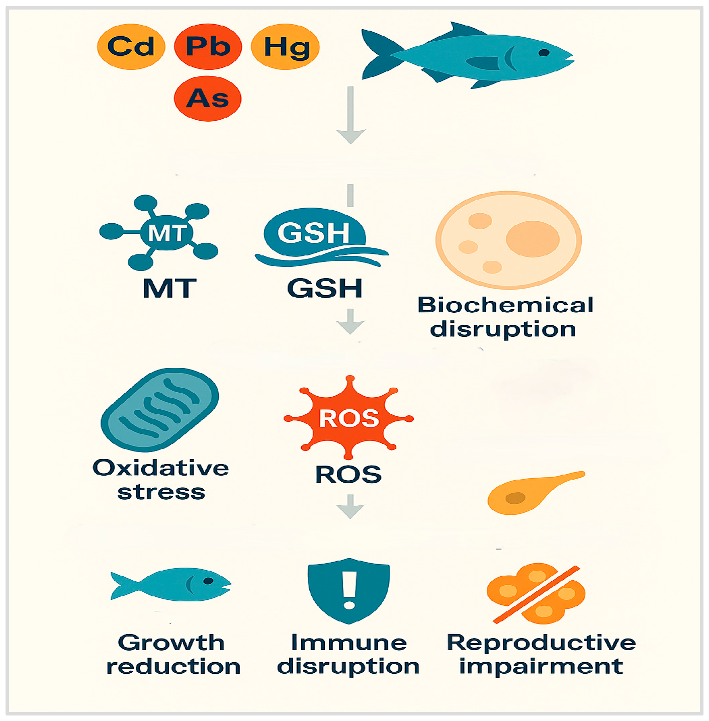
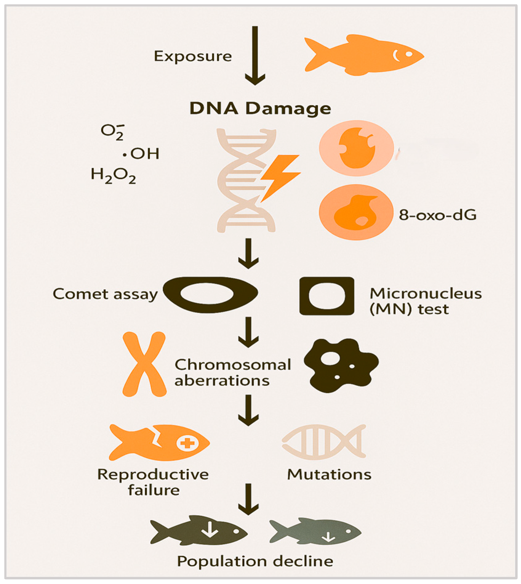
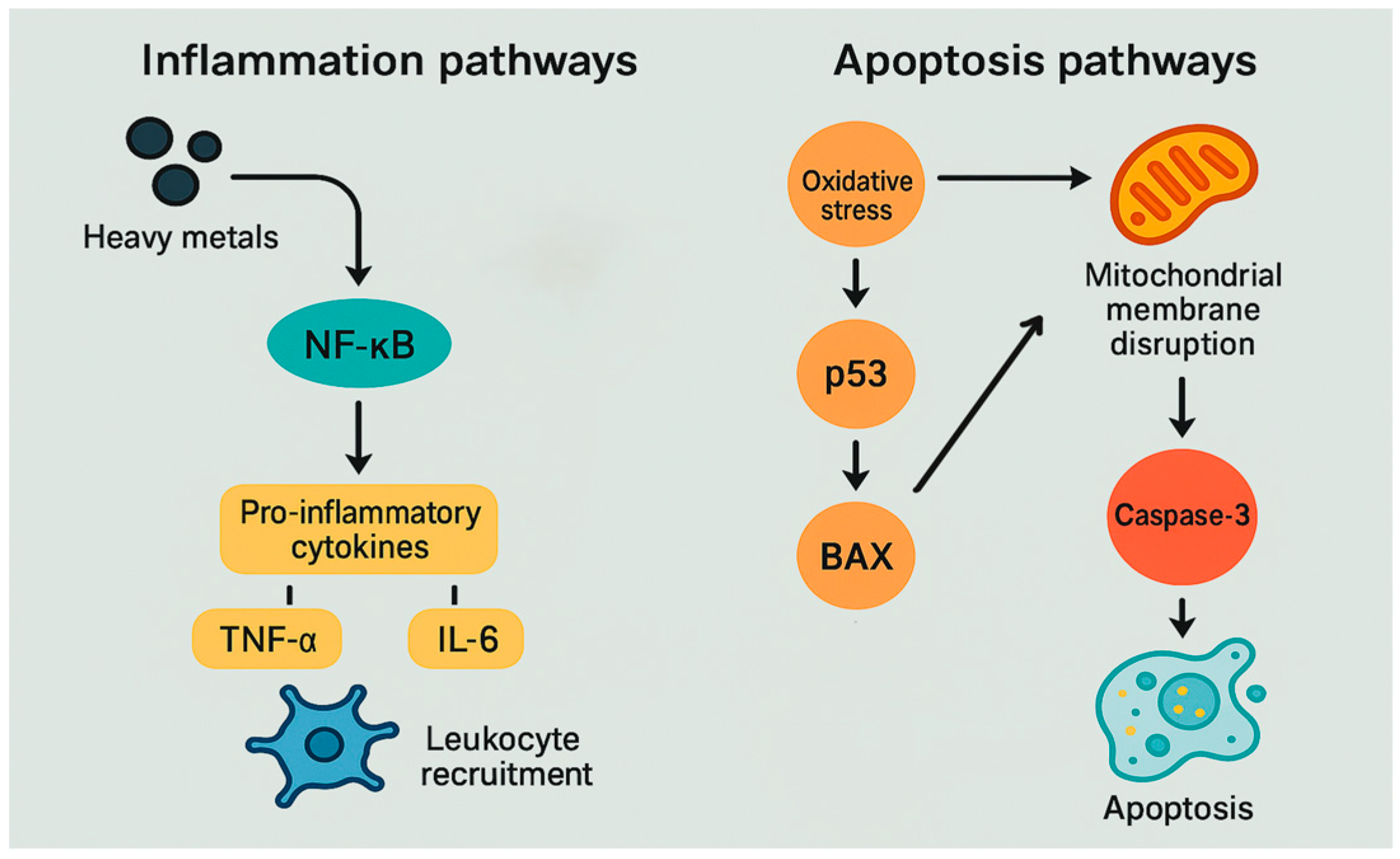
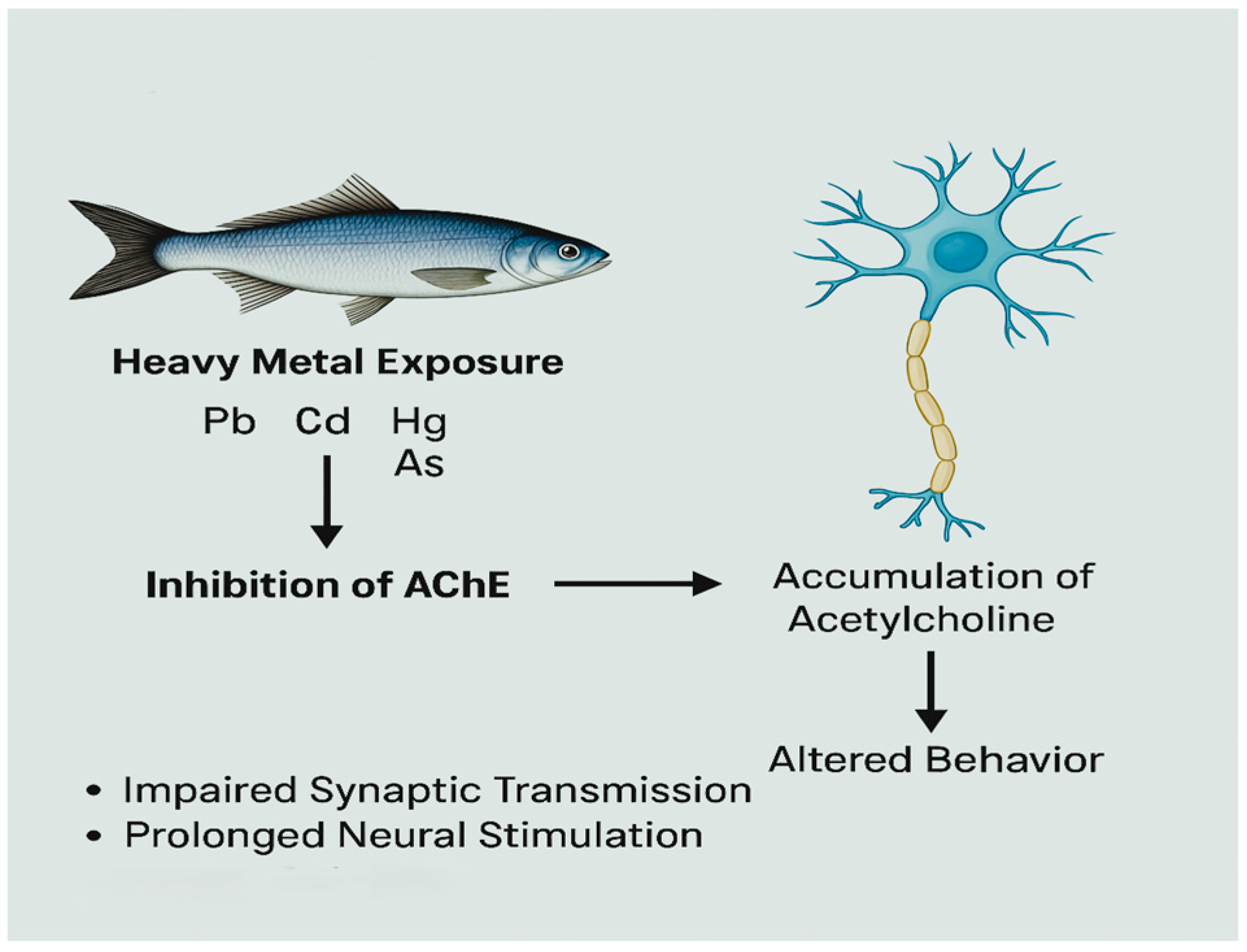
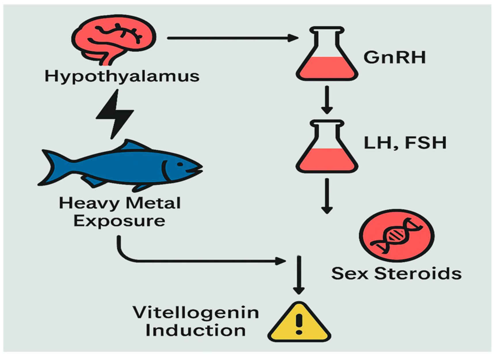
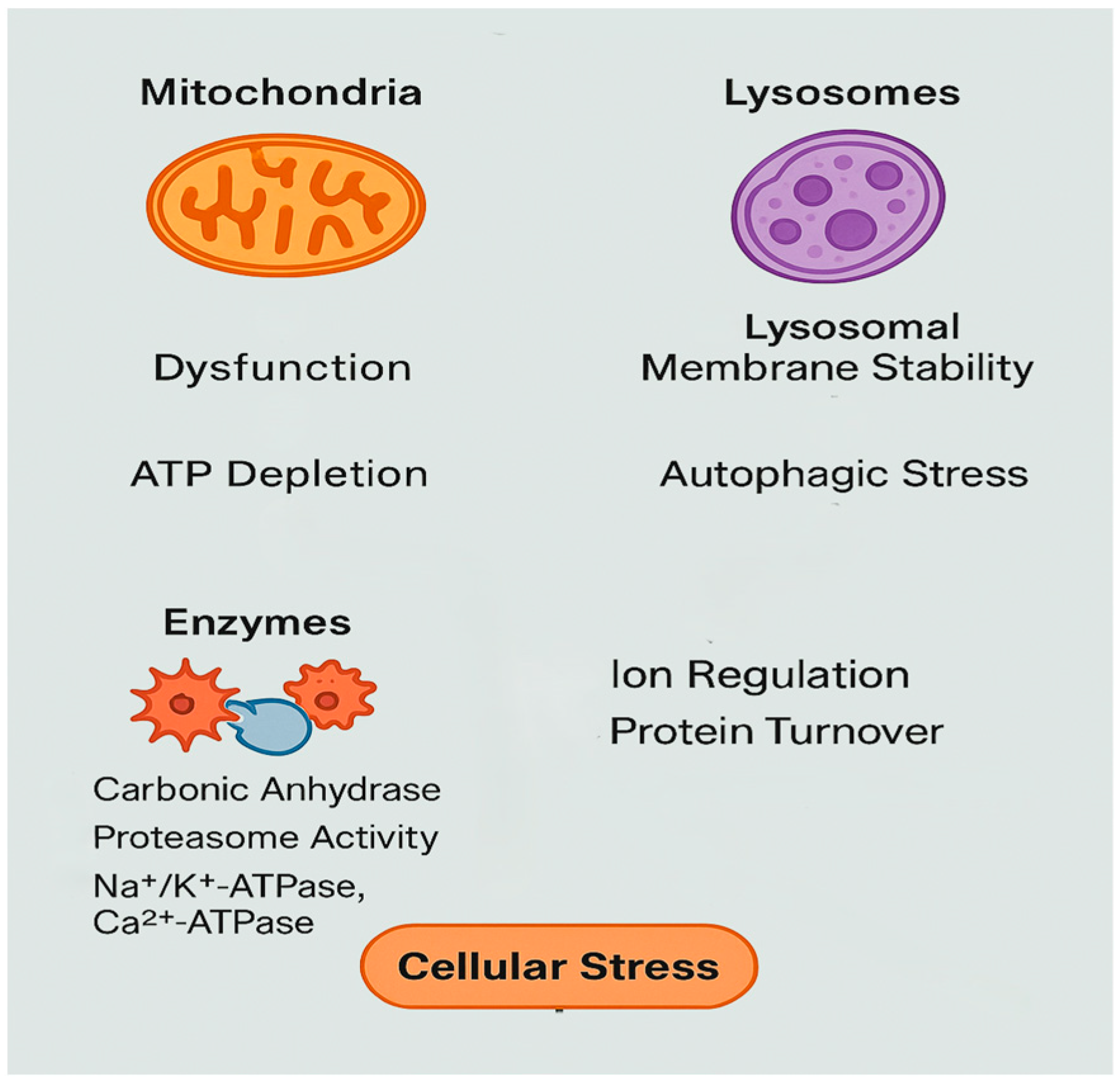
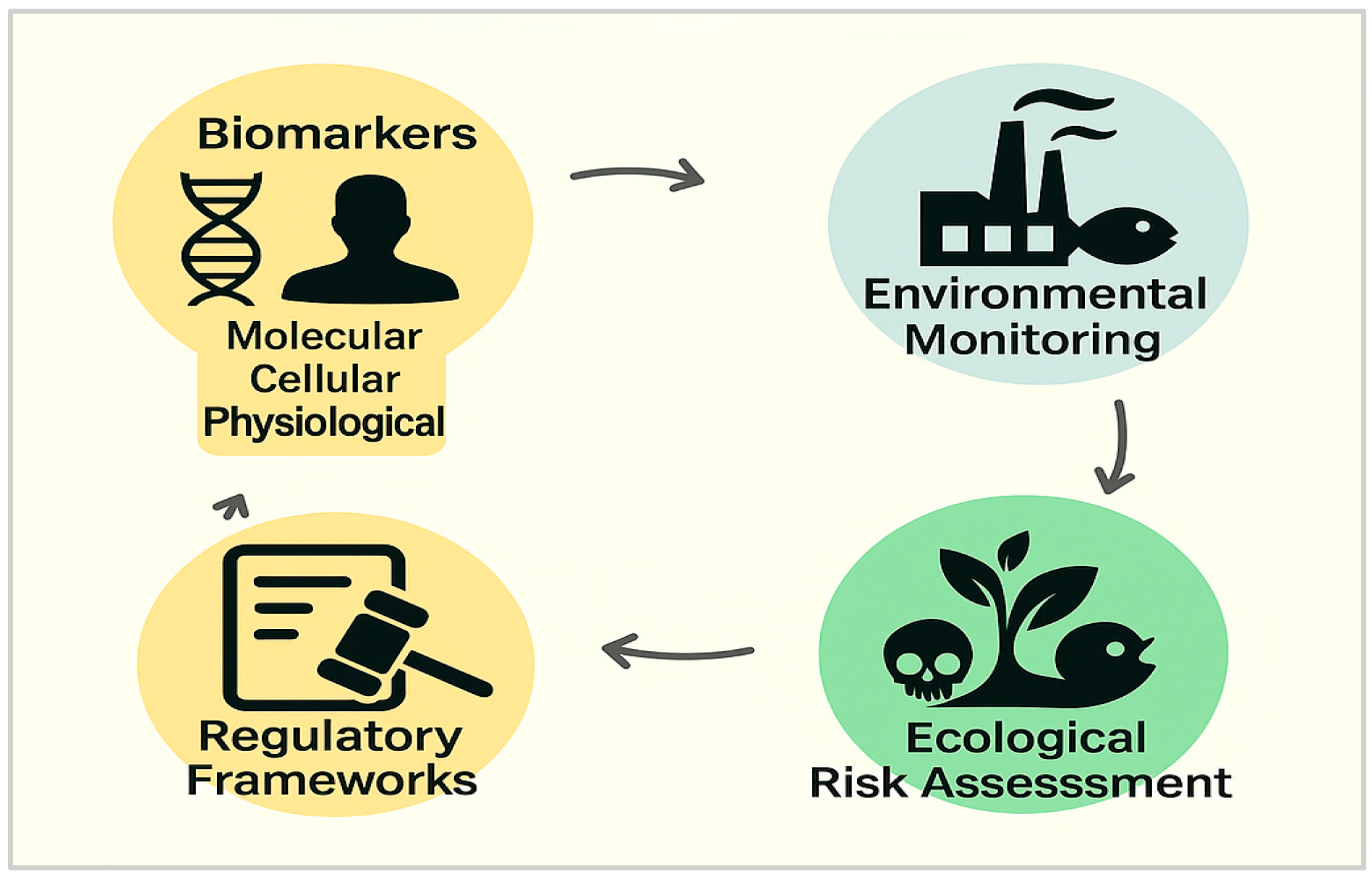
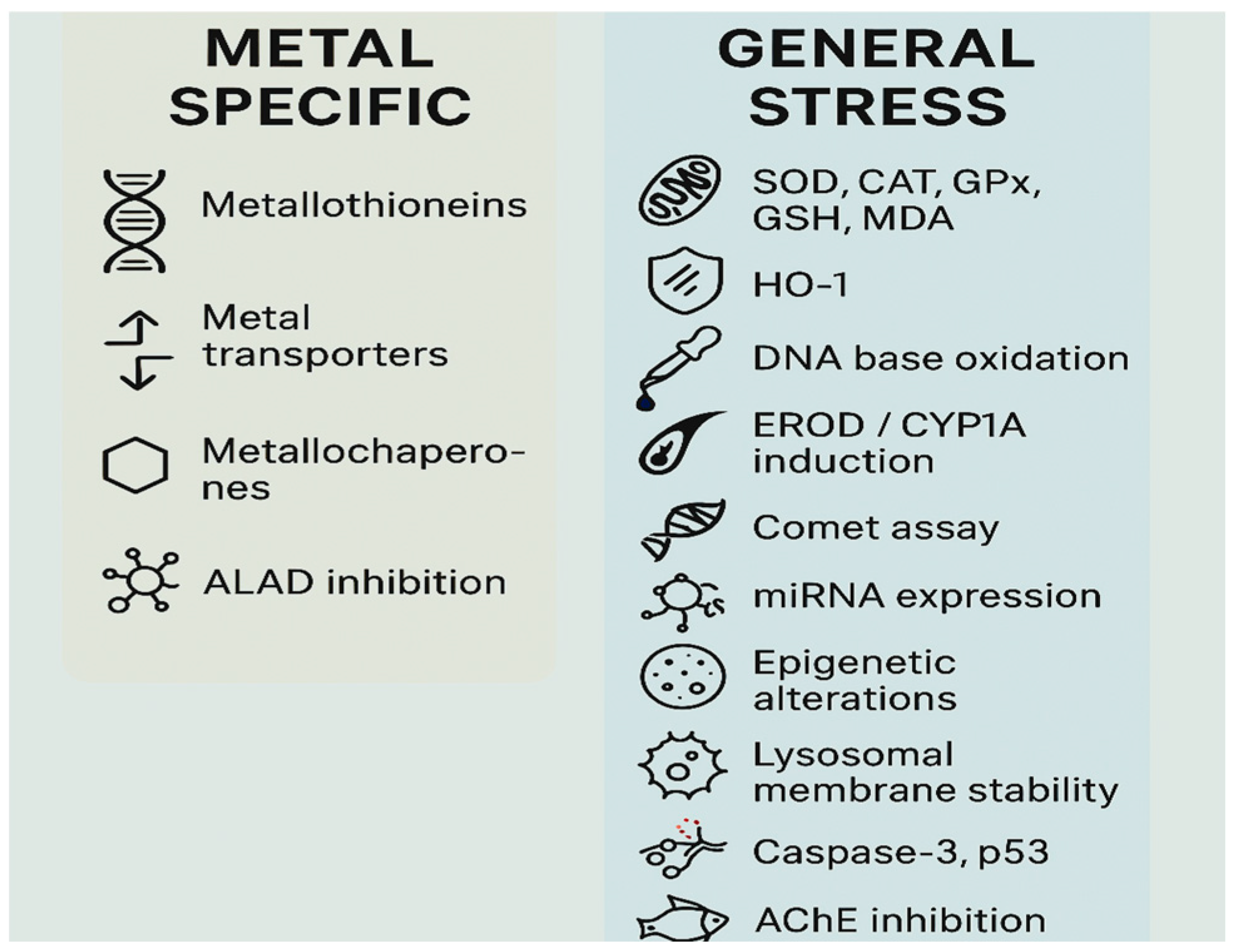
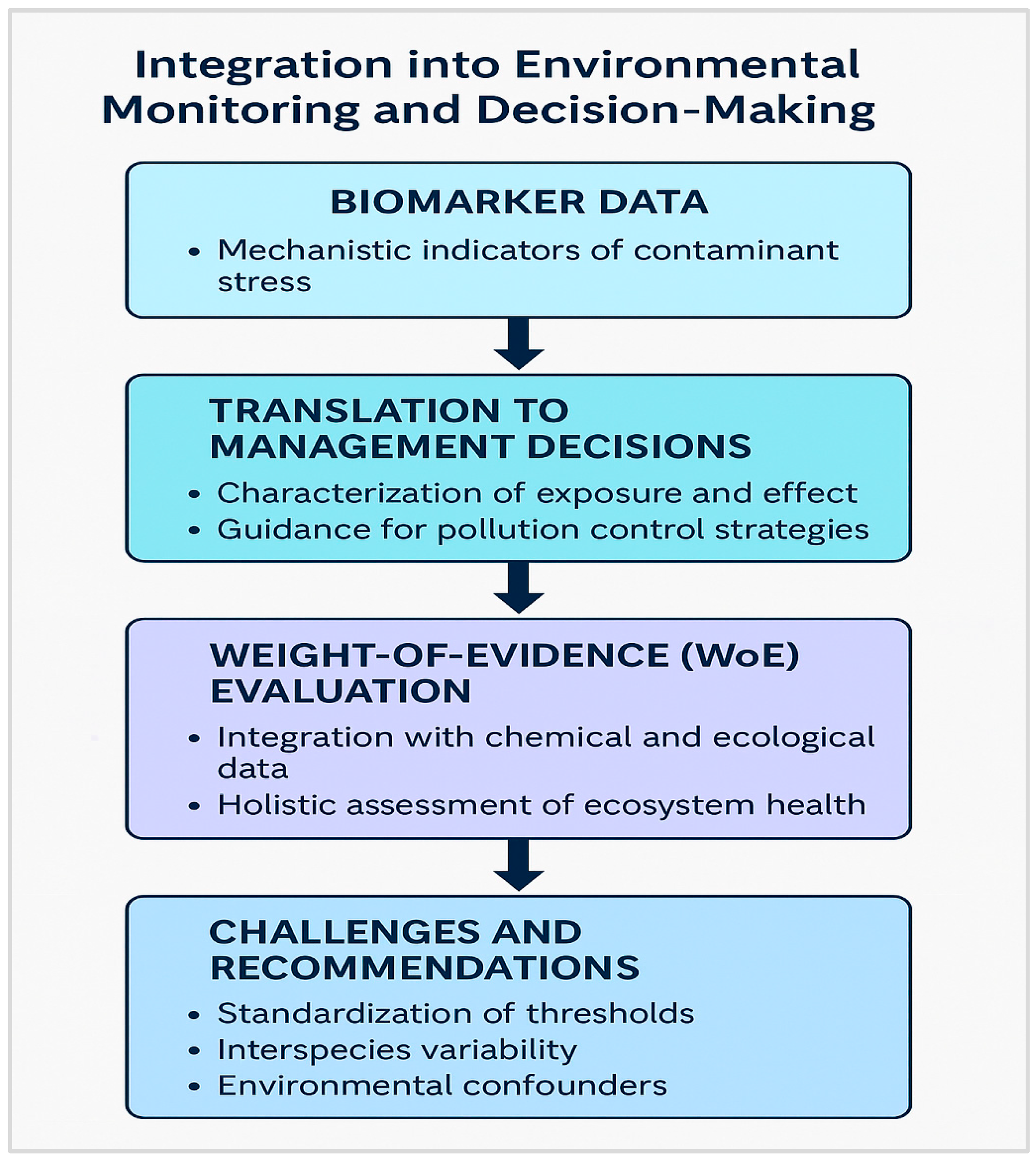
Figure 1. Overview of HM toxicity mechanisms.
Fishes 10 00339 g001
Figure 2. Overview of oxidative stress and antioxidant responses.
Fishes 10 00339 g002
Figure 3. Cellular detoxification and stress outcomes.
Fishes 10 00339 g003
Figure 4. DNA damage and genotoxic effects.
Fishes 10 00339 g004
Figure 5. Molecular reprogramming and stress adaptation.
Fishes 10 00339 g005
Figure 6. Inflammation and apoptosis pathways.
Fishes 10 00339 g006
Figure 7. Neurotoxicity and cholinergic disruption.
Fishes 10 00339 g007
Figure 8. Endocrine disruption pathways.
Fishes 10 00339 g008
Figure 9. Cellular disruption key drivers.
Fishes 10 00339 g009
Figure 10. Biomarkers in marine monitoring and risk assessment.
Fishes 10 00339 g010
Figure 11. HM-specific biomarkers and some general stress biomarkers.
Fishes 10 00339 g011
Figure 12. Role of biomarkers in environmental assessment and decision-making.
Fishes 10 00339 g012
Table 1. Biomarkers of HMs exposure: sensitivity, specificity, and early-warning potential.
Biomarker/
Mechanism
Mechanistic CategoryIndicative EffectSpecificity to MetalsBiomarker Type Early-Warning PotentialLimitations
Metallothioneins (MTs)DetoxificationMetal sequestrationHighExposureHighWidely used in marine monitoring. Baseline varies by species, tissue, and environmental conditions.
Metal transporters (DMT1, ZIP, and ZnT ATP7A/B)
Metallochaperones (ATOX1, CCS, andCOX17)
DetoxificationMetal homeostasisHighExposureHighBaseline variability and limited routine application.
ALAD inhibitionEnzymatic inhibitionDisrupted heme synthesisHigh
(Pb-specific)
EffectMediumInhibited by metals (Pb); also affected by diet and nutritional status.
Heme Oxygenase-1 (HO-1)Oxidative StressCytoprotective responseModerateEffectHighInduced by multiple ROS-generating agents.
Malondialdehyde (MDA) and protein carbonylsOxidative StressOxidative membrane damageModerateEffectMediumInduced by various pollutants and stressors.
Mitochondrial function (membrane potential and ATP levels)Cellular and energy disruptionBioenergetic failureModerateEffectMediumNot specific to HMs; technical demands for measurement.
Proteasome activityCellular and energy disruptionProtein turnoverModerateEffectMediumSensitive but non-specific.
Metabolomics/Lipidomics profilesCellular/molecular changesMetabolic healthModerateEffectMediumExpensive, high complexity, and not routine.
Epigenetic alterations (DNA methylation and histone acetylation)GenotoxicityAltered gene regulationModerateEffectLowComplex and not standardized for routine use.
Aromatase and 17β-HSD expressionEndocrine disruptionSteroidogenesis disruptionModerateEffectLowSpecies- and sex-dependent expression; limited field use.
SOD, CAT, GPx, and GSHOxidative stressAntioxidant defenseModerateEffectModerateInduced by multiple stressors.
MicroRNA (miRNAs)Gene regulationPost-transcriptional modulationLow to moderateEffectHighNot yet standardized for field use.
Glutathione system (GSH, GST, GPx, and GR)DetoxificationAntioxidant defenseLow to moderateEffectMediumModulated by multiple stressors.
DNA strand breaks and 8-oxodGGenotoxicityDNA damageLowEffectMediumNot metal-specific; general genotoxicity.
Lysosomal membrane stability (LMS)Cellular integritySubcellular dysfunctionLowEffectMediumSensitive, but not specific; influenced by many factors.
Micronucleus test (MN)GenotoxicityChromosomal damageLowEffectMediumNot metal-specific; general genotoxicity.
HSPs and HIF-1αStress adaptationStress adaptationLowEffectMediumBroadly inducible; lacks exposure specificity.
Cytokine expression
(NF-κB, TNF-α, IL-6)
InflammationImmune activationLowEffectMediumNot unique to metals; can reflect disease and other stress.
Na+/K+-ATPase, Ca2+-ATPaseNeurotoxicityIon regulation disruptionLowEffectMediumActivity influenced by multiple environmental factors.
Caspase-3, p53, BAXApoptosisProgrammed cell deathLowEffectLowActivation occurs under multiple stress types; not metal specific.
May indicate irreversible damage.
AChE inhibitionNeurotoxicityNeuro-
transmission impairment
LowEffectLowInhibited by metals and pesticides.
Carbonic anhydrase (CA)Ion regulationpH/ion imbalanceLowEffectLowInfluenced by salinity, pH, and other contaminants.
Vitellogenin (VTG) and
plasma E2/T
Endocrine disruptionEstrogenic response Moderate (Cd and As)ConditionLowInduced by metals and organic EDCs.
EROD, CYP1A, and GSTDetoxification and biotransformationPhase I/II detoxification and CYP1A inductionLowEffectLowEROD is more relevant to organics; not HM-specific.
AMPKStress adaptationEnergy metabolism shiftLowEffectLowStill experimental; ecological significance not fully established.
IL-1β, CRPInflammationChronic inflammationLowEffectLowRequires co-assessment with other immune parameters for interpretation.
Brain cytokines and 8-oxodGNeurotoxicityNeuro-inflammation and damageLowEffectLowLimited baseline data; currently experimental.
ALT and AST leakageCellular and energy disruptionMembrane integrity lossLowEffectLowNon-specific.
Lysozyme activity, leukocyte counts, and immunoglobulinsImmunological and hematologicalImmune function and physiological stress responseLowEffectLowBroad; not specific for metals.
Gut microbiota compositionSystemic/metabolic healthMetabolic healthLowEffectLowField application limited and not yet standardized.
Histopathological lesionsTissue alterationStructural damage to gill, liver, and kidneyLowConditionLowRequires expert interpretation; not specific to HMs.
Gonadosomatic Index (GSI)Reproductive indicatorReproductive impairmentLowConditionLowSensitive to seasonal and reproductive cycles; low specificity to metal stress.
Hepatosomatic Index (HSI)Metabolic indicatorLiver enlargement from stressLowConditionLowAffected by nutritional status and multiple stressors; non-specific.
Growth and reproduction endpointsLife-history traitsGrowth inhibition and reduced fecundityLowConditionLowIntegrative but slow-response indicators; influenced by multiple environmental and biological variables.
Disclaimer/Publisher’s Note: The statements, opinions and data contained in all publications are solely those of the individual author(s) and contributor(s) and not of MDPI and/or the editor(s). MDPI and/or the editor(s) disclaim responsibility for any injury to people or property resulting from any ideas, methods, instructions or products referred to in the content.

