Article

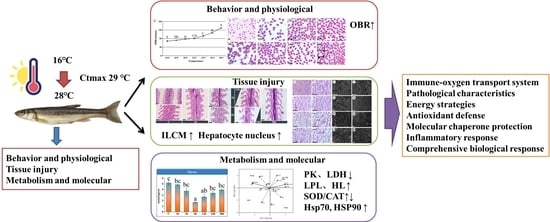
Behavioral, Hematological, Histological, Physiological Regulation and Gene Expression in Response to Heat Stress in Amur Minnow (Phoxinus lagowskii)

Fish Physiology and Resource Utilization Laboratory, Harbin Normal University, Harbin 150025, China
*
Author to whom correspondence should be addressed.
Fishes 2025, 10(7), 335; https://doi.org/10.3390/fishes10070335
Submission received: 21 May 2025 / Revised: 30 June 2025 / Accepted: 4 July 2025 / Published: 8 July 2025
(This article belongs to the Special Issue Environmental Physiology of Aquatic Animals)

Abstract

Rising water temperatures due to climate change pose a significant threat to Phoxinus lagowskii, a cold-water fish that is ecologically vital to the high-latitude regions of China. This study assessed heat stress effects on behavioral, hematological, histological, physiological, and molecular responses in P. lagowskii. The critical maximum temperature (CTmax) was determined using the loss of equilibrium (LOE) method, with the CTmax reaching 29 °C. Elevated temperatures lead to an increase in the OBR. Fish were subjected to acute heat stress at 28 °C (below CTmax) for 48 h, with samples collected during the 48 h period. RBC, WBC, HGB, and HCT significantly increased during heat stress but decreased 12 h after heat stress. The levels of serum cortisol and blood glucose after heat stress were significantly higher than those in the control group. After heat stress, the height of the ILCM in the gills increased significantly, and the liver exhibited vacuolar degeneration and hypopigmentation. The activities of Na+-K+-ATPase and Ca2+-Mg2+-ATPase in the gills initially increased and then decreased over the duration of heat stress. Most enzyme activities (PK, LDH, PFK, and HK) decreased during heat stress, while LPL and HL levels increased, indicating that lipid metabolism was the primary utilization process under heat stress. There was an increase in SOD activity at 12 h, followed by a decrease at 24 h, and an increase in CAT activity under heat stress. Integrated biomarker response (IBR) and principal component analysis (PCA) were employed to synthesize multi-level responses. The IBR values reached their peak at 3 h and 48 h of heat stress. We observed an upregulation of heat shock proteins (Hsp70, Hsp90, and Hsc70) as well as interleukin-10 (IL-10) in response to heat stress. Our findings offer novel insights into the mechanisms underlying the heat stress response in P. lagowskii, thereby enhancing our understanding of the effects of heat stress on cold-water fish.
Key Contribution: This study systematically elucidates the multi-level response mechanism of Phoxinus lagowskii, a significant cold-water fish species in high-latitude regions, to high-temperature stress. Research has established that the critical heat stress threshold for this species is 29 °C. It has been demonstrated that acute heat stress at 28 °C can induce a series of physiological and pathological changes, including considerable respiratory and metabolic disorders, abnormal blood indicators, and damage to gill and liver tissues. Additionally, this study confirms for the first time that lipid metabolism in P. lagowskii is significantly impacted under heat stress, revealing that the upregulation of heat shock proteins (Hsp70/Hsp90) and antioxidant enzymes (SOD/CAT) constitutes the key molecular mechanism underlying its response to heat stress.

Graphical Abstract

1. Introduction

For fish, temperature is one of the most critical physical variables, and significant fluctuations in water temperature can result in a series of physiological responses [1,2,3]. The rapid increase in water temperature and the rising frequency of heat waves present substantial challenges to the aquaculture industry [1,2,3,4,5], particularly for cold-water species such as the Amur minnow (Phoxinus lagowskii). As a cold-water fish that is widely distributed in northeast Asia and has significant commercial value, P. lagowskii has great breeding potential and is relatively sensitive to temperature fluctuations [6]. Fish exhibit a remarkable sensitivity to even minuscule fluctuations in water temperature, and certain species are capable of detecting temperature variations as slight as 0.03 °C [7]. When water temperature deviates beyond a specific range, it triggers a series of stress responses in fish, including abnormal behavior, physiological dysfunction, tissue cell lesions, and biochemical reactions, which may ultimately result in mortality [8,9]. When fish are subjected to acute heat stress, their state of motion becomes chaotic and unbalanced, with the corresponding temperature referred to as the critical thermal maximum (CTmax) [10]. This metric is commonly employed to measure the upper limit of thermal tolerance in animals [11]. CTmax serves as one of the most widely used physiological indicators for quantifying the thermal tolerance of fish [12]. An integrated analysis of CTmax measurements alongside other physiological variables can yield a more nuanced and comprehensive understanding of the organism’s response mechanisms to high-temperature environments [13]. Fish blood is readily available and responds quickly to factors affecting homeostasis, thus serving as an indicator of fish health [14]. External stimuli such as heat stress can induce changes in the physiological phenotype, particularly in the blood [15]. Blood components such as total red blood cell count (RBC), total white blood cell count (WBC), and hemoglobin concentration (HGB) are critical indicators for evaluating the physiological status of fish [16]. When ambient temperature increases, fish can adapt to these changes by modifying the composition of their blood [17].
Gills are frequently exposed to the external environment, making them susceptible to temperature fluctuations and other stressors [18]. Consequently, they often serve as a primary target for stress responses [19]. Heat stress has been shown to cause gill damage, including epithelial hyperplasia and congestion, in species such as Banded killifish (Fundulus diaphanus) [20] and Olive flounder (Paralichthys olivaceus) [21]. In addition, rapid increases in temperature can lead to significant reductions in Na+/K+-ATPase activity in the gills of juvenile salmonids [22]. To date, there have been no documented effects of prolonged heat stress on the gills of P. lagowskii. Additionally, the liver, a key metabolic and thermogenic organ, is highly sensitive to heat stress and plays a vital role in metabolism and immunity [23]. Heat stress elevates the metabolic rate and peripheral blood flow in fish, leading to the production of reactive oxygen species (ROS) associated with lipid and protein reactions, which in turn activate antioxidant defense mechanisms [24,25,26,27]. Antioxidant enzymes such as superoxide dismutase (SOD) and catalase (CAT) help mitigate ROS accumulation and subsequent oxidative damage [28,29]. Acid phosphatase (ACP) and alkaline phosphatase (ALP) are lysosomal enzymes linked to the innate immune system of fish. Furthermore, as phosphatase levels often rise in response to physical, chemical, and microbial stress, they are regarded as potential indicators of stress [30]. Moreover, fish exposed to elevated temperatures can disrupt energy metabolism, altering glucose and fatty acid levels, among other factors [31,32]. Interleukin-10 (IL-10) is a multifunctional cytokine that plays a crucial role in immunity by inhibiting the production of pro-inflammatory cytokines and reactive oxygen and nitrogen radicals [33]. Studies in teleosts, including Zebrafish (Danio rerio) [34], Common carp (Cyprinus carpio) [35], and Crucian carp (Carassius auratus) [36], have focused on their characteristics and anti-inflammatory effects; however, their role under heat stress conditions remains unreported. Heat shock proteins (Hsps) were first identified in Drosophila [37] as a response to elevated temperatures, functioning as molecular chaperones to protect cellular proteins during biosynthesis and refolding [38]. Studies have demonstrated that increased water temperature induces Hsp expression in mussels (Mytilus trossulus) [39] and stream fish (Fathead Minnow Pimephales promelas, Brown trout Salmo trutta and Rock bass Ambloplites rupestris) [40]. Hsps are considered to play a pivotal role in responding to environmental stresses such as elevated temperature [41] and hypoxia [42], and they have been recognized as indicators of stress [38].
Phoxinus lagowskii is a cold-water fish species widely distributed across northern China, Russia, and North Korea [43]. Its annual production is approximately 1.7 to 2 million tons, generating about 500 million renminbi (RMB) in economic value. This species has been designated as one of the ten characteristic aquatic germplasm species by the Ministry of Agriculture and Rural Affairs of China (2022). P. lagowskii is omnivorous and is distinguished by its tender meat, delicious flavor, and high nutritional value, attributes that make it highly favored by consumers and indicate promising prospects for aquaculture [6]. The optimal living temperature for P. lagowskii ranges from 16 to 24 °C. For cold-water fish, acute heat stress poses a significant challenge [44]. Although the critical thermal maximum (CTmax) has been established at 29 °C, the experimental conditions in this study were maintained at 28 °C, which is below the CTmax threshold, to ensure the survival of the fish. We hypothesize that acute heat stress, occurring near the critical thermal maximum, will elicit significant behavioral stress responses, induce hematological disturbances indicative of physiological adaptation, cause measurable histological damage in gill and liver tissues, and activate integrated physiological and molecular defense mechanisms in P. lagowskii. This study aims to comprehensively characterize the multi-level responses of P. lagowskii, encompassing behavioral, hematological, histological, physiological, and molecular aspects, to acute heat stress approaching its CTmax. The results of our study contribute to the understanding of response mechanisms under heat stress and provide practical solutions for improving the survival rate of P. lagowskii in summer aquaculture.

2. Materials and Methods

2.1. Experimental Fish

In this study, we utilized 184 adult P. lagowskii (mean weight: 42.75 ± 6.4 g; mean length: 16.5 ± 2.3 cm; approximately 1:1 sex ratio) sourced from a local farm in Harbin. The experiment was conducted in June 2023, during which the fish were acclimated for 14 days in water maintained at a temperature of 17 ± 2 °C, with dissolved oxygen levels at 7.0 ± 0.5 mg/L and ammonia nitrogen concentrations at 0.15 ± 0.04 mg/L. The fish were fed twice daily with commercial feed at 7:00 AM and 7:00 PM until satiation. At the onset of the experiment, healthy fish of comparable size were randomly selected. All experimental procedures received approval from the Biological Sciences Animal Ethics Committee of Harbin Normal University (permit number: HNUARIA2024023).

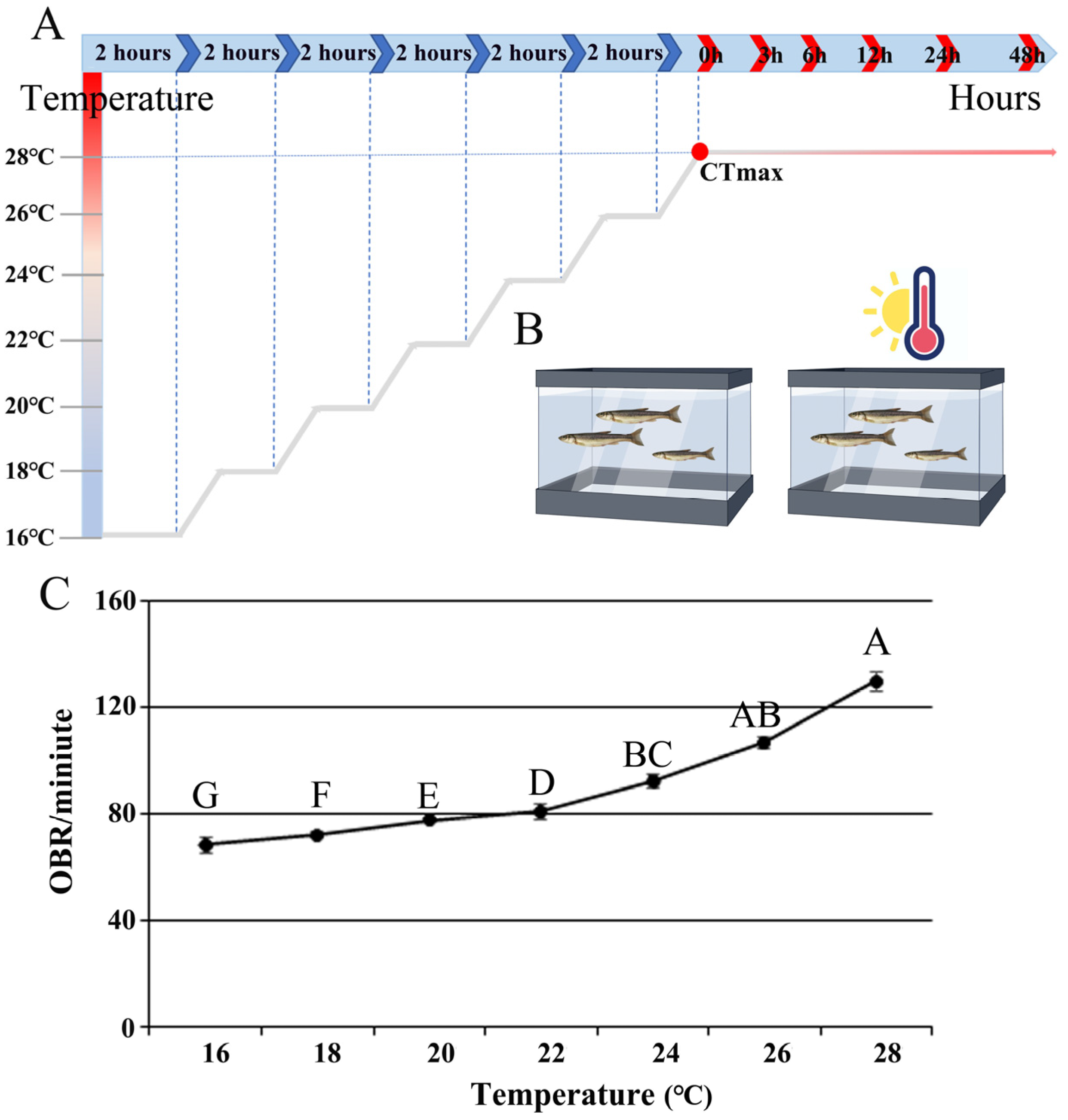
2.2. Measurements of CTmax and Behavioral Studies

Thirty fish were randomly selected and placed in the test tank for the critical thermal maximum (CTmax) test. The CTmax of acclimated P. lagowskii was estimated using the critical thermal methodology described by [45]. All fish were starved for 24 h prior to the CTmax test. The fish were transferred from the acclimation tank to a separate tank of the same temperature, which was sufficiently sized to allow for free movement. Each fish was subjected to thermal ramping regimes at a rate of 1 °C/min (fast rate) using aquarium heaters (YRK, SUNSUN, Zhoushan, China). During the procedure, the tank water was vigorously aerated to prevent thermal stratification and to maintain oxygen saturation. Heating continued until a persistent loss of equilibrium (LOE) was observed, indicated by a fish’s inability to right itself after being manually turned ventral side up. The temperature at LOE was recorded as the CTmax, while the water temperature at the start of the trial and at CTmax was measured with a digital thermometer (±0.1 °C). In this study, the opercular beat rate (OBR) was used to evaluate the behavioral stress response with 10 fish placed in the tank. The most accurate technique for observing free-swimming fish involves the use of stereo video [46]. The OBR of P. lagowskii was estimated using the methodology described by [47,48]. Before filming, three digital cameras were mounted overhead on tripods outside the experimental tanks. To minimize stress from the camera’s presence, the cameras were positioned 1.5 m away from the tanks. After initiating the recording, all experimenters left the room, capturing a 10 min video. The first 3 min (adaptation phase) and the last 2 min (potential disturbance from the experimenter’s return) were excluded, leaving the middle 5 min for analysis. Both observers were blinded to the treatment groups by using anonymized video filenames, and they had no involvement in the experimental design or the handling of the fish.

2.3. Fish Treatments and Sample Collection

The temperature of each tank was controlled using a thermostatic heating rod and monitored by a thermometer twice per hour. Adjustments were made as necessary, following the method described by [49]. Fish were randomly assigned to a control group (16 °C) and an experimental group (28 °C), with 100 fish in each group. The water temperature in the experimental group was increased from 16 °C to 28 °C at a rate of 6 °C per hour and maintained at 28 °C for 48 h. Fish samples were taken at six different time points: 1 h, 3 h, 6 h, 12 h, 24 h, and 48 h. Histological observations were conducted on gill and liver samples from three fish among these nine samples, while the remaining liver samples were stored at −80 °C for subsequent experiments.

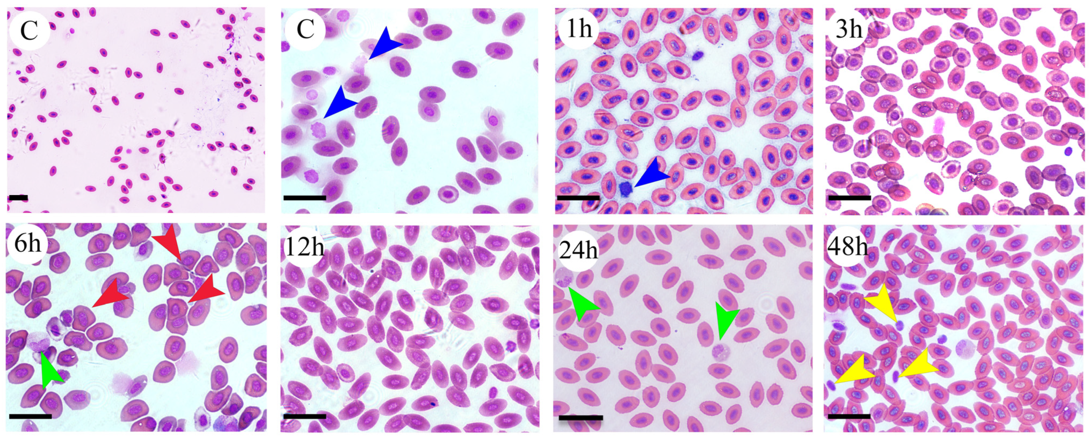
2.4. Hematological Study

Blood samples were collected at 0, 1, 3, 6, 12, 24, and 48 h (n = 6 biological replicates). The fish was anesthetized using MS-222 at a concentration of 200 mg/L. Blood samples from the caudal vein were obtained using a 1.0 mL syringe and transferred to Eppendorf tubes. Heparin sodium was added to prevent blood coagulation. Subsequently, the blood was analyzed using an appropriately pre-calibrated automatic hematology analyzer (Rayto RT-7600, Shenzhen, China). Blood parameters, including white blood cell (WBC) count, red blood cell (RBC) count, hemoglobin concentration (HGB), and hematocrit value (HCT), were determined for all groups. Serum cortisol concentration and glucose levels were measured using Fish Cortisol ELISA kits (FH96020, Shanghai, China) and Nanjing Jiancheng kits (Nanjing Jiancheng, A154-1-1, Nanjing, China), respectively, as per the manufacturer’s instructions.

2.5. Diff-Quick Staining

Blood was collected via the caudal vein using a 1.0 mL syringe and sodium heparin as an anticoagulant and subsequently stained with the Diff-Quick Stain Kit (Solarbio, Beijing, China). The Diff-Quick fixative was applied for 20 s, followed by staining with Diff-Quick Stain I for 5 to 10 s and Diff-Quick Stain II for 10 to 20 s. After rinsing with water, the samples were either directly observed under a microscope or dehydrated through a series of ethanol concentrations (95% ethanol, 100% ethanol I, and 100% ethanol II for 5 s, 5 s, and 30 s, respectively). The samples were then cleared with xylene for 1 min and prepared for observation.

2.6. Observation of the Gill and Liver Histopathology

Gill and liver samples fixed in Bouin’s fixative for over 24 h were dehydrated, embedded, sectioned, and stained for histological analysis using a Zeiss Axio Imager A2 microscope (Carl Zeiss, Jena, Germany). Samples fixed in 4% glutaraldehyde solution for more than 4 h at 4 °C were subsequently washed with phosphate buffer (pH 7.4, 0.1M), dehydrated with ethanol, dried, and coated using a Hitachi E-1010 gold plating machine (Tokyo, Japan). Ultrastructural analysis was conducted utilizing a Hitachi S-3400 N scanning electron microscope. This study analyzed the protruding lamella (PL) length, PL thickness, PL height, PL basal length, the distance between adjacent lamellae, and the height of the interlamellar cell mass (ILCM), as previously described by [50].

2.7. Determination of Relevant Enzyme Activities

The tissues were homogenized in pre-cooled phosphate buffer (1:9, w/v) and subsequently centrifuged to obtain the supernatant. The levels of Na+/K+-ATPase, Ca2+/Mg2+-ATPase, total ATPase (T-ATPase), total peroxidase (SOD), catalase (CAT), acid phosphatase (ACP), alkaline phosphatase (ALP), malondialdehyde (MDA), total cholesterol (T-CHO), triglycerides (TG), hexokinase (HK), phosphofructokinase (PFK), pyruvate kinase (PK), lactate dehydrogenase (LDH), lipoprotein lipase (LPL), and hepatic lipase (HL) were measured using the protocols provided by Nanjing Jiancheng Kits. Principal component analysis (PCA) was employed to visualize the relationship between the duration of heat stress treatment and the biomarkers. All data were logarithmically transformed to ensure equal weighting of the elements. The data were analyzed using Microsoft Excel, and the PCA graphs were generated with CANOCO 5.0.

2.8. Integrated Biomarker Response (IBR)

IBR analysis facilitates the integration of biomarker responses into standardized metrics for assessing contaminant stress levels [51]. In the present study, this index was utilized to evaluate the effects of various durations of heat stress treatments on enzyme activity in the liver of P. lagowskii [52]. Star plots for each biomarker were generated across different tissues to elucidate the multi-biomarker response in the fish. The mean values of IBR were calculated using the following formula:
A i = S i 2 sin β   ( S i   cos β + S i + 1   sin β )
where β = Arc tan (Si + 1 sin α/SiSi + 1 cos α) and α = 2π/n, Sn + 1 = S1 where Ai represents the area connecting the two scores (S), Si and Si + 1 are two consecutive clockwise scores (radius coordinates) of a given star plot, and n represents the number of biomarkers used for calculations. The IBR index for each treatment was then standardized to calculate the mean value of each biomarker.

2.9. Gene Expression Analysis with qPCR

Total RNA was extracted from the liver and kidney of pangasius using RNAiso Plus (Takara, Shiga, Japan). The purity and integrity of the total RNA were assessed using agarose gel electrophoresis. The isolated total RNA was quantified, and its purity was examined by measuring the 260/280 ratios using a UV spectrophotometer. Following DNase treatment, RNA was reverse transcribed into cDNA using the PrimeScript cDNA synthesis kit (Takara). Quantitative PCR was performed according to established protocols using a quantitative thermal cycler (CFX96 Real-Time System, BIO-RAD, Hercules, CA, USA). The amplification was conducted in a final volume of 10 μL, which contained 1 μL of cDNA product, 0.5 μL (5 pmole) of forward and reverse primers, 5 μL of TB Green® Premix Ex Taq II (Takara), and 3 μL of PCR-grade water. β-actin was utilized as the internal reference gene due to its stability [53]. The qPCR conditions included initial denaturation at 95 °C for 5 min, followed by 40 cycles of amplification at 95 °C for 10 s, and extension at each specified annealing temperature (as detailed in Table 1) for 10 s with scanning. The primers used for RT-PCR are presented in Table 1, showing an efficiency range of 0.95 to 1.05. Gene expression levels were analyzed using the comparative CT method (2−ΔΔCT) as described by [54].

2.10. Data Analysis

All data are presented as mean values ± standard error (SE). The normality of the data and the homogeneity of variances were assessed using the Kolmogorov–Smirnov test and Levene’s test, respectively. Statistical analyses were performed using SPSS Statistics 20.0. A one-way analysis of variance (ANOVA) was conducted to compare differences between treatment groups, followed by Duncan’s test. For all tests, a significance level of p < 0.05 was adopted.

3. Results

3.1. Changes in Behavioral and Hematological Parameters

In this study, the opercular beat rate (OBR) of P. lagowskii exhibited a marked increase with prolonged exposure to heat stress (Figure 1). At a relatively low temperature of 16 °C, the OBR value was at its minimum, while at 28 °C, it reached its maximum. This observation suggests a potential positive correlation between temperature and OBR. Regarding hematological indicators, white blood cells (WBCs), red blood cells (RBCs), hemoglobin (HGB), and hematocrit (HCT) were measured during various stages of heat stress (Table 2). The results indicated that WBC counts in P. lagowskii increased after 24 h of heat stress. An increase in RBC counts was observed at 12 and 24 h, followed by a decrease at 48 h. Initially, HGB concentrations were low after heat stress treatment, reaching their lowest point at 3 h (p < 0.05), before gradually increasing to a peak at 24 h (p < 0.05). HCT level increased after 12 h of heat shock in P. lagowskii, after which it returned to baseline levels (p < 0.05).
Following Diff-Quick staining, the erythrocytes of P. lagowskii were observed to be round to oval in shape, exhibiting smooth edges, with the nucleus centrally located within the cell (Figure 2). As indicated by the long arrow in the figure, fish lymphocytes display irregular cell margins and condensed nuclear chromatin and possess the highest nucleoplasmic ratio among all leukocytes. Neutrophils are characterized by their round shape and irregularly shaped nuclei, containing moderately condensed chromatin. Mononuclear cells are also round but exhibit uneven margins, often containing a few clear round vacuoles in the cytoplasm and displaying loose nuclear chromatin. The short arrows in the figure point to platelets, which are smaller than red blood cells and typically appear round or oval. Furthermore, the morphology of erythrocytes was significantly altered following heat stress, particularly at the 6 h mark, where the edges of the erythrocytes were no longer rounded, resulting in severe deformation of their morphology (Figure 2).

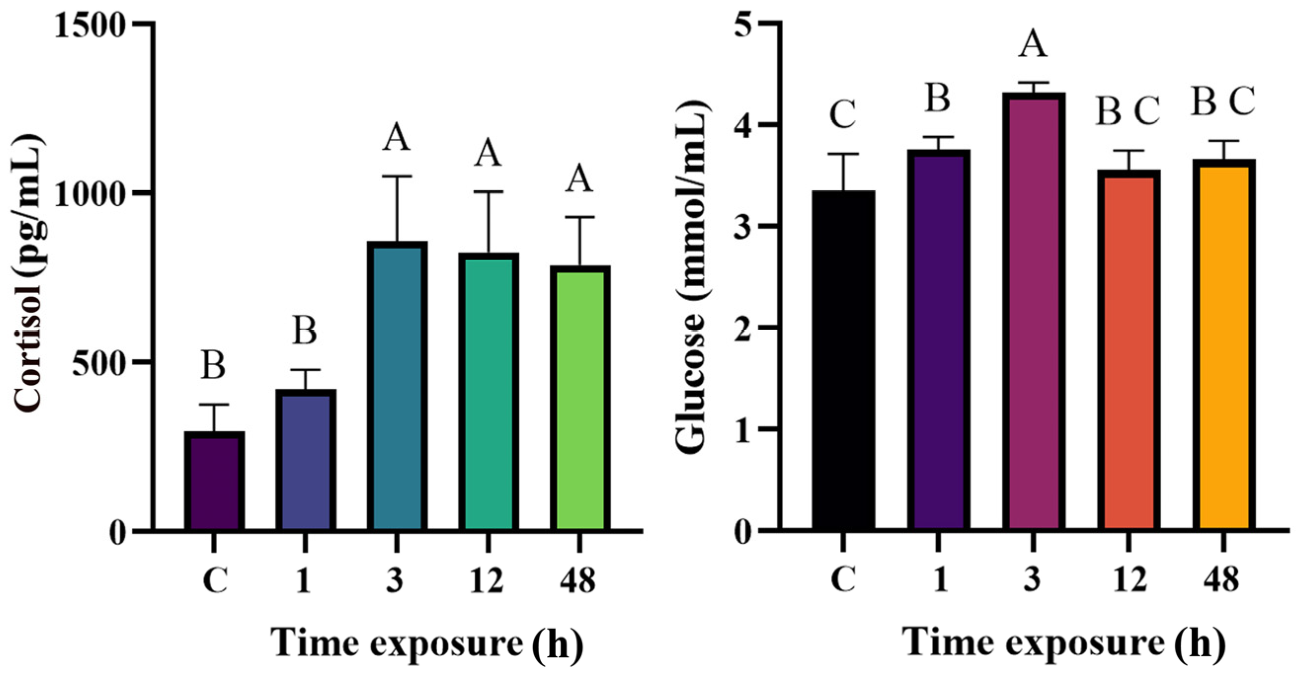
3.2. Effects of Heat Stress on Serum Cortisol and Glucose During 48 h Period

Serum cortisol levels increased markedly with temperature (Figure 3). The cortisol concentrations in the 3 h, 12 h, and 48 h groups were significantly elevated compared to the control group (p = 0.000021, 0.000057, and 0.000053, respectively). Additionally, an increase in serum glucose was observed at 3 h, followed by a decrease at 12 h (Figure 3). The glucose levels at 1 h and 3 h were significantly higher than those in the control group (p = 0.037 and 0.024, respectively).

3.3. Effects of Heat Stress on Activity of ATPase in Gills During 48 h Period

In this study, the activities of Na+/K+-ATPase and Ca2+/Mg2+-ATPase in the gills of P. lagowskii initially increased and subsequently decreased with the duration of heat stress (Figure 4). Notably, total ATPase activity was significantly elevated at both 3 h and 24 h compared to the control group (Figure 4).

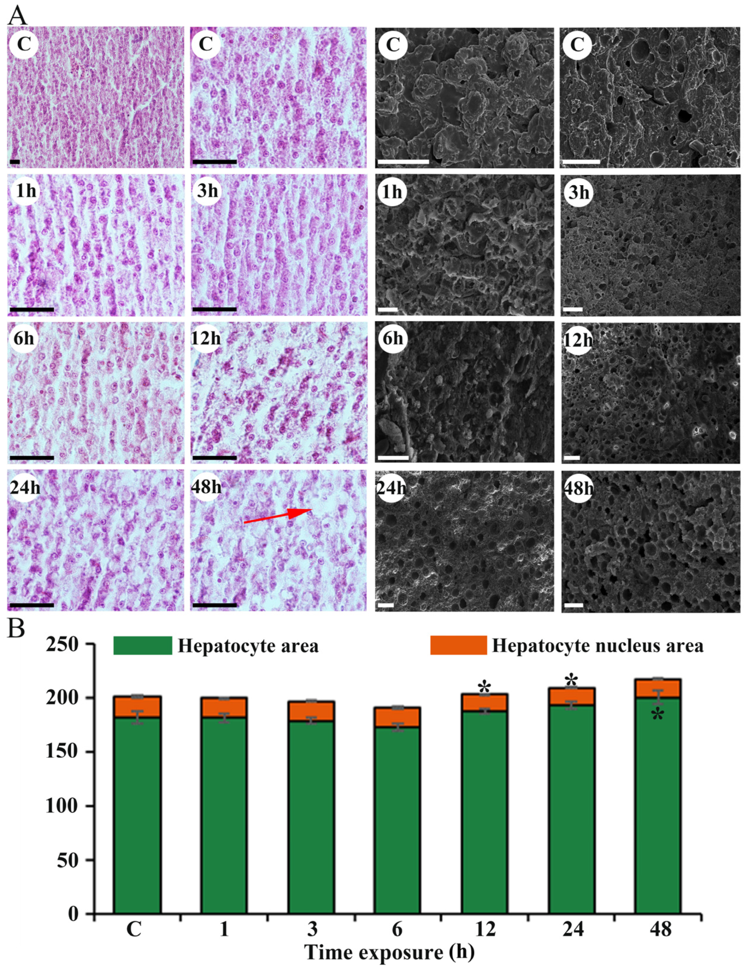
3.4. Effects of Heat Stress Duration on Gill and Liver Damage

During heat stress, fish exhibited several morphological changes in their gills (Figure 4) and liver (Figure 5). The gill structure in control fish displayed a normal appearance of the primary and secondary filaments (Figure 4). The thickness of the primary lamella (PL) was significantly increased at both 1 h and 3 h of heat stress compared to the control group (Figure 4 and Table 3). In the 1 h treatment group, the height of the PL decreased significantly (p < 0.05) compared to controls, while in the 6 h treatment group, it increased significantly (p < 0.05) compared to controls. An increase in the basal length of the PL was observed in P. lagowskii at 1 h, followed by a decrease at 3 h. The distance between adjacent lamellae significantly increased at 6 h of heat stress compared to the control group (Figure 4 and Table 3). The height of the ILCM was also significantly increased in the stressed group at 24 and 48 h post-stress. Furthermore, we analyzed the histological changes in the liver following heat stress in fish. The cells in the control group appeared morphologically intact and homogeneous in size. In contrast, the other treatment groups exhibited varying degrees of cellular damage (Figure 5). Compared to the control group, the liver cells of fish subjected to heat stress were loosely arranged, with some cells appearing vacuolated (Figure 5). Histological examination revealed distinct pathological alterations, including cytoplasmic vacuolization, loss of cellular boundaries, and nuclear condensation. The area of hepatocytes significantly increased at both 24 h (p < 0.05, Figure 5) and 48 h (p < 0.05, Figure 5) compared to the control group. Additionally, the area of hepatocytes at 12, 24, and 48 h was significantly greater than at 6 h (p = 0.043, 0.00144, 0.0041). Moreover, the area of hepatocyte nuclei was reduced after heat stress treatment compared to the control group, peaking at 12 h (Figure 5), indicating nuclear condensation as a characteristic response to heat stress.

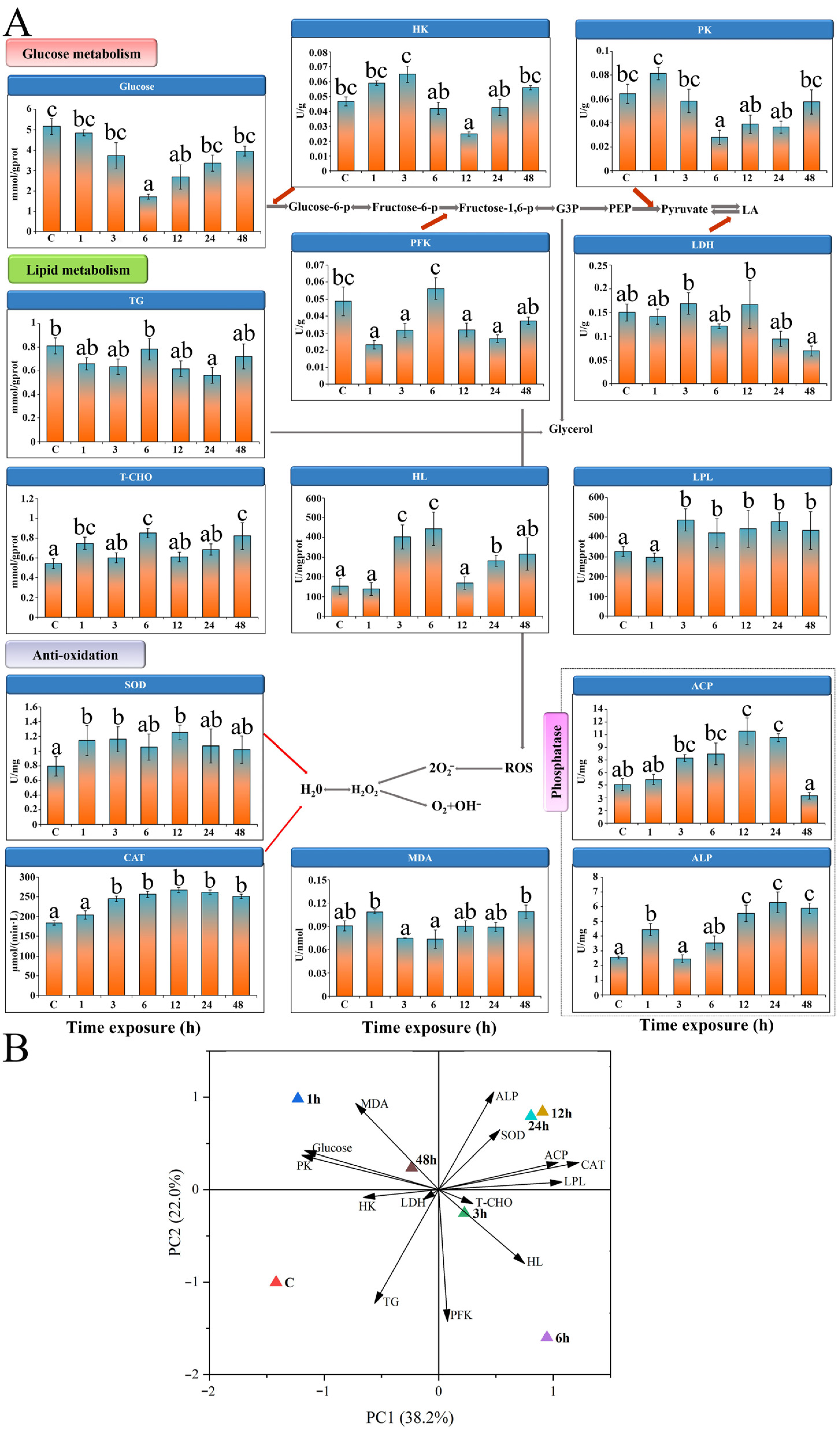
3.5. Enzyme Activities

3.5.1. Glycolytic Enzymes

After exposure to heat stress, the glucose content in the liver of P. lagowskii was diminished across all treated groups, with the most pronounced reduction observed at 6 h (p < 0.05, Figure 6). In comparison to the control group, hexokinase (HK) activity exhibited a significant decline at 12 h (p < 0.05), followed by an increase at 48 h; however, this change was not statistically significant at that time point (Figure 6). The activities of pyruvate kinase (PK) and phosphofructokinase (PFK) displayed contrasting trends: PFK activity increased initially and then decreased post-heat stress treatment, whereas PK activity showed the opposite pattern, with both enzymes peaking at 6 h (p < 0.05, Figure 6). Additionally, there was a significant reduction in lactate dehydrogenase (LDH) activity after 48 h of heat stress (p < 0.05, Figure 6).

3.5.2. Lipid Metabolism

Heat stress significantly affected both total cholesterol (T-CHO) and triglyceride (TG) levels in the liver; notably, TG levels decreased while T-CHO levels increased (p < 0.05, Figure 6). Following heat stress treatment, hepatic lipase (HL) activity exhibited a significant increase at both 3 h and 6 h (p < 0.05, Figure 6). Additionally, lipoprotein lipase (LPL) activity was significantly elevated in all experimental groups except for the 1 h group.

3.5.3. Antioxidant Enzyme Activities

The activities of comprehensive antioxidant enzymes, specifically superoxide dismutase (SOD) and catalase (CAT), exhibited significant alterations under heat stress treatment, with their activities being activated (p < 0.05, Figure 6). With the exception of the 48 h mark, acid phosphatase (ACP) activity was enhanced with prolonged exposure to heat stress. Alkaline phosphatase (ALP) activity showed a significant increase compared to the control group, except at the 3 h and 6 h intervals (p < 0.05, Figure 6). Notably, heat stress treatment for 48 h led to a significant decrease in ACP activity while simultaneously resulting in a significant increase in ALP activity (p < 0.05, Figure 6).

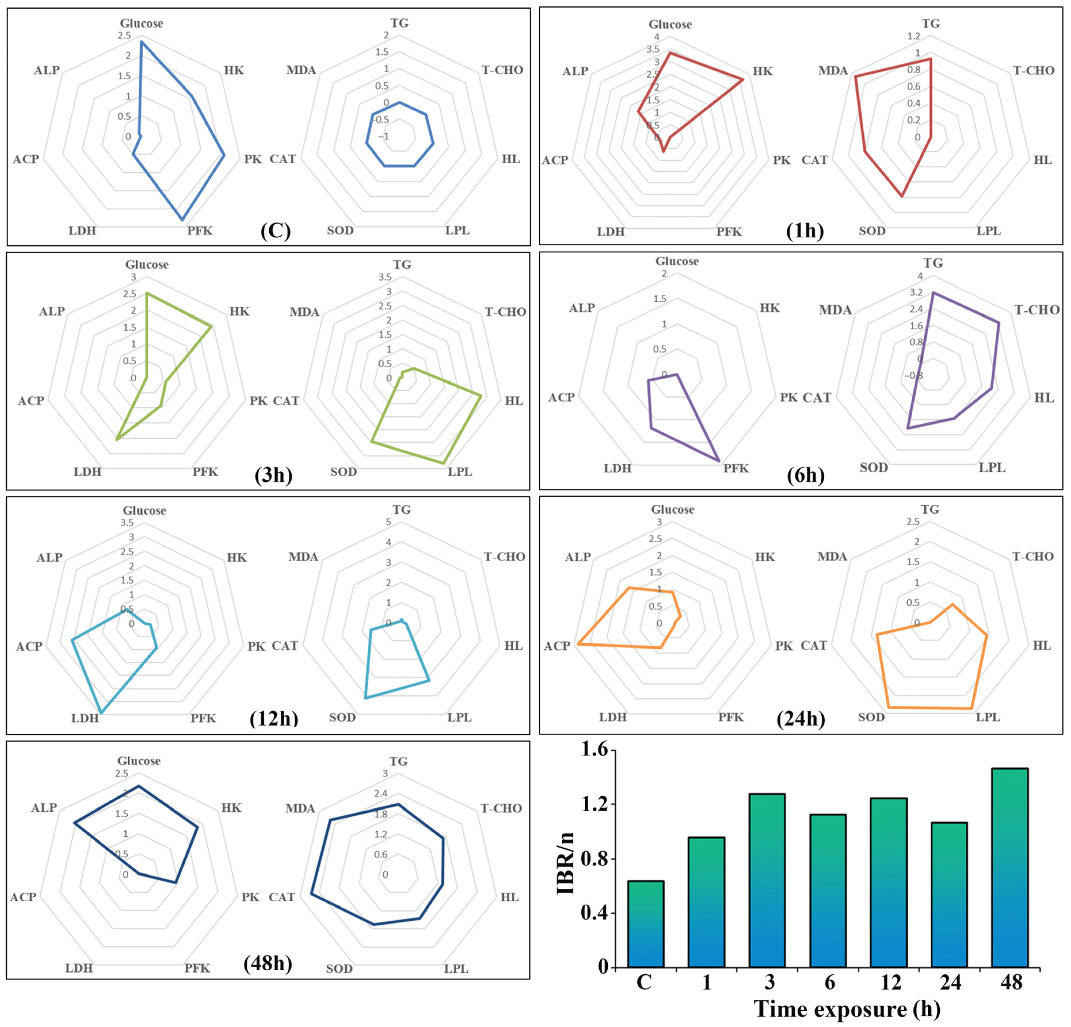
3.6. PCA and IBR Analysis

Principal component analysis (PCA) methods were employed to investigate the associations between enzymes involved in glucose metabolism, lipid metabolism, oxidative stress, and immunity in response to heat stress at various time points for P. lagowskii (Figure 6). The PCA ordination demonstrated a distinct separation between the control and heat stress groups. Furthermore, we identified that antioxidant-related indices (SOD, CAT, MDA) and phosphatase activities (ACP, ALP) exhibited positive correlations with the duration of heat stress. The integration of biological effects was conducted using the IBR (Figure 7). Notably, the highest IBR values were recorded at 48 h of heat stress, followed by those at 3 h. Biomarkers (SOD, CAT, MDA, and ALP) had a greater contribution to the IBR calculation at 48 h. In contrast, IBR values were lower after 1 h of heat stress, and the star plot indicated that T-CHO, HL, and LPL had minimal contributions to the stress response.

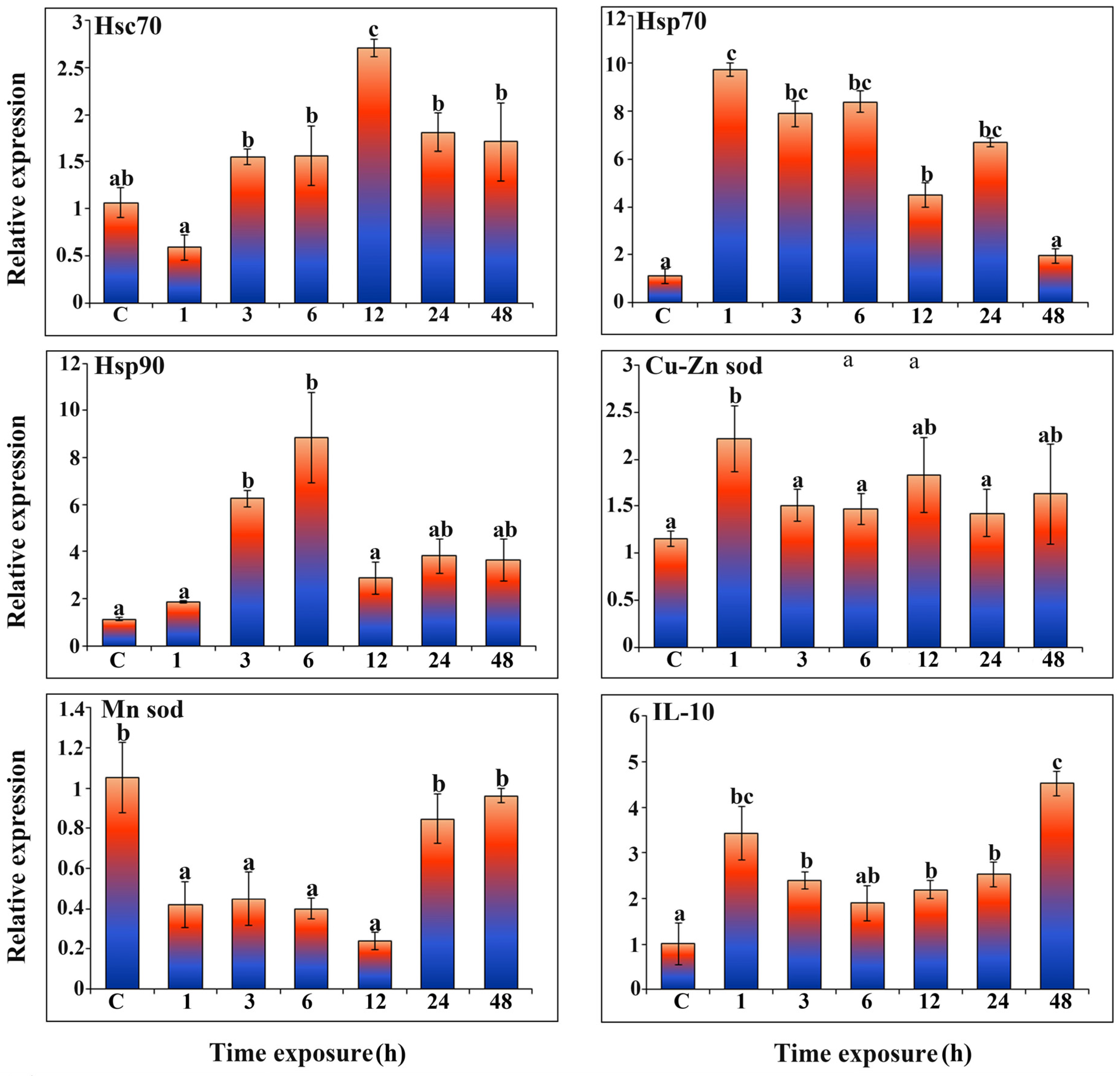
3.7. Expression of Antioxidant-Related Genes and Hsp Member Family Genes

Hsc70 expression was significantly elevated (p < 0.05) in all experimental groups, with the exception of the 1 h group, which did not show a statistically significant difference (p < 0.05) when compared to the control group (Figure 8). Conversely, Hsp70 expression exhibited an opposing trend to that of Hsc70, being significantly higher (p < 0.05) in all groups except for the 48 h group, which similarly did not demonstrate a significant difference (p < 0.05) relative to the control group. Following heat stress treatment, the expressions of Hsp90, Cu-Zn SOD, and IL-10 were all greater than those observed in the control group (p < 0.05). Furthermore, the expression of the Mn SOD gene was significantly lower than that of the control group (p < 0.05), with the exception of the 24 h and 48 h groups.

4. Discussion

Fish enhance their survival during heat stress by modifying their behavior and physiology [55]. OBR is frequently utilized as a non-invasive indicator of stress levels in fish [56]. The results indicated that the OBR of P. lagowskii increased with rising temperatures, thereby facilitating effective gas exchange between blood and water [57]. Similar findings were reported in the reef fish Fransmadam (Boopsoidea inornate), which exhibited behavioral responses to physiological stress, evidenced by an increased OBR at a threshold temperature of 25 °C [10]. Additionally, the OBR of Janitor fish (Ancistrus sp. orange) accelerated when the water temperature reached 26 °C [58]. Our research results align with these findings, further reinforcing the effectiveness of OBR as an indicator of heat stress. The eye pulsation rate of juvenile Chinook salmon in autumn also increases with rising water temperatures and serves as an indicator of physiological stress; the observed increase suggests that a sudden temperature rise may induce physiological stress [59]. Our findings corroborate that elevated temperatures can induce physiological stress in P. lagowskii.
In this study, we found that red and white blood cell counts, as well as HGB concentration and HCT percentage, were significantly increased during heat stress. This finding aligns with reports indicating that RBC, WBC, HGB, and HCT counts were significantly elevated in hybrid yellow catfish (Tachysurus fulvidraco) subjected to heat stress [60]. These data collectively indicate that changes in blood cell parameters represent a significant physiological response of fish to heat stress. HGB serves as the primary oxygen-carrying protein in RBCs, influencing both the transport capacity of blood oxygen and the quantity and functionality of RBCs [61,62]. Our research findings support this perspective, indicating that HGB plays a critical role in oxygen transport under conditions of heat stress. Previous studies have demonstrated that elevated water temperatures increase the metabolic rate of experimental fish, leading to the production of more red blood cells and hemoglobin, thereby enhancing oxygen transport to meet the heightened metabolic demands [61]. Interestingly, our study revealed a decrease in RBC, HGB, and HCT counts after 12 h of heat stress. Furthermore, we observed that serum cortisol and blood glucose levels were significantly higher in the heat-stressed group compared to the control group. This elevation may result from the fish’s efforts to maintain homeostasis during heat stress by increasing cortisol levels to mobilize glucose reserves and elevate blood pressure [63]. Similarly, both glucose and cortisol levels were found to be elevated in Nile tilapia (Oreochromis niloticus) following heat stress [64]. Our research findings align with these established results, further validating the universality of endocrine responses induced by heat stress. Overall, these results suggest that heat stress activates nonspecific immunity in P. lagowskii and aids in maintaining homeostasis through the mobilization of glucose reserves and elevation of cortisol levels. Based on these findings, we recommend that aquaculture practices emphasize the importance of ensuring adequate oxygen transport and energy supply within the critical time frame of 6 to 12 h post-heat stress. Additionally, it is essential to monitor the compensatory balance following this period.
Given that the gills are in direct contact with the respiratory system and serve as a vital immune organ [65], we selected the gills as the experimental organ. Although morphological and structural changes in gills can reflect responses to temperature, the specific morphological changes under heat stress in cold-water fish remain unclear. Previous investigations into the adaptation of Crucian carp (C. carassius) and Goldfish (C. auratus) to temperatures below 15 °C have shown that the gills are covered by a mass of cells known as the ILCM, suggesting that the ILCM serves as an indicator of the corresponding temperature in fish [66]. Furthermore, an increase in ILCM may be advantageous as it reduces the ion regulation costs associated with replenishing salts lost through gill diffusion [66,67]. This mechanism may elucidate the increase in ILCM observed in fish subjected to heat stress. We observed considerable changes in gill morphology following heat stress. Compared to the control group, the distance between adjacent lamellae decreased, and the height of the ILCM increased significantly after 48 h of heat stress. A similar fusion of gill lamellae was observed in heat-stressed Pikeperch (Sander lucioperca) [68]. Additionally, heat stress altered the gill morphology of Lenok trout (Brachymystax lenok tsinlingensis) [69], Icefish (Chionodraco hamatus), and Emerald rockcod Trematomus bernacchii [70]. Our results, in conjunction with these reports, confirm that one of the phenomena observed in fish responding to heat stress after exposure to elevated temperatures is the alteration in gill morphology. Damage to gill filaments reduces respiratory capacity and impairs osmoregulation, leading to abnormal physiological conditions [71]. Moreover, adenosine triphosphatases (ATPases) are potentially useful biochemical markers of pollution stress in aquatic organisms [72]. Consistent with previously reported mechanisms, our data indicate that Na+-K+-ATPase activity increases with the duration of heat stress in the present study, suggesting its significant osmoregulatory role in high-temperature environments [73]. The activity of Ca2+-Mg2+-ATPase decreased after reaching its peak at 6 h. It is plausible that high temperatures contribute to the disruption of Ca2+ homeostasis through free oxygen radicals, which leads to oxidative cell damage [74]. These results demonstrate that heat stress significantly affects the gills of P. lagowskii, characterized by morphological remodeling and an increase in chloride cells within the gill epithelium, resulting in a marked increase in Na+-K+-ATPase activity.
The liver is a crucial metabolic and thermogenic organ, serving as a significant indicator of oxidative stress [75]. Histological observations of the liver revealed that sustained heat stress resulted in liver damage characterized by vacuolar degeneration and hypopigmentation in P. lagowskii. These findings are consistent with those reported in the liver of Pikeperch (S. lucioperca), where heat stress was shown to induce liver damage, including vacuolar degeneration and hyperpigmentation, at 29 °C. Notably, histological damage was evident after 3 h, with the most severe effects observed at 48 h [76]. Our results indicate that elevated temperatures can cause liver damage in cold-water fish. In unstable environments, glycometabolism serves as the primary means of energy acquisition [77]. Notably, we observed an increase in the activities of HK and PFK in liver tissue following heat stress. Similarly, Kumar et al. (2011) reported that the PK activity in the liver of Rohu (Labeo rohita) was elevated after exposure to heat stress [78]. The increased PK enzyme activity in the liver suggests enhanced production of pyruvate, which serves as an energy source to mitigate the effects of heat stress [5]. Fish subjected to 6 h of heat stress exhibited significantly lower levels of HK and PFK enzyme activities at subsequent time points post-exposure. Previous studies have indicated that heat stress can lead to an insufficient ATP supply in fish, resulting in decreased HK and PFK activity [5]. LDH plays a crucial role in anaerobic glycolysis, and its activity is positively correlated with anaerobic glycolysis during heat stress [79]. Research has shown that LDH activity in the liver of Rainbow trout (Oncorhynchus mykiss) increases following heat stress [79]. Interestingly, our study found that LDH activity at 48 h post-exposure was lower than that of the control group, indicating a transition in the glucose metabolic pathway from anaerobic fermentation to aerobic oxidation during the later stages of heat stress. This study demonstrates that under short-term heat stress, the liver of P. lagowskii shifts from glycolysis to cholesterol oxidation, a process influenced by energy supply and oxidative damage.
The content of total T-CHO and TG is crucial for lipid metabolism in fish and plays a significant role in resisting external stress [80]. TG serves as an important energy source, and its levels increase under hypoxic stress to help the organism maintain energy balance [81]. In P. lagowskii, the concentration of TG did not change significantly under heat stress, suggesting that this species does not utilize TG as a primary energy source. The T-CHO levels were significantly higher at 6 h and 48 h compared to other groups, indicating that high temperatures facilitate lipid metabolism. In the present study, both high HL and LPL activities increased to varying degrees with prolonged heat stress. These results suggest that fish can shift from glucose metabolism to lipid metabolism when faced with environmental stress [82]. ALP and ACP are involved in the degradation of exogenous proteins, carbohydrates, and lipids, and they are sensitive to temperature changes [83]. The current findings indicate that heat stress induces ACP and ALP activity, and the enhanced phosphatase activity suggests increased transport of metabolites across the cell membrane [84]. However, ACP activity was significantly lower at 48 h than at 24 h, likely due to prolonged heat stress causing instability of the lysosomal membrane [85], which ultimately leads to a loss of lysosomal function [86].
In the present study, both SOD and CAT activities were found to be elevated in the liver following heat stress treatment compared to the control group. Our findings indicate that the activity of antioxidant enzymes in fish diminishes with prolonged exposure to heat stress, and similar trends were observed in olive flounder (Paralichthys olivaceus) and turbot (Scophthalmus maximus) [15]. Furthermore, the results of PCA and IBR suggest that antioxidant enzymes serve as important biomarkers for heat stress in fish, which could be instrumental in elucidating the mechanisms underlying the heat stress response. The IBR methodology provided a comprehensive visualization of integrated biomarker responses, enabling quantitative comparisons of the relative contributions of various biomarkers (SOD, CAT, MDA, ALP, T-CHO, HDL, and LPL) across different durations of stress. In the present study, the IBR in the high-temperature treatment group was found to be higher than that in the control group, which is consistent with previous research on Astyanax lacustris, which confirmed that the IBR index exhibited a higher value at elevated temperatures compared to the control group [87]. Similarly, in the study of Amphiprion ocellaris, after exposure to heat stress for 7 days, a significantly higher IBR value was observed at 30 °C compared to the control. This finding aligns with the elevated scores recorded for biomarkers at 30 °C, which corresponds with their increase and suggests a heightened demand for cytoprotection against heat stress [88]. Interestingly, in A. ocellaris, IBR values subsequently stabilized, and no significant differences were detected between the control and elevated temperature for the remaining sampling times, such as at 28 days [88]. This stabilization may indicate acclimation, suggesting that the adaptation to prolonged high temperatures warrants further investigation in future studies. This integrative analysis confirmed that antioxidant enzymes were the primary indicators during extended heat exposure (48 h), while markers of lipid metabolism exhibited minimal responses, thereby assisting in the prioritization of biomarkers for future monitoring studies. Previous research has demonstrated that these enzymes play a crucial role in shielding marine mussels from oxidative stress induced by reactive oxygen species [89] and may serve as valuable biomarkers for assessing the rearing environment [90].
Gene expression and enzyme activity of antioxidant mechanisms, as well as markers of oxidative damage, are common indicators used to assess the redox status of fish subjected to short-term or long-term temperature treatments [91]. The regulation of enzyme activity through translational and post-translational mechanisms leads to inconsistencies between gene expression and protein activity [92]. Notably, superoxide dismutase (SOD) exists in two isozymes, Cu-Zn SOD and Mn SOD [76]. Heat stress treatment significantly elevated the expression of Cu-Zn SOD in the liver of P. lagowskii compared to the control group. This trend parallels that observed in the Yellow drum (Nibea albiflora), indicating a strong correlation between increased Cu-Zn SOD mRNA expression and the upregulation of SOD activity [93]. Conversely, the expression of Mn-SOD was found to be lower than that of the control under identical conditions, with no correlation evident between the expression level and the enzymatic activity of antioxidant enzymes. In Milkfish (Chanos chanos), Cu-Zn SOD expression exhibited synergistic effects with temperature, while Mn-SOD showed no significant changes in response to acute low-temperature stimulation [94]. These findings suggest that reactive oxygen species (ROS) generated by heat stress originate from the cytoplasm and can induce Cu-Zn SOD expression [95].
Heat shock proteins (Hsps) play a crucial role in cellular repair mechanisms, with their upregulation aiding in the restoration of damaged protein structures. This process is essential for maintaining the structural integrity and functional capacity of the organism under heat stress [96]. Hsp70, recognized as a biomarker, functions as a redox sensor that activates reactive oxygen species (ROS) scavengers [97]. Additionally, Hsc70, a significant member of the heat shock protein 70 family [98], has been identified as a sensitive biomarker. Our study revealed that the expressions of Hsc70, Hsp70, and Hsp90 were significantly elevated compared to the control group following heat stress, indicating that high temperatures stimulate the expression of these proteins [99,100,101]. Similar responses have been documented in aquatic species, including the grass carp (C. idella) [23]. Interleukin-10 (IL-10) is recognized as a pleiotropic regulatory cytokine and one of the most critical anti-inflammatory cytokines, as evidenced in studies involving grass carp (C. idella) [102], Rainbow trout (Oncorhynchus mykiss) [103] and carp [33]. Our data demonstrated that IL-10 expression was significantly elevated following heat stress treatment compared to the control, peaking at 48 h. This finding suggests that heat stress triggers an inflammatory response in the liver of P. lagowskii, with the inflammatory response intensifying as the duration of heat stress increases.

5. Conclusions

This study provides a comprehensive analysis of the multi-level responses of P. lagowskii to acute heat stress approaching its CTmax of 28 °C. The OBR increased significantly with temperature, indicating respiratory stress. Hematological parameters demonstrated dynamic changes, including transient increases in WBC and RBC counts, as well as hemoglobin concentration, followed by subsequent declines. Notably, erythrocyte morphology exhibited severe deformation after 6 h of heat stress. Heat stress induced significant gill remodeling, characterized by increased ILCM height and lamellar fusion, which may impair respiratory efficiency. Liver tissues displayed vacuolization, nuclear condensation, and hepatocyte enlargement, suggesting metabolic disruption and cellular damage. Glycolytic enzyme activities (HK, PFK, and PK) fluctuated, while lipid metabolism shifted toward cholesterol utilization. Antioxidant defenses, including SOD and CAT, were activated to counteract oxidative stress; however, prolonged exposure (48 h) resulted in declining ACP activity, indicating lysosomal dysfunction. Heat shock proteins (Hsc70, Hsp70 and Hsp90) and the anti-inflammatory cytokine IL-10 were upregulated, reflecting cellular protection and immune modulation. IBR analysis confirmed that antioxidant enzymes were the most sensitive indicators of prolonged heat stress. These findings underscore the physiological challenges faced by P. lagowskii under acute heat stress and emphasize the necessity for thermal management strategies in aquaculture to mitigate climate change impacts on cold-water species. Future studies should investigate long-term acclimation potential and genetic adaptation mechanisms.

Author Contributions

W.M. conducted the investigation and was responsible for writing—review and editing; J.W. conducted the investigation and performed data analysis; Y.Z. conducted the investigation and performed data analysis; S.F. performed data analysis; Y.H. conducted the investigation and performed data analysis; Q.L. performed data analysis. All authors have read and agreed to the published version of the manuscript.

Funding

This work was supported by the National Natural Science Foundation of China (32170523) and Outstanding Youth Program of Natural Science Foundation of Heilongjiang Province (YQ2022C026).

Institutional Review Board Statement

This study was approved by Comments of the laboratory animal care Committee of Harbin Normal University Approval (Code: HNUARIA2024023 2023-03-06).

Informed Consent Statement

Not applicable.

Data Availability Statement

All datasets generated for this study are included in the article.

Conflicts of Interest

The authors declare no conflicts of interest.

References

  1. Chen, Y.; Wu, X.; Li, P.; Liu, Y.; Song, M.; Li, F.; Ou, J.; Lai, J. Integrated metabolomic and transcriptomic responses to heat stress in a high-altitude fish, Triplophysa siluroides. Fish Shellfish Immunol. 2023, 142, 109118. [Google Scholar] [CrossRef] [PubMed]
  2. Saravia, J.; Paschke, K.; Oyarzún-Salazar, R.; Cheng, C.C.; Navarro, J.M.; Vargas-Chacoff, L. Effects of warming rates on physiological and molecular components of response to CTMax heat stress in the Antarctic fish Harpagifer antarcticus. J. Therm. Biol. 2021, 99, 103021. [Google Scholar] [CrossRef] [PubMed]
  3. Roychowdhury, P.; Aftabuddin, M.; Pati, M.K. Thermal stress altered growth performance and metabolism and induced anaemia and liver disorder in Labeo rohita. Aquac. Res. 2020, 51, 1406–1414. [Google Scholar] [CrossRef]
  4. Zhang, Y.; Nie, H.; Huo, Z.; Yan, X. Heat hardening enhances the energy metabolism activity and oxidative defense ability of Manila clam Ruditapes philippinarum under different thermal stress. Aquaculture 2024, 595, 741511. [Google Scholar] [CrossRef]
  5. Zhao, H.; Ke, H.; Zhang, L.; Zhao, Z.; Lai, J.; Zhou, J.; Huang, Z.; Li, H.; Du, J.; Li, Q. Integrated analysis about the effects of heat stress on physiological responses and energy metabolism in Gymnocypris chilianensis. Sci. Total Environ. 2022, 806, 151252. [Google Scholar] [CrossRef]
  6. Chen, X.; Wang, Q.; Guo, Z.; Zhao, Y.; Gao, Y.; Yu, T.; Chen, Y.; Zhang, D.; Wang, G. Effects of dietary oxidized fish oil on growth performance and antioxidant defense mechanism of juvenile Rhynchocypris lagowski Dybowski. Aquaculture 2019, 512, 734368. [Google Scholar] [CrossRef]
  7. Bartolini, T.; Butail, S.; Porfiri, M. Temperature influences sociality and activity of freshwater fish. Environ. Biol. Fishes 2015, 98, 825–832. [Google Scholar] [CrossRef]
  8. Liu, Z.M.; Zhu, X.L.; Lu, J.; Cai, W.J.; Ye, Y.-P.; Lv, Y.P. Effect of high temperature stress on heat shock protein expression and antioxidant enzyme activity of two morphs of the mud crab Scylla paramamosain. Comp. Biochem. Phys. A 2018, 223, 10–17. [Google Scholar] [CrossRef]
  9. Liu, F.; Qu, Y.K.; Wang, A.M.; Yu, Y.B.; Yang, W.P.; Lv, F.; Nie, Q. Effects of carotenoids on the growth performance, biochemical parameters, immune responses and disease resistance of yellow catfish (Pelteobagrus fulvidraco) under high-temperature stress. Aquaculture 2019, 503, 293–303. [Google Scholar] [CrossRef]
  10. Allison, C.; Muller, C.; Childs, A.R.; Froneman, W.; Bailey, L.A.; Potts, W.M. When cooling is worse than warming: Investigations into the thermal tolerance of an endemic reef fish, Boopsoidea inornata. Afr. J. Mar. Sci. 2021, 43, 239–249. [Google Scholar] [CrossRef]
  11. Morgan, R.; Finnoen, M.H.; Jutfelt, F. CTmax is repeatable and doesn’t reduce growth in zebrafish. Sci. Rep. 2018, 8, 7099. [Google Scholar] [CrossRef] [PubMed]
  12. Vinagre, C.; Dias, M.; Roma, J.; Silva, A.; Madeira, D.; Diniz, M.S. Critical thermal maxima of common rocky intertidal fish and shrimps-A preliminary assessment. J. Sea Res. 2013, 81, 10–12. [Google Scholar] [CrossRef]
  13. Morrison, S.M.; Mackey, T.E.; Durhack, T.; Jeffrey, J.D.; Wiens, L.M.; Mochnacz, N.J.; Hasler, C.T.; Enders, E.C.; Treberg, J.R.; Jeffries, K.M. Sub-lethal temperature thresholds indicate acclimation and physiological limits in brook trout Salvelinus fontinalis. J. Fish Biol. 2020, 97, 583–587. [Google Scholar] [CrossRef] [PubMed]
  14. Elahee, K.B.; Bhagwant, S. Hematological and gill histopathological parameters of three tropical fish species from a polluted lagoon on the west coast of Mauritius. Ecotoxicol. Environ. Saf. 2007, 68, 361–371. [Google Scholar] [CrossRef]
  15. Lu, Y.; Wu, Z.; Song, Z.; Xiao, P.; Liu, Y.; Zhang, P.; You, F. Insight into the heat resistance of fish via blood: Effects of heat stress on metabolism, oxidative stress and antioxidant response of olive flounder Paralichthys olivaceus and turbot Scophthalmus maximus. Fish Shellfish Immunol. 2016, 58, 125–135. [Google Scholar] [CrossRef]
  16. Shah, N.; Khisroon, M.; Shah, S.S.A. Assessment of copper, chromium, and lead toxicity in fish (Ctenopharyngodon idella Valenciennes, 1844) through hematological biomarkers. Environ. Sci. Pollut. Res. Int. 2020, 27, 33259–33269. [Google Scholar] [CrossRef]
  17. Radoslav, D.; Aleksandar, I.; Rajko, G.; Goran, T.; Danijela, Ć.; Svjetlana, L. Effect of thermal stress of short duration on the red blood cell parameters of Barbus balcanicus Kotlik, Tsigenopulos, Rab, Berrebi, 2002. Afr. J. Biotechnol. 2013, 12, 2484–2491. [Google Scholar]
  18. Forgati, M.; Kandalski, P.K.; Herrerias, T.; Zaleski, T.; Machado, C.; Souza, M.; Donatti, L. Effects of heat stress on the renal and branchial carbohydrate metabolism and antioxidant system of Antarctic fish. J. Comp. Physiol. B 2017, 187, 1137–1154. [Google Scholar] [CrossRef]
  19. Harper, C.; Wolf, J.C. Morphologic Effects of the Stress Response in Fish. ILAR J. 2009, 50, 387–396. [Google Scholar] [CrossRef]
  20. Rombough, P.J.; Garside, E.T. Hypoxial death inferred from thermally induced injuries at upper lethal temperatures, in the banded killifish, Fundulus diaphanus (LeSueur). Can. J. Zool. 1977, 55, 1705–1719. [Google Scholar] [CrossRef]
  21. Liu, Y.; Ma, D.; Zhao, C.; Wang, W.; Zhang, X.; Liu, X.; Liu, Y.; Xiao, Z.; Xu, S.; Xiao, Y.; et al. Histological and enzymatic responses of Japanese flounder (Paralichthys olivaceus) and its hybrids (P. olivaceus female symbol × P. dentatus male symbol) to chronic heat stress. Fish Physiol. Biochem. 2014, 40, 1031–1041. [Google Scholar]
  22. Bernard, B.; Leguen, I.; Mandiki, S.N.M.; Cornet, V.; Redivo, B.; Kestemont, P. Impact of temperature shift on gill physiology during smoltification of Atlantic salmon smolts (Salmo salar L.). Comp. Biochem. Physiol. A 2020, 244, 110685. [Google Scholar] [CrossRef] [PubMed]
  23. Cui, Y.; Liu, B.; Xie, J.; Xu, P.; Tsion, H.M.H.; Zhang, Y. The effect of hyperthermia on cell viability, oxidative damage, and heat shock protein expression in hepatic cells of grass carp (Ctenopharyngodon idellus). J. Therm. Biol. 2013, 38, 355–361. [Google Scholar] [CrossRef]
  24. Yu, J.; Liu, F.; Yin, P.; Zhu, X.; Cheng, G.; Wang, N.; Lu, A.; Luan, W.; Zhang, N.; Li, J.; et al. Integrating miRNA and mRNA expression profiles in response to heat stress-induced injury in rat small intestine. Funct. Integr. Genom. 2011, 11, 203–213. [Google Scholar] [CrossRef]
  25. Lushchak, V.I. Environmentally induced oxidative stress in aquatic animals. Aquat. Toxicol. 2011, 101, 13–30. [Google Scholar] [CrossRef]
  26. Ahmad, I.; Maria, V.L.; Oliveira, M.; Serafim, A.; Bebianno, M.J.; Pacheco, M.; Santos, M.A. DNA damage and lipid peroxidation vs. protection responses in the gill of Dicentrarchus labrax L. from a contaminated coastal lagoon (Ria de Aveiro, Portugal). Sci. Total. Environ. 2008, 406, 298–307. [Google Scholar] [CrossRef]
  27. Regoli, F.; Giuliani, M.E. Oxidative pathways of chemical toxicity and oxidative stress biomarkers in marine organisms. Mar. Environ. Res. 2014, 93, 106–117. [Google Scholar] [CrossRef]
  28. Grim, J.M.; Miles, D.R.; Crockett, E.L. Temperature acclimation alters oxidative capacities and composition of membrane lipids without influencing activities of enzymatic antioxidants or susceptibility to lipid peroxidation in fish muscle. J. Exp. Biol. 2010, 213, 445–452. [Google Scholar] [CrossRef]
  29. Moniruzzaman, M.; Saha, N.C. Consequences of sodium dodecyl sulfate exposure on the antioxidant status and steroidogenesis in fish gonad. Environ. Sci. Pollut. Res. Int. 2021, 28, 19247–19259. [Google Scholar] [CrossRef]
  30. Subramanian, S.; Ross, N.W.; Mackinnon, S.L. Comparison of the biochemical composition of normal epidermal mucus and extruded slime of hagfish (Myxine glutinosa L.). Fish Shellfish Immunol. 2008, 25, 625–632. [Google Scholar] [CrossRef]
  31. Mahmoud, M.A.; Kassab, M.S.; Zaineldin, A.I.; Amer, A.A.; Gewaily, M.S.; Darwish, S.; Dawood, M.A.O. Mitigation of heat stress in striped catfish (Pangasianodon hypophthalmus) by dietary allicin: Exploring the growth performance, stress biomarkers, antioxidative, and immune responses. Aquac. Res. 2023, 2023, 8292007. [Google Scholar] [CrossRef]
  32. Liu, S.; Chen, S.; Lu, C.; Qi, D.; Qi, H.; Wang, Y.; Zhao, K.; Tian, F. Fatty acid metabolism and antioxidant capacity in Gymnocypris przewalskii (Kessler, 1876) response to thermal stress. J. Therm. Biol. 2023, 116, 103650. [Google Scholar] [CrossRef] [PubMed]
  33. Piazzon, M.C.; Savelkoul, H.S.; Pietretti, D.; Wiegertjes, G.F.; Forlenza, M. Carp il10 has anti-inflammatory activities on phagocytes, promotes proliferation of memory T cells, and regulates B cell differentiation and antibody secretion. J. Immunol. 2015, 194, 187–199. [Google Scholar] [CrossRef]
  34. Zhang, D.C.; Shao, Y.Q.; Huang, Y.Q.; Jiang, S.G. Cloning, characterization and expression analysis of interleukin-10 from the zebrafish (Danio rerion). J. Biochem. Mol. Biol. 2005, 38, 571–576. [Google Scholar] [CrossRef]
  35. Savan, R.; Igawa, D.; Sakai, M. Cloning, characterization and expression analysis of interleukin-10 from the common carp, Cyprinus carpio L. Eur. J. Biochem. 2003, 270, 4647–4654. [Google Scholar]
  36. Grayfer, L.; Hodgkinson, J.W.; Hitchen, S.J.; Belosevic, M. Characterization and functional analysis of goldfish (Carassius auratus L.) interleukin-10. Mol. Immunol. 2011, 48, 563–571. [Google Scholar] [CrossRef]
  37. Ashburner, M.; Bonner, J.J. The induction of gene activity in Drosophila by heat shock. Cell 1979, 17, 241–254. [Google Scholar] [CrossRef]
  38. Wang, X.R.; Wang, C.; Ban, F.X.; Zhu, D.T.; Liu, S.S.; Wang, X.W. Genome-wide identification and characterization of HSP gene superfamily in whitefly (Bemisia tabaci) and expression profiling analysis under temperature stress. Insect Sci. 2019, 26, 44–57. [Google Scholar] [CrossRef]
  39. Somero, G.H.A.G. Evidence for protein damage at environmental temperatures: Seasonal changesin levels of ubiquitin conjugates and hsp70 in the intertidal mussel Mytilus trossulus. J. Exp. Biol. 1995, 198, 1509–1518. [Google Scholar]
  40. Susan, C.; Fader, Z.; Spotila, J.R. Seasonal variation in heat shock proteins (hsp 70) in stream fish under natural conditions. J. Therm. Biol. 1994, 19, 335–341. [Google Scholar]
  41. Dong, Y.; Dong, S.; Meng, X. Effects of thermal and osmotic stress on growth, osmoregulation and Hsp70 in sea cucumber (Apostichopus japonicus Selenka). Aquaculture 2008, 276, 179–186. [Google Scholar] [CrossRef]
  42. Mu, W.; Wen, H.; Li, J.; He, F. Cloning and expression analysis of a HSP70 gene from Korean rockfish (Sebastes schlegeli). Fish Shellfish Immunol. 2013, 35, 1111–1121. [Google Scholar] [CrossRef] [PubMed]
  43. Sun, Q.; Wang, D.; Wei, Q. The complete mitochondrial gemone of Phoxinus lagowskii (Teleostei, Cypriniformes: Cyprinidae). Mitochondrial DNA 2014, 27, 830–831. [Google Scholar] [CrossRef] [PubMed]
  44. Hoseini, S.; Adineh, H.; Kulikov, E.; Vatnikov, Y.; Telezhenkova, A.; Yousefi, M. Effects of dietary autolyzed yeast and quercetin on growth performance, antioxidant and immunological parameters, and resistance to heat stress in rainbow trout. Aquaculture 2025, 600, 742257. [Google Scholar] [CrossRef]
  45. van der Walt, K.A.; Porri, F.; Potts, W.M.; Duncan, M.I.; James, N.C. Thermal tolerance, safety margins and vulnerability of coastal species: Projected impact of climate change induced cold water variability in a temperate African region. Mar. Environ. Res. 2021, 169, 105346. [Google Scholar] [CrossRef]
  46. Martinez-Palacios, C.; Tovar, E.; Taylor, J.; Ríos-Durán, M.; Ross, L. Effect of temperature on growth and survival of Chirostoma estor estor, Jordan 1879, monitored using a simple video technique for remote measurement of length and mass of larval and juvenile fishes. Aquaculture 2002, 209, 369–377. [Google Scholar] [CrossRef]
  47. Zhang, Z.; Fu, Y.; Guo, H.; Zhang, X. Effect of environmental enrichment on the stress response of juvenile black rockfish Sebastes schlegelii. Aquaculture 2021, 533, 736088. [Google Scholar] [CrossRef]
  48. Brown, C.; Gardner, C.; Braithwaite, V. Differential stress responses in fish from areas of high- and low-predation pressure. J. Comp. Physiol. B 2005, 175, 305–312. [Google Scholar] [CrossRef]
  49. Fan, Z.; Mao, J.; Wang, Y.; Tang, A.; Hang, Y.; Tian, Y.; Wang, X.; Hao, Z.; Ding, J.; Chang, Y. Transcriptomic WGCNA analyses reveal endoplasmic reticulum response of Patinopecten yessoensis under acute heat stress. Aquaculture 2024, 589, 740938. [Google Scholar] [CrossRef]
  50. Yang, Y.; Wang, Z.; Wang, J.; Lyu, F.; Xu, K.; Mu, W. Histopathological, hematological, and biochemical changes in high-latitude fish Phoxinus lagowskii exposed to hypoxia. Fish Physiol. Biochem. 2021, 47, 919–938. [Google Scholar] [CrossRef]
  51. Beliaeff, B.; Burgeot, T. Integrated biomarker response: A useful tool for ecological risk assessment. Environ. Toxicol. Chem. 2002, 21, 1316–1322. [Google Scholar] [CrossRef] [PubMed]
  52. Mu, W.; Wang, C.; Pan, X.; Jin, W.; Wang, Y.; Shi, B. TiO2 nanoparticles and multi-walled carbon nanotubes monitoring and bioremediation potential using ciliates Pseudocohnilembus persalinus. Ecotoxicol. Environ. Saf. 2020, 187, 109825. [Google Scholar]
  53. Wang, J.; Yang, Y.; Wang, Z.; Xu, K.; Xiao, X.; Mu, W. Comparison of effects in sustained and diel-cycling hypoxia on hypoxia tolerance, histology, physiology and expression of clock genes in high latitude fish Phoxinus lagowskii. Comp. Biochem. Phys. A 2021, 260, 111020. [Google Scholar] [CrossRef] [PubMed]
  54. Livak, K.J.; Schmittgen, T.D. Analysis of relative gene expression data using real-time quantitative PCR and the 2−ΔΔCT method. Methods 2001, 25, 402–408. [Google Scholar] [CrossRef]
  55. Lee, C.G.; Farrell, A.P.; Lotto, A.; MacNutt, M.J.; Hinch, S.G.; Healey, M.C. The effect of temperature on swimming performance and oxygen consumption in adult sockeye (Oncorhynchus nerka) and coho (O. kisutch) salmon stocks. J. Exp. Biol. 2003, 206, 3239–3251. [Google Scholar] [CrossRef]
  56. Pounder, K.C.; Mitchell, J.L.; Thomson, J.S.; Pottinger, T.G.; Buckley, J.; Sneddon, L.U. Does environmental enrichment promote recovery from stress in rainbow trout? Appl. Anim. Behav. Sci. 2016, 176, 136–142. [Google Scholar] [CrossRef]
  57. de Araújo, E.R.L.; da Silva e Silva, J.; Lopes, L.M.; Torres, M.F.; da Costa, B.M.P.A.; do Amarante, C.B.; Hamoy, M.; Barbas, L.A.L.; Sampaio, L.A. Geraniol and citronellol as alternative and safe phytoconstituents to induce immobilization and facilitate handling of fish. Aquaculture 2021, 537, 736517. [Google Scholar] [CrossRef]
  58. Berame, J.S. Physiological adaptations and behavioral responses of Janitor fish (Ancistrus sp. orange) to warming temperature. Int. J. Environ. Sci. Nat. Resour. 2019, 16, 58–63. [Google Scholar] [CrossRef]
  59. Bellgraph, B.J.; McMichael, G.A.; Mueller, R.P.; Monroe, J.L. Behavioural response of juvenile Chinook salmon Oncorhynchus tshawytscha during a sudden temperature increase and implications for survival. J. Therm. Biol. 2010, 35, 6–10. [Google Scholar] [CrossRef]
  60. Dagoudo, M.; Mutebi, E.T.; Qiang, J.; Tao, Y.F.; Zhu, H.J.; Ngoepe, T.K.; Xu, P. Effects of acute heat stress on haemato-biochemical parameters, oxidative resistance ability, and immune responses of hybrid yellow catfish (Pelteobagrus fulvidraco × P. vachelli) juveniles. Vet. Res. Commun. 2023, 47, 1217–1229. [Google Scholar] [CrossRef]
  61. Bao, J.; Qiang, J.; Tao, Y.; Li, H.; He, J.; Xu, P.; Chen, D. Responses of blood biochemistry, fatty acid composition and expression of microRNAs to heat stress in genetically improved farmed tilapia (Oreochromis niloticus). J. Therm. Biol. 2018, 73, 91–97. [Google Scholar] [CrossRef] [PubMed]
  62. Ma, Y.; Zhang, Q.; Dai, Z.; Li, J.; Li, W.; Fu, C.; Wang, Q.; Yin, W. Structural optimization and prospect of constructing hemoglobin oxygen carriers based on hemoglobin. Heliyon 2023, 9, e19430. [Google Scholar] [CrossRef]
  63. Basu, N.; Nakano, T.; Grau, E.G.; Iwama, G.K. The effects of cortisol on heat shock protein 70 levels in two fish species. Gen. Comp. Endocrinol. 2001, 124, 97–105. [Google Scholar] [CrossRef]
  64. Dawood, M.A.O.; Eweedah, N.M.; El-Sharawy, M.E.; Awad, S.S.; Van Doan, H.; Paray, B.A. Dietary white button mushroom improved the growth, immunity, antioxidative status and resistance against heat stress in Nile tilapia (Oreochromis niloticus). Aquaculture 2020, 523, 735229. [Google Scholar] [CrossRef]
  65. Jiao, W.; Han, Q.; Xu, Y.; Jiang, H.; Xing, H.; Teng, X. Impaired immune function and structural integrity in the gills of common carp (Cyprinus carpio L.) caused by chlorpyrifos exposure: Through oxidative stress and apoptosis. Fish Shellfish Immunol. 2019, 86, 239–245. [Google Scholar] [CrossRef]
  66. Tzaneva, V.; Bailey, S.; Perry, S.F. The interactive effects of hypoxemia, hyperoxia, and temperature on the gill morphology of goldfish (Carassius auratus). Am. J. Physiol. Regul. Integr. Comp. Physiol. 2011, 300, R1344–R1351. [Google Scholar] [CrossRef]
  67. Tzaneva, V.; Perry, S.F. The control of breathing in goldfish (Carassius auratus) experiencing thermally induced gill remodelling. J. Exp. Biol. 2010, 213, 3666–3675. [Google Scholar] [CrossRef]
  68. Chen, Y.; Liu, E.; Li, C.; Pan, C.; Zhao, X.; Wang, Y.; Ling, Q. Effects of heat stress on histopathology, antioxidant enzymes, and transcriptomic profiles in gills of pikeperch Sander lucioperca. Aquaculture 2021, 534, 736277. [Google Scholar] [CrossRef]
  69. Li, P.; Liu, Q.; Li, J.; Wang, F.; Wen, S.; Li, N. Transcriptomic responses to heat stress in gill and liver of endangered Brachymystax lenok tsinlingensis. Comp. Biochem. Physiol. Part D Genom. Proteom. 2021, 38, 100791. [Google Scholar] [CrossRef]
  70. Garofalo, F.; Santovito, G.; Amelio, D. Morpho-functional effects of heat stress on the gills of Antarctic, T. bernacchii and C. hamatus. Mar. Pollut. Bull. 2019, 141, 194–204. [Google Scholar] [CrossRef]
  71. Esam, F.; Khalafalla, M.M.; Gewaily, M.S.; Abdo, S.; Hassan, A.M.; Dawood, M.A.O. Acute ammonia exposure combined with heat stress impaired the histological features of gills and liver tissues and the expression responses of immune and antioxidative related genes in Nile tilapia. Ecotoxicol. Environ. Saf. 2022, 231, 113187. [Google Scholar] [CrossRef] [PubMed]
  72. Pham, B.; Miranda, A.; Allinson, G.; Nugegoda, D. Evaluating the non-lethal effects of organophosphorous and carbamate insecticides on the yabby (Cherax destructor) using cholinesterase (AChE, BChE), Glutathione S-Transferase and ATPase as biomarkers. Ecotoxicol. Environ. Saf. 2017, 143, 283–288. [Google Scholar] [CrossRef] [PubMed]
  73. Lian-Tien, S.; Chen, G.-R.; Chang, C.-F. Characteristics of blood parameters and gill Na+-K+-ATPase in chilled comatose tilapia cultured in various salinities. Camp. Eiochem. Physiol. 1994, 107A, 641–646. [Google Scholar] [CrossRef]
  74. Yoloğlu, E. Assesment of Na+/K+-ATPase Mg2+-ATPase Ca2+-ATPase, and total-ATPase activities in gills of freshwater mussels exposed to penconazole. Commagene J. Biol. 2019, 3, 88–92. [Google Scholar] [CrossRef]
  75. Mustafa, S.A.; Karieb, S.S.; Davies, S.J.; Jha, A.N. Assessment of oxidative damage to DNA, transcriptional expression of key genes, lipid peroxidation and histopathological changes in carp Cyprinus carpio L. following exposure to chronic hypoxic and subsequent recovery in normoxic conditions. Mutagenesis 2015, 30, 107–116. [Google Scholar] [CrossRef]
  76. Wang, Y.; Li, C.; Pan, C.; Liu, E.; Zhao, X.; Ling, Q. Alterations to transcriptomic profile, histopathology, and oxidative stress in liver of pikeperch (Sander lucioperca) under heat stress. Fish Shellfish Immunol. 2019, 95, 659–669. [Google Scholar] [CrossRef]
  77. Leung, L.Y.; Woo, N.Y. Influence of dietary carbohydrate level on endocrine status and hepatic carbohydrate metabolism in the marine fish Sparus sarba. Fish Physiol. Biochem. 2012, 38, 543–554. [Google Scholar] [CrossRef]
  78. Kumar, S.; Sahu, N.P.; Pal, A.K.; Subramanian, S.; Priyadarshi, H.; Kumar, V. High dietary protein combats the stress of Labeo rohita fingerlings exposed to heat shock. Fish Physiol. Biochem. 2011, 37, 1005–1019. [Google Scholar] [CrossRef]
  79. Quan, J.; Kang, Y.; Li, L.; Zhao, G.; Sun, J.; Liu, Z. Proteome analysis of rainbow trout (Oncorhynchus mykiss) liver responses to chronic heat stress using DIA/SWATH. J. Proteom. 2021, 233, 104079. [Google Scholar] [CrossRef]
  80. Montero, D.; Izquierdo, M.S.; Tort, L.; Robaina, L.; Vergara, J.M. High stocking density produces crowding stress altering some physiological and biochemical parameters in gilthead seabream, Sparus aurata, juveniles. Fish Physiol. Biochem. 1999, 20, 53–60. [Google Scholar] [CrossRef]
  81. Weber, J.M. Metabolic fuels: Regulating fluxes to select mix. J. Exp. Biol. 2011, 214, 286–294. [Google Scholar] [CrossRef] [PubMed]
  82. Li, M.; Wang, X.; Qi, C.; Li, E.; Du, Z.; Qin, J.G.; Chen, L. Metabolic response of Nile tilapia (Oreochromis niloticus) to acute and chronic hypoxia stress. Aquaculture 2018, 495, 187–195. [Google Scholar] [CrossRef]
  83. Liu, S.; Jiang, X.; Hu, X.; Gong, J.; Hwang, H.; Mai, K. Effects of temperature on non-specific immune parameters in two scallop species: Argopecten irradians (Lamarck 1819) and Chlamys farreri (Jones & Preston 1904). Aquac. Res. 2004, 35, 678–682. [Google Scholar]
  84. Naveed, A.; Janaiah, C.; Venkateshwarlu, P. The effects of lihocin toxicity on protein metabolism of the fresh water edible fish, Channa punctatus (Bloch). J. Toxicol. Environ. Health Sci 2010, 3, 18–23. [Google Scholar]
  85. Chen, M.; Yang, H.; Delaporte, M.; Zhao, S. Immune condition of Chlamys farreri in response to acute temperature challenge. Aquaculture 2007, 271, 479–487. [Google Scholar] [CrossRef]
  86. Naqshbandi, A.K.M.; Rizwan, S.; Yusufi, A.N.K.; Khan, F. Studies on the protective effect of fish oil against cisplatin induced hepatotoxicity. Biol. Med 2011, 3, 86–97. [Google Scholar]
  87. Pereira, D.M.C.; Resende, A.C.; Schleger, I.C.; Neundorf, A.K.A.; Romão, S.; de Souza, M.R.D.P.; Herrerias, T.; Donatti, L. Integrated biomarker response index as an ally in the observation of metabolic biomarkers in muscle of Astyanax lacustris exposed to thermal variation. Biochimie 2023, 210, 3–13. [Google Scholar] [CrossRef]
  88. Madeira, C.; Madeira, D.; Diniz, M.S.; Cabral, H.N.; Vinagre, C. Thermal acclimation in clownfish: An integrated biomarker response and multi-tissue experimental approach. Ecol. Indic. 2016, 71, 280–292. [Google Scholar] [CrossRef]
  89. Prego-Faraldo, M.V.; Vieira, L.R.; Eirin-Lopez, J.M.; Mendez, J.; Guilhermino, L. Transcriptional and biochemical analysis of antioxidant enzymes in the mussel Mytilus galloprovincialis during experimental exposures to the toxic dinoflagellate Prorocentrum lima. Mar. Environ. Res. 2017, 129, 304–315. [Google Scholar] [CrossRef]
  90. Prego-Faraldo, M.V.; Méndez, J.; Laffon, B.; Valdiglesias, V.J.E.R.J. Cellular and molecular biomarkers for assessing the harmful effects of marine toxins in bivalve mollusks. Environ. Res. J. 2016, 10, 337–357. [Google Scholar]
  91. Chang, C.H.; Mayer, M.; Rivera-Ingraham, G.; Blondeau-Bidet, E.; Wu, W.Y.; Lorin-Nebel, C.; Lee, T.H. Effects of temperature and salinity on antioxidant responses in livers of temperate (Dicentrarchus labrax) and tropical (Chanos Chanos) marine euryhaline fish. J. Therm. Biol. 2021, 99, 103016. [Google Scholar] [CrossRef] [PubMed]
  92. O’Brien, K.M. Mitochondrial biogenesis in cold-bodied fishes. J. Exp. Biol. 2011, 214, 275–285. [Google Scholar] [CrossRef] [PubMed]
  93. Song, H.; Xu, D.; Tian, L.; Chen, R.; Wang, L.; Tan, P.; You, Q. Overwinter mortality in yellow drum (Nibea albiflora): Insights from growth and immune responses to cold and starvation stress. Fish Shellfish Immunol. 2019, 92, 341–347. [Google Scholar] [CrossRef]
  94. Chang, C.H.; Wang, Y.C.; Lee, T.H. Hypothermal stress-induced salinity-dependent oxidative stress and apoptosis in the livers of euryhaline milkfish, Chanos chanos. Aquaculture 2021, 534, 736280. [Google Scholar] [CrossRef]
  95. Igor, N.; Zelko, T.J.M.; Folz, R.J. Superoxide dismutase multigene family: A comparison of the CuZn-SOD (SOD1), Mn-SOD (SOD2), and EC-SOD (SOD3) gene structures, evolution, and expression. Free Radic. Biol. Med. 2002, 3, 337–349. [Google Scholar]
  96. Xu, K.; Xu, H.; Han, Z. Genome-wide identification of hsp70 genes in the large yellow croaker (Larimichthys crocea) and their regulated expression under cold and heat stress. Genes 2018, 9, 590. [Google Scholar] [CrossRef]
  97. Madeira, D.; Narciso, L.; Cabral, H.N.; Vinagre, C.; Diniz, M.S. Influence of temperature in thermal and oxidative stress responses in estuarine fish. Comp. Biochem. Physiol. A Mol. Integr. Physiol. 2013, 166, 237–243. [Google Scholar] [CrossRef]
  98. Boutet, I.; Tanguy, A.; Rousseau, S.; Auffret, M.; Moraga, D. Molecular identification and expression of heat shock cognate 70 (hsc70) and heat shock protein 70 (hsp70) genes in the Pacific oyster Crassostrea gigas. Cell Stress Chaperones 2003, 8, 76–85. [Google Scholar] [CrossRef]
  99. Li, C.; Wang, Y.; Wang, G.; Chen, Y.; Guo, J.; Pan, C.; Liu, E.; Ling, Q. Physicochemical changes in liver and Hsc70 expression in pikeperch Sander lucioperca under heat stress. Ecotoxicol. Environ. Saf. 2019, 181, 130–137. [Google Scholar] [CrossRef]
  100. Cheng, C.H.; Yang, F.F.; Liao, S.A.; Miao, Y.T.; Ye, C.X.; Wang, A.L.; Tan, J.W.; Chen, X.Y. High temperature induces apoptosis and oxidative stress in pufferfish (Takifugu obscurus) blood cells. J. Therm. Biol. 2015, 53, 172–179. [Google Scholar] [CrossRef]
  101. Parsell, D.A.; Lindquist, S. The function of heat-shock proteins in stress tolerance: Degradation and reactivation of damaged proteins. Annu. Rev. 1993, 27, 437–496. [Google Scholar] [CrossRef] [PubMed]
  102. Wei, H.; Yang, M.; Zhao, T.; Wang, X.; Zhou, H. Functional expression and characterization of grass carp IL-10: An essential mediator of TGF-β1 immune regulation in peripheral blood lymphocytes. Mol. Immunol. 2013, 53, 313–320. [Google Scholar] [CrossRef] [PubMed]
  103. Inoue, Y.; Kamota, S.; Ito, K.; Yoshiura, Y.; Ototake, M.; Moritomo, T.; Nakanishi, T. Molecular cloning and expression analysis of rainbow trout (Oncorhynchus mykiss) interleukin-10 cDNAs. Fish Shellfish Immunol. 2005, 18, 335–344. [Google Scholar] [CrossRef] [PubMed]
Figure 1. Results of critical thermal maximum (CTmax) (A,B). Opercular beat rate (OBR) of P. lagowskii during heating (C). All data are presented as mean values ± standard error (n = 6). Different letters indicate significant differences from groups (p < 0.05).
Fishes 10 00335 g001
Figure 2. Diff-Quick-stained blood offers a quick diagnosis. Black scale bar = 50 μm. Red arrows indicate irregular cell edges, blue arrows indicate dense nuclear chromatin, yellow arrows indicate the presence of platelets, and green arrows indicate eosinophils. C: control.
Fishes 10 00335 g002
Figure 3. Changes in plasma cortisol and serum glucose in P. lagowskii exposed to 28 °C for 48 h. All data are presented as mean ± SEM (n = 8). Different letters indicate statistically significant differences (Duncan test, p < 0.05).
Fishes 10 00335 g003
Figure 4. Representative light microscope (LM) (A) as well as scanning electron micrographs (SEM) (B) of the P. lagowskii gills after heat stress. Black scale bars = 50 μm. White scale bars = 100 μm. Scanning electron micrographs reveals effect of heat stress times on the activity of Na+/K+-ATPase, Ca2+/Mg2+-ATPase and total-ATPase (T-ATPase) in P. lagowskii gills (C), data expressed as mean ± SEM (n = 6). Distinct alphabetical superscripts denote statistically significant differences (p < 0.05) among experimental timepoints (Duncan’s test, p < 0.05).
Fishes 10 00335 g004
Figure 5. Representative light microscope (LM) as well as scanning electron micrographs (SEM) of the P. lagowskii liver after heat stress (A). Black scale bars = 50 μm. White scale bars = 30 μm. Scanning electron micrographs reveal hepatocyte and nucleus area in liver sections in heat stress (B). Data represents the mean ± SEM (n = 6). A * indicates significant differences (Duncan test, p < 0.05).
Fishes 10 00335 g005
Figure 6. Changes in the activities of glucometabolizing enzymes, lipid-metabolizing enzymes, antioxidant enzymes and phosphatases during heat stress (A). All data are presented as mean values ± SEM (n = 6). Different letters indicate statistically significant differences (Duncan test, p < 0.05). Principal component analysis of enzyme activities in fish treated with heat stress at different times (B).
Fishes 10 00335 g006
Figure 7. Star plots of standardized biomarker scores for different enzymes exposed to 16 °C as well as 28 °C for 1 h, 3 h, 6 h, 12 h, 24 h and 48 h, and histograms of total IBR scores.
Fishes 10 00335 g007
Figure 8. Gene expression analysis of antioxidant genes, Hsps family and anti-inflammatory factors. All data are presented as mean values ± SEM (n = 6). Different letters indicate statistically significant differences (Duncan test, p < 0.05).
Fishes 10 00335 g008
Table 1. Specific primers in P. lagowskii used for qRT-PCR.
Target GenePrimer Sequences (5′–3′)TM (℃)Product (bp)
hsp90F: CCCAGTTCATCGGATACCCAATCAC
R: TTCCTCCTTCTCAGCCTTCTCCTC
60.0101
hsc70F: CGCACTTTGTCCTCCAGCACTC
R: CCACGGAAGAGATCGGCATTGAG
61.5122
Hsp70F: GCCCTCATCAAACGCAAC
R: CCCTCTCTCCCTCATACACCT
55109
Cu-Zn SODF: TGGTAATGTGACGGCTGGTGAAAG
R: TCCCTTACCCAAGTCATCCTCCTTC
55.5136
Mn SODF: TGACGACCCAAGTCTCCCTTCAG
R: AGCTCACCCTGTGGTTCTCCTC
60.7118
IL-10F: GAGTGTTGCTCATTTGTGGAA
R: TGGTTCTAAGTCGTCATTGGA
53.7105
β-actinF: CGGATTCGCTGGAGATGATGCTC
R: TGGTGACAATACCGTGCTCAATGG
60.5176
Table 2. Hematological analysis in heat stress in P. lagowskii. Data represents the mean ± SEM (n = 6). A * indicates significant differences (Duncan test, p < 0.05).
Control Groups (16 °C)Heat Stress Groups (28 °C)
C1 h3 h6 h12 h24 h48 h
WBC (1 × 10⁶ cells mg−1)4.97 ± 1.035.31 ± 0.219.96 ± 1.138.06 ± 1.459.26 ± 1.8111.26 ± 2.32 *19.02 ± 4.95 *
RBC (1 × 10⁹ cells mg−1)2.63 ± 0.182.52 ± 0.212.14 ± 0.282.56 ± 0.273.26 ± 0.50 *3.68 ± 0.22 *2.59 ± 0.31
HGB (gL−1)116.83 ± 9.2996.66 ± 6.9287.16 ± 12.79 *103.83 ± 12.03128.66 ± 21.91133.66 ± 4.98 *119.16 ± 8.36
HCT (%)45.04 ± 1.5841.63 ± 1.23 *44.7 ± 2.3945.1 ± 3.6555.2 ± 2.28 *49.7 ± 1.8143.57 ± 4.87 *
Table 3. Gill changes under heat stress. PL: protruding lamella; ILCM: interlamellar cell mass. Data represents the mean ± SEM (n = 6). A * indicates significant differences (Duncan test, p < 0.05).
Control Groups (16 °C)Heat Stress Groups (28 °C)
C1 h3 h6 h12 h24 h48 h
PL thickness (μm)24.25 ± 0.5920.17 ± 0.73 *20.58 ± 0.63 *24.31 ± 0.5721.56 ± 0.5721.30 ± 0.6621.70 ± 0.52
PL height (μm)113.14 ± 3.1396.52 ± 2.26 *106.80 ± 2.27125.61 ± 2.58 *108.35 ± 1.60110.33 ± 2.06102.37 ± 1.76
PL basal length (μm)203.30 ± 2.11164.34 ± 3.14 *217.93 ± 2.66214.15 ± 4.53233.00 ± 4.57 *210.87 ± 2.38207.26 ± 3.29
Distance between
adjacent lamella (μm)
20.91 ± 0.5520.98 ± 0.7822.26 ± 0.5423.17 ± 0.68 *21.21 ± 0.4420.31 ± 0.6420.29 ± 1.06
ILCM height (μm)81.54 ± 1.8285.67 ± 1.9382.54 ± 2.1887.21 ± 1.2084.96 ± 1.1290.54 ± 1.40 *91.29 ± 0.83 *
Disclaimer/Publisher’s Note: The statements, opinions and data contained in all publications are solely those of the individual author(s) and contributor(s) and not of MDPI and/or the editor(s). MDPI and/or the editor(s) disclaim responsibility for any injury to people or property resulting from any ideas, methods, instructions or products referred to in the content.

