Article

Evolutionary Dynamics and Functional Conservation of amh Signaling in Teleost Lineages

1
MOE Key Laboratory of Marine Genetics and Breeding, College of Marine Life Sciences, Ocean University of China, Qingdao 266003, China
2
Key Laboratory of Tropical Aquatic Germplasm of Hainan Province, Sanya Oceanographic Institution, Ocean University of China, Sanya 572024, China
3
Laboratory for Marine Fisheries Science and Food Production Processes, Qingdao Marine Science and Technology Center, Qingdao 266237, China
4
Shandong Key Laboratory of Marine Seed Industry, Ocean University of China, Qingdao 266003, China
*
Author to whom correspondence should be addressed.
These authors contributed equally to this work.
Fishes 2025, 10(7), 327; https://doi.org/10.3390/fishes10070327
Submission received: 9 April 2025 / Revised: 30 June 2025 / Accepted: 1 July 2025 / Published: 3 July 2025
(This article belongs to the Special Issue Sex and Reproductive Regulation in Marine Animals)

Abstract

The anti-Müllerian hormone (amh) and its receptor, amhr2, along with the downstream bone morphogenetic protein receptors (bmprs), have been recognized as the central regulators in teleost sex determination (SD) and differentiation. However, their evolution and function in reproduction among diverse teleost lineages may represent species-specific patterns and still need more explanation. In this study, systematic investigations of amh signaling genes, including amh, amhy (Y-linked paralog of amh), amhr2, bmpr1, and bmpr2, were conducted among teleost species. The results revealed generally conserved gene copy number, phylogeny, structure, and synteny, among teleost amh signaling genes. Notably, significantly accelerated evolutionary rates (dN/dS) were found in teleost amhy compared to amh, and amh exhibited faster molecular evolution in amhy-SD teleosts than in non-amhy-SD teleosts, suggesting their enhanced evolutionary plasticity in teleosts. Expression profiling identified testis-biased expression of the most amh signaling genes in fish species with different SD genes and mechanisms, including Lateolabrax maculatus and Dicentrarchus labrax from Order Perciformes, Cynoglossus semilaevis and Paralichthys olivaceus from Order Pleuronectiformes, and Salmo salar and Oncorhynchus mykiss from Order Salmoniformes, with ovary-biased expression also found in Salmoniformes. A weighted gene co-expression network analysis further uncovered strong species-specific functional interactions between amh signaling components and genes of germ-cell development, the meiotic process, etc. Collectively, the integrated evidence from this study supports the hypothesis that amh signaling provides the key molecules governing sex differentiation in a species-specific manner in diverse teleost lineages, independent of its SD role, and interacts with functions of both testis and ovary development.
Key Contribution: Evolutionary and functional divergences in amh signaling were characterized in a species-specific manner among teleost lineages.

1. Introduction

The conserved pattern of two sexes is one of the most common and interesting features in biology. To date, almost 30 different master sex-determination (MSD) genes have been characterized in diverse vertebrates, including mammals, birds, amphibians, and teleosts, mainly including the transcription factors (dmrt1, sox family), transforming growth factor-β (TGF-β) signaling pathway (amh, amhr2, bmpr1b, gsdf, gdf6), steroidogenic pathway (cyp19a1, hsd17b1, fshrY), etc. [1,2,3,4,5,6,7]. Among these MSD genes, anti-Müllerian hormone (amh) is a distant member of the TGF-β family, and amh-amh receptor (amhr2, bmpr1, bmpr2) signaling is the essential regulatory hub for primary sex determination in many species, and exclusively in teleost lineages [4].
Notably, the biological function of amh has diverged between mammalian and teleost lineages. In mammals, with sry as the primary MSD gene, amh is mainly expressed in the Sertoli cells of fetal and adult testis and in the granulosa cells of postnatal ovaries [8]. Amh induces the regression of Müllerian ducts during male fetal development, whereas female fetuses retain Müllerian ducts which develop into the fallopian tubes, uterus, and upper vagina [9]. However, teleost lineages have lost the Müllerian duct structures during evolution [10], and the retention of amh in teleosts suggests its putative neofunctionalization in fish reproductive development beyond ancestral roles in ductal regression [4]. For example, in some teleost species, the amh paralog represents a Y-chromosomal duplication (amhy) generated via mechanisms including allelic diversification and duplication with neofunctionalization, positioned downstream in testicular differentiation cascades [4,11]. Therefore, even if they are not required for primary sex determination in mammals, the amh signaling genes have been identified as the MSD genes in many teleost species, such as in silversides Odontesthes (amhy, 12 species), pikes Esox (amhby, seven species), sticklebacks Gasterosteidae (amhy, four species), rockfishes Sebastes (amhy, three species), pufferfishes Takifugu (amhr2, nine species), cichlids Amphilophus (amhr2Y, 12 species) [1,2,11,12,13,14,15], as well as in Nile tilapia Oreochromis niloticus (amhy) [12], olive flounder Paralichthys olivaceus (amhy) [16], Atlantic herring Clupea harengus (bmpr1bbY) [17], and Pacific halibut Hippoglossus stenolepis (bmpr1ba) [18]. For instance, amhy overexpression triggered female-to-male sex reversal in black rockfish S. schlegelii, displaying its fundamental role in driving testis differentiation [15]; a truncated bmpr1bb paralog resulting from gene duplication acts as the MSD gene in C. harengus, which can transduce amh signaling to induce testicular development, even under ligand-deficient conditions [17].
Other than the primary sex determination function in teleosts, amh signaling also regulates germ-cell proliferation and spermatogenesis, and participates in sexual differentiation and gonadal maintenance through signaling cascades. Specifically, the amh and amhy genes encode AMH, which binds to the receptor AMHR2 to form a complex. AMHR2 transmits signals to BMPR1, triggering downstream signaling [19], and BMPR1 receives these signals and phosphorylates/activates SMAD proteins, which modulate specific gene expression and play crucial roles in sexual differentiation [4]. For example, in P. olivaceus [20] and Japanese eel Anguilla japonica [21], amh exhibits active expression in immature testes, but declines before spermatogenesis. Persistent amh expression may negatively impact sperm production, suggesting its critical role in spermatogenesis regulation [22]. In half-smooth tongue sole Cynoglossus semilaevis, amh shows higher expression in testicular tissue, while ovarian expression remains relatively low [23]. Elevated amh expression is also observed in sex-reversed pseudo-males, indicating its importance in sexual differentiation, gonadal development, and sex reversal [23]. In medaka Oryzias latipes, similar amh expression levels occur in both ovaries and testes, primarily localized in Sertoli cells and granulosa cells, respectively, highlighting its significance in gonadal formation and functional maintenance in both sexes [19].
Although the sex-determination role of amh signaling is well supported in teleost lineages, most studies have focused on their structure or point mutations and SD roles in certain species, and specific issues remain unanswered and frequently discussed [1,4,14]. For example, what is the evolutionary trend toward establishing amh signaling to control sex determination in certain fishes but not in others? How does the amh signaling function with the canonical or non-canonical sex-related genes in diverse teleost lineages? Therefore, to explore the evolutionary patterns (dN/dS) and species-specific regulatory networks of amh signaling in teleost lineages, here, the amh signaling genes were characterized through the copy number, phylogeny, conserved structure, and synteny, a molecular evolution analysis was conducted among teleost lineages, and their expression and interactive co-expression network was investigated with the examples of Chinese seabass Lateolabrax maculatus and European seabass Dicentrarchus labrax, both representing the putative polygenic SD mode without an MSD gene, P. olivaceus (amhy MSD) and C. semilaevis (dmrt1 MSD), both representing a sex-reversal scenario from genetic female to phenotypic pseudo-male under environmental stimuli, and Atlantic salmon Salmo salar and rainbow trout Oncorhynchus mykiss, both representing the fourth-round whole genome duplication with the non-canonical sdY (sexually dimorphic gene on Y chromosome) as their MSD gene. These findings will lay a solid foundation for a more systematic understanding of these members in fish amh signaling pathways, and for further investigations into the different functions of fish amh signaling members in sex determination or differentiation, along with their underlying mechanisms.

2. Materials and Methods

2.1. Identification of amh Signaling Genes

The amh signaling genes from 87 teleosts (representing 28 orders), were retrieved from the Ensembl (http://asia.ensembl.org) and NCBI (http://www.ncbi.nlm.nih.gov) databases (Table 1 and Table S1). The species used in this study generally followed the teleost MSD lists reviewed by Kitano et al. (2024) [2] and Liu and Gao (2025) [1], with the amh signaling MSD species over-represented for systematic comparison. Local BLASTN and BLASTP analyses (e-value = 1 × 10⁻⁵) were performed to confirm the amh signaling genes, and copy numbers of these genes were visualized using TBtools v2.056 [24]. Mammalian species were used only as outgroups for phylogeny and gene structure analysis, not for systematic comparison with the teleost lineages.

2.2. Phylogenetic and Conserved Domain Analysis of amh Signaling Genes

Phylogenetic analysis was performed using amino acid sequences of amh signaling genes. Sequences were aligned using the Clustal W algorithm in MEGA-X [25], and the maximum-likelihood (ML) phylogenetic tree was constructed with IQ-TREE (Auto substitution model, bootstrap value = 1000) [26]. The tree file was imported into ITOL (https://itol.embl.de/, accessed on 13 June 2024) for visualization [27]. Conserved domain analysis was conducted with amino acid sequences from human/mouse and teleosts using MEME (https://meme-suite.org/meme/tools/meme, accessed on 13 June 2024) with parameters setting to 8 motifs, minimum width = 6, and maximum width = 200 [28]. The resulting XML file and IQ-TREE phylogeny were imported into TBtools [24] to generate domain diagrams using the Gene Structure View function. Differential domain sequences were analyzed via SMART (http://smart.embl.de/, accessed on 13 June 2024) to predict and annotate divergent domains.

2.3. Synteny Analysis of amh Signaling Genes

The conserved syntenic regions flanking amh signaling genes were retrieved from Genomicus (https://www.genomicus.bio.ens.psl.eu/genomicus-110.01/cgi-bin/search.pl, accessed on 21 June 2024) for two mammals (H. sapiens, M. musculus) and seven teleosts (P. olivaceus, C. semilaevis, D. rerio, T. rubripes, G. aculeatus, O. niloticus, E. lucius). For L. maculatus, not archived in the Genomicus database, the syntenic regions were identified from its genome (Accession: GWHCAYQ00000000) with the GTF files locally.

2.4. Molecular Evolution Analysis of amh Signaling Genes

Molecular evolution testing was conducted with coding sequences (CDS) of amh signaling genes from teleosts using PAML (Phylogenetic Analysis by Maximum Likelihood) package [29]. Codon alignment was performed using Clustal W in MEGA-X, followed by ML tree construction. EasyCodeML was employed for site- and branch-model analyses [29]. In site model (SM) tests, comparative molecular evolution analyses were conducted across three categories: (1) the amhy genes from teleosts in which amhy functions as the MSD gene, (2) the amh genes from amhy-MSD teleosts, and (3) the amh genes from non-amhy-MSD teleosts. The branch model (BM) tests were further conducted, mainly as defined by the ω value (dN/dS) variability of different branches in the phylogenetic tree. Three hierarchical evolutionary comparisons were conducted. Firstly, amhy sequences from amhy-MSD teleosts were labeled as the foreground branches (ω1), with homologous amh sequences across all species serving as the background references (ω0) to investigate lineage-specific selection pressures. Subsequently, amh in amhy-MSD teleosts was labeled as the foreground branches (ω1) against amh in non-amhy-MSD teleosts (ω0) to assess functional divergence between these two subgroups. There were two models for BM tests: one-ratio model assumed that the ω values of all branches in the phylogenetic tree were equal, and two-ratio model assumed that the ω values of the foreground branches (ω1) and background branches (ω0) were different. 2ΔlnL was used to test the significance of the likelihood ratio tests (LRTs) between the one-ratio model and two-ratio model via χ2 analysis.

2.5. Expression Patterns of amh Signaling Genes

Transcriptome data from mature gonads of L. maculatus, D. labrax, P. olivaceus, C. semilaevis, S. salar, and O. mykiss (GenBank accession Nos. PRJEB52776, PRJNA413516, PRJEB47409, PRJEB57191, and Cheng et al., 2022 [30]) were employed to obtain the expression profiles of amh signaling genes. For transcriptome analysis, the raw data were first optimized by trimmomatic [31], to remove adapters, low-quality reads, and the reads with least base numbers. Next, the clean read mapping and quantification were performed through the Hisat2-StringTie pipeline [32], and the fragments per kilobase of exon model per million mapped fragments (FPKM) values were extracted for each gene. Data were normalized as log10(FPKM + 1) and visualized via TBtools [24] HeatMap function.

2.6. Weighted Gene Co-Expression Network Analysis of amh Signaling Genes

Weighted gene co-expression network analysis (WGCNA) was employed to characterize the co-expression networks with the amh signaling genes, with different tissue transcriptomes from L. maculatus (12 tissues of heart, liver, kidney, brain, muscle, intestine, stomach, adipose, spleen, gill, testis, ovary), P. olivaceus (11 tissues of heart, liver, spleen, kidney, brain, gill, muscle, intestine, stomach, testis, ovary), and S. salar (8 tissues of brain, liver, muscle, gill, kidney, intestine, testis, ovary) samples, as described by Zhu et al. (2023) [33]. Briefly, through the WGCNA R package v1.73 [34], the gene dendrogram was used for module detection by the dynamic tree cut method (minimum module size = 200, cutting height = 0.99 and deepSplit = F). The testis- or ovary-specific module was identified through module-trait correlation (p < 0.05). Each node (gene) in the module was usually connected with several nodes through the edges with different weight values. The top 20 genes having the highest connectivity (weight value) with each of the amh signaling genes were used for functional annotation through Gene Ontology (GO), and GO terms were enriched with the EnrichPipeline at levels 3, 4, 5 (p < 0.05) [35]. Cytoscape 3.10.3 was employed for visualization of the co-expression network [36].

3. Results and Discussion

3.1. Copy Number of amh Signaling Genes in Teleosts

The amh signaling was generally conserved in the teleosts, with the five genes, amh, amhr2, bmpr1a/1b, and bmpr2, identified (Table 1). In the 87 teleost species (representing 28 orders), most species have a single locus of amh and amhr2, with some exceptions. For example, the Odontesthes (three species) and Sebastes (three species) lineages, as well as P. olivaceus and lumpfish Cyclopterus lumpus, all have one extra locus of amh, named amhy, mostly as their MSD gene, while many teleost species lack amhr2 (19 species), indicating the possible replacement of the amhr2 function by other candidate receptors, like bmprs (Table 1) [4]. In addition, a diversified copy number of bmpr1 was found among the teleost lineages; for instance, 37 teleosts contained three or more paralogs (bmpr1aa, bmpr1ba, bmpr1bb, and bmpr1ab), 29 teleosts retained two bmpr1s (consistent with mammals), 13 teleosts possessed only one bmpr1, and eight teleosts lost the bmpr1 (Table 1). Moreover, 37 teleosts carried two or more bmpr2 paralogs (bmpr2a and bmpr2b), indicative of gene duplication, while 11 teleosts lost bmpr2 (Table 1). Collectively, amh in teleost lineages showed a tendency for duplication, while amhr2 was prone to loss, and bmpr1/bmpr2 exhibited both duplication and loss events. Notably, approximately half of the examined teleost species exhibiting amhr2 deletion concurrently showed bmpr2 loss, although their mechanistic linkage remains unresolved. Recent studies in C. harengus highlighted that bmpr1b could induce testis development by transducing amh signaling in the absence of ligands, and the crosstalk between the amh and bmp pathways made the amh/amhr2/bmpr1 backbone the main axis for the gonadal TGF-β signaling [4]. Therefore, how the diversity of amh-bmpr redundance and cross-talk physiologically interacts with the canonical gonadal gene regulatory network warrants further investigation.

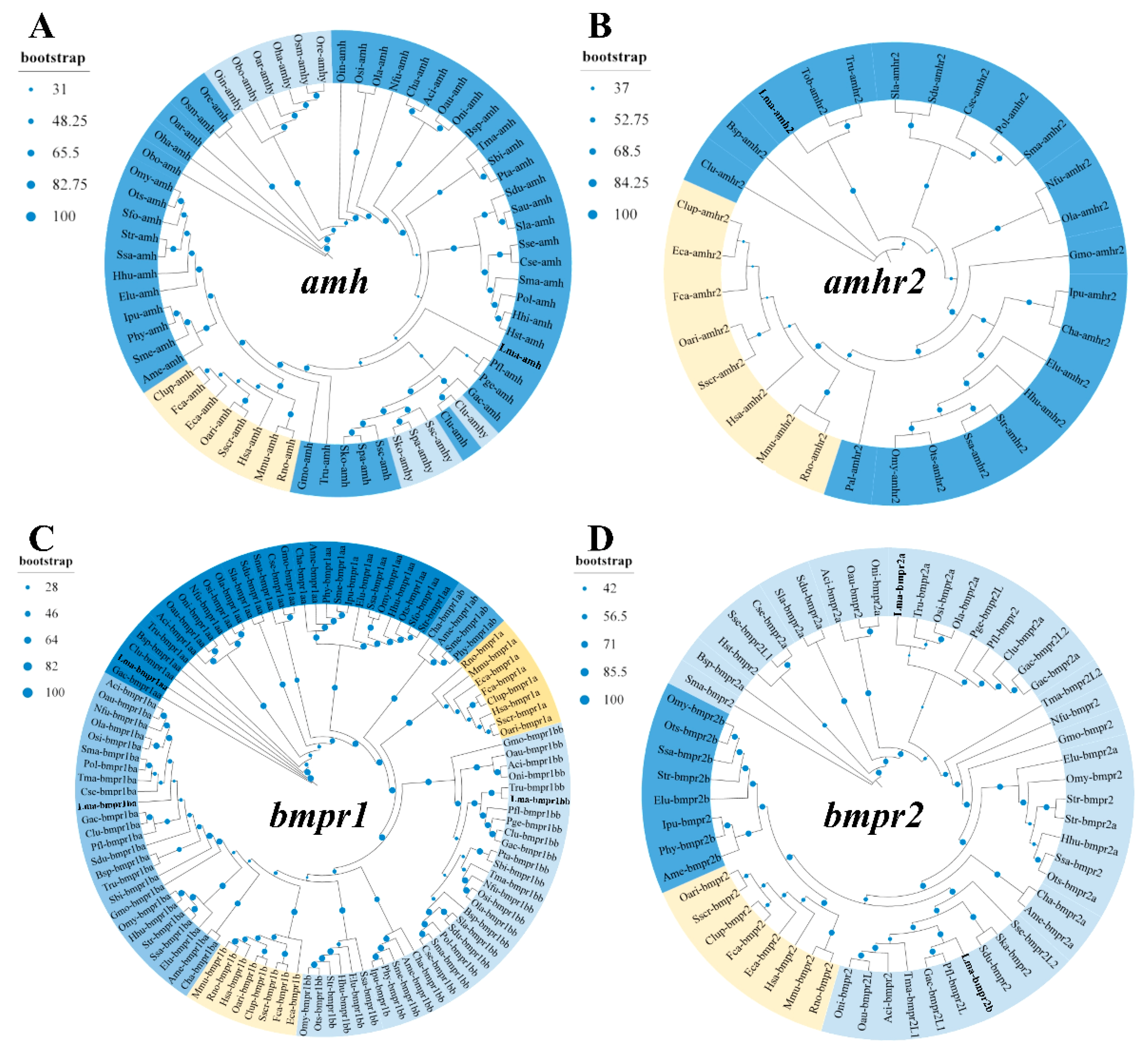
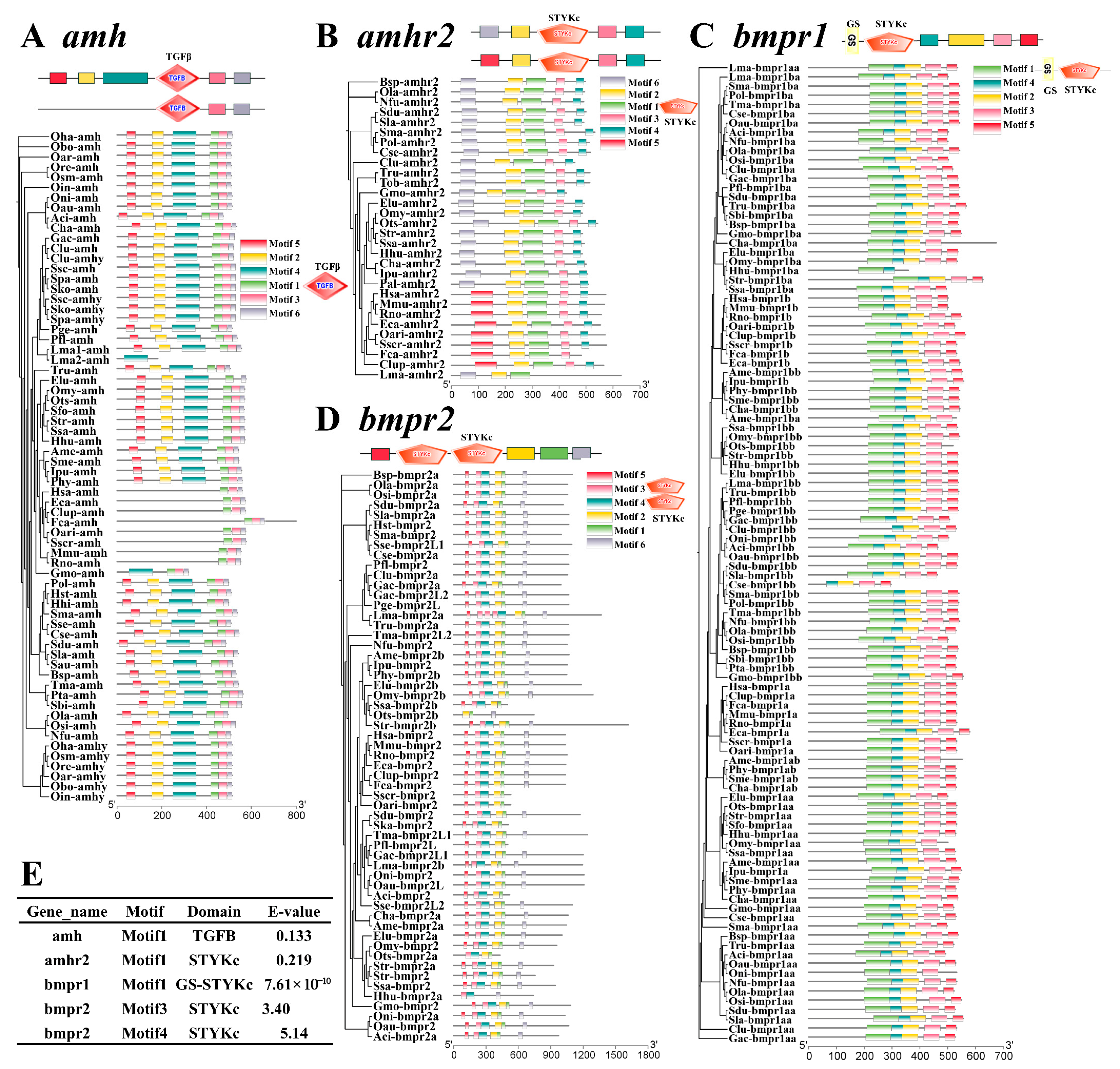
3.2. Phylogeny and Functional Domain of amh Signaling Genes

Through phylogeny reconstruction, mammalian amh genes were clustered into a distinct clade, while the teleost amhy genes from the Odontesthes and Sebastes lineages formed a monophyletic group (Figure 1A), indicating their duplication before the split of these species. Conversely, the amhy and amh from C. lumpus did not resolve into separate clades, suggesting the recent emergence of amhy in C. lumpus. For amhr2, mammalian amhr2 formed a conserved cluster, while teleost amhr2 clustered into another clade (Figure 1B). For bmpr1, mammalian bmpr1a and bmpr1b clustered independently, mirroring their teleost paralogs (Figure 1C). In addition, mammalian bmpr2 exhibited strong sequence conservation, while teleost bmpr2a clustered with mammalian bmpr2, and bmpr2b formed a divergent clade (except in L. maculatus), indicating prolonged divergence (Figure 1D). These phylogenies demonstrated strong evolutionary conservation of mammalian amh signaling genes, whereas the teleost lineages exhibited well-conserved amh/amhy and amhr2, alongside ambiguous differentiation among bmpr1 and bmpr2.
Through a functional domain analysis, compared with mammalian amh having motifs 1/3/6, teleost amh was found to contain the extra motifs 2/4/5, and they both had the motif 1 identified as the TGF-β domain (Figure 2A). For teleost amhr2, motif 6 was replaced by motif 5 (Activin_recp domain) in mammals, while they both contained motif 1 as the STYKc domain (Figure 2B). Teleost amhr2 lacked the Activin_recp domain, a hydrophilic, cysteine-rich ligand-binding module conserved in both type I and type II TGF-β receptors of mammals, suggesting potential functional differentiation in teleosts. In addition, bmpr1 and bmpr2 were generally conserved between mammals and teleosts in functional structure, both containing the STYKc domain (Figure 2C,D), supporting their primary role in functional redundancy rather than substantial neofunctionalization. All these phylogeny and functional structure results suggested that amh signaling is generally conserved between mammalian and teleost lineages.

3.3. Conserved Synteny of amh Signaling Genes

Through a genomic synteny analysis, the human/mouse amh, amhr2, bmpr1a, bmpr1b, and bmpr2 genes exhibited strong syntenic conservation (Figure 3), with minor exceptions, like the absence of peak3 upstream of amh in mice (Figure 3A) and loss of Atf7 upstream of amhr2 in humans (Figure 3B). Moreover, the teleost lineages displayed variable synteny of amh signaling genes, with flanking genes often rearranged due to insertions of uncharacterized genes. For example, the amh flanking genes (downstream, oaz1, org, lingo3, and upstream, dot1l, pkbp8, tmem59, rex1b, slc35a3) (Figure 3A) and amhr2 flanking genes (downstream, pcbp2, map3k12, myg1, and upstream, cdcap7l, sp1, tarbp2) (Figure 3B) were retained but repositioned among the teleost species. This was also observed among the teleost bmpr1a, bmpr1b, and bmpr2 (Figure 3C–E). The lower degree of conservation of gene synteny in teleosts compared to mammals may be attributed to several factors, as exemplified by the additional whole-genome duplication events in teleosts that promoted genomic reorganization and synteny loss [37], whereas the mammalian genomes retained stability through constrained diploid inheritance patterns, enhancing syntenic relationship preservation [38]. Therefore, the synteny analysis revealed exceptional chromosomal colinearity in mammalian amh/amhr2 loci, with teleost homologs maintaining moderate syntenic conservation despite localized genomic rearrangements. These findings collectively suggested that amh signaling exhibited heightened evolutionary constraints in mammals compared to teleosts, with relaxed selection pressures permitting limited structural diversification while preserving core functionality through paralogous compensation mechanisms.

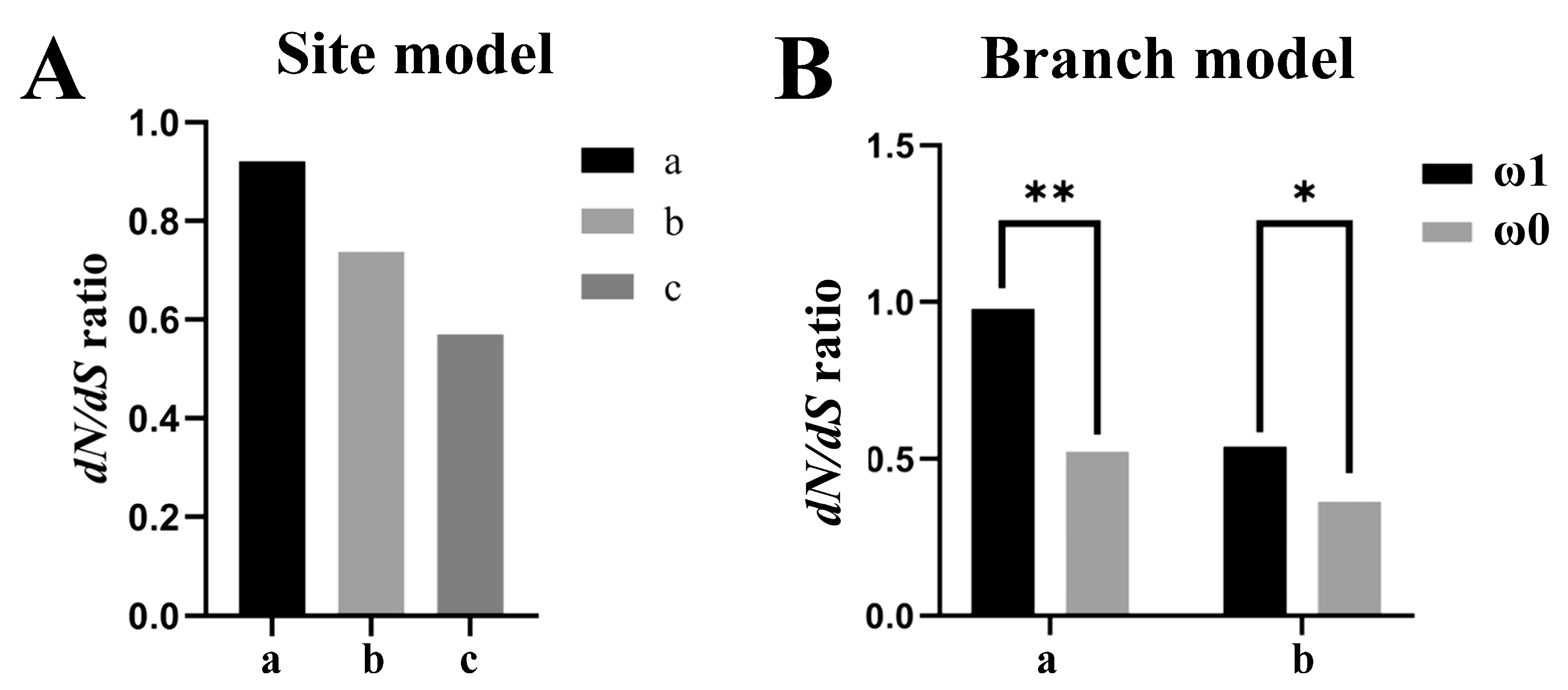
3.4. Molecular Evolution of amh Signaling Genes

To investigate the evolutionary rates (dN/dS, ω) of amh signaling genes, a molecular evolution analysis with site model (SM) and branch model (BM) tests from PAML were conducted. Among the results, the amh signaling genes exhibited dN/dS (ω) ratios consistently below one across the examined taxa (Figure 4), indicating strong purifying selection acting to eliminate deleterious nonsynonymous mutations. In the teleosts examined with both SM and BM tests, the duplicated amhy genes presented a higher ω value than that of the original amh genes, while the amh genes from the amhy-MSD teleosts also evolved faster than the amh genes from the non-amhy-MSD teleosts (Figure 4A,B). These results indicated functional redundancy or tolerance of amino acid substitutions in amhy-associated teleost species, in which mutations may not critically disrupt the duplicated protein function. Therefore, in teleosts utilizing amhy as the MSD gene, the amhy normally originated from a Y-chromosomal duplication event and acquired critical mutations to assume dominant sex-determination functionality [11]. Notably, amhy displayed a higher dN/dS ratio compared to its ancestral paralog, amh, reflecting weaker selective constraints and a greater tolerance for amino acid substitutions, which may facilitate the retention of functionally significant polymorphisms. Comparative analyses may further demonstrate elevated evolutionary rates in teleost amh signaling genes relative to their mammalian orthologs, a phenomenon likely linked to functional divergence. In mammals, these genes maintain conserved roles in fetal male reproductive development, particularly through AMH-mediated regression of Müllerian ducts during testicular differentiation [9]. In contrast, teleost lineages exhibit substantial functional plasticity, with amh signaling components acquiring novel roles as master regulators of sexual differentiation in multiple species [11,12,13].

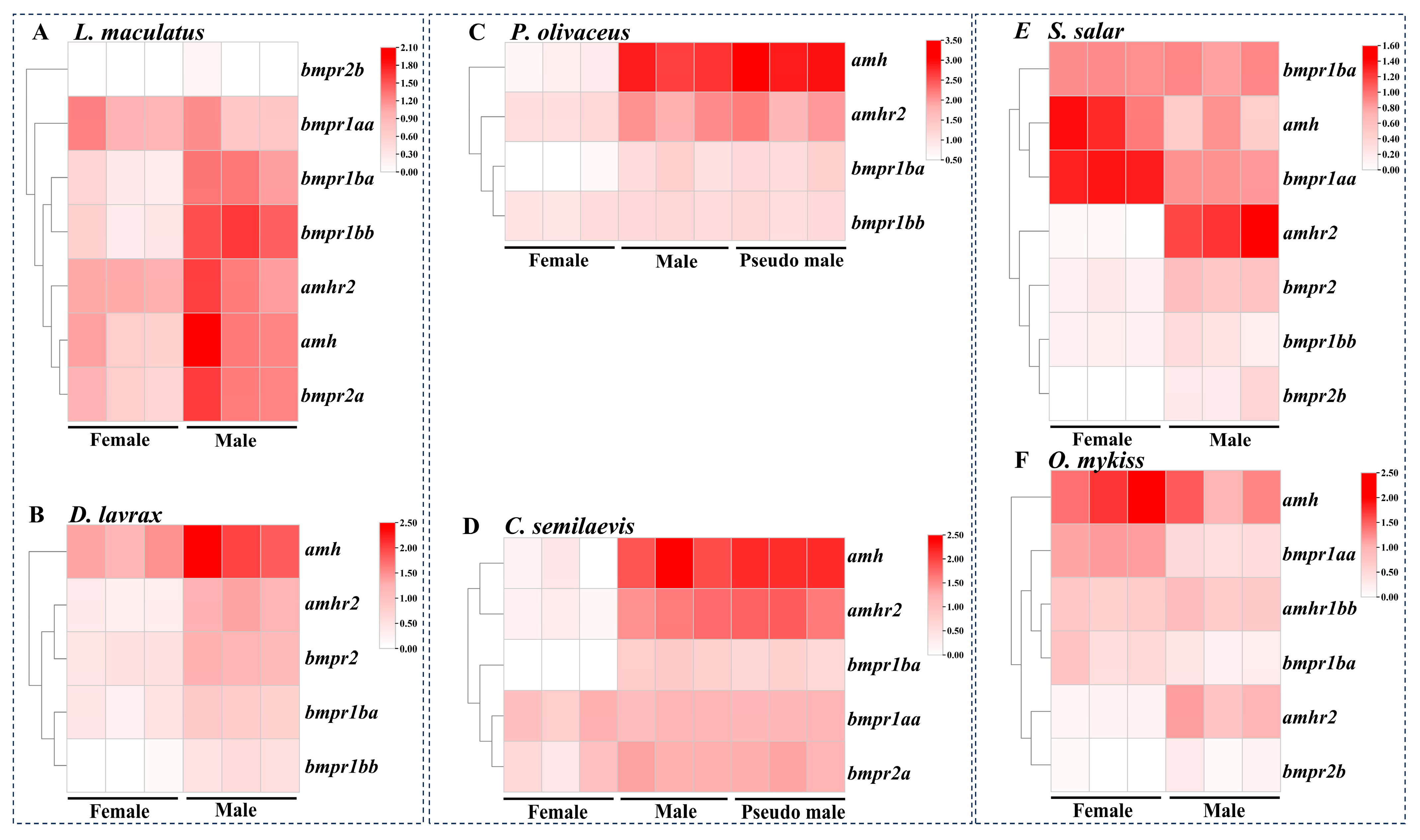
3.5. Expression Profiles of amh Signaling Genes in Teleost Species

We further investigated the expression profiles of amh signaling genes in teleost lineages with different SD patterns and mechanisms, including L. maculatus and D. labrax, both representing a putatively polygenic SD mode without an MSD gene; P. olivaceus (amhy MSD) and C. semilaevis (dmrt1 MSD), both representing a sex-reversal scenario from genetic female to phenotypic pseudo-male under environmental stimuli; and S. salar and O. mykiss, both representing the extra fourth-round whole-genome duplication with the non-canonical sdY as their MSD gene. Generally, the amh signaling genes (amh, amhr2, bmpr1, and bmpr2) exhibited testis-biased expression (Figure 5), with some species-specific exceptions. For example, in L. maculatus and D. labrax, amh/amhr2 and most of the bmprs showed testis-biased expression, except for bmpr1aa and bmpr2b in L. maculatus (Figure 5A,B). Similarly, in P. olivaceus and C. semilaevis, amh/amhr2 and bmpr1ba were testis-biased, but not with other bmprs (bmpr1bb in P. olivaceus and bmpr1aa, bmpr2a in C. semilaevis) (Figure 5C,D). Moreover, no difference was observed between genetic males and sex-reversed pseudo-males of the two flatfishes. In S. salar and O. mykiss, amhr2 and bmpr2 were testis-biased, while amh and bmpr1aa were ovary-biased, and bmpr1ba/1bb was not bias-expressed (Figure 5E,F). Interestingly, several Salmoniformes amh signaling genes displayed female-specific up-regulation patterns, with amh and bmpr1aa showing predominantly ovarian expression during sexual maturation, highlighting a conserved pathway architecture with divergent functional deployment across teleost lineages. These results suggested that even with similar functions in sexual development and sex determination, the amh signaling genes presented divergent expression patterns across teleost lineages, in species-specific ways.

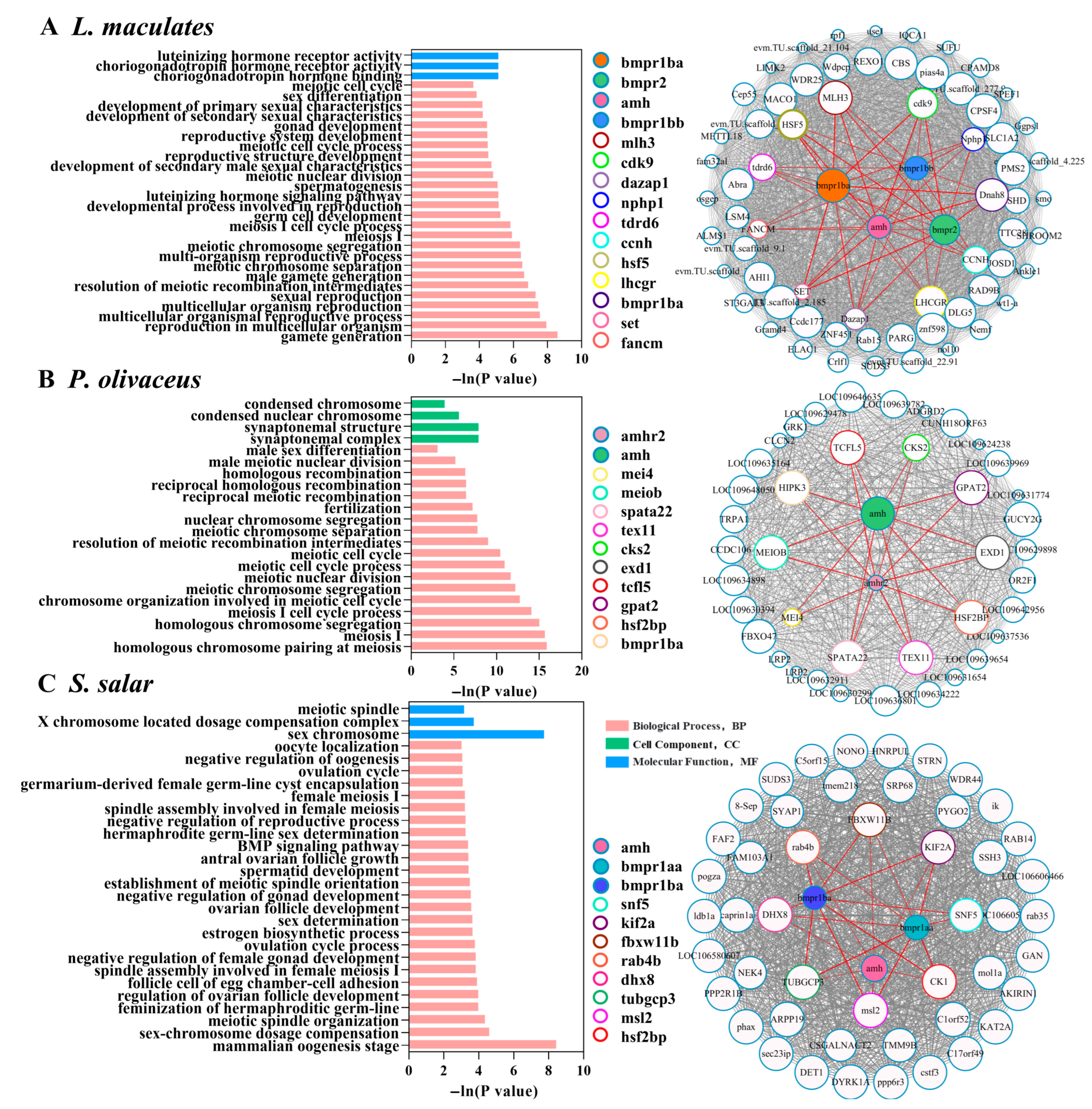
3.6. Gene Co-Expression Network of amh Signaling Genes in Teleost Species

To further understand the regulatory mechanisms for amh signaling genes in teleost sexual development, WGCNA was performed with different tissue-specific transcriptomes for L. maculatus (12 tissues), P. olivaceus (11 tissues), and S. salar (eight tissues), respectively, and testis- or ovary-specific gene co-expression modules were identified (Figure 6). In the L. maculatus testis-specific module, four amh signaling genes (amh, bmpr1ba/1bb, and bmpr2) were identified, and their top 20 co-expressing genes were enriched with GO functions of “gamete generation”, “reproductive process”, “spermatogenesis”, “sexual characteristics”, “meiotic recombination”, “meiosis”, etc. (Figure 6A). Specifically, many amh signaling co-expressed genes are involved in the spermatogenesis process (Figure 6A). For example, daz-associated protein 1 (dazap1), a germline-specific RNA-binding protein, is essential for primordial germ cell differentiation during spermatogenesis, and its deficiency could cause complete spermatogenic arrest and azoospermia [39]. Nephrocystin-1 (nphp1) is required for the differentiation of early elongating spermatids into spermatozoa [40]. Luteinizing hormone/choriogonadotropin receptor (lhcgr) is the receptor for lutropin-choriogonadotropic hormone [41]. The chromatin remodeler tudor domain-containing protein 6 (tdrd6) is involved in mRNA splicing in prophase I spermatocytes [42], and tdrd6-deficient diplotene spermatocytes revealed high numbers of splicing defects, such as aberrant usage of introns and exons, as well as aberrant representation of splice junctions [42]. Heat shock factor 5 (hsf5) is essential for male fertility, spermatogenesis, and meiotic prophase progression in spermatocytes [43], which operates as a constitutive transcriptional regulator in male germ cells, governing meiotic prophase I progression [43]. Dynein axonemal heavy chain 8 (dnah8) generates the protein component of the outer dynein arms (ODAs) in the sperm flagellum [44]. Notably, dysregulation of Fanconi anemia group M protein (fancm) precipitates dual reproductive pathologies: premature ovarian failure through impaired oocyte meiotic progression, and spermatogenic collapse, manifesting as non-obstructive azoospermia with Sertoli cell-only syndrome—an autosomal recessive infertility disorder characterized by complete germ cell aplasia despite intact somatic niche cells [45]. Therefore, all these genes co-expressed with amh, bmpr1ba, bmpr1bb, and bmpr2 collectively form an evolutionarily conserved regulatory network for spermatogenesis in L. maculates, which does not even have the MSD gene (Figure 6A).
In the P. olivaceus testis-specific module, only amh and amhr2 were identified, corresponding to their testis-biased expression (Figure 5C), and their top 20 co-expressing genes had enriched GO functions of “male sex differentiation”, “meiotic cell cycle”, “recombination”, “synaptonemal complex”, “chromosome separation”, etc. (Figure 6B). Many meiosis-related genes were co-expressed with amh and amhr2 (Figure 6B). For instance, spermatogenesis-associated 22 (spata22) is a meiosis-specific protein required for homologous recombination in meiosis I [46]. Testis-expressed 11 (tex11) could regulate crossing-over during meiosis [47]. Meiosis-specific with OB domain-containing protein (meiob) is required for homologous recombination in meiosis I [48]. Meiosis-specific coiled-coil protein 4 (mei4) is required for DNA double-strand breaks (DSBs) formation in unsynapsed regions during meiotic recombination [49]. Heat shock factor 2-binding protein (hsf2bp) is involved in meiotic double-strand break repair [50]. Moreover, spermatogenesis genes were also associated with the amh/amhr2 in the P. olivaceus testis; for instance, transcription factor-like 5 protein (tcfl5) plays a role in early spermatogenesis [51]. Glycerol-3-phosphate acyltransferase 2 (gpat2) is required for the primary processing step during piRNA biosynthesis [52]. PiRNA biogenesis protein exd1 is required for the processing of piRNAs during spermatogenesis [53]. Therefore, all these genes interacted with amh and amhr2, collectively forming the putatively evolutionarily conserved regulatory network for male gamete production in P. olivaceus, with amhy as the MSD gene (Figure 6B), probably with both genetic males and sex-reversed pseudo-males, which frequently display differences in sperm quality [54].
Conversely, no testis-specific module was found in S. salar, while an ovary-specific module, containing amh, bmpr1aa, and bmpr1ba, was identified, and their top 20 co-expressing genes had enriched GO functions of “female meiosis I”, “regulation of oogenesis”, “ovarian follicle development”, “estrogen biosynthetic process”, “ovulation cycle process”, etc. (Figure 6C). Many female sexual development-related genes were co-expressed with amh, bmpr1aa, and bmpr1ba (Figure 6C). For example, meiotic regulation in salmonid ovaries involves a sophisticated interplay of microtubule dynamics and chromatin remodeling mechanisms, with casein kinase 1 (ck1) ensuring precise chromosomal alignment through phosphorylation-mediated activation of FAM110A, thereby facilitating its spindle interaction [55]. This process is complemented by the centrosomal protein γ-tubulin complex protein 3 (TUBGCP3/GCP3), a core constituent of both γ-tubulin small complexes (γ-TuSCs) and ring complexes (γ-TuRCs), which orchestrates microtubule nucleation, essential for bipolar spindle assembly [56]. Simultaneously, the ubiquitination machinery demonstrates critical regulatory functions, as evidenced by F-box and WD repeat domain containing 11 (FBXW11)’s mediation of CEP68 degradation to enable centriole separation [57], while kinesin family member 2a (kif2a) emerges as a microtubule depolymerase coordinating spindle dynamics with chromosome congression during meiotic progression [58]. Notably, these mitotic regulators functionally converge with RNA processing mechanisms through DEAH-box helicase 8 (dhx8), which exhibits dual roles in mRNA splicing fidelity and mitotic checkpoint regulation [59]. The system further integrates chromatin organization via the MSL complex, where MSL complex subunit 1 (msl1)-dependent dimerization establishes a prerequisite platform for MSL complex subunit 2 (msl2)-mediated X-chromosome recognition and subsequent chromatin spreading, potentially maintaining dosage compensation during oogenesis [60]. Therefore, all these genes co-expressed with amh, bmpr1aa, and bmpr1ba, collectively forming a non-canonical regulatory network for ovary and oocyte development in S. salar (Figure 6C), and the fourth-round genome duplication may provide more materials (duplicated bmprs) for their functional diversification in Salmoniformes.
Collectively, the integrated evidence from the expression and gene networks supported the hypothesis that amh signaling may serve as the key molecular complex governing sex differentiation in a species-specific manner in diverse teleost lineages, independent of its sex-determination role, and interacting with functions of both testis and ovary development. Therefore, its heightened evolutionary and functional flexibility potentially explains the remarkable sexual plasticity characteristic of teleost lineages.

4. Conclusions

The molecular insights reported here contextualize the evolutionary dynamics of amh signaling in teleosts, and revealed frequent lineage-specific paralog expansions (e.g., amhy neofunctionalization), contrasting with the extreme conservation observed in mammalian homologs. The amh signaling exhibits weaker evolutionary constraint in teleosts, correlating with accelerated substitution rates and functional repurposing from ancestral roles in Müllerian duct regression to novel sex-determination mechanisms. This evolutionary plasticity is exemplified by the recurrent co-option of amh/amhr2 components into master sex-determining switches through subfunctionalization and regulatory element rewiring, establishing these genes as central nodes in the emergence of diverse neo-sex determination systems across the teleost phylogeny. Further investigations with more fish species and functional verification are warranted.

Supplementary Materials

The following supporting information can be downloaded at https://www.mdpi.com/article/10.3390/fishes10070327/s1, Table S1: The copy numbers of amh signaling genes in selected teleost and mammalian species.

Author Contributions

Conceptualization, J.C.; methodology, L.Z., K.H., and Q.Z.; validation, L.Z., K.H., and Q.Z.; formal analysis, L.Z., K.H., and Q.Z.; resources, F.W. and W.L. (Weigang Li); data curation, F.W. and W.L. (Weigang Li); writing—original draft preparation, L.Z.; writing—review and editing, J.C.; visualization, L.Z., K.H., W.L. (Wei Lu), and Q.Z.; supervision, J.C.; project administration, W.L. (Wei Lu); funding acquisition, J.C. and W.L. (Wei Lu). All authors have read and agreed to the published version of the manuscript.

Funding

This research was funded by the National Key Research and Development Program of China (2024YFD2400901, 2022YFD2400503), the Key Research and Development Program of Shandong Province (2022LZGC016), the Natural Science Foundation of Shandong Province (ZR2022MC100), the China Agriculture Research System (CARS-47-G06), and the National Marine Genetic Resource Center, China.

Institutional Review Board Statement

This study was conducted in accordance with the Institutional Animal Care and Use Committee of Ocean University of China (IACUC-OUC), and it does not contain any studies with human participants. (approval date: 25 February 2023).

Informed Consent Statement

Not applicable.

Data Availability Statement

The transcriptome datasets used in this study can be retrieved from the NCBI database or made available on request.

Acknowledgments

This research was supported by the High-Performance Computing Platform of YZBSTCACC, Sanya, China, the Large Instrument Sharing Platform of College of Marine Life Sciences, Ocean University of China, and the Laoshan Laboratory, Qingdao, China.

Conflicts of Interest

The authors declare no conflicts of interest.

References

  1. Liu, Z.; Gao, D. The Cause–Effect model of master sex determination gene acquisition and the evolution of sex chromosomes. Int. J. Mol. Sci. 2025, 26, 3282. [Google Scholar] [CrossRef] [PubMed]
  2. Kitano, J.; Ansai, S.; Takehana, Y.; Yamamoto, Y. Diversity and convergence of sex-determination mechanisms in teleost fish. Annu. Rev. Anim. Biosci. 2024, 12, 233–259. [Google Scholar] [CrossRef]
  3. Wang, J.; Tao, W.; Kocher, T.D.; Wang, D. Sex chromosome turnover and biodiversity in fishes. J. Genet. Genom. 2024, 51, 1351–1360. [Google Scholar] [CrossRef] [PubMed]
  4. Pan, Q.; Kay, T.; Depincé, A.; Adolfi, M.; Schartl, M.; Guiguen, Y.; Herpin, A. Evolution of master sex determiners: TGF-β signalling pathways at regulatory crossroads. Philos. Trans. R. Soc. Lond. B Biol. Sci. 2021, 376, 20200091. [Google Scholar] [CrossRef] [PubMed]
  5. Nagahama, Y.; Chakraborty, T.; Paul-Prasanth, B.; Ohta, K.; Nakamura, M. Sex determination, gonadal sex differentiation, and plasticity in vertebrate species. Physiol. Rev. 2021, 101, 1237–1308. [Google Scholar] [CrossRef] [PubMed]
  6. Taslima, K.; Kikuchi, K.; Hosoya, S. Genomic architecture and sex chromosome systems of commercially important fish species in Asia—Current status, knowledge gaps and future prospects. Rev. Aquac. 2024, 16, 1918–1946. [Google Scholar] [CrossRef]
  7. Zhu, Z.; Younas, L.; Zhou, Q. Evolution and regulation of animal sex chromosomes. Nat. Rev. Genet. 2025, 26, 59–74. [Google Scholar] [CrossRef]
  8. Mullen, R.D.; Ontiveros, A.E.; Moses, M.M.; Behringer, R.R. AMH and AMHR2 mutations: A spectrum of reproductive phenotypes across vertebrate species. Dev. Biol. 2019, 455, 1–9. [Google Scholar] [CrossRef]
  9. Moses, M.M.; Behringer, R.R. A gene regulatory network for Müllerian duct regression. Environ. Epigenet. 2019, 5, dvz017. [Google Scholar] [CrossRef]
  10. Adolfi, M.C.; Nakajima, R.T.; Nóbrega, R.H.; Schartl, M. Intersex, hermaphroditism, and gonadal plasticity in vertebrates: Evolution of the Müllerian duct and amh/amhr2 signaling. Annu. Rev. Anim. Biosci. 2019, 7, 149–172. [Google Scholar] [CrossRef]
  11. Hattori, R.S.; Strüssmann, C.A.; Fernandino, J.I.; Somoza, G.M. Genotypic sex determination in teleosts: Insights from the testis-determining amhy gene. Gen. Comp. Endocrinol. 2013, 192, 55–59. [Google Scholar] [CrossRef] [PubMed]
  12. Liu, X.; Dai, S.; Wu, J.; Wei, X.; Zhou, X.; Chen, M.; Tan, D.; Pu, D.; Li, M.; Wang, D. Roles of anti-Müllerian hormone and its duplicates in sex determination and germ cell proliferation of Nile tilapia. Genetics 2022, 220, iyab237. [Google Scholar] [CrossRef] [PubMed]
  13. Hattori, R.S.; Somoza, G.M.; Fernandino, J.I.; Colautti, D.C.; Miyoshi, K.; Gong, Z.; Yamamoto, Y.; Strüssmann, C.A. The duplicated Y-specific amhy gene is conserved and linked to maleness in silversides of the genus Odontesthes. Genes 2019, 10, 679. [Google Scholar] [CrossRef]
  14. Jeffries, D.L.; Mee, J.A.; Peichel, C.L. Identification of a candidate sex determination gene in Culaea inconstans suggests convergent recruitment of an Amh duplicate in two lineages of stickleback. J. Evol. Biol. 2022, 35, 1683–1695. [Google Scholar] [CrossRef]
  15. Song, W.; Xie, Y.; Sun, M.; Li, X.; Fitzpatrick, C.K.; Vaux, F.; O’Malley, K.G.; Zhang, Q.; Qi, J.; He, Y. A duplicated amh is the master sex-determining gene for Sebastes rockfish in the Northwest Pacific. Open Biol. 2021, 11, 210063. [Google Scholar] [CrossRef]
  16. Hattori, R.S.; Kumazawa, K.; Nakamoto, M.; Nakano, Y.; Yamaguchi, T.; Kitano, T.; Yamamoto, E.; Fuji, K.; Sakamoto, T. Y-specific amh allele, amhy, is the master sex-determining gene in Japanese flounder Paralichthys olivaceus. Front. Genet. 2022, 13, 1007548. [Google Scholar] [CrossRef]
  17. Rafati, N.; Chen, J.; Herpin, A.; Pettersson, M.E.; Han, F.; Feng, C.; Wallerman, O.; Rubin, C.J.; Péron, S.; Cocco, A.; et al. Reconstruction of the birth of a male sex chromosome present in Atlantic herring. Proc. Natl. Acad. Sci. USA 2020, 117, 24359–24368. [Google Scholar] [CrossRef] [PubMed]
  18. Jasonowicz, A.J.; Simeon, A.; Zahm, M.; Cabau, C.; Klopp, C.; Roques, C.; Iampietro, C.; Lluch, J.; Donnadieu, C.; Parrinello, H.; et al. Generation of a chromosome-level genome assembly for Pacific halibut (Hippoglossus stenolepis) and characterization of its sex-determining genomic region. Mol. Ecol. Resour. 2022, 22, 2685–2700. [Google Scholar] [CrossRef]
  19. Klüver, N.; Pfennig, F.; Pala, I.; Storch, K.; Schlieder, M.; Froschauer, A.; Gutzeit, H.O.; Schartl, M. Differential expression of anti-Müllerian hormone (AMH) and anti-Müllerian hormone receptor type II (AMHRII) in the teleost medaka. Dev. Dyn. 2007, 236, 271–281. [Google Scholar] [CrossRef]
  20. Wang, W.; Liang, S.; Zou, Y.; Wu, Z.; Wang, L.; Liu, Y.; You, F. Amh dominant expression in Sertoli cells during the testicular differentiation and development stages in the olive flounder Paralichthys olivaceus. Gene 2020, 755, 144906. [Google Scholar] [CrossRef]
  21. Lin, C.J.; Jeng, S.R.; Lei, Z.Y.; Yueh, W.S.; Dufour, S.; Wu, G.C.; Chang, C.F. Involvement of transforming growth factor beta family genes in gonadal differentiation in Japanese eel, Anguilla japonica, according to sex-Related gene expressions. Cells 2021, 10, 3007. [Google Scholar] [CrossRef]
  22. Pfennig, F.; Standke, A.; Gutzeit, H.O. The role of Amh signaling in teleost fish--Multiple functions not restricted to the gonads. Gen. Comp. Endocrinol. 2015, 223, 87–107. [Google Scholar] [CrossRef]
  23. Shanshan, L.; Bing, S.; Zhuo, L.; Jing, Z.; Songlin, C. Cloning and expression of anti-Müllerian hormone gene in half- smooth tongue-sole, Cynoglossus semilaevis. J. Fish. Sci. China 2013, 20, 35–43, (In Chinese with English Abstract). [Google Scholar]
  24. Chen, C.; Chen, H.; Zhang, Y.; Thomas, H.R.; Frank, M.H.; He, Y.; Xia, R. TBtools: An integrative toolkit developed for interactive analyses of big biological data. Mol. Plant 2020, 13, 1194–1202. [Google Scholar] [CrossRef] [PubMed]
  25. Kumar, S.; Stecher, G.; Li, M.; Knyaz, C.; Tamura, K. MEGA X: Molecular evolutionary genetics analysis across computing platforms. Mol. Biol. Evol. 2018, 35, 1547–1549. [Google Scholar] [CrossRef] [PubMed]
  26. Nguyen, L.T.; Schmidt, H.A.; von Haeseler, A.; Minh, B.Q. IQ-TREE: A fast and effective stochastic algorithm for estimating maximum-likelihood phylogenies. Mol. Biol. Evol. 2015, 32, 268–274. [Google Scholar] [CrossRef]
  27. Letunic, I.; Bork, P. Interactive Tree Of Life (iTOL) v5: An online tool for phylogenetic tree display and annotation. Nucleic Acids Res. 2021, 49, W293–W296. [Google Scholar] [CrossRef] [PubMed]
  28. Bailey, T.L.; Johnson, J.; Grant, C.E.; Noble, W.S. The MEME suite. Nucleic Acids Res. 2015, 43, W39–W49. [Google Scholar] [CrossRef]
  29. Gao, F.; Chen, C.; Arab, D.A.; Du, Z.; He, Y.; Ho, S.Y.W. EasyCodeML: A visual tool for analysis of selection using CodeML. Ecol. Evol. 2019, 9, 3891–3898. [Google Scholar] [CrossRef]
  30. Cheng, J.; Yang, F.; Liu, S.; Zhao, H.; Lu, W.; Zhang, Q. Transcriptomic analysis reveals functional interaction of mRNA–lncRNA–miRNA in steroidogenesis and spermatogenesis of gynogenetic Japanese flounder (Paralichthys olivaceus). Biology 2022, 11, 213. [Google Scholar] [CrossRef]
  31. Bolger, A.M.; Lohse, M.; Usadel, B. Trimmomatic: A flexible trimmer for Illumina sequence data. Bioinformatics 2014, 30, 2114–2120. [Google Scholar] [CrossRef] [PubMed]
  32. Pertea, M.; Kim, D.; Pertea, G.M.; Leek, J.T.; Salzberg, S.L. Transcript-level expression analysis of RNA-seq experiments with HISAT, StringTie and Ballgown. Nat. Protoc. 2016, 11, 1650–1667. [Google Scholar] [CrossRef] [PubMed]
  33. Zhu, Q.; Li, M.; Lu, W.; Wang, Y.; Li, X.; Cheng, J. transcriptomic modulation reveals the specific cellular response in Chinese sea bass (Lateolabrax maculatus) gills under salinity change and alkalinity stress. Int. J. Mol. Sci. 2023, 24, 5877. [Google Scholar] [CrossRef] [PubMed]
  34. Langfelder, P.; Horvath, S. WGCNA: An R package for weighted correlation network analysis. BMC Bioinf. 2008, 9, 559. [Google Scholar] [CrossRef]
  35. Gene Ontology Consortium. The Gene Ontology in 2010: Extensions and refinements. Nucleic Acids Res. 2010, 38, D331–D335. [Google Scholar] [CrossRef] [PubMed]
  36. Shannon, P.; Markiel, A.; Ozier, O.; Baliga, N.S.; Wang, J.T.; Ramage, D.; Amin, N.; Schwikowski, B.; Ideker, T. Cytoscape: A software environment for integrated models of biomolecular interaction networks. Genome Res. 2003, 13, 2498–2504. [Google Scholar] [CrossRef]
  37. Amores, A.; Force, A.; Yan, Y.-L.; Joly, L.; Amemiya, C.; Fritz, A.; Ho, R.K.; Langeland, J.; Prince, V.; Wang, Y.-L.; et al. Zebrafish hox clusters and vertebrate genome evolution. Science 1998, 282, 1711–1714. [Google Scholar] [CrossRef]
  38. Sharman, A.C.; Holland, P.W. Conservation, duplication, and divergence of developmental genes during chordate evolution. Neth. J. Zool. 1995, 46, 47–67. [Google Scholar] [CrossRef]
  39. Razavi, S.M.; Sabbaghian, M.; Jalili, M.; Divsalar, A.; Wolkenhauer, O.; Salehzadeh-Yazdi, A. Comprehensive functional enrichment analysis of male infertility. Sci. Rep. 2017, 7, 15778. [Google Scholar] [CrossRef]
  40. Jiang, S.T.; Chiou, Y.Y.; Wang, E.; Lin, H.K.; Lee, S.P.; Lu, H.Y.; Wang, C.K.; Tang, M.J.; Li, H. Targeted disruption of Nphp1 causes male infertility due to defects in the later steps of sperm morphogenesis in mice. Hum. Mol. Genet. 2008, 17, 3368–3379. [Google Scholar] [CrossRef]
  41. Xu, Y.; Chen, Y.; Li, N.; Hu, X.; Li, G.; Ding, Y.; Li, J.; Shen, Y.; Wang, X.; Wang, J. Novel compound heterozygous variants in the LHCGR gene identified in a subject with Leydig cell hypoplasia type 1. J. Pediatr. Endocrinol. Metab. 2018, 31, 239–245. [Google Scholar] [CrossRef] [PubMed]
  42. Akpınar, M.; Lesche, M.; Fanourgakis, G.; Fu, J.; Anastassiadis, K.; Dahl, A.; Jessberger, R. TDRD6 mediates early steps of spliceosome maturation in primary spermatocytes. PLoS Genet. 2017, 13, e1006660. [Google Scholar] [CrossRef]
  43. Liu, M.; Wang, L.; Li, Y.; Zhi, E.; Shen, G.; Jiang, X.; Li, D.; Zhao, X.; Ruan, T.; Jiang, C.; et al. HSF5 deficiency causes male infertility involving spermatogenic arrest at meiotic prophase i in humans and mice. Adv. Sci. 2024, 11, e2402412. [Google Scholar] [CrossRef]
  44. Whitfield, M.; Thomas, L.; Bequignon, E.; Schmitt, A.; Stouvenel, L.; Montantin, G.; Tissier, S.; Duquesnoy, P.; Copin, B.; Chantot, S.; et al. Mutations in DNAH17, encoding a sperm-specific axonemal outer dynein arm heavy chain, cause isolated male infertility due to asthenozoospermia. Am. J. Hum. Genet. 2019, 105, 198–212. [Google Scholar] [CrossRef] [PubMed]
  45. Tsui, V.; Lyu, R.; Novakovic, S.; Stringer, J.M.; Dunleavy, J.E.M.; Granger, E.; Semple, T.; Leichter, A.; Martelotto, L.G.; Merriner, D.J.; et al. Fancm has dual roles in the limiting of meiotic crossovers and germ cell maintenance in mammals. Cell Genom. 2023, 3, 100349. [Google Scholar] [CrossRef]
  46. Petrillo, C.; Barroca, V.; Ribeiro, J.; Lailler, N.; Livera, G.; Keeney, S.; Martini, E.; Jain, D. shani mutation in mouse affects splicing of Spata22 and leads to impaired meiotic recombination. Chromosoma 2020, 129, 161–179. [Google Scholar] [CrossRef]
  47. Ghieh, F.; Passet, B.; Poumerol, E.; Castille, J.; Calvel, P.; Vilotte, J.L.; Sellem, E.; Jouneau, L.; Mambu-Mambueni, H.; Garchon, H.J.; et al. A partial deletion within the meiosis-specific sporulation domain SPO22 of Tex11 is not associated with infertility in mice. PLoS ONE 2024, 19, e0309974. [Google Scholar] [CrossRef]
  48. Zhu, X.; Hu, K.; Cheng, H.; Wu, H.; Li, K.; Gao, Y.; Lv, M.; Xu, C.; Geng, H.; Shen, Q.; et al. Novel MEIOB pathogenic variants including a homozygous non-canonical splicing variant, cause meiotic arrest and human non-obstructive azoospermia. Clin. Genet. 2024, 105, 99–105. [Google Scholar] [CrossRef]
  49. Kumar, R.; Ghyselinck, N.; Ishiguro, K.; Watanabe, Y.; Kouznetsova, A.; Höög, C.; Strong, E.; Schimenti, J.; Daniel, K.; Toth, A.; et al. MEI4—A central player in the regulation of meiotic DNA double-strand break formation in the mouse. J. Cell Sci. 2015, 128, 1800–1811. [Google Scholar] [CrossRef]
  50. Brandsma, I.; Sato, K.; van Rossum-Fikkert, S.E.; van Vliet, N.; Sleddens, E.; Reuter, M.; Odijk, H.; van den Tempel, N.; Dekkers, D.H.W.; Bezstarosti, K.; et al. HSF2BP interacts with a conserved domain of BRCA2 and is required for mouse spermatogenesis. Cell Rep. 2019, 27, 3790–3798.e3797. [Google Scholar] [CrossRef]
  51. Säflund, M.; Özata, D.M. The MYBL1/TCFL5 transcription network: Two collaborative factors with central role in male meiosis. Biochem. Soc. Trans. 2023, 51, 2163–2172. [Google Scholar] [CrossRef] [PubMed]
  52. Shiromoto, Y.; Kuramochi-Miyagawa, S.; Nagamori, I.; Chuma, S.; Arakawa, T.; Nishimura, T.; Hasuwa, H.; Tachibana, T.; Ikawa, M.; Nakano, T. GPAT2 is required for piRNA biogenesis, transposon silencing, and maintenance of spermatogonia in mice. Biol. Reprod. 2019, 101, 248–256. [Google Scholar] [CrossRef]
  53. Yang, Z.; Chen, K.M.; Pandey, R.R.; Homolka, D.; Reuter, M.; Janeiro, B.K.; Sachidanandam, R.; Fauvarque, M.O.; McCarthy, A.A.; Pillai, R.S. PIWI slicing and EXD1 drive biogenesis of nuclear piRNAs from cytosolic targets of the mouse piRNA pathway. Mol. Cell 2016, 61, 138–152. [Google Scholar] [CrossRef]
  54. Zong, W.; Wang, Y.; Zhang, L.; Lu, W.; Li, W.; Wang, F.; Cheng, J. DNA methylation mediates sperm quality via piwil1 and piwil2 regulation in Japanese flounder (Paralichthys olivaceus). Int. J. Mol. Sci. 2024, 25, 5935. [Google Scholar] [CrossRef]
  55. Perez, C.A.; Burocziova, M.; Jenikova, G.; Macurek, L. CK1-mediated phosphorylation of FAM110A promotes its interaction with mitotic spindle and controls chromosomal alignment. EMBO Rep. 2021, 22, e51847. [Google Scholar] [CrossRef]
  56. Ge, C.; Lin, C.; Zhang, M.; Yuan, J.; Feng, X.; Hao, Z.; Zhang, S.; Tian, Q. Tubgcp3 is a mitotic regulator of planarian epidermal differentiation. Gene 2021, 775, 145440. [Google Scholar] [CrossRef] [PubMed]
  57. Pagan, J.K.; Marzio, A.; Jones, M.J.; Saraf, A.; Jallepalli, P.V.; Florens, L.; Washburn, M.P.; Pagano, M. Degradation of Cep68 and PCNT cleavage mediate Cep215 removal from the PCM to allow centriole separation, disengagement and licensing. Nat. Cell Biol. 2015, 17, 31–43. [Google Scholar] [CrossRef]
  58. Yi, Z.Y.; Ma, X.S.; Liang, Q.X.; Zhang, T.; Xu, Z.Y.; Meng, T.G.; Ouyang, Y.C.; Hou, Y.; Schatten, H.; Sun, Q.Y.; et al. Kif2a regulates spindle organization and cell cycle progression in meiotic oocytes. Sci. Rep. 2016, 6, 38574. [Google Scholar] [CrossRef] [PubMed]
  59. English, M.A.; Lei, L.; Blake, T.; Wincovitch, S.M.; Sood, R.; Azuma, M.; Hickstein, D.; Liu, P.P. Incomplete splicing, cell division defects, and hematopoietic blockage in dhx8 mutant zebrafish. Dev. Dyn. 2012, 241, 879–889. [Google Scholar] [CrossRef]
  60. Hallacli, E.; Lipp, M.; Georgiev, P.; Spielman, C.; Cusack, S.; Akhtar, A.; Kadlec, J. Msl1-mediated dimerization of the dosage compensation complex is essential for male X-chromosome regulation in Drosophila. Mol. Cell 2012, 48, 587–600. [Google Scholar] [CrossRef]
Figure 1. Phylogeny of amh signaling genes. (A) amh phylogenetic tree with mammalian amh in yellow, teleost amh in dark blue, and amhy in light blue; (B) amhr2 phylogenetic tree with mammalian amhr2 in yellow and teleost amhr2 in dark blue; (C) bmpr1 phylogenetic tree, with mammalian bmpr1a in dark yellow, mammalian bmpr1b in light yellow, and the teleost bmpr1aa, bmpr1ab, bmpr1ba, and bmpr1bb in dark to light blue; (D) bmpr2 phylogenetic tree, with mammalian bmpr2 in yellow, teleost bmpr2a in light blue, and bmpr2b in dark blue.
Fishes 10 00327 g001
Figure 2. Conserved domain of amh signaling genes in teleosts, as follows: (A) amh, (B) amhr2, (C) bmpr1, (D) bmpr2, and (E) the functional domain and motif in each of the amh signaling genes. The colored boxes on the horizontal lines represent the motifs successfully predicted in amino acid sequences and the abscissa axes represent the lengths of amino acid sequences. The colored text boxes above each motif structure diagram represent the predicted functional domain of the corresponding genes.
Fishes 10 00327 g002
Figure 3. Conserved synteny of amh signaling genes in teleost lineages, as follows: (A) amh, (B) amhr2, (C) bmpr1a, (D) bmpr1b, and (E) bmpr2. The top corner of the pentagon indicates gene orientation, and the white pentagon represents genes that are not annotated in the database.
Fishes 10 00327 g003
Figure 4. Tests of molecular evolution of amh signaling genes in teleost lineages using PAML. (A) Site model tests in teleosts, as follows: (a) amhy genes, (b) amh genes from teleost species with amhy duplication, and (c) amh genes from species without amhy duplication. (B) Branch model tests in teleosts, as follows: (a) between amhy gene (ω1) and amh gene (ω0), and (b) between amh gene with amhy duplication (ω1) and amh gene without amhy duplication (ω0). Black and gray columns indicate ω (dN/dS) values. * represents p < 0.05, and ** represents p < 0.01.
Fishes 10 00327 g004
Figure 5. Expression profiles of amh signaling genes in gonads of different teleost species, as follows: (A) L. maculatus; (B) D. labrax; (C) P. olivaceus; (D) C. semilaevis; (E) S. salar; and (F) O. mykiss, all of which are shown as the log2(FPKM+1) on the heatmaps.
Fishes 10 00327 g005
Figure 6. WGCNA of amh signaling genes in the testis-specific modules of (A) L. maculatus and (B) P. olivaceus, and in ovary-specific module of (C) S. salar, with transcriptomes of different tissues. The top 20 genes with the highest connectivity (weight value) to each amh signaling gene in the candidate modules were included, with their GO enrichment represented. The circle size indicates connectivity and circle frame colors indicate different functional pathways. The red line indicates the interaction between the amh signaling genes and the candidate functional genes.
Fishes 10 00327 g006
Table 1. Copy numbers of amh signaling genes in selected teleost species.
OrderFamilyGenusSpeciesamhamhr2bmpr1bmpr2
AnabantiformesOsphronemidaeBettasplendens1131
CichliformesCichlidaeAmphilophuscitrinellus1132
CichliformesCichlidaeOreochromisaureus1012
CichliformesCichlidaeOreochromisniloticus1022
CichliformesCichlidaeHaplochromisburtoni1132
CichliformesCichlidaePundamilianyererei1132
CichliformesCichlidaeMaylandiazebra1121
CichliformesCichlidaeAstatotilapiacalliptera1132
CichliformesCichlidaeNeolamprologusbrichardi0031
Perciformes AcropomatiformesLateolabracidaeLateolabraxmaculatus1132
Perciformes AcanthuriformesMoronidaeDicentrarchuslabrax1121
PerciformesPercidaePercaflavescens1112
PerciformesPercidaePseudocaranxgeorgianus1011
PerciformesPercidaeSanderlucioperca1332
PerciformesLabridaeLabrusbergylta1132
PerciformesCentropomidaeLatescalcarifer1122
PerciformesBovichthyidaeCottopercagobio1132
PerciformesAnabantidaeAnabastestudineus2132
PerciformesPomacentridaeAmphiprionocellaris1132
PerciformesSciaenidaeLarimichthyscrocea1121
PerciformesPomacentridaeAmphiprionpercula1121
PerciformesPomacentridaeAcanthochromispolyacanthus1321
CarangiformesCarangidaeSerioladumerili1132
CarangiformesCarangidaeSeriolaaureovittata1000
CarangiformesCarangidaeSeriolalalandi1122
ScorpaeniformesCyclopteridaeCyclopteruslumpus2131
ScorpaeniformesSebastidaeSebastesschlegelii2112
ScorpaeniformesSebastidaeSebastespachycephalus2111
ScorpaeniformesSebastidaeSebasteskoreanus2111
Scombriformes ScombridaeThunnusmaccoyii1012
PleuronectiformesSoleidaeSoleasenegalensis1002
PleuronectiformesCynoglossidaeCynoglossussemilaevis1131
PleuronectiformesScophthalmidae Scophthalmusmaximus1131
PleuronectiformesParalichthyidaeParalichthysolivaceus2120
PleuronectiformesPleuronectidaeHippoglossushippoglossus1000
PleuronectiformesPleuronectidaeHippoglossusstenolepis1001
CyprinodontiformesNothobranchiidaeNothobranchiusfurzeri1121
CyprinodontiformesNothobranchiidaeNothobranchiuskadleci1121
CyprinodontiformesPoeciliidaePoeciliaformosa1122
CyprinodontiformesPoeciliidaePoeciliareticulata1121
CyprinodontiformesPoeciliidaePoecilialatipinna1122
CyprinodontiformesPoeciliidaeXiphophoruscouchianus1011
CyprinodontiformesPoeciliidaeXiphophorusmaculatus1122
CyprinodontiformesRivulidaeKryptolebiasmarmoratus1121
CyprinodontiformesFundulidaeFundulusheteroclitus1121
CyprinodontiformesCyprinodontidaeCyprinodonvariegatus2121
BeloniformesAdrianichthyidaeOryziaslatipes1131
BeloniformesAdrianichthyidaeOryziassinensis1031
BeloniformesAdrianichthyidaeOryziasmelastigma1121
BeloniformesAdrianichthyidaeOryziasjavanicus1121
Atheriniformes AtherinopsidaeOdontesthesbonariensis2121
Atheriniformes AtherinopsidaeOdontestheshatcheri2100
Atheriniformes AtherinopsidaeOdontesthesincisa2000
GasterosteiformesGasterosteidaeGasterosteusaculeatus1131
SyngnathiformesSyngnathidaeSyngnathoidesbiaculeatus1110
SyngnathiformesSyngnathidaePhyllopteryxtaeniolatus1110
SyngnathiformesSyngnathidaeHippocampuscomes2121
TetraodontiformesTetraodontidaeTakifugurubripes1131
TetraodontiformesTetraodontidaeTakifuguobscurus0100
TetraodontiformesTetraodontidaeTetraodonnigroviridis1021
ClupeiformesClupeidaeClupeaharengus1141
ClupeiformesClupeiformesDenticepsclupeoides2122
GadiformesGadidaeGadusmorhua1131
GadiformesGadidaeGadusmacrocephalus1131
CharaciformesCharacidaePygocentrusnattereri1142
CharaciformesCharacidaeAstyanaxmexicanus1042
SiluriformesSiluridaeSilurusmeridionalis1110
SiluriformesPangasiidaePangasianodonhypophthalmus1111
SiluriformesIctaluridaeIctaluruspunctatus1121
SalmoniformesSalmonidae Salvelinusfontinalis1010
SalmoniformesSalmonidae Oncorhynchustshawytscha1122
SalmoniformesSalmonidaeOncorhynchusmykiss1132
SalmoniformesSalmonidaeSalmosalar1132
SalmoniformesSalmonidaeSalmotrutta1133
SalmoniformesSalmonidaeHuchohucho1131
SalmoniformesSalmonidaeOncorhynchuskisutch1143
EsociformesEsocidaeEsoxlucius1132
OsmeriformesOsmeridaePlecoglossusaltivelis1100
BeryciformesHolocentridaeMyripristismurdjan1132
OsteoglossiformesOsteoglossidaeArapaimagigas1021
OsteoglossiformesMormyridaeParamormyropskingsleyae1232
CypriniformesCyprinidaeCyprinuscarpio1084
CypriniformesCyprinidaeCarassiusauratus1084
CypriniformesCyprinidaeDaniorerio1042
LepisosteiformesLepisosteidaeLepisosteusoculatus1121
SynbranchiformesMastacembelidaeMastacembelusarmatus1121
GymnotiformesElectrophoridaeElectrophoruselectricus1142
Disclaimer/Publisher’s Note: The statements, opinions and data contained in all publications are solely those of the individual author(s) and contributor(s) and not of MDPI and/or the editor(s). MDPI and/or the editor(s) disclaim responsibility for any injury to people or property resulting from any ideas, methods, instructions or products referred to in the content.

