Article

Changes in Subcellular Responses in the Digestive Gland of the Freshwater Mussel Unio crassus from a Historically Contaminated Environment

1
Ruđer Bošković Institute, Division for Marine and Environmental Research, Bijenička Cesta 54, 10000 Zagreb, Croatia
2
Department of Biology, Faculty of Science, University of Zagreb, Horvatovac 102a, 10000 Zagreb, Croatia
3
Vodovod i Kanalizacija d.o.o. Karlovac, Gažanski Trg 8, 47000 Karlovac, Croatia
4
Faculty of Chemical Engineering and Technology, University of Zagreb, Trg Marka Marulića 19, 10000 Zagreb, Croatia
*
Authors to whom correspondence should be addressed.
Fishes 2025, 10(7), 317; https://doi.org/10.3390/fishes10070317
Submission received: 9 May 2025 / Revised: 11 June 2025 / Accepted: 17 June 2025 / Published: 2 July 2025
(This article belongs to the Section Environment and Climate Change)

Abstract

Utilizing a multi-biomarker approach, we assessed the potential adverse effects of pollutants on subcellular responses in the digestive gland of the freshwater mussel Unio crassus from a historically contaminated lowland section (KIZ) of the river Mrežnica compared to its less impacted upstream karstic section (REF) and their seasonality (spring vs. autumn). This approach accounted for the diverse modes of action of pollutants by including biomarkers of metal exposure (metallothioneins, MT), general stress (total cytosolic proteins, TP), antioxidative capacity (catalase, CAT; glutathione, GSH; glutathione-S-transferase, GST), oxidative damage (malondialdehyde, MDA), and neurotoxicity (acetylcholinesterase, AChE). Only in spring, MT concentrations were 15% higher at the REF site (4.38 ± 1.06 µg mg proteins−1) compared to the KIZ site (3.69 ± 0.63 µg mg proteins−1), likely related to elevated Cd bioaccumulation due to the karstic substrate. Regardless of the season, mussels from KIZ showed consistently lower TP and GSH, with significantly higher CAT, GST, and MDA levels, indicating elevated stress, activation of antioxidant defenses, and oxidative damage from chronic exposure to pro-oxidant pollutants, including metal(loid)s and organic contaminants (e.g., ibuprofen, nicotine). Compared to the REF site, AChE activity at the KIZ site was higher in late spring and lower in early autumn, indicating seasonal variability in AChE activity at the contamination-impacted location driven by fluctuating exposure to neurotoxicants, such as drugs and insecticides. Overall, biomarker responses indicated that mild historical pollution, reinforced by current low-capacity sources, has an observable impact on mussel health, posing long-term risks to sediment-dwelling aquatic organisms.
Key Contribution: Mild freshwater contamination, including historical pollution, can cause visible subcellular biochemical alterations in wild mussel populations, highlighting oxidative stress and neurotoxicity as lasting physiological effects. The multi-biomarker approach proved essential for detecting subtle yet biologically relevant physiological alterations in Unio crassus, offering a comprehensive assessment of organism health and improving the interpretation of complex environmental pollution effects.

Graphical Abstract

1. Introduction

Traces of historical industrial contamination can still be found in numerous aquatic environments around the globe [1,2,3,4,5]. The Mrežnica River in Croatia serves as an example, as it has been exposed to various sources of industrial pollution for over a century, which were stopped or reduced more than a decade ago [2]. Our recent study of water/sediment contamination of the Mrežnica River, during the same seasons and at the same sites as chosen for the current study, confirmed the presence of contaminants that may be associated not only with current but also historical sources of pollution, such as increased concentrations of metals and certain organic contaminants in the water and sediments that are typical for the currently inactive textile industry and coal combustion [1,6]. Prior to the current study, we first investigated the bioaccumulation of metals (total and cytosolic) in the digestive gland of the same specimens of the sediment-dwelling endangered bivalve species, Unio crassus, from the Mrežnica River, which were used in this study [7]. Due to their direct exposure to the pollutants stored in the sediments, increased concentrations of many non-essential and potentially highly toxic elements were detected in the mussel specimens from the industrial zone of Karlovac town [7]. However, the sole measurement of bioaccumulated pollutants, such as metals, in bivalve organs does not present sufficient information to estimate their potential negative effects at the subcellular level or on the health of the organisms. On the other hand, the analysis of subcellular molecular/biochemical biomarkers involved in important cellular processes, such as detoxification or antioxidative defense, can be used to detect the earliest disturbances of homeostasis that may have occurred in the organisms due to exposure to different types of contaminants [8].
The best-known biomarkers for metal exposure are metallothioneins (MT), thermostable cytosolic proteins with low molecular mass, which are important for the regulation of essential and the detoxification of non-essential elements in mussels and other organisms [9]. Many studies suggest that analyzing MT induction is a valuable method to detect the responses of organisms exposed to metals [10,11,12]. The subcellular presence of excess metals in a non-detoxified form can be associated with the onset of negative events, including the increased production of reactive oxygen species (ROS), which consequently pose a significant threat to cellular integrity by causing damage to proteins, DNA, and polyunsaturated fatty acids in the cell membranes of organisms [13,14]. To counteract the negative effects of oxidative stress (OS), aquatic organisms, including freshwater mussels, show a variety of changes in (non-)enzymatic antioxidant defenses following exposure to pollutants with oxidative potential [8,15]. These antioxidant defense systems include, in particular, catalase (CAT), an oxidoreductase enzyme that catalyzes the oxidation of various electron donor substrates and is specific to the degradation of H2O2 [14]; glutathione (GSH), with the important function of neutralizing reactive oxygen intermediates and free radicals that are constantly formed during metabolism [13]; and glutathione S-transferase (GST), a phase II detoxification enzyme that catalyzes the binding of GSH to potentially cell-damaging xenobiotics and converts them into more water-soluble molecules to facilitate their excretion [16]. The overproduction of ROS caused by metallic or organic contaminants can trigger a process known as lipid peroxidation (LPO), and one of the best-known by-products of LPO is malondialdehyde (MDA), which is often used as a reliable marker for the assessment of cell membrane damage [17]. Cell membrane damage can interfere with the release, reuptake, and degradation of neurotransmitters, such as acethylcholinesterase (AChE), leading to imbalances in neuromuscular synaptic transmission. Inhibition of AChE has been successfully used as a biomarker for the neurotoxicity of various pollutants, such as pesticides, detergents, and pharmaceuticals [16].
Previous research on the effects of environmental stressors on U. crassus is scarce. To our knowledge, only three laboratory studies have been conducted, one observing behavioral changes in U. crassus exposed to volatile organic solvents [18], the other measuring four immunological/biochemical parameters in the digestive gland of mussels exposed to zinc/copper pyrithione [19], and the third measuring hemocyte counts, histological alterations, and five biomarkers of OS following exposure to nano-CeO2 [20]. In addition, research on other freshwater mussels has also focused on a limited number of biomarkers in mussel tissues, both in laboratory and environmental studies [21,22,23]. Accordingly, the main aim of our study was to assess the susceptibility of an endangered freshwater mussel species, U. crassus, to mild sediment contamination, including from historical sources, using a set of different subcellular biomarkers. As a target organ, we have chosen digestive gland, as, compared to the gills and hemolymph, it offers greater sensitivity for biomarker analyses due to higher contaminant accumulation, metabolic activity, and ability to integrate long-term exposure [10,24,25]. The specific aim was to identify potential adverse effects and subcellular biochemical changes caused by pollutants stored in the sediment and bioaccumulated in mussel tissue. The studied set of biomarkers included biomarkers of metal exposure (MT), general stress (total cytosolic proteins, TP), antioxidative capacity (CAT, GSH, and GST), oxidative damage (MDA), and exposure to organic pollutants (AChE). We hypothesized that mussels from the historically contaminated site will exhibit altered biomarker responses indicative of metal exposure, oxidative stress, and neurotoxicity compared to those from the less impacted reference site. To our knowledge, the results reported in this study are the first data from a multi-biomarker approach for the wild population of the mussel species U. crassus under real environmental conditions. The influence of various biotic and abiotic factors, such as the season of sampling (e.g., reproductive status of mussels, water temperature), on biomarker responses is also discussed, as they may contribute to fluctuations in biomarker activity/concentration and thus mask the effects of pollutants [10].

2. Materials and Methods

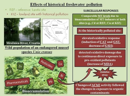
2.1. Study Area and Sampling of Mussels

Sampling of the thick-shelled river mussel U. crassus was carried out in the Mrežnica River in Croatia at two sites. These are the reference site (REF) (N 45°26′28.40″ E 15°30′15.39″), which is located about 2 km upstream from the town of Duga Resa and represents a karst location with low anthropogenic impact, and the polluted site, which is located in Karlovac town’s industrial zone, Mala Švarča (KIZ), and represents a location historically contaminated with a mixture of pollutants from various anthropogenic sources and is nowadays exposed only to low-pollution-capacity facilities (N 45°27′49.05″ E 15°32′30.09″) (Figure 1).
The river section from Duga Resa to Karlovac was for decades exposed to various industrial wastewaters, including from the textile industry and the production of medical supplies; approximately a decade ago, the textile industry ceased to operate, and the majority of the remaining industrial wastewaters were connected to the wastewater treatment plant of Karlovac town, with treated wastewater being discharged into the Kupa River downstream from Karlovac [1]. Only a few small facilities still discharge their wastewaters into the Mrežnica River (e.g., a heating plant, a car wash, a hospital, and production of medical supplies made of cotton and personal care products) [1]. The data confirming the contamination status of the selected sampling sites originating largely from historical sources, i.e., elemental concentrations in water (dissolved/particulate) and sediments, as well as concentrations of organic contaminants and other valuable information about REF and KIZ sites, can be found in our previously published papers [1,6]. Excerpted results from these studies, which are important for our discussion, are shown in Table 1.
The specimens of U. crassus can burrow up to 20 cm into the riverbed’s sediment, but they are usually visible on the sediment’s surface [26]. The granulometric analysis of the sediments in 2021 showed that the average proportions of clay amounted to 9.5–11.9%, silt 47–56%, and sand 34–44% [1]. The mussels were collected manually by a diver during two seasons in 2021, including late spring (early June, a period of higher water levels/flows) and early autumn (early October, a period of lower water levels/flows). Sampling collection was performed during stable weather conditions. In June 2021, sampling was conducted on sunny days with an average air temperature of 22.8 °C, while in October 2021 the weather was also clear and dry, with an average air temperature of 9.5 °C. There were no significant precipitation events or sudden weather changes during the sampling periods, ensuring consistency and minimizing potential environmental variability. Because U. crassus is a protected mussel species, sampling was carried out with the permission of the state authorities. In total, 30 mussels were collected and transported to the laboratory in dark and cool containers. Average lengths of mussels (cm) were as follows: REFspring: 46.7 ± 3.65; KIZspring: 59.0 ± 2.83; REFautumn: 46.1 ± 3.76; KIZautumn: 61.7 ± 3.52. Thereafter, the digestive glands were dissected, frozen into liquid nitrogen, and kept at −80°C until further treatment. Composite samples, each containing digestive glands from four to five mussels, were prepared for biomarker analyses: REFspring = 7; KIZspring = 10; REFautumn = 6; KIZ autumn = 6. Digestive glands are commonly used mussel tissue to assess environmental quality because they are a major storage and detoxification organ that plays an important role in cell homeostasis [10,24,25].

2.2. Processing of the Mussel Digestive Gland Samples

For the analyses of MT and TP, the digestive glands were cut into small pieces, and a cooled homogenization buffer 20 mM Tris/HCl buffer (Sigma Aldrich, St. Louis, MO, USA; pH 8.6 at 22 °C) containing 0.01% β-mercaptoethanol (Sigma Aldrich, St. Louis, MO, USA), protease inhibitors (0.006 mM leupeptin and 0.5 mM phenylmethylsulfonyl fluoride; Alfa Aesar, Karlsruhe, Germany), and 0.5 M saccharose (only for MT analysis; Sigma Aldrich, St. Louis, MO, USA) was added (mass:volume = 1:4). The suspension was homogenized with a Potter–Elvehjem homogenizer (Glas-Col, Terre Haute, IN, USA; used for all sample preparations). The homogenates were then centrifuged at 50,000× g for 120 min at +4 °C (Avanti J-E centrifuge; Beckman Coulter, Indianapolis, IN, USA), and the resulting supernatant (S50) was used for MT and TP analyses. For GSH analysis, a piece of digestive gland tissue was homogenized in ice-cold 5% sulphosalicylic acid (SSA) (Kemika, Zagreb, Croatia) (mass:volume = 1:5) and then centrifuged at 10,000× g for 10 min at +4 °C (Biofuge Fresco, Heraeus, Hanau, Germany). The resulting supernatant (S10-SSA) was used for GSH analysis. Another piece of digestive gland tissue for the analyses of CAT, GST, MDA, and AChE was homogenized in ice-cold 50 mM potassium phosphate buffer (PPB) (Kemika, Zagreb, Croatia; pH 8.1 at 4 °C) (mass:volume = 1:5), and then the following centrifugation steps (Avanti J-E centrifuge) were performed: 3000× g for 10 min at +4 °C, and the resulting supernatant (S3-PPB) was used for MDA analysis; and 10,000× g for 30 min at +4 °C, and the obtained supernatant (S10-PPB) was used for analyzing CAT, GST, and AChE activity. The supernatants obtained for all biomarker measurements were stored at –80 °C until further analysis.

2.3. Biomarker Analyses

MTs were determined according to the modified method of Viarengo et al. [27], which is described in Bourdineaud et al. [28]. The supernatant S50 (250 µL) was used for two-step ethanol/chloroform precipitation (CARLO ERBA Reagents, Cornaredo, Italy, and Fischer Chemical, Loughborough, UK, respectively). The resulting pellets were then washed with a solution of 87% ethanol and 1% chloroform in the homogenization buffer. After centrifugation at 12,000× g for 6 min at 2 °C, the pellets were dried under a stream of nitrogen. The MT-containing pellets were reconstituted by adding 35 μL each of 0.25 M NaCl (Merck, Darmstadt, Germany) and a solution of 4 mM EDTA/1 M HCl (both Merck, Darmstadt, Germany). The thiol group’s content was then analyzed using 5,5-dithiobis-2-nitrobenzoic acid (DTNB) (Sigma Aldrich, St. Louis, MO, USA) dissolved in 0.2 M PPB/2 M NaCl (Kemika, Zagreb, Croatia, and Merck, Darmstadt, Germany, respectively), pH 8. The samples were transferred to a microplate, and the absorbance was measured at 412 nm. Reduced GSH was used as the reference standard (2.5–25 μg GSH), and the MT content was expressed per TP content (μg MT mg−1 TP).
The quantification of TP concentrations was performed according to the method of Bradford [29], which was adapted for use in microplates. A volume of 300 µL of CBB reagent (Coomassie brilliant blue G-250; Serva, Heidelberg, Germany) was added to 10 µL of 20-fold-diluted S50 samples in 20 mM Tris/HCl buffer. After 10 min of incubation, the absorbance was read at 595 nm. Bovine serum albumin (BSA) (Serva, Heidelberg, Germany) was used as a reference standard (0.0625–0.500 mg mL−1 BSA).
CAT activity was determined according to a micromethod described by Li and Schellhorn [30], with some modifications. A solution of PPB (50 mM, pH 7.0; Kemika, Croatia) and 30% hydrogen peroxide (Suprapur®, Merck, Darmstadt, Germany) was prepared to yield 10.34 mM H2O2, and a volume of 290 µL of this solution was then added to 10 µL of a 15-times-diluted sample (S10-PPB) in 50 mM PPB (pH 7.0). The analysis was performed in UV-transparent 96-well microplates, and absorbance measurements were taken every 10 s for 1 min at 25 °C. The specific enzyme activity was calculated using a molar extinction coefficient of 43.6 M−1 cm−1 and expressed as μmol H2O2 degraded per minute per mg of proteins. Protein concentrations in the S10-PPB fractions were determined using the Bradford method [29].
The total concentration of GSH was measured in five-times-diluted S10-SSA supernatants using a spectrophotometric DTNB-GSSG reductase recycling assay, as outlined by Tietze [31]. The protocol for the microtiter plate assay was adapted from Rahman et al. [32]. All solutions were prepared in 0.1 M PPB (Kemika, Zagreb, Croatia) with the addition of 1 mM EDTA disodium salt (Merck, Darmstadt, Germany), maintaining a pH of 7.5. A volume of 150 μL of a solution containing DTNB (0.108 mM, Sigma Aldrich, St. Louis, MO, USA) and GR (glutathione reductase, 0.171 U mL−1; Sigma Aldrich, St. Louis, MO, USA) was added to the sample (five-times-diluted supernatants S10-SSA in Mili Q) in the plate. The plate was then mixed and left in the dark for 5 min. Subsequently, 50 μL of NADPH solution (0.192 mM; Sigma Aldrich, St. Louis, MO, USA) was added, and the absorbance at 412 nm was measured at 1-min intervals for 5 min at 25 °C. GSH standards (3.125–12.5 nM mL−1; Sigma Aldrich, St. Louis, MO, USA) were prepared in 1% SSA. Results were expressed as nmol of GSH per gram of wet tissue mass.
GST was analyzed according to the method of Habig et al. [33], which we modified slightly and adapted for use in microplates. The samples (S10-PPB) were diluted five times in 0.1 M sodium phosphate buffer (pH 6.5; Merck, Germany), and 5 µL of the diluted sample was added to the microtiter plate. Subsequently, 10 µL of a 20 mM GSH solution (co-substrate, final concentration 1 mM; Sigma Aldrich, St. Louis, MO, USA) and 185 µL of a 1 mM 1-chloro-2,4-dinitrobenzene solution (CDNB; Sigma Aldrich, St. Louis, MO, USA) were added, and the absorbance at 340 nm and 25 °C was measured at 15 s intervals for 3 min. Enzyme activity was expressed as µmol per minute per mg of proteins (determined in S10-PPB fractions using the Bradford method [29]), and the absorbance coefficient of 9.6 mM−1 cm−1 was used for the calculations.
The MDA concentration was determined spectrophotometrically after the reaction of MDA with 2-thiobarbituric acid (TBA) [34] according to the protocol described in Mijošek et al. [35]. A mixture of 1% butylated hydroxytoluene (BHT, Sigma-Aldrich, St. Louis, MO, USA) in ethanol (CARLO ERBA Reagents, Cornaredo, Italy) and 10% trichloroacetic acid (TCA, Kemika, Zagreb, Croatia) in Milli-Q water (BHT/TCA = 1:100) was added to the S3-PPB supernatant. The samples were vortexed and cooled for 15 min, followed by centrifugation at 4000× g for 15 min at 4 °C using a Biofuge Fresco centrifuge (Heraeus, Hanau, Germany). The resulting supernatants were transferred to 1.5 mL Eppendorf® tubes, and TBA (Alfa Aesar, Karlsruhe, Germany) dissolved in Milli-Q water was added. The tubes were heated for 30 min at 100 °C, producing a pink fluorescent product. After cooling, the samples were transferred to a microplate. The absorbance was measured at a wavelength of 535 nm, and a calibration curve was generated using five concentrations (0.9–15 μM) of MDA (Aldrich, St. Louis, MO, USA). The homogenization buffer served as a blank and underwent the same treatment as the samples. Results were expressed as nmol of MDA per gram of wet tissue mass.
AChE analysis was performed according to the method described by Ellman et al. [36], adapted for use in microtiter plates. A reaction mixture consisting of 10 µL of sample (S10-PPB) and 250 µL of 0.1 M PPB (pH 7.4 at 25 °C) (Kemika, Zagreb, Croatia) with 0.1 mM DTNB (Sigma Aldrich, St. Louis, MO, USA) was incubated in the dark for 15 min. The measurement began with the addition of 25 µL of 10 mM acetylthiocholine iodide and immediate observation of the increase in absorbance at 412 nm and 25 °C, with absorbance values measured at 15 s intervals for 3 min. Enzymatic activity was quantified as nmol of hydrolyzed acetylthiocholine per minute per mg of proteins (determined in S10-PPB fractions using the Bradford method [29]) using the absorbance coefficient of 13.6 mM−1 cm−1 for calculations.
Absorbance readings for all biochemical measurements were performed using the Infinite M200 microplate spectrophotometer (Tecan, Männedorf, Switzerland). Each composite sample was analyzed in triplicate for all biomarkers to ensure analytical reproducibility. For each analytical method, blank samples were included and processed alongside biological samples. Depending on the biomarker, blank values were subtracted from the corresponding sample measurements to correct for background levels and ensure data accuracy.

2.4. Statistical Analysis

SigmaPlot 11.0 and Statistica 12.5 for Windows were used for statistical analyses. Basic calculations and graphs were performed/created in Microsoft Office Excel 2011 for Windows. To assess the variability of biomarker values in the digestive gland of freshwater mussels, U. crassus, across two seasons and two sites, a two-way ANOVA with a post hoc LSD test was performed. Prior to analysis, data were log-transformed to meet the assumption of normal distribution and variance homogeneity, where necessary. Results were considered statistically significant at p < 0.05. Pearson correlation analysis was applied to calculate correlation coefficients (r) between MT and certain metal levels.

3. Results and Discussion

In order to assess biological responses and detect potential adverse effects of pollutants in native populations of endangered U. crassus mussels living in a lowland river section historically exposed to a mixture of pollutants from different anthropogenic sources, mainly from industry, we used the multi-biomarker approach. Results of statistical analyses between sites and seasons are shown in Table 2. We discuss this in light of information on the bioaccumulation of metals in the digestive glands of the same mussel samples used in this study, which we have published previously [7]. This approach involved the analysis of a set of subcellular molecular/biochemical biomarkers that may indicate the earliest disturbances of cellular homeostasis. The data presented here are, to our knowledge, the first comparative data on the concentrations/activities of seven biomarkers considered in this study in the digestive gland of U. crassus exposed to pollution under real environmental conditions and the first data altogether for MTs, AChE, and CAT in U. crassus. As mentioned above, the only data available for the remaining four biomarkers were previously reported for the digestive gland of U. crassus exposed to Zn/Cu pyrithione (MDA, GSH, and TP [19]) or nano-CeO2 (GSH, GST, and TP [20]) in the laboratory.

3.1. Changes in Biomarkers of Metal Exposure and General Stress

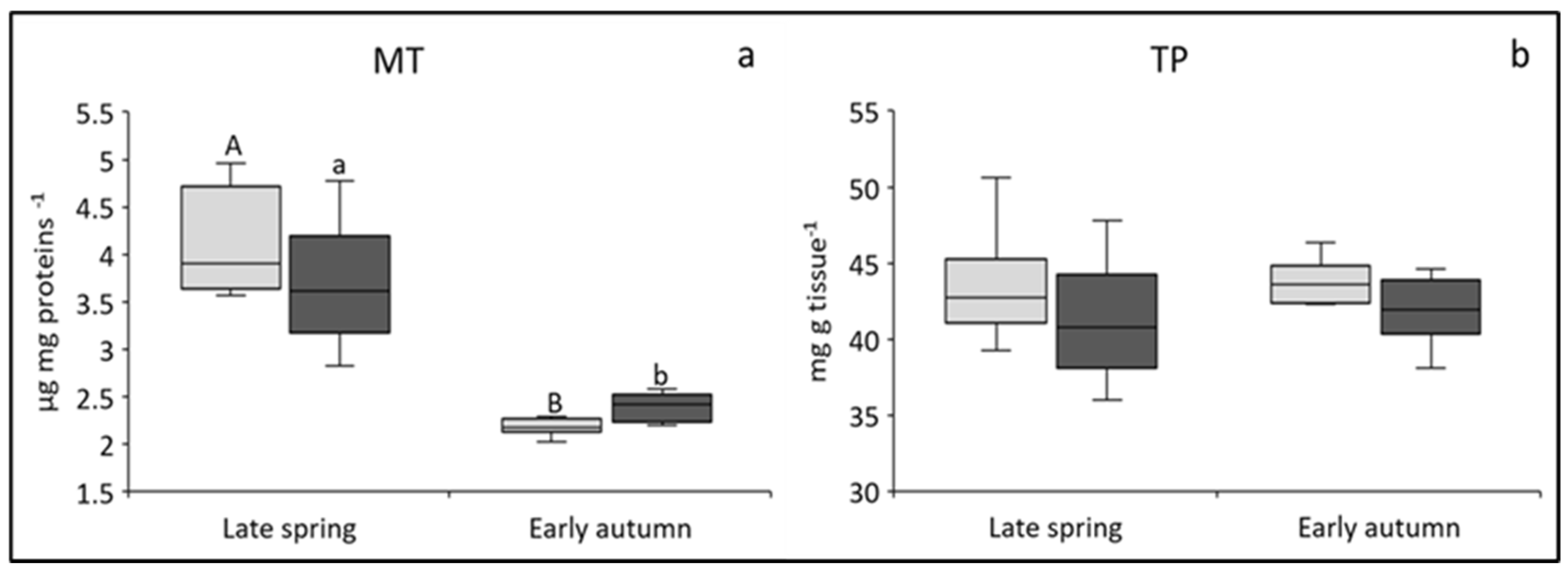
3.1.1. MT Concentrations

In our study, the average MT concentrations in the digestive gland of U. crassus were 4.38 ± 1.06 µg mg proteins−1 at the REF site and 3.69 ± 0.63 µg mg proteins−1 at the KIZ site in the spring campaign and 2.25 ± 0.25 µg mg proteins−1 at the REF site and 2.39 ± 0.15 µg mg proteins−1 at the KIZ site in the autumn campaign (Figure 2a). No significant differences in MT concentrations were observed between the sites in either season. Similarly, in the digestive gland of the duck mussel Anodonta anatina, no differences in MT concentrations were detected among three sites with different contamination levels [24]. Although the induction of MT is considered a biomarker of exposure to metals [9], numerous studies examining MTs in mussels inhabiting chronically polluted sites have shown that MT concentrations did not correspond to the elevated environmental levels of metals [10].
Although there were no significant differences in MT concentrations between the sites, approximately 15% higher MT concentrations were observed in the digestive gland of U. crassus at the REF site compared to the KIZ site in spring, concurrent with approximately ten times higher concentrations of bioaccumulated Cd in the cytosol of the digestive gland of the same mussel specimens (Table 3 [7]). Several studies have shown that Cd is a good inducer of MTs in various mussels, including freshwater species [9,11,24], and the results of this study also suggest that certain Cd concentrations may represent a potential trigger for MT synthesis in the digestive gland of U. crassus. However, the lack of significant differences in MT concentrations between sites may be due to increased bioaccumulation of several other well-known or potential MT inducers observed in the digestive gland cytosol of the same mussels at the KIZ site in spring (Bi, Pb, Cu; Table 3 [7]), which could have caused some MT increase at that site also, thus attenuating the difference (r(Pb, MT) = 0.29).
Unlike Cd, the potential of some of these elements to induce MT synthesis is still questionable, as often discussed for Pb [9], although some metals (e.g., Ag, Cu) have been proven to bind to MTs [37]. However, Cu, an element that was elevated in these same mussels from the KIZ site in both samplings (approximately twice higher; Table 3 [7]), has been shown to be a weak inducer of MT synthesis in many mussel species either at high short-term exposure [11] or moderate long-term exposure [38]. Our results therefore also suggest a probable lower induction potential of Cu (r = 0.38) for MT synthesis than of Cd (r = 0.61), at least at the Cu levels measured in this study.
In autumn, the MT content was mildly increased (p > 0.05) in the digestive gland of mussels from the KIZ site, which was simultaneously accompanied by a decrease in Cd bioaccumulation in the same mussels as those studied here at the REF site compared to spring (the difference between the two sites was several times lower) and an increase in Ag, Bi, and Pb bioaccumulation in the mussels at the KIZ site (Table 3 [7]). The attenuation of the difference in MT levels between the two sites, and even the change of direction, could thus be attributed, at least partly, to the change in dynamics of metal bioaccumulation. On the other hand, metals are probably not the only triggers of MT synthesis, and other non-metallic pollutants could also influence MT induction. Because the contamination of the KIZ site, not only with metals but also with various organic contaminants, has been stronger than the contamination of the REF site ([1,6]; Table 1), and due to the fact that organic contaminants can induce MT synthesis in mussels [15], the slightly increased MT content in the digestive gland of mussels from the KIZ site in autumn could have occurred, to some extent, due to the additional contribution of organic contaminants with pro-oxidative activity at this site.

3.1.2. TP Concentrations

In general, proteins respond to environmental changes and stress by enabling metabolic processes and biochemical reactions to counteract adversity [35]. In our study, the average cytosolic TP concentrations in the digestive gland of U. crassus were 43.6 ± 3.96 mg g tissue−1 at the REF site and 41.2 ± 3.81 mg g tissue−1 at the KIZ site in the spring campaign and 42.6 ± 3.97 mg g tissue−1 at the REF site and 41.8 ± 2.26 mg g tissue−1 tissue at the KIZ site in autumn (Figure 2b). Although there were no significant differences between the TP content at the REF and KIZ sites in either season, the average TP concentrations were always slightly lower at the KIZ site. High levels of pollutants can suppress protein synthesis and disrupt normal metabolism [35]. In the historically contaminated section of the Mrežnica River, pollution stress could have been caused, among other things, by the increased accumulation of several non-essential elements in the digestive gland of the same specimens of U. crassus as that studied here in both seasons (Ag, Bi, Cu, Cs, Pb, Sb, Tl, U; Table 3 [7]). Although some metals are important elements for various biochemical and physiological processes in organisms, all of them, and especially non-essential ones, can cause adverse effects when present in certain concentrations [24,35,39]. The digestive gland plays a pivotal role in the metabolism and detoxification of metals, resulting in an increased demand for protein to meet energy requirements, with increased protein degradation serving as a valuable measure of the extent of toxicity [40]. However, increased exposure to organic pollutants (pesticides and drugs), which were detected in higher concentrations at this site compared to the REF site in the year of mussel sampling ([1,6]; Table 1), could have also contributed to the general stress of the studied mussels.

3.2. Changes in Biomarkers of Antioxidative Capacity (CAT, GSH), Xenobiotic Detoxification (GST), and Oxidative Damage (MDA)

The generation of ROS is an integral part of cellular metabolism, which necessitates the presence of antioxidant defense mechanisms to mitigate their harmful effects [14]. However, various (non)essential elements and organic contaminants can also act as pro-oxidants in mussel organisms [23]. Accordingly, literature reports suggest that both inorganic and organic contaminants can penetrate the cellular lipid membrane, disrupt phospholipid arrangement, and alter membrane fluidity, leading to an increase in LPO and thus increased MDA concentrations. Higher concentrations of many metals in both seasons (e.g., Cr, Cs, Cu, Fe, Pb, Rb, U, V, Table 1) were found in the sediments of the Mrežnica River at the KIZ site compared to the REF site, as well as pharmaceuticals in the water only in spring (total concentrations in Table 1; examples of compounds detected only at the KIZ site in spring: desvenlafaxine, 94 ng L−1; ibuprofen, 109 ng L−1; paracetamol, 34 ng L−1; amphetamine, 5 ng L−1; and caffeine, 100 ng L−1) and pesticides in the water in both seasons (total concentrations in Table 1, e.g., nicotine, REFspring, 198 ng L−1, KIZspring, 309 ng L−1, REFautumn, 347 ng L−1, KIZautumn, 578 ng L−1; terbutryn, detected only at the KIZ site in spring, 104 ng L−1) [1,6]). These could have jointly induced ROS production, consequently exacerbating OS in the studied mussels. The difference between sites in terms of water contamination with pharmaceuticals was especially pronounced in spring, with 40 times higher total drug concentration at the KIZ site ([6]; Table 1). Furthermore, as shown above, increased bioaccumulation of many of the studied metals (Ag, Al, Bi, Cs, Cu, Mo, Pb, Sb, V, Tl, and U) was observed at the KIZ site in the digestive gland of the same mussel specimens as those studied here in both seasons [7], and these metals include those with high oxidative potential [14], such as Cu, Pb, and V (Table 3 [7]). Therefore, the four biomarkers of OS (CAT, GSH, GST, MDA) were utilized to assess the effects of the mentioned contaminants on antioxidant responses and oxidative damage in wild mussels, U. crassus, from the two sites on the Mrežnica River.

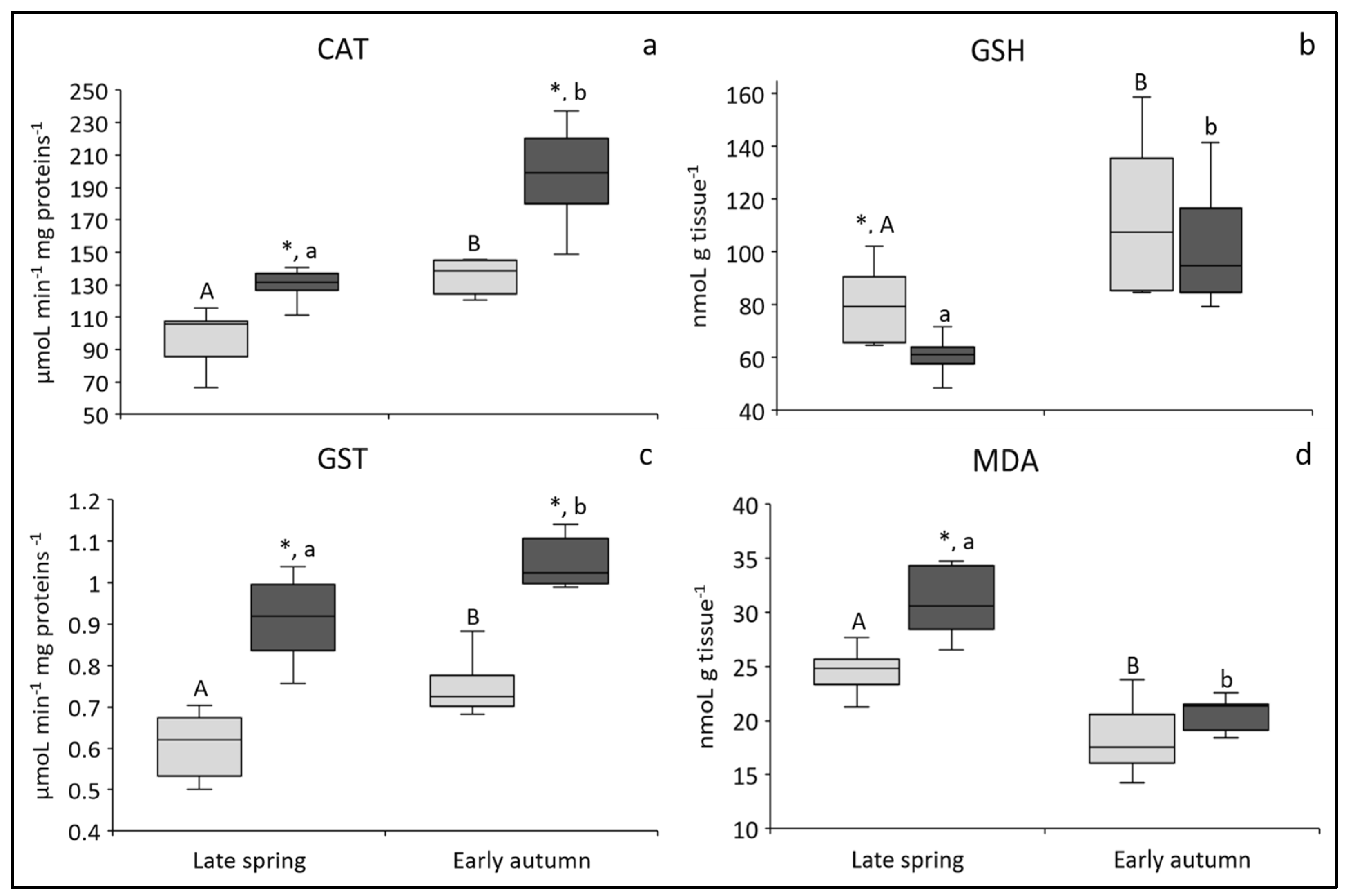
3.2.1. CAT Activity: Biomarker of Antioxidative Capacity

In our study, the average CAT activities in the digestive gland of U. crassus were 96.5 ± 17.5 µmol min−1 mg proteins−1 at the REF site and 131 ± 8.71 µmol min−1 mg proteins−1 at the KIZ site in the spring campaign and 141 ± 23.7 µmol min−1 mg proteins−1 at the REF site and 198 ± 29.3 µmol min−1 mg proteins−1 at the KIZ site in autumn (Figure 3a). Significant spatial differences were observed in both seasons (p = 0.002), with increased enzyme activity detected in the digestive glands of mussels from the KIZ site. Increased CAT activities in bivalves exposed to metals have also been observed in freshwater mussel species D. polymorpha and D. bugensis exposed to Cr and Ni [41]. The early activation of CAT may indicate that this enzyme represents the first line of defense against OS caused by inorganic and organic contaminants.

3.2.2. GSH Concentration: Biomarker of Antioxidative Capacity

In our study, the average GSH concentrations in the digestive gland of U. crassus were 79.8 ± 14.3 nmol g−1 tissue at the REF site and 60.2 ± 6.66 nmol g−1 at the KIZ site in the spring and 112 ± 28.7 nmol g−1 at the REF site and 103 ± 24.4 nmol g−1 at the KIZ site in the autumn sampling campaign (Figure 3b). In general, lower GSH levels were detected at the KIZ site than at the REF site, although the difference between sites was significant only during spring sampling (p < 0.01). GSH, as the most important cellular thiol, plays a crucial role in protecting cells against metal toxicity by forming complexes with metal ions, such as Cd, Cu, and Pb. These metals have a tendency to form covalent bonds with sulfhydryl groups of proteins and peptides [13]. Thus, an increase in GSH content is sometimes observed in aquatic organisms as a result of metal exposure, as observed by Machado et al. [42] in the freshwater unionid mussel Lasmigona costata. Moreover, GSH has been suggested to be involved in conjugation reactions with various pesticides. Accordingly, GSH increases were reported in the digestive gland of Unio tumidus at a site with increased polycyclic aromatic hydrocarbons, polychlorobiphenyls, and organochlorine pesticide concentrations in the sediment [17].
In contrast, it is assumed that the GSH content decreases under OS due to its antioxidant function [13]. Therefore, the finding of GSH depletion in the digestive gland of mussels living in a historically polluted site indicates the occurrence of OS in these organisms caused by either metals or organic pollutants detected at this site, as listed above, or their combination [1,6]. The results obtained are consistent with the findings of several other studies in which GSH depletion was observed in mussels chronically exposed to elevated concentrations of pro-oxidant pollutants, both organic and inorganic. Leonard et al. [14] reported that prooxidant metals like Cu and Pb, which were present in higher concentrations in the digestive gland of the same specimens of U. crassus from the KIZ site as those studied here (Table 3 [7]), could be associated with decreased GSH concentrations. On the other hand, significant depletion of GSH was observed in the digestive glands of U. tumidus transplanted to a site polluted with organochlorine pesticides [43]. Finally, there is convincing evidence that GSH levels are significantly affected by metallic and organic pollutants and that the response of this antioxidant may depend on the specific pollutant, the exposure’s duration, the contaminant’s concentration, the exposed organ, and the individual physiology of a given animal species, among other factors.

3.2.3. GST Activity: Biomarker of Xenobiotic Detoxification

In our study, the average GST activities in the digestive gland of U. crassus were 0.605 ± 0.080 µmol min−1 mg−1 proteins at the REF site and 0.911 ± 0.090 µmol min−1 mg−1 at the KIZ site in the spring campaign and 0.750 ± 0.070 µmol min−1 mg−1 at the REF site and 1.05 ± 0.05 µmol min−1 mg−1 at the KIZ site in autumn (Figure 3c). The differences between the two sites were significant in both sampling seasons. Furthermore, as already mentioned, the increased GST activity was accompanied by lower GSH content. Although metals are not natural substrates for GST, several studies have documented the induction of GST, sometimes concomitant with the decrease of GSH, in mussels exposed to metals [44] or collected from metal-polluted sites [45]. Therefore, the increase in GST activity in the digestive gland of U. crassus at the KIZ site could possibly serve as a mechanism for metal detoxification, ultimately leading to a decrease in GSH concentrations.
However, GST is primarily involved in the conjugation of glutathione with organic electrophilic compounds (e.g., pesticides), forming less reactive and more polar glutathione S-conjugates [16]. A study on Unio tigridis exposed to the pesticide chlorpyrifos showed a progressive rise in GST activity along the concentration and duration of exposure gradient [46]. The fact that pesticides were also detected in higher concentrations in the water at the KIZ site compared to REF site of the Mrežnica River ([1]; Table 1) could be a plausible and even probable cause for the higher GST activity in the digestive gland of U. crassus from this particular site.

3.2.4. MDA Concentration: Biomarker of Oxidative Damage

In our study, the average MDA concentrations in the digestive gland of U. crassus were 24.5 ± 2.1 nmol g−1 tissue at the REF site and 31.7 ± 5.1 nmol g−1 at the KIZ site in the spring campaign and 18.4 ± 3.6 nmol g−1 at the REF site and 19.9 ± 3.2 nmol g−1 at the KIZ site in autumn (Figure 3d). The difference between sites was statistically significant in spring (p < 0.001) and showed about 23% higher average values at KIZ compared to the REF site. An increase in MDA was observed in the digestive gland of U. mancus [23], in U. tumidus [17], and in U. crassus [19] in association with metal pollution. On the other hand, in a laboratory study by Köprücü et al. [22] on the freshwater mussel Unio elongatulus eucirrus, an increase in MDA was observed following exposure to cypermethrin, while Khudhur and Shekha [21] observed a significant induction of both MDA and CAT activity with decreased GSH concentrations in U. pictorum exposed to 1-naphthyl methyl carbamate, thus confirming that increased MDA production can occur as a result of exposure to organic contaminants.
Overall, the results obtained for CAT, GSH, GST, and MDA activities/concentrations in the digestive gland of U. crassus from the Mrežnica River have pointed to the same conclusion, namely, to the notable consequences of OS in U. crassus at the historically contaminated site, which were exhibited in the form of an induced antioxidative defense and detoxification system and already presented oxidative damage. Although the spatial differences were observable in both seasons, the results for GSH and MDA showed more pronounced differences between the sites in spring, when the difference in drug contamination between sites was also multifold greater ([6]; Table 1). Such rapid changes in the responses of the biomarkers of OS in the digestive gland of U. crassus point to their sensitivity to changes in pollution levels of prooxidant metals/organic compounds and thus may serve as a reliable component of water pollution assessment studies.

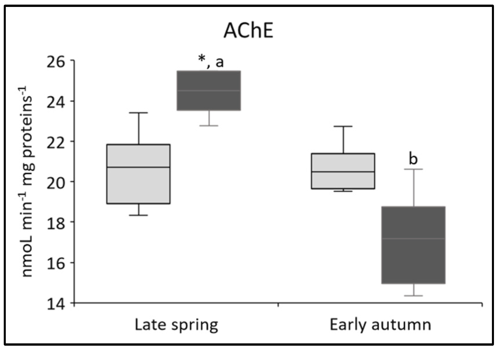
3.3. Changes in Biomarker of Neurotoxicity (AChE)

One enzyme that is particularly sensitive to organic contamination is AChE. It is mainly present in postsynaptic neurons and catalyzes the hydrolysis of the neurotransmitter acetylcholine, which terminates the nerve impulse and allows the cell to return to its resting state [47]. Some organic compounds, such as organophosphorus pesticides and carbamates, are known to interact with the active site of AChE and inhibit its activity, which can potentially lead to behavioral changes, paralysis, and even death [16]. In our study, the average AChE activity in the digestive gland of U. crassus was 20.6 ± 2.0 nmol min−1 mg−1 of proteins at the REF site and 27.3 ± 6.7 nmol min−1 mg−1 at the KIZ site in the spring campaign and 20.2 ± 2.1 nmol min−1 mg−1 at the REF site and 17.1 ± 2.5 nmol min−1 mg−1 at the KIZ site in autumn (Figure 4). The differences were significant in both seasons, with higher AChE activity at the KIZ site in spring and higher AChE activity at the REF site in autumn.
In spring, AChE activity in the digestive gland of U. crassus was significantly higher at the KIZ site compared to the REF site (p = 0.008). The study by Zhang et al. [48] suggests a possible role of AChE induction in regulating the apoptotic process leading to cell membrane disruption and the subsequent release of AChE into the cytoplasm. Kaizer et al. [49] also suggested that higher AChE activity may be due to increased LPO, which has the potential to affect the integrity and functionality of the cholinergic system. In the spring period, drug concentrations, including the non-steroidal anti-inflammatory compound ibuprofen, were detected in much higher concentrations at the KIZ site (ibuprofen: 109 ng L−1) than at the REF site, where ibuprofen itself was not even detected ([6]). As ibuprofen (at 1 μg L−1) has been reported to cause significant oxidative damage in D. polymorpha [50], AChE induction in this season in the digestive gland of U. crassus could be hypothesized to be a consequence of the drug-promoted LPO and increased MDA concentrations (Figure 3d).
In autumn, however, decreased AChE activity was observed at the KIZ site. These results were not surprising, as AChE inhibition is actually used as an indicator of neurotoxicity caused by the presence of organophosphate and carbamate pesticides in the environment [47]. A study on U. tigridis exposed to different concentrations of chlorpyrifos, a common component of organophosphate pesticides, showed a decrease in AChE activity with increasing pesticide dose and exposure duration [46]. Moreover, the mechanism of action of some anxiolytic/antidepressant drugs involves the inhibition of certain neurotransmitters, so it is not unreasonable to assume that AChE could also be affected by their presence. Recent studies have shown that AChE can be inhibited not only by organic compounds but also by heavy metals [39,51], which could potentially interact with the acetylcholine receptor and affect AChE binding efficiency, leading to inhibition of its activity [47]. In a study by Georgieva et al. [51], decreased AChE activity was found in the Chinese pond mussels Sinanodonta woodiana, which were exposed to different environmental pollutants, including metals and metalloids. The KIZ site on the Mrežnica River proved to be a site with higher levels of both inorganic and organic contaminants, including anxiolytic/antidepressant drugs in the water, compared to REF site in both seasons, but higher levels of insecticides (i.e., nicotine, REFspring 198 ng L−1, REFautumn 347 ng L−1, KIZspring 309 ng L−1, KIZautumn 578 ng L−1, [1]; proven inhibitor of AChE activity [52]) were recorded in autumn compared to spring ([1,6]; Table 1). Generally higher bioaccumulation of the number of metals in the digestive gland of the same specimens of U. crassus as those studied here was also observed at the KIZ site, which was especially prominent in autumn for several elements (Ag, Bi, Cs, Pb, Sb, Tl, U; Table 3 [7]). Thus, it can be presumed that the synergistic action of these contaminants has caused slight inhibition of AChE at the KIZ site in autumn. The results of our study therefore support the usefulness of AChE as a biomarker of neurotoxicity, demonstrating its applicability for the recognition of the effects of complex mixtures of pollutants.

3.4. Season as a Common Factor Influencing Biomarker Responses

Seasonal differences in site characteristics, including abiotic factors, such as temperature and oxygen levels and resulting changes in the bioavailability of metals and other contaminants, as well as biotic factors, such as annual growth, feeding activity, and reproductive cycles of mussel species, may contribute to seasonal differences in biomarker responses [10,39]. Seasonal differences in temperature and food availability, which vary according to river regime, can also significantly affect the biology of mussels [8,39].
In this study, data on biomarker activities and concentrations gathered from two sampling campaigns carried out at different times of the year (late spring—early June 2021; early autumn—early October 2021) showed a significant seasonality of biomarker responses in the digestive gland of U. crassus (Figure 2, Figure 3 and Figure 4). This was especially obvious for MTs and MDA with higher concentrations in spring (Figure 2a; Figure 3d) (p < 0.001) and CAT, GSH, and GST with higher activities/concentrations in autumn (Figure 3a–c) (p < 0.001). TPs (Figure 2b) did not show seasonal differences, whereas seasonal differences of AChE (Figure 3) seemed to indicate the above-discussed difference in environmental exposure to contaminants.
One of the factors that may have caused higher MT and MDA concentrations in late spring is probably the higher water temperature in June compared to October. Several studies have shown that higher water temperature leads to increased MT and MDA concentrations in mussel species [39]. In addition, a known MT inducer, the non-essential element Cd, was found to accumulate at higher concentrations in the digestive gland of the same specimens of U. crassus as those studied here in spring at both sites (Table 3 [7]), which could also explain higher MT concentrations in that season. Also, several authors have pointed out that concentrations of these two biomarkers could be influenced by factors like organic matter, nutrients, phytoplankton blooms, and feeding activities [10]. As expected, higher nutrient levels were reported in spring than autumn 2021 in the Mrežnica River (e.g., nitrates, total phosphorus; Table 4 [1]), which probably caused mussels’ greater feeding activities and consequently increased metabolic rates and increased MDA and MT levels in the digestive gland of U. crassus. It should, however, be emphasized that U. crassus reproduces during spring and summer months [53], as confirmed in our study by the presence of glochidia (unpublished results). Thus, increased MT concentrations could perhaps reflect increased MT synthesis during the development of glochidia.
Regarding the three biomarkers characteristic of OS, which were higher in autumn, these results contradicted previously reported studies, which generally observed higher activity and concentration of these biomarkers in spring related to the development of gonads and higher temperature, which contribute to increased sensitivity to OS [15,39]. However, in the same mussel specimens as those studied here, increased cytosolic concentrations in the digestive gland were reported for several non-essential metals during the autumn sampling campaign compared to spring at both sites (Ag, Cs; Table 3 [7]). In addition, concentrations of insecticides (i.e., nicotine, the concentrations presented above) in the water of the Mrežnica River were also higher in autumn compared to the spring campaign at both sites ([1]; Table 1). The combination of increased exposure to metals and organic pollutants could have additionally induced antioxidative mechanisms in the digestive gland of U. crassus in autumn. Because the adaptability or saturation of antioxidant defenses against prooxidant challenges depends on the intensity and variability of environmental factors [8], the above-mentioned seasonal variability of contamination levels may serve as a plausible explanation for the increase in OS biomarkers in autumn. The observed seasonal variations highlight the importance of investigating the seasonality of biomarker responses and determining the values typical of different seasons to provide a basis for future comparisons and achieve a more reliable assessment of the consequences of environmental pollution.

4. Conclusions

The multi-biomarker approach utilized in this study was the first of its kind for the wild population of the endangered mussel species U. crassus under real environmental conditions. The obtained results have demonstrated the usefulness of the applied approach for the recognition of early signs of organism health disturbances. Among investigated biomarkers, MTs, as a biomarker of metal exposure, showed the least responsiveness to environmental exposure to contaminants, which is consistent with their well-known susceptibility to many other biological and ambient factors. Contrary, all of the other biomarkers, as indicators of oxidative stress and neurotoxicity in mussels, seem to be directly influenced by the contaminant exposure level in water/sediment, with the seemingly fast response to changes in the contaminant level or type. Furthermore, the observed fluctuating seasonal responses confirmed the necessity of considering, whenever possible, the sampling season and other accompanying factors, both biotic (e.g., age, size, reproductive phase) and abiotic (e.g., water temperature, dissolved oxygen, nutrient level, etc.) in the assessment of the biomarker levels and health status of the studied mussels. Our results emphasized the susceptibility of U. crassus mussels to even moderately polluted freshwater environments due to their continuous direct contact with the pollutants stored in the sediments. This further accentuates the need to monitor freshwater affected by industrial pollution sources long after they stop operating, as well as supervision of sensitive aquatic organisms living in such environments.

Author Contributions

Conceptualization, D.I.; methodology, Z.K., Z.D., T.M.P. and B.B.; formal analysis, Z.K. and Z.D.; investigation, Z.K., Z.D., J.L., K.T., T.M.P., B.B. and D.I.; resources, Z.K., J.L., K.T. and D.I.; data curation, Z.K. and D.I.; writing—original draft preparation, Z.K. and D.I.; writing—review and editing, Z.K., Z.D., J.L. and D.I.; visualization, D.I.; supervision, Z.D. and D.I.; funding acquisition, Z.D., project administration, Z.D. All authors have read and agreed to the published version of the manuscript.

Funding

This work has been supported by the Croatian Science Foundation under the project “Metal-binding biomolecules and health disturbances of freshwater organisms exposed to industrial wastes” (METABIOM; Grant number: IP-2019-04-2636).

Institutional Review Board Statement

The authors state that the ordinance on the protection of animals used for scientific purposes (NN 55/2013) issued by the Republic of Croatia was followed during this study.

Informed Consent Statement

Not applicable.

Data Availability Statement

The datasets used and/or analyzed during the current study are available from the corresponding author upon reasonable request.

Conflicts of Interest

Krešimira Trgovčić is employed by the company Vodovod i kanalizacija d.o.o. Karlovac and this company is listed as her affiliation in the paper. However, as Krešimira Trgovčić was involved in the METABIOM project as an external collaborator, we consider that there is no potential commercial or financial conflict of interest due to her employment with the said company, as the work she carried out in the project (diving, sampling and identification of mussels) is in no way related to her work in the company. Therefore, we have declared for all authors that there is no conflict of interest.

References

  1. Dragun, Z.; Stipaničev, D.; Fiket, Ž.; Lučić, M.; Udiković Kolić, N.; Puljko, A.; Repec, S.; Šoštarić Vulić, Z.; Ivanković, D.; Barac, F.; et al. Yesterday’s contamination—A problem of today? The case study of discontinued historical contamination of the Mrežnica River (Croatia). Sci. Total Environ. 2022, 848, e157775. [Google Scholar] [CrossRef] [PubMed]
  2. Frančišković-Bilinski, S.; Bilinski, H.; Maldini, K.; Milović, S.; Zhang, Q.; Appel, E. Chemical and magnetic tracing of coal slag pollutants in karstic river sediments. Environ. Earth Sci. 2017, 76, 476. [Google Scholar] [CrossRef]
  3. Hurley, R.R.; Rothwell, J.J.; Woodward, J.C. Metal contamination of bed sediments in the Irwell and Upper Mersey catchments, northwest England: Exploring the legacy of industry and urban growth. J. Soils Sediments 2017, 17, 2648–2665. [Google Scholar] [CrossRef]
  4. Hutchinson, S.M.; Rothwell, J.J. Mobilisation of sediment-associated metals from historical Pb working sites on the River Sheaf, Sheffield, UK. Environ. Pollut. 2008, 155, 61–71. [Google Scholar] [CrossRef] [PubMed]
  5. Suschka, J.; Ryborz, S.; Leszczynska, I. Surface water and sediment contamination in an old industrial region of Poland—Two critical examples. Water Sci. Technol. 1994, 29, 107–114. [Google Scholar] [CrossRef]
  6. Stipaničev, D.; Dragun, Z.; Repec, S.; Ivanković, D.; Barac, F.; Kiralj, Z.; Kralj, T.; Valić, D. Dynamics of drug contamination of the river-water in the rural, semirural and urban areas of the Mrežnica River in Croatia during COVID-19 pandemic (2020–2021). Environ. Sci. Pollut. Res. 2023, 30, 93652–93666. [Google Scholar] [CrossRef]
  7. Kiralj, Z.; Dragun, Z.; Lajtner, J.; Trgovčić, K.; Valić, D.; Ivanković, D. Accumulation of metal(loid)s in the digestive gland of the mussel Unio crassus Philipsson, 1788: A reliable detection of historical freshwater contamination. Environ. Pollut. 2023, 334, 122164. [Google Scholar] [CrossRef]
  8. Regoli, F.; Pellegrini, D.; Cicero, A.M.; Nigro, M.; Benedetti, M.; Gorbi, S.; Fattorini, D.; D’Errico, G.; Di Carlo, M.; Nardi, A.; et al. A multidisciplinary weight of evidence approach for environmental risk assessment at the Costa Concordia wreck: Integrative indices from Mussel Watch. Mar. Environ. Res. 2014, 96, 92–104. [Google Scholar] [CrossRef]
  9. Amiard, J.C.; Amiard-Triquet, C.; Barka, S.; Pellerin, J.; Rainbow, P.S. Metallothioneins in aquatic invertebrates: Their role in metal detoxification and their use as biomarkers. Aquat. Toxicol. 2006, 76, 160–202. [Google Scholar] [CrossRef]
  10. Ivanković, D.; Pavičić, J.; Erk, M.; Filipović-Marijić, V.; Raspor, B. Evaluation of the Mytilus galloprovincialis Lam. digestive gland metallothionein as a biomarker in a long-term field study: Seasonal and spatial variability. Mar. Pollut. Bull. 2005, 50, 1303–1313. [Google Scholar] [CrossRef]
  11. Ivanković, D.; Pavičić, J.; Beatović, V.; Klobučar, R.S.; Klobučar, G.I.V. Inducibility of metallothionein biosynthesis in the whole soft tissue of zebra mussels Dreissena polymorpha exposed to cadmium, copper, and pentachlorophenol. Environ. Toxicol. 2010, 25, 198–211. [Google Scholar] [CrossRef]
  12. Raspor, B.; Pavičić, J. Induction of metallothionein-like proteins in the digestive gland of Mytilus galloprovincialis after a chronic exposure to the mixture of trace heavy metals. Chem. Speciat. Bioavailab. 1991, 3, 39–46. [Google Scholar] [CrossRef]
  13. Ercal, N.; Gurer-Orhan, H.; Aykin-Burns, N. Toxic metals and oxidative stress. Part I: Mechanisms involved in metal-induced oxidative damage. Curr. Top. Med. Chem. 2001, 1, 529–539. [Google Scholar] [CrossRef] [PubMed]
  14. Leonard, S.S.; Harris, G.K.; Shi, X. Metal-induced oxidative stress and signal transduction. Free Radic. Biol. Med. 2004, 37, 1921–1942. [Google Scholar] [CrossRef] [PubMed]
  15. Viarengo, A.; Burlando, B.; Dondero, F.; Marro, A.; Fabbri, R. Metallothionein as a tool in biomonitoring programmes. Biomarkers 1999, 4, 455–466. [Google Scholar]
  16. Gonçalves, A.M.M.; Rocha, C.P.; Marques, J.C.; Gonçalves, F.J.M. Enzymes as useful biomarkers to assess the response of freshwater communities to pesticide exposure—A review. Ecol. Indic. 2021, 122, 107303. [Google Scholar] [CrossRef]
  17. Cossu, C.; Doyotte, A.; Babut, M.; Exinger, A.; Vasseur, P. Antioxidant biomarkers in freshwater bivalves, Unio tumidus, in response to different contamination profiles of aquatic sediments. Ecotoxicol. Environ. Saf. 2000, 45, 106–121. [Google Scholar] [CrossRef]
  18. Sárkány-Kiss, A.; Herczeg, I.; Palombi, B.; Grigorszky, I.; Antal, L.; Bácsi, I.; Mozsár, A.; Kalmár, A.F.; Nagy, S.A. Toxicity tests of chlorinated hydrocarbons on the river mussel, Unio crassus (Bivalvia, Unionidae). North-West. J. Zool. 2012, 8, 358–361. [Google Scholar]
  19. Třešňáková, N.; Çağlan Günal, A.; Başaran Kankılıç, G.; Paçal, E.; Nihan Tavşanoğlu, Ü; Uyar, R.; Erkoç, F. Sub-lethal toxicities of zinc pyrithione, copper pyrithione alone and in combination to the indicator mussel species Unio crassus Philipsson, 1788 (Bivalvia, Unionidae). Chem. Ecol. 2020, 36, 292–308. [Google Scholar] [CrossRef]
  20. Türkmen, E.U.; Arslan, P.; Erkoç, F.; Günal, A.Ç.; Duran, H. The cerium oxide nanoparticles toxicity induced physiological, histological and biochemical alterations in freshwater mussels, Unio crassus. J. Trace Elem. Med. Biol. 2024, 83, 127371. [Google Scholar] [CrossRef]
  21. Khudhur, S.M.; Shekha, Y.A. Histopathological and biochemical biomarker response of mussel, Unio pictorum, to carbamate pesticide carbaryl: A laboratory study. Indian J. Anim. Res. 2019. [Google Scholar] [CrossRef]
  22. Köprücü, K.; Yonar, S.M.; Şeker, E. Effects of cypermethrin on antioxidant status, oxidative stress biomarkers, behavior, and mortality in the freshwater mussel Unio elongatulus eucirrus. Fish. Sci. 2010, 76, 1007–1013. [Google Scholar] [CrossRef]
  23. Paçal, E.; Gümüş, A.; Günal, Ç.; Erkmen, B.; Arslan, P.; Yıldırım, Z.; Erkoç, F. Oxidative stress response as biomarker of exposure of a freshwater invertebrate model organism (Unio mancus Lamarck, 1819) to antifouling copper pyrithione. Pestic. Fitomed. 2022, 37, 63–76. [Google Scholar] [CrossRef]
  24. Falfushynska, H.I.; Gnatyshyna, L.L.; Gyori, J.; Stoliar, O.B. Metallothioneins, Caspase-3 and oxidative stress responses in the multi-marker study of freshwater mussel inhabiting sites of various human impact. Turk. J. Fish. Aquat. Sci. 2014, 14, 591–603. [Google Scholar] [CrossRef]
  25. Raspor, B.; Dragun, Z.; Erk, M. Examining the suitability of mussel digestive gland to serve as a biomonitoring target organ. Arh. Hig. Rada Toksikol. 2005, 56, 141–149. [Google Scholar] [PubMed]
  26. Zając, K.; Zając, T.A.; Adamski, P.; Bielański, W.; Ćmiel, A.M.; Lipińska, A.M. Dispersal and mortality of translocated thick-shelled river mussel Unio crassus Philipsson, 1788 adults revealed by radio tracking. Aquat. Conserv. Mar. Freshw. Ecosyst. 2018, 29, 331–520. [Google Scholar] [CrossRef]
  27. Viarengo, A.; Ponzano, E.; Dondero, F.; Fabbri, R. A simple spectrophotometric method for metallothionein evaluation in marine organisms: An application to Mediterranean and Antarctic molluscs. Mar. Environ. Res. 1997, 44, 69–84. [Google Scholar] [CrossRef]
  28. Bourdineaud, J.-P.; Štambuk, A.; Šrut, M.; Radić Brkanac, S.; Ivanković, D.; Lisjak, D.; Sauerborn Klobučar, R.; Dragun, Z.; Bačić, N.; Klobučar, G. Gold and silver nanoparticles effects to the earthworm Eisenia fetida—The importance of tissue over soil concentrations. Drug Chem. Toxicol. 2021, 44, 21–29. [Google Scholar] [CrossRef]
  29. Bradford, M. A rapid and sensitive method for the quantitation of microgram quantities of protein utilizing the principle of protein-dye binding. Anal. Biochem. 1976, 72, 248–254. [Google Scholar] [CrossRef]
  30. Li, Y.; Schellhorn, H. Rapid kinetic microassay for catalase activity. J. Biomol. Tech. 2007, 18, 185–187. [Google Scholar]
  31. Tietze, F. Enzymic method for quantitative determination of nanogram amounts of total and oxidized glutathione: Applications to mammalian blood and other tissues. Anal. Biochem. 1969, 27, 502–522. [Google Scholar] [CrossRef] [PubMed]
  32. Rahman, I.; Kode, A.; Biswas, S. Assay for quantitative determination of glutathione and glutathione disulfide levels using enzymatic recycling method. Nat. Protoc. 2006, 1, 3159–3165. [Google Scholar] [CrossRef]
  33. Habig, W.H.; Pabst, M.J.; Jakoby, W.B. Glutathione S-transferase. J. Biol. Chem. 1974, 249, 7130–7139. [Google Scholar] [CrossRef] [PubMed]
  34. Botsoglou, N.A.; Fletouris, D.J.; Papageorgiou, G.E.; Vassilopoulos, V.N.; Mantis, A.J.; Trakatelliss, A.G. Rapid, sensitive, and specific thiobarbituric acid method for measuring lipid peroxidation in animal tissue, food, and feedstuff samples. J. Agric. Food Chem. 1994, 42, 1931–1937. [Google Scholar] [CrossRef]
  35. Mijošek, T.; Filipović Marijić, V.; Dragun, Z.; Krasnići, N.; Ivanković, D.; Erk, M. Evaluation of multi-biomarker response in fish intestine as an initial indication of anthropogenic impact in the aquatic karst environment. Sci. Total Environ. 2019, 660, 1079–1090. [Google Scholar] [CrossRef]
  36. Ellman, G.L.; Courtney, K.D.; Andres, V.; Featherstone, R.M. A new and rapid colorimetric determination of acetylcholinesterase activity. Biochem. Pharmacol. 1961, 7, 88–95. [Google Scholar] [CrossRef]
  37. Dragun, Z.; Kiralj, Z.; Pećnjak, A.; Ivanković, D. The study of acidic/basic nature of metallothioneins and other metal-binding biomolecules in the soluble hepatic fraction of the northern pike (Esox lucius). Int. J. Biol. Macromol. 2024, 256, 128209. [Google Scholar] [CrossRef]
  38. Lecoeur, S.; Videmann, B.; Berny, P. Evaluation of metallothionein as a biomarker of single and combined Cd/Cu exposure in Dreissena polymorpha. Environ. Res. 2004, 94, 184–191. [Google Scholar] [CrossRef]
  39. Yoloğlu, E.; Uçkun, M.; Uçkun, A.A. Metal accumulation and biochemical variations in the freshwater mussels (Unio mancus) collected from Atatürk Dam Lake, Turkey. Biochem. Syst. Ecol. 2018, 79, 60–68. [Google Scholar] [CrossRef]
  40. Radwan, E.H.; Mawgood, A.A.; Ghonim, A.Z.; Nagar, R.E. The possibility of using the fresh water bivalve, Spathopsis rubens, in the Nile River, El Mahmoudia water stream as bioindicator for pollution. Int. J. Limnol. 2018, 1, 1–23. [Google Scholar] [CrossRef]
  41. Potet, M.; Devin, S.; Pain-Devin, S.; Rousselle, P.; Giambérini, L. Integrated multi-biomarker responses in two dreissenid species following metal and thermal cross-stress. Environ. Pollut. 2016, 218, 39–49. [Google Scholar] [CrossRef] [PubMed]
  42. Machado, A.A.D.S.; Wood, C.M.; Bianchini, A.; Gillis, P.L. Responses of biomarkers in wild freshwater mussels chronically exposed to complex contaminant mixtures. Ecotoxicology 2014, 23, 1345–1358. [Google Scholar] [CrossRef] [PubMed]
  43. Doyotte, A.; Cossu, C.; Jacquin, M.-C.; Babut, M.; Vasseur, P. Antioxidant enzymes, glutathione and lipid peroxidation as relevant biomarkers of experimental or field exposure in the gills and the digestive gland of the freshwater bivalve Unio tumidus. Aquat. Toxicol. 1997, 39, 93–110. [Google Scholar] [CrossRef]
  44. Pytharopoulou, S.; Grintzalis, K.; Sazakli, E.; Leotsinidis, M.; Georgiou, C.D.; Kalpaxis, D.L. Translational responses and oxidative stress of mussels experimentally exposed to Hg, Cu and Cd: One pattern does not fit at all. Aquat. Toxicol. 2011, 105, 157–165. [Google Scholar] [CrossRef]
  45. Vidal-Liñán, L.; Bellas, J.; Etxebarria, N.; Nieto, O.; Beiras, R. Glutathione S-transferase, glutathione peroxidase and acetylcholinesterase activities in mussels transplanted to harbour areas. Sci. Total Environ. 2014, 470–471, 107–116. [Google Scholar] [CrossRef] [PubMed]
  46. Hanna, N.S.; Shekha, Y.A. Acute toxicity of chlorpyrifos on the freshwater bivalves (Unio tigridis) and effects on bioindicators. Baghdad Sci. J. 2023, 21, 53–61. [Google Scholar] [CrossRef]
  47. Bainy, A.C.D.; Gennari De Medeiros, M.H.; Di Mascio, P.; de Almeida, E.A. In vivo effects of metals on the acetylcholinesterase activity of the Perna perna mussel’s digestive gland. Rev. Biotemas 2006, 19, 35–39. [Google Scholar]
  48. Zhang, X.J.; Yang, L.; Zhao, Q.; Caen, J.P.; He, H.Y.; Jin, Q.H.; Guo, L.H.; Alemany, M.; Zhang, L.Y.; Shi, Y.F. Induction of acetylcholinesterase expression during apoptosis in various cell types. Cell Death Differ. 2002, 9, 790–800. [Google Scholar] [CrossRef]
  49. Kaizer, R.R.; Corrêa, M.C.; Spanevello, R.M.; Morsch, V.M.; Mazzanti, C.M.; Gonçalves, J.F.; Schetinger, M.R.C. Acetylcholinesterase activation and enhanced lipid peroxidation after long-term exposure to low levels of aluminum on different mouse brain regions. J. Inorg. Biochem. 2005, 99, 1865–1870. [Google Scholar] [CrossRef]
  50. André, C.; Gagné, F. Cumulative effects of ibuprofen and air emersion in zebra mussels Dreissena polymorpha. Environ. Toxicol. Pharmacol. 2017, 55, 156–164. [Google Scholar] [CrossRef]
  51. Georgieva, E.; Antal, L.; Stoyanova, S.; Aranudova, D.; Velcheva, I.; Iliev, I.; Vasileva, T.; Bivolarski, V.; Mitkovska, V.; Chassovnikarova, T.; et al. Biomarkers for pollution in caged mussels from three reservoirs in Bulgaria: A pilot study. Heliyon 2022, 8, e10916. [Google Scholar] [CrossRef] [PubMed]
  52. Chang, P.-L.; Taylor, J.J. Effect of chronic administration of acetylcholinesterase activity in the hypothalamus and medulla oblongata of the rat brain. An ultrastructural study. Brain Res. 1973, 54, 75. [Google Scholar] [CrossRef] [PubMed]
  53. Selak, L. Biološke i Ekološke Značajke Slatkovodnog Školjkaša Unio crassus Philipsson, 1788. Bachelor’s Thesis, Faculty of Science, University of Zagreb, Zagreb, Croatia, 2016. [Google Scholar]
Figure 1. Study area with marked sampling sites on the Mrežnica River in Croatia (site upstream from the town of Duga Resa (REF); historically contaminated site: industrial zone of the city of Karlovac (KIZ)). Sources of the maps: https://gisportal.hr/ (accessed on 30 January 2025); https://d-maps.com/carte.php?num_car=2175&lang=en. (accessed on 1 March 2023).
Fishes 10 00317 g001
Figure 2. Concentrations of (a) metallothionein (biomarker of exposure to metals, MT) and (b) total proteins (biomarker of general stress, TP) in the digestive gland of the mussel species Unio crassus, collected at the REF site (light gray boxes) and at the KIZ site (dark gray boxes) in the Mrežnica River during two sampling seasons (late spring and early autumn 2021) (n = 7 for the REF site in late spring; n = 10 for the KIZ site in late spring; n = 6 for both sites in early autumn). Significant differences between seasons within each site are indicated by different letters: A, B for the REF site and a, b for the KIZ site (p < 0.05, two-way ANOVA and post hoc LSD test).
Fishes 10 00317 g002
Figure 3. Biomarkers of antioxidative capacity and xenobiotic detoxification: (a) activities of catalase (CAT), (b) total glutathione (GSH) concentrations, and (c) the activity of glutathione S-transferase (GST); and biomarker of oxidative damage: (d) malondialdehyde (MDA) concentration in the digestive gland of the mussel species Unio crassus collected at the REF site (light gray boxes) and at the KIZ site (dark gray boxes) in the Mrežnica River during two sampling seasons (late spring and early autumn 2021) (n = 7 for the REF site in late spring; n = 10 for the KIZ site in late spring; n = 6 for both sites in early autumn). Significant differences between sites within each season are indicated with an asterisk (*), and differences between seasons within each site are indicated by different letters: A, B for the REF site and a, b for the KIZ site (p < 0.05, two-way ANOVA and post hoc LSD test).
Fishes 10 00317 g003
Figure 4. Biomarker of pesticide and metal exposure/neurotoxicity (acethylcholinesterase (AChE) activity) in the digestive gland of the mussel species Unio crassus collected at the reference (REF) site (light gray boxes) and at the Karlovac industrial zone (KIZ) site (dark gray boxes) in the Mrežnica River during two sampling seasons (late spring and early autumn 2021) (n = 7 for the REF site in late spring; n = 10 for the KIZ site in late spring; n = 6 for both sites in early autumn). Significant differences between sites within each season are indicated with an asterisk (*), and differences between seasons within the KIZ site are indicated by the letters a and b (p < 0.05, two-way ANOVA and post hoc LSD test).
Fishes 10 00317 g004
Table 1. Concentrations of selected metals in the sediments and selected organic contaminants in the water of the Mrežnica River at two mussel sampling sites (REF—reference site; KIZ—historically contaminated site—industrial zone of the city of Karlovac) in April 2021 and September 2021. The presented data are excerpts from our previously published papers, where they are discussed in detail [1,6].
PeriodSampling Site
REFKIZ
Metals in the Sediment [1]
Bi/µg g−1April 2021
September 2021
0.330 ± 0.112
0.254 ± 0.030
0.416 ± 0.152
0.405 ± 0.052
Cd/µg g−1April 2021
September 2021
0.509 ± 0.067
0.516 ± 0.047
0.537 ± 0.021
0.582 ± 0.058
Cr/µg g−1April 2021
September 2021
52.6 ± 17.5
37.9 ± 5.4
81.0 ± 13.9
84.4 ± 3.1
Cs/µg g−1April 2021
September 2021
2.02 ± 0.85
1.87 ± 0.19
4.13 ± 0.94
5.04 ± 0.41
Cu/µg g−1April 2021
September 2021
19.4 ± 6.0
15.3 ± 2.5
31.7 ± 8.9
38.9 ± 1.8
Fe/mg g−1April 2021
September 2021
11.7 ± 3.7
9.69 ± 1.24
21.9 ± 5.4
20.1 ± 1.7
Ni/µg g−1April 2021
September 2021
25.8 ± 6.6
23.6 ± 3.0
48.8 ± 5.8
54.2 ± 8.3
Pb/µg g−1April 2021
September 2021
13.3 ± 6.0
9.95 ± 0.86
26.9 ± 21.8
20.6 ± 1.7
Rb/µg g−1April 2021
September 2021
28.0 ± 12.7
25.4 ± 2.7
38.8 ± 11.7
45.2 ± 5.2
Sb/µg g−1April 2021
September 2021
0.537 ± 0.229
0.434 ± 0.062
0.799 ± 0.337
4.19 ± 5.66
U/µg g−1April 2021
September 2021
1.18 ± 0.48
1.08 ± 0.12
3.75 ± 0.76
3.94 ± 0.37
V/µg g−1April 2021
September 2021
38.6 ± 15.4
36.6 ± 4.8
63.0 ± 11.1
71.1 ± 6.5
Zn/µg g−1April 2021
September 2021
49.1 ± 19.9
36.1 ± 2.1
93.2 ± 30.2
68.2 ± 1.1
Organic contaminants in the water [1,6]
Herbicides/µg L−1 AApril 2021
September 2021
0.007
0.002
0.029
0.002
Insecticides/µg L−1 BApril 2021
September 2021
0.198
0.347
0.309
0.578
Total drugs/ng L−1 CApril 2021
September 2021
10.6
443
429
491
A Metolachlor, neburon, terbutryn, atrazine, and its metabolites. B Nicotine, methiocarb metabolites. C Ibuprofen, topiramate, lorazepam, trazodone.
Table 2. Two-way ANOVA results and post hoc LSD test for biomarker responses in the digestive gland of Unio crassus showing the effects of site, season, and their interaction. For each biomarker, degrees of freedom (DF), F-values, and corresponding p-values are presented. Statistically significant effects (p < 0.05) are indicated in bold.
DFMTTPCATGSHGSTMDAAChE
FpFpFpFpFpFpFp
Site11.190.2861.250.27536.8<0.0014.150.05289.6<0.0018.970.0061.090.307
Season147.2<0.0010.1680.68556.9<0.00127.5<0.00119.4<0.00138.1<0.0019.340.005
Interaction12.780.1070.9460.3412.220.1490.5270.4750.0190.8893.890.0608.010.009
Table 3. Cytosolic concentrations (expressed on wet mass basis; average ± SD) of selected (non)essential elements in the digestive gland of the mussel species Unio crassus sampled at the two sites in the Mrežnica River (reference site (REF): upstream of the town of Duga Resa; contaminated site (KIZ): Karlovac city industrial zone) during two sampling campaigns (June and October 2021). The presented data are excerpts from our previously published paper, where they are discussed in detail [7].
PeriodSampling Site
REFKIZ
Ag/ng g−1June 2021
October 2021
3.81 ± 0.55
5.22 ± 1.13
5.47 ± 0.79
11.8 ± 5.5
Bi/ng g−1June 2021
October 2021
2.55 ± 0.34
2.37 ± 0.57
3.89 ± 0.34
3.72 ± 0.78
Cd/ng g−1June 2021
October 2021
1149 ± 284
222 ± 47.8
96.0 ± 12.3
49.9 ± 7.9
Cu/µg g−1June 2021
October 2021
1.41 ± 0.21
0.99 ± 0.13
3.67 ± 0.57
2.02 ± 0.25
Cs/ng g−1June 2021
October 2021
0.67 ± 0.11
0.86 ± 0.06
1.54 ± 0.21
2.95 ± 0.30
Pb/ng g−1June 2021
October 2021
31.7 ± 6.6
18.6 ± 1.3
48.2 ± 8.1
66.5 ± 18.8
Sb/ng g−1June 2021
October 2021
1.06 ± 0.10
0.77 ± 0.08
2.41 ± 0.14
2.27 ± 0.17
Tl/ng g−1June 2021
October 2021
1.42 ± 0.28
1.52 ± 0.12
1.96 ± 0.24
2.33 ± 0.19
U/ng g−1June 2021
October 2021
24.4 ± 3.1
17.5 ± 2.9
43.6 ± 3.8
41.1 ± 2.9
V/ng g−1June 2021
October 2021
129 ± 8.6
102 ± 20.4
166 ± 18
141 ± 16
Table 4. Selected physico-chemical parameters and nutrient concentrations in the water of the Mrežnica River at two mussel sampling sites (REF—reference site; KIZ –Karlovac industrial zone) in April 2021 and September 2021 (n = 1). The presented data are excerpts from our previously published paper, where they are discussed in detail [1].
PeriodSampling Site
REFKIZ
Dissolved oxygen
/mg L−1
April 2021
September 2021
10.1
11.4
9.24
11.3
Conductivity
/µS cm−1
April 2021
September 2021
375
333
366
327
pHApril 2021
September 2021
8.30
8.08
8.38
8.26
Nitrates
/mg N L−1
April 2021
September 2021
0.28
0.07
0.29
0.08
Total phosphorus
/mg P L−1
April 2021
September 2021
0.017
0.013
0.022
0.003
Disclaimer/Publisher’s Note: The statements, opinions and data contained in all publications are solely those of the individual author(s) and contributor(s) and not of MDPI and/or the editor(s). MDPI and/or the editor(s) disclaim responsibility for any injury to people or property resulting from any ideas, methods, instructions or products referred to in the content.

