Article

Expression Conditions of Melanogenic Enzymes and Immune Molecular Markers in Atlantic Salmon Muscle During Different Productive Stages

by
Raúl Cortés
1,
Cristián A. Valenzuela
2,
Andrés Johnson
1,
Ariel Valenzuela
3,
Juan Antonio Valdés
4,5 and
Sebastián Escobar-Aguirre
1,*
1
Laboratorio Biología Molecular Marina, Facultad de Agronomía y Sistemas Naturales, Pontificia Universidad Católica de Chile, Santiago 7820436, Chile
2
Grupo de Marcadores Inmunológicos, Laboratorio de Genética e Inmunología Molecular, Instituto de Biología, Pontificia Universidad Católica de Valparaíso, Valparaíso 2374631, Chile
3
Laboratorio de Piscicultura y Patología Acuática, Departamento Oceanografía, Facultad de Ciencias Naturales y Oceanográficas, Universidad de Concepción, Concepción 4070386, Chile
4
Laboratorio de Biotecnología Molecular, Facultad de Ciencias de la Vida, Universidad Andrés Bello, Santiago 8370035, Chile
5
Interdisciplinary Center for Aquaculture Research (INCAR), University of Concepción, Concepcion 4030000, Chile
*
Author to whom correspondence should be addressed.
Fishes 2025, 10(7), 302; https://doi.org/10.3390/fishes10070302
Submission received: 13 May 2025 / Revised: 16 June 2025 / Accepted: 18 June 2025 / Published: 20 June 2025
(This article belongs to the Section Welfare, Health and Disease)

Abstract

Melanosis, or melanized focal changes (MFCs), is a significant issue in the Atlantic salmon (Salmo salar) farming industry, causing economic losses due to fillet downgrading. Nevertheless, molecular mechanisms underlying melanosis remain poorly understood, particularly in Chilean aquaculture. This study aimed to characterize transcriptional regulation associated with melanogenesis and inflammation in salmon muscle tissues exhibiting melanosis. Samples were collected from fish at three productive stages, all with MFCs in muscle in common: freshwater at two different origins and accumulated thermal units “ATUs” (300 ATUs post-antibiotic overdose, “Security”; 600 ATUs post-vaccination, “Vaccination”) and seawater harvest (“Harvest”). Gene expression analysis by qPCR targeted melanogenesis-related genes (mitf, tyr, and dct) and immune markers (arg2, inos2, and il-1β). Results revealed significant transcriptional modulation in freshwater samples, including downregulation of mitf, upregulation of dct, and changes in immune-related genes (arg2 and inos2). In contrast, seawater (“Harvest”) samples showed significant upregulation of tyr and dct, but no significant immune gene modulation. These findings indicate distinct molecular responses depending on the MFCs’ development stage, emphasizing early stages as critical points for intervention. Unlike recent studies, which have predominantly focused on samples from the harvest stage, this work uniquely integrates results from both the freshwater phase and the harvest stage.
Key Contribution: This study describes the expression patterns of melanogenesis-related enzymes in muscle tissues exhibiting melanosis, using samples collected from freshwater and harvest stages. This research represents the first approach to characterizing the molecular mechanisms of melanosis in Atlantic salmon in Chilean aquaculture. Specifically, it provides the first recorded investigation into the expression of melanogenesis-related enzymes under productive conditions, considering three distinct situations at productive stages.

1. Introduction

Melanosis is an accumulation of melanin in tissues, including muscle or viscera. The spots were previously known as “black spots” and are currently termed melanized focal changes (MFCs) [1,2,3,4]. MFCs are a relevant cause of product downgrades, becoming a constant challenge in salmon farming, leading to increased production costs. Managing melanosis involves additional processing steps, such as fillet trimming, which reduces yields and heightens the risk of microbiological contamination due to increased handling [5]. Despite its significant impact on the Chilean salmon industry, there is a notable absence of official and consolidated records quantifying its effects on production. However, for the first time, Aquabench has provided our team with official industry-wide data, monitoring processing plants across all salmon farming companies in Chile. Their results indicate a melanosis prevalence of 19.5%, 22.69%, and 21.9% in fillets from 2022 to 2024, with the anterior ventral area being the most affected (Information provided by Aquabench).
Among the possible causes, it is related to a chronic inflammatory process characterized initially by an increase in M1 iNOS+ macrophages (red spots), followed by an upregulation of M2 macrophage (black spots) polarization with a consequent increase of the arg2 gene expression [4,6]. On the other hand, fractures in the abdominal region have been identified as a plausible occurrence, often linked to nutritional deficiencies [7,8]. This condition has also been associated with the development of MFCs [9,10,11]. The causes that promote the formation of MFCs have been widely addressed from a pathological perspective [1,6,10,12]; however, few studies have focused on aspects related to melanogenesis itself [13].
In mammals and fish, key factors in melanin synthesis include microphthalmia-associated transcription factor (mitf), which regulates pigment-related enzymes such as tyrosinase (encoded by the tyr gene), have been described [14]. Tyrosinase, expressed mainly in pigment-producing cells, catalyzes the oxidation of tyrosine to form DOPA (dihydroxyphenylalanine) inside melanosomes. Subsequently, the intermediate compound dopaquinone is converted by dopachrome tautomerase (dct) into melanin [15]. A clear example of this is the publication by Edvardsen et al. [16], who, by blocking the expression of the tyr gene by genome editing, showed an albino phenotype in Atlantic salmon Salmo salar embryos, as well as Guyonneau et al. [17], who, by generating DCT-deficient mice, showed a phenotype with deficient pigmentation. These genes can be regulated to adjust melanin production [18].
Based on existing research, there is a consensus that melanosis has a multifactorial origin that cannot be easily addressed [15]. However, no peer-reviewed evidence addresses this issue from Chile, and existing investigations have not pinpointed the various physiological systems that may be involved. Therefore, a greater understanding of molecular mechanisms that prove this condition in salmon is necessary, which can be focused on as possible control and/or solution pathways to this challenge. Furthermore, existing evidence highlights the connection between phagocytic cells and melanin’s role in neutralizing oxidative radicals generated during phagocytosis [19], clearly underscoring the relationship between the immune system and melanogenesis in melanosis.
Considering the significance of the melanosis issue in Chile, this research aimed to investigate descriptively the mRNA expression of genes associated with melanogenesis in samples obtained in three different field conditions exhibiting melanosis and those without melanotic pigmentation. Furthermore, the study explored the expression of inos2 and arg2 genes, which are associated with macrophage polarization and function as markers of inflammation and tissue repair, respectively, alongside other inflammatory markers such as il-1β.
The study was conducted according to the Declaration of Helsinki and approved by the Institutional Review Board (or Ethics Committee) of the Pontificia Universidad Católica de Chile (protocol code 220424004 and date of approval 24 May 2023).

2. Materials and Methods

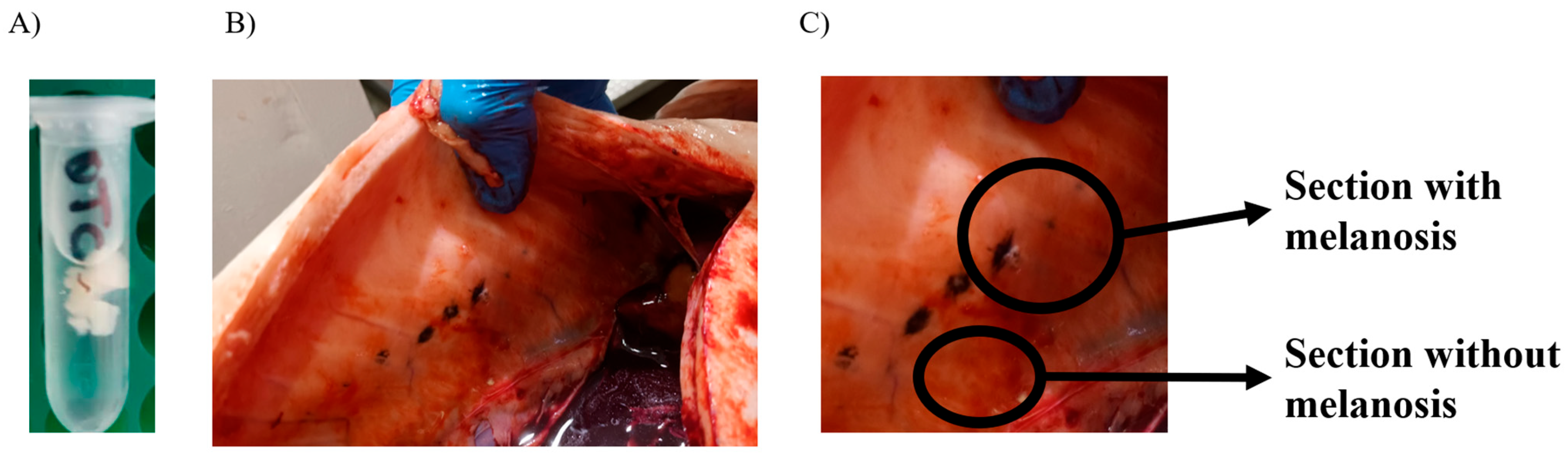
2.1. Tissue Sampling

Samples of Salmo salar used in this study were obtained from three different sources and were kindly provided by a pharmaceutical company specializing in salmon aquaculture, which conducts monitoring programs for its products. As part of its ongoing research efforts, selected samples from its program were used in this study through a collaborative initiative, contributing to the first approach to this topic within the Chilean industry. The sampling procedures were carried out by veterinarians certified by Sernapesca, who are officially accredited as “Certificadores de la condición sanitaria”, ensuring compliance with industry health standards. The sampling process considered only the external injection point visible on the belly of fish sourced from freshwater sites, along with ten samples collected at a processing plant with the support of a salmon company at the processing plant.
The first group of samples was collected from a “Safety Experiment” from a Fish pharmaceutical company located in Puerto Varas, Chile, and sampled at 300 degree-days after an oxytetracycline overdose testing assay. The second group was obtained at 600 degree-days post-vaccination (before their transfer to seawater) from a Chilean fish farm in the “Los Ríos” region. Finally, after the stunning procedure, the third group of harvest samples was collected from a processing plant in Puerto Montt (Chile). Due to the operational conditions of the processing plant, which primarily handles food for human consumption, further in-depth analyses were not feasible. As a result, only tissue samples from the thoracic region with focal melanosis were selected for this study (Figure 1B,C). These three sample groups are safety, vaccination, and harvest (Table 1). Considering that the cause of MFCs is not part of this research and that the sample information is confidential, the samples were delivered in a “blind” manner to avoid bias during the analyses, considering only seemingly healthy fish.
The experimental design of this study considered a limited number of samples due to the challenges of obtaining them under productive conditions in Chile. The study implemented a statistical analysis that accounted for repeated measures to address this limitation. By using a small number of fish but identifying each individual and its corresponding samples with and without melanosis, this approach minimized genetic background effects when comparing independent samples [14,20]. Additionally, the methodology was designed to maximize representativeness, ensuring that the findings remain robust and meaningful despite the sample size constraints.
This procedure involved identifying the MFCs in muscle and extracting the sample from the melanosis tissue and the adjacent area without melanosis, which was used as a control (Figure 1A,C). To minimize the effect of genetic background when comparing independent samples, both melanotic and non-melanotic tissue were identified from each fish and included in the analysis. After obtaining and identifying each sample, they were stored at 4 °C in Eppendorf tubes with RNAlater (SigmaTM, Darmstadt, Germany), previously determined for each tissue sample, and immediately sent to the Marine Molecular Biology Laboratory at the Faculty of Agronomy and Natural Systems, Pontificia Universidad Católica de Chile, where they were stored at −20 °C until processing.

2.2. Tissue Homogenization and RNA Isolation

The tissue homogenization was performed using 50 mg of the sample with the Mini-BeadBeater 24 (BioSpec Products, Inc., Bartlesville, OK, USA). Samples were placed in 2.0 mL screw tubes with standard caps (SSIbio, Lodi, CA, USA, cat. 2641-0B). Each tube contained six 1.5 mm stainless steel beads (BioSpec, cat. 11079115ss) and 700 µL of TRIzol (Invitrogen, Carlsbad, CA, USA). The samples were shaken at 2800 rpm for 30 s in the bead beater, followed by a cooling step for 60 s. This process was repeated three times to achieve complete tissue homogenization. The sample was then transferred to a 1.5 mL Eppendorf tube and centrifuged at 12,000 rpm for 5 min. The supernatant was removed and mixed in another tube with 100% ethanol at a 1:1 ratio. Subsequently, total RNA was extracted using the “Direct-zol RNA Miniprep Plus” kit (Zymo Research, Irvine, CA, USA) following the protocol until eluting the RNA sample in 50 µL of nuclease-free water and stored at −80 °C. The RNA concentration of the samples was then quantified and aliquoted to a concentration of 100 ng/µL with nuclease-free water.

2.3. Quantitative Real-Time PCR and Statistical Analysis
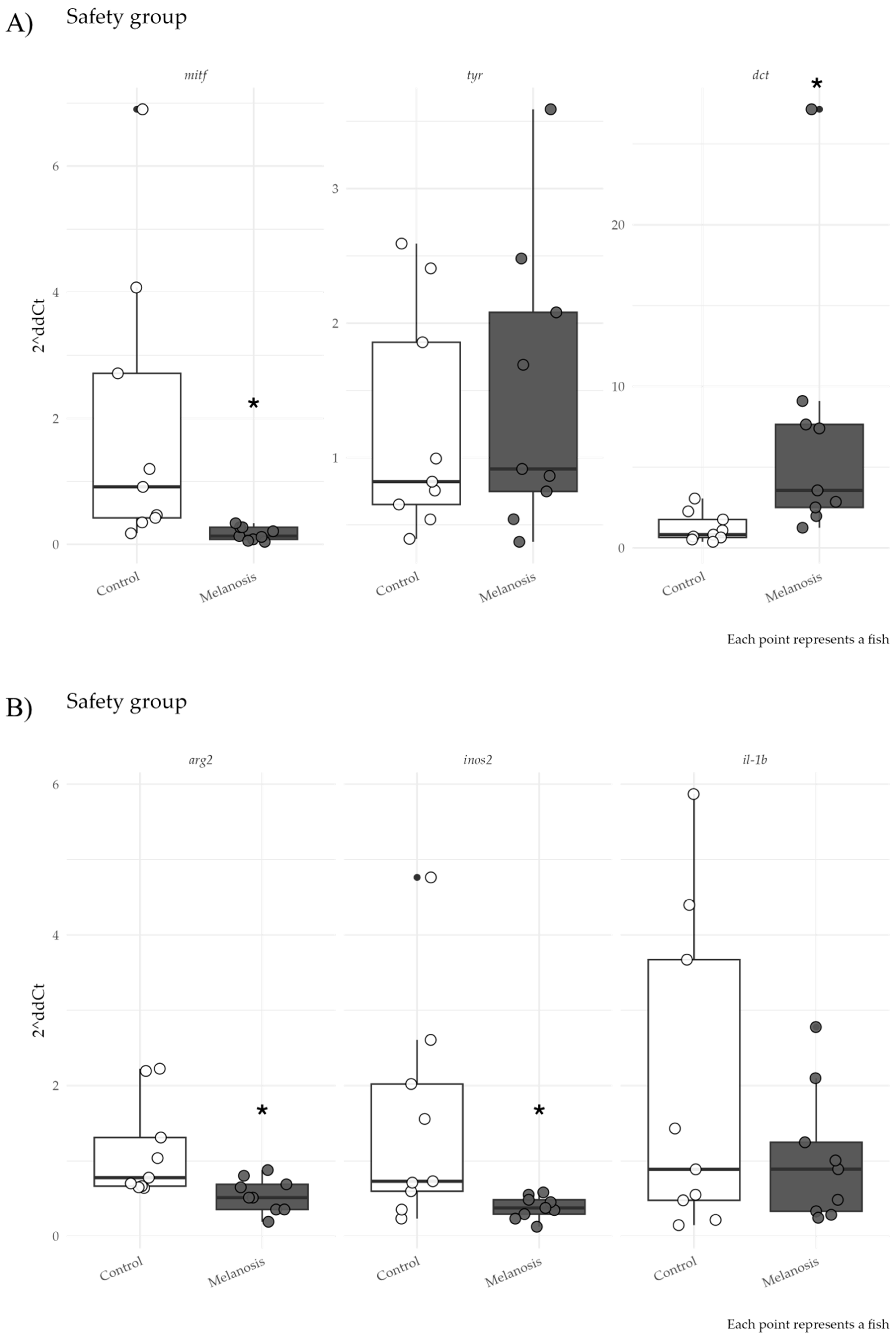
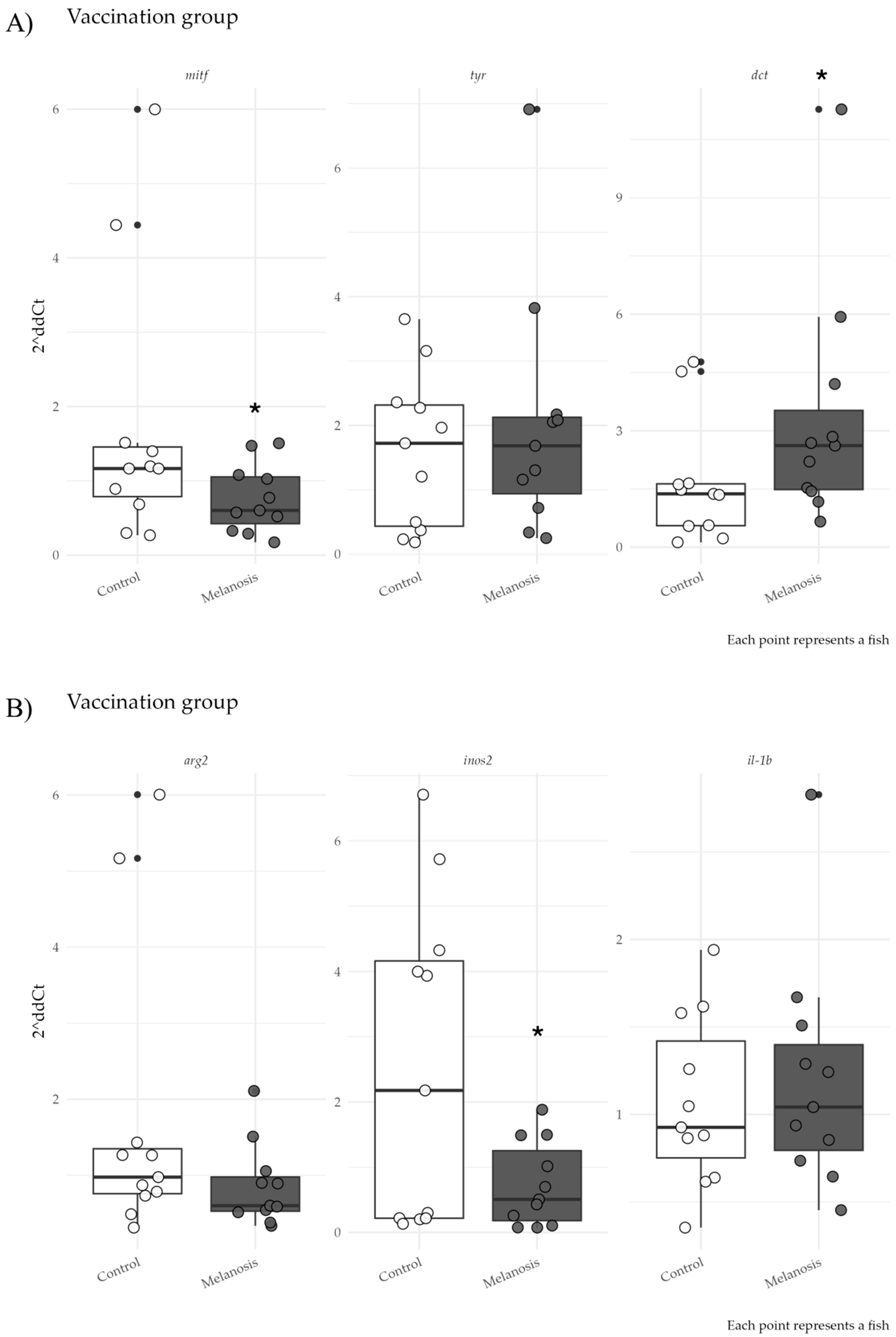
The HiScript II One Step Qrt-PCR SYBR Green kit (Vazyme, Nanjing, China) was used for the qPCR on tissue samples, employing a Dlab Real-Time PCR System Accurate 96 (Dlab, Beijing, China). An aliquot of 100 ng/µL was used as a template for all analyses. Primer sequences are listed in Table 2.
The data associated with the qPCR results obtained from related tissue samples were analyzed using a paired t-test. We considered comparing the transcriptional regulation of the studied genes in tissues with and without melanosis using a repeated measures comparison. The analysis began by evaluating the assumptions necessary for a parametric analysis (homogeneity of variance and normality of residuals). When the ΔCt data met the assumptions, the repeated measures t-test was used. When any assumptions were not met, the Wilcoxon signed-rank test was used. The ∆Ct values of tissues with and without melanosis identified in each fish were compared and graphed as 2∆∆Ct.

3. Results

The qPCR showed a significant downregulation of mitf (t = −5.703; p = 0.00045) and upregulation of dct (t = 3.27; p = 0.01135) in MFC tissue compared to controls for the “Safety” group (Figure 2A). In contrast, tyr showed no significant differences. Regarding immune-related genes, arg2 (W = 1; p = 0.0078) and K (t = −2.749; p = 0.0251) were significantly downregulated in MFC tissue, whereas il-1β showed no significant changes.
A similar trend occurred in the “Vaccination” group (Figure 3). Pigmentation-related genes showed significant downregulation of mitf (t = −3.951; p = 0.0027) and upregulation of dct (t = 2.626; p = 0.0253) in MFC tissue. Again, tyr showed no significant differences. For immune genes, inos2 was significantly downregulated (W = 7; p = 0.0185), whereas arg2 downregulation was not statistically significant, and il-1β remained unchanged.
In the “Harvest” group (Figure 4B), mitf expression did not differ significantly between melanosis and non-melanosis tissues. However, both tyr (t = 2.945; p = 0.0163) and dct (t = 7; p < 0.001) showed significant upregulation in MFCs samples. Regarding immune-related genes (Figure 4A), arg2, inos2, and il-1β showed no significant transcriptional differences between MFCs and control tissues.

4. Discussion

This study represents the first investigation analyzing melanosis in Salmo salar across both the freshwater stage and the harvest phase (seawater), providing valuable insights into the transcriptional regulation mechanisms in muscular tissues with and without MFCs in Salmo salar from different developmental stages and sources. However, this work presents some limitations that we want to highlight. It is a descriptive study that shows expression patterns in three different groups, as the sample sources were obtained through a monitoring program by a pharmaceutical salmon farming company. Additionally, the samples with and without melanosis from each fish were analyzed using a repeated measures model, which considers individual-level transcriptional regulation changes and the intrinsic correlation among repeated measurements. Leveraging this relationship reduces unexplained variability, enhancing statistical power and precision [24].
Significant differences and gene expression patterns were identified, potentially shedding light on key stages and regulatory points associated with the MFCs. MITF is a transcription factor that regulates the expression of several key genes involved in melanogenesis, such as tyr and dct. It is considered one of the most important transcription factors in melanogenesis [25] and the central target of multiple signal transduction pathways [26,27]. As a transcriptional activator, MITF controls the expression of crucial melanogenesis-related genes, including tyr, tyrp1, and dct, thereby promoting melanin synthesis [28]. The significant negative regulation of the mitf gene in tissues with melanosis in safety and vaccination groups supports the hypothesis that melanogenesis is modulated at the transcriptional level in the presence of melanosis. This negative regulation represents an inhibition of melanin production post-stimulus of the melanosis focus [29]. On the other hand, the significant upregulation of the dct gene suggests active melanization. In contrast, the lack of significant changes in tyr expression is likely due to the negative regulation of mitf [30].
Regarding genes related to melanization in the harvest group, the lack of significant differences in mitf expression suggests that this gene is not differentially regulated in the same way at an early stage of melanosis formation. However, the significant upregulation of tyr and dct in tissues with melanosis reinforces the idea that these genes play an important role in the pigmentary response of the tissue and could be regulated differently [31], maybe related to the size of the MFCs or possibly associated with a longer development time of MFCs.
From an immunological perspective, fish muscle possesses immune activity and is an active site of immune responses against pathogens [32]. In addition, it has been described that resident macrophages are present in muscle tissue, are involved in promoting the inflammatory response, or participate in tissue repair. Within the cellular phenotypes of macrophages, we can find the pro-inflammatory M1 and anti-inflammatory M2 cells that participate in tissue repair [33]. These phenotypes are well characterized in fish, where molecular markers have been described for each of them, finding inducible nitric oxide synthase (iNOS) as the marker of M1 macrophages and arginase 2 (arg2) as the marker of M2 macrophages [34]. In the present study, both groups, security and vaccination, showed a negative regulation of the genes arg2 and inos2 in tissues with melanosis. This suggests that these genes indicate the proximity in time to the inflammatory process that could have triggered the melanosis focus, showing a negative regulation that compensates for the activity. Both genes may play a role in reducing repair and inflammation associated with the melanosis focus [2].
Considering that melanosis could be triggered by inflammatory processes, the gene expression of il-1β, an early response pro-inflammatory cytokine characterized in fish, was evaluated [35,36]. In this work, no significant differences in the expression of il-1β in security and vaccination groups were observed. This could indicate that this molecule is not modulated after 300 ATUs post-stimulus. It may play a different role in earlier stages in what has been termed red spots, which occur in tissues before melanosis [12].
Unlike the other groups, the “Harvest” samples did not show significant differences in the transcriptional regulation of arg2, inos2, and il-1β between tissues with and without melanosis. This absence of differences could be attributed to the processes that triggered MFCs occurring over extended periods compared to those found in the smolt fish samples, where samples were obtained at 300 and 600 ATUs post-melanosis initiation by OTC injection and vaccination, respectively.
Although it is only a descriptive study, we hope it will be the first step towards focusing the study of melanosis through the investigation of enzymes involved in pigmentation. This research represents an effort to analyze MFCs from the dual perspective of melanogenesis and immune response related to macrophage polarization. It integrates molecular insights related to pigmentation and immune responses, offering a comprehensive understanding of the underlying mechanisms. However, it is important to note that this is a brief and focused study, limited explicitly to analyzing selected genes associated with pigmentation and inflammatory responses. Additionally, this study is the first to describe MFCs in both freshwater environments during the early developmental stages of salmon and at the harvest stage, the culmination of the production cycle [15]. This dual-phase approach underscores the importance of investigating the phenomenon across different life stages and conditions to provide actionable insights for the industry.

5. Conclusions

In summary, this study provides critical insights into the transcriptional and immunological mechanisms underlying melanosis in Salmo salar. Regarding gene expression patterns, a clear distinction emerged between freshwater and harvest-stage samples, suggesting a different regulatory mechanism. These changes likely represent later stages of MFCs’ development, potentially influenced by prolonged exposure to stimuli and the increased size of MFCs. The findings underscore that pigmentation-related genes are active in the pigmentary response during advanced stages, while immune responses stabilize over time.
This study highlights these distinctions and emphasizes the importance of considering developmental stages and temporal factors when investigating melanosis in aquaculture systems. These insights offer valuable contributions to understanding the molecular basis of melanosis and inform strategies for its management in the industry. Identifying specific genes that are differentially regulated in MFCs provides a starting point for developing solution strategies to mitigate this condition in farmed salmon, thereby improving both the health and quality of fish in production systems in Chile and other countries.

Author Contributions

Conceptualization, R.C. and S.E.-A.; methodology, R.C. and A.J.; statistical analysis, R.C.; writing—original draft preparation, R.C. and C.A.V.; writing—review and editing, S.E.-A., C.A.V., A.J., A.V. and J.A.V.; project administration, R.C.; funding acquisition, R.C. All authors have read and agreed to the published version of the manuscript.

Funding

This research was funded by the Fondecyt Postdoctoral program, grant number 3230212.

Institutional Review Board Statement

The study was conducted according to the Declaration of Helsinki and approved by the Institutional Review Board (or Ethics Committee) of the Pontificia Universidad Católica de Chile (protocol code 220424004 and date of approval 24 May 2023).

Informed Consent Statement

Not applicable.

Data Availability Statement

The original contributions presented in this study are included in the article. Further inquiries can be directed to the corresponding author.

Acknowledgments

The authors would like to express their heartfelt gratitude to Amanda Escorza for her invaluable technical support during the study. ANID funded this research through the Fondecyt Postdoctoral program, which provided essential financial assistance. We also thank Aquabench for their invaluable cooperation in providing relevant information on the context of melanosis in Chilean salmon farming. We would also like to include our acknowledgments to Gabriela Salas, Claudia Rivera, and to Rodrigo Alarcón, Jose Fernando Gutiérrez and Rodrigo Díaz Arquimed for their support during the research.

Conflicts of Interest

The authors declare no conflicts of interest.

References

  1. Bjorgen, H.; Kumar, S.; Gunnes, G.; Press, C.M.; Rimstad, E.; Koppang, E.O. Immunopathological characterization of red focal changes in Atlantic salmon (Salmo salar) white muscle. Vet. Immunol. Immunopathol. 2020, 222, 110035. [Google Scholar] [CrossRef] [PubMed]
  2. Krasnov, A.; Moghadam, H.; Larsson, T.; Afanasyev, S.; Morkore, T. Gene expression profiling in melanised sites of Atlantic salmon fillets. Fish Shellfish Immunol. 2016, 55, 56–63. [Google Scholar] [CrossRef] [PubMed]
  3. Koppang, E.O.; Haugarvoll, E.; Hordvik, I.; Aune, L.; Poppe, T.T. Vaccine-associated granulomatous inflammation and melanin accumulation in Atlantic salmon, Salmo salar L., white muscle. J. Fish Dis. 2005, 28, 13–22. [Google Scholar] [CrossRef] [PubMed]
  4. Bjorgen, H.; Wessel, O.; Fjelldal, P.G.; Hansen, T.; Sveier, H.; Saebo, H.R.; Enger, K.B.; Monsen, E.; Kvellestad, A.; Rimstad, E.; et al. Piscine orthoreovirus (PRV) in red and melanised foci in white muscle of Atlantic salmon (Salmo salar). Vet. Res. 2015, 46, 89. [Google Scholar] [CrossRef]
  5. Moretro, T.; Moen, B.; Heir, E.; Hansen, A.A.; Langsrud, S. Contamination of salmon fillets and processing plants with spoilage bacteria. Int. J. Food Microbiol. 2016, 237, 98–108. [Google Scholar] [CrossRef]
  6. Malik, M.S.; Bjorgen, H.; Nyman, I.B.; Wessel, O.; Koppang, E.O.; Dahle, M.K.; Rimstad, E. PRV-1 Infected Macrophages in Melanized Focal Changes in White Muscle of Atlantic Salmon (Salmo salar) Correlates with a Pro-Inflammatory Environment. Front. Immunol. 2021, 12, 664624. [Google Scholar] [CrossRef]
  7. Bjarne, H.; Thomas, L.; Tone-Kari, Ø.; Odd Helge, R.; Laura Martinez, R.; Bente, R. Improved fillet quality in harvest-size Atlantic salmon fed high n-3 canola oil as a DHA-source. Aquaculture 2022, 560, 738555. [Google Scholar] [CrossRef]
  8. Sissener, N.H.; Waagbø, R.; Rosenlund, G.; Tvenning, L.; Susort, S.; Lea, T.B.; Oaland, Ø.; Chen, L.; Breck, O. Reduced n-3 long chain fatty acid levels in feed for Atlantic salmon (Salmo salar L.) do not reduce growth, robustness or product quality through an entire full scale commercial production cycle in seawater. Aquaculture 2016, 464, 236–245. [Google Scholar] [CrossRef]
  9. Larsen, H.A.; Austbo, L.; Morkore, T.; Thorsen, J.; Hordvik, I.; Fischer, U.; Jirillo, E.; Rimstad, E.; Koppang, E.O. Pigment-producing granulomatous myopathy in Atlantic salmon: A novel inflammatory response. Fish Shellfish Immunol. 2012, 33, 277–285. [Google Scholar] [CrossRef]
  10. Bjorgen, H.; Haldorsen, R.; Oaland, O.; Kvellestad, A.; Kannimuthu, D.; Rimstad, E.; Koppang, E.O. Melanized focal changes in skeletal muscle in farmed Atlantic salmon after natural infection with Piscine orthoreovirus (PRV). J. Fish Dis. 2019, 42, 935–945. [Google Scholar] [CrossRef]
  11. Berg, A.; Yurtseva, A.; Hansen, T.; Lajus, D.; Fjelldal, P.G. Vaccinated farmed Atlantic salmon are susceptible to spinal and skull deformities. J. Appl. Ichthyol. 2012, 28, 446–452. [Google Scholar] [CrossRef]
  12. Brimsholm, M.; Ronning, L.; Rimstad, E.; Koppang, E.O.; Bjorgen, H. Diffuse melanization of the red skeletal musculature in farmed Atlantic salmon (Salmo salar L.). J. Fish Dis. 2023, 46, 453–458. [Google Scholar] [CrossRef] [PubMed]
  13. Bjorgen, H.; Brimsholm, M.; Asserson, C.F.; Skaar, K.; Knutsen, G.M.; Oaland, O.; Haldorsen, R.; Fjelldal, P.G.; Hansen, T.; Rimstad, E.; et al. Deciphering the pathogenesis of melanized focal changes in the white skeletal muscle of farmed Atlantic salmon (Salmo salar). J. Fish Dis. 2024, 47, e13988. [Google Scholar] [CrossRef] [PubMed]
  14. Sunderland, K.; Keselman, H.; Algina, J.; Lix, L.; Wilcox, R. Conventional and Robust Paired and Independent-Samples t Tests: Type I Error and Power Rates. J. Mod. Appl. Stat. Methods 2003, 2, 481–496. [Google Scholar] [CrossRef]
  15. Bjorgen, H.; Rimstad, E.; Koppang, E.O. Melanisation in Salmonid Skeletal Muscle: A Review. J. Fish Dis. 2025, 48, e14063. [Google Scholar] [CrossRef]
  16. Edvardsen, R.B.; Leininger, S.; Kleppe, L.; Skaftnesmo, K.O.; Wargelius, A. Targeted mutagenesis in Atlantic salmon (Salmo salar L.) using the CRISPR/Cas9 system induces complete knockout individuals in the F0 generation. PLoS ONE 2014, 9, e108622. [Google Scholar] [CrossRef]
  17. Guyonneau, L.; Murisier, F.; Rossier, A.; Moulin, A.; Beermann, F. Melanocytes and pigmentation are affected in dopachrome tautomerase knockout mice. Mol. Cell Biol. 2004, 24, 3396–3403. [Google Scholar] [CrossRef]
  18. Wang, F.; Ma, W.; Fan, D.; Hu, J.; An, X.; Wang, Z. The biochemistry of melanogenesis: An insight into the function and mechanism of melanogenesis-related proteins. Front. Mol. Biosci. 2024, 11, 1440187. [Google Scholar] [CrossRef]
  19. Rozanowska, M.; Sarna, T.; Land, E.J.; Truscott, T.G. Free radical scavenging properties of melanin interaction of eu- and pheo-melanin models with reducing and oxidising radicals. Free Radic. Biol. Med. 1999, 26, 518–525. [Google Scholar] [CrossRef]
  20. Jennings, M.J.; Zumbo, B. The Robustness of Validity and Efficiency of the Related Samples t-Test in the Presence of Outliers. Psicológica 2002, 23, 415–450. [Google Scholar]
  21. Thorsen, J.; Hoyheim, B.; Koppang, E.O. Isolation of the Atlantic salmon tyrosinase gene family reveals heterogenous transcripts in a leukocyte cell line. Pigment Cell Res. 2006, 19, 327–336. [Google Scholar] [CrossRef] [PubMed]
  22. Larsen, H.A.; Austbo, L.; Konig, M.; Sorum, H.; Rimstad, E.; Koppang, E.O. Transcription of the tyrosinase gene family in an Atlantic salmon leukocyte cell line (SHK-1) is influenced by temperature, but not by virus infection or bacterin stimulation. Dev. Comp. Immunol. 2013, 41, 50–58. [Google Scholar] [CrossRef] [PubMed]
  23. Rozas-Serri, M.; Pena, A.; Maldonado, L. Gene expression associated with immune response in Atlantic salmon head-kidney vaccinated with inactivated whole-cell bacterin of Piscirickettsia salmonis and pathogenic isolates. Fish Shellfish Immunol. 2019, 93, 789–795. [Google Scholar] [CrossRef]
  24. Oberfeld, D.; Franke, T. Evaluating the robustness of repeated measures analyses: The case of small sample sizes and nonnormal data. Behav. Res. Methods 2013, 45, 792–812. [Google Scholar] [CrossRef] [PubMed]
  25. Vachtenheim, J.; Borovansky, J. “Transcription physiology” of pigment formation in melanocytes: Central role of MITF. Exp. Dermatol. 2010, 19, 617–627. [Google Scholar] [CrossRef]
  26. de la Serna, I.L.; Ohkawa, Y.; Higashi, C.; Dutta, C.; Osias, J.; Kommajosyula, N.; Tachibana, T.; Imbalzano, A.N. The microphthalmia-associated transcription factor requires SWI/SNF enzymes to activate melanocyte-specific genes. J. Biol. Chem. 2006, 281, 20233–20241. [Google Scholar] [CrossRef]
  27. Lee, A.; Lim, J.; Lim, J.S. Emerging roles of MITF as a crucial regulator of immunity. Exp. Mol. Med. 2024, 56, 311–318. [Google Scholar] [CrossRef]
  28. Wan, P.; Hu, Y.; He, L. Regulation of melanocyte pivotal transcription factor MITF by some other transcription factors. Mol. Cell Biochem. 2011, 354, 241–246. [Google Scholar] [CrossRef]
  29. Lee, W.H.; Ha, Y.; Park, J.I.; Joh, W.B.; Park, M.; Kim, J.K.; Jeon, H.K.; Kim, Y.J. Triglochin maritima Extracts Exert Anti-Melanogenic Properties via the CREB/MAPK Pathway in B16F10 Cells. Mar. Drugs 2024, 22, 532. [Google Scholar] [CrossRef]
  30. Fang, D.; Tsuji, Y.; Setaluri, V. Selective down-regulation of tyrosinase family gene TYRP1 by inhibition of the activity of melanocyte transcription factor, MITF. Nucleic Acids Res. 2002, 30, 3096–3106. [Google Scholar] [CrossRef]
  31. Wang, P.; Li, Y.; Hong, W.; Zhen, J.; Ren, J.; Li, Z.; Xu, A. The changes of microRNA expression profiles and tyrosinase related proteins in MITF knocked down melanocytes. Mol. Biosyst. 2012, 8, 2924–2931. [Google Scholar] [CrossRef] [PubMed]
  32. Valenzuela, C.A.; Zuloaga, R.; Poblete-Morales, M.; Vera-Tobar, T.; Mercado, L.; Avendano-Herrera, R.; Valdes, J.A.; Molina, A. Fish skeletal muscle tissue is an important focus of immune reactions during pathogen infection. Dev. Comp. Immunol. 2017, 73, 1–9. [Google Scholar] [CrossRef] [PubMed]
  33. Wentzel, A.S.; Petit, J.; van Veen, W.G.; Fink, I.R.; Scheer, M.H.; Piazzon, M.C.; Forlenza, M.; Spaink, H.P.; Wiegertjes, G.F. Transcriptome sequencing supports a conservation of macrophage polarization in fish. Sci. Rep. 2020, 10, 13470. [Google Scholar] [CrossRef] [PubMed]
  34. Wiegertjes, G.F.; Wentzel, A.S.; Spaink, H.P.; Elks, P.M.; Fink, I.R. Polarization of immune responses in fish: The ‘macrophages first’ point of view. Mol. Immunol. 2016, 69, 146–156. [Google Scholar] [CrossRef]
  35. Ho, P.-Y.; Byadgi, O.; Wang, P.-C.; Tsai, M.-A.; Liaw, L.-L.; Chen, S.-C. Identification, Molecular Cloning of IL-1? and Its Expression Profile during Nocardia seriolae Infection in Largemouth Bass, Micropterus salmoides. Int. J. Mol. Sci. 2016, 17, 1670. [Google Scholar] [CrossRef]
  36. Hu, B.; Chen, B.; Mao, M.; Chen, M.; Liu, X.; Cui, Q.; Liu, Y.; Jiang, C. Molecular characterization and expression analysis of the interleukin 1b gene in Pacific cod (Gadus macrocephalus). Dev. Comp. Immunol. 2018, 88, 213–218. [Google Scholar] [CrossRef]
Figure 1. Photographs of samples used. (A) Photographs of a sample from safety showing the injection point melanized 300 UTAs from the treatment, (B) harvest samples, and (C) images at 1.5× magnification representing the sampling process. For each identified fish, a section of muscle tissue was taken without melanosis (control) and with melanosis (melanosis).
Fishes 10 00302 g001
Figure 2. Relative gene expression in samples from the safety group. The upper panel shows pigmentation-related genes, and the lower panel shows immune-related genes. * Means statistical differences between melanized tissue from related samples. Number of pairs for repeated analysis: 9.
Fishes 10 00302 g002
Figure 3. Relative gene expression in samples from the vaccination group. The upper panel shows pigmentation-related genes, and the lower panel shows immune-related genes. * Means statistical differences between melanized tissue from related samples. Number of pairs for repeated analysis: 11.
Fishes 10 00302 g003
Figure 4. Relative gene expression in samples from the harvest group. The upper panel shows pigmentation-related genes, and the lower panel shows immune-related genes. * Means statistical differences between melanized tissue from related samples. Number of pairs for repeated analysis: 10.
Fishes 10 00302 g004
Table 1. Description of the samples used. The weight data are expressed in grams, and K is the Fulton condition factor (Weight/Length3) × 100.
GroupnStageWeightCoefficient of VariationK
Safety9Freshwater144.6110%0.81
Vaccination11Freshwater209.0922%1.20
Harvest10Seawater6058.2018%1.29
Table 2. Primer sequences.
GenePrimers Sequence (5′→3′)Product bpAccession Number or Reference
mitfgattgagagaagacggaggttt100 bpXM_014136388.2
gcctttgttccaacgcatac
tyrtgggaaacaaggtcctgggctac111 bp[21]
actgccagatcagctgagcctc
dcttctcactctgcagccaatgac85 bp [22]
cagacttcctcatccactcatcaaa
inos2catcggcaggattcagtggtccaat136 bp[6]
ggtaatcgcagaccttaggtttcctc
arg2cctgaaggacttgggtgtccagta109 bp [6]
ccgctgcttccttgacaagaggt
il-1βatcaccatgcgtcacattgc90 bp[23]
gtccttgaactcggttccca
β-actincccatctacgagggttacgc112 bpAF012125.1
tgaaactgtaaccgcgctct
Disclaimer/Publisher’s Note: The statements, opinions and data contained in all publications are solely those of the individual author(s) and contributor(s) and not of MDPI and/or the editor(s). MDPI and/or the editor(s) disclaim responsibility for any injury to people or property resulting from any ideas, methods, instructions or products referred to in the content.

