Article

Imidacloprid Exposure Induced Impaired Intestinal Immune Function in Procambarus clarkii: Involvement of Oxidative Stress, Inflammatory Response, and Autophagy

by
Zhaolin Li
1,†,
Yong Shi
1,2,†,
Kai Xie
1,
Lei Zhong
1,
Yi Hu
1,* and
Kaijian Chen
1,*
1
Fisheries College, Hunan Agricultural University, Changsha 410128, China
2
State Key Laboratory of Freshwater Ecology and Biotechnology, Institute of Hydrobiology, Chinese Academy of Sciences, Wuhan 430072, China
*
Authors to whom correspondence should be addressed.
These author contributed equally to this work.
Fishes 2025, 10(3), 131; https://doi.org/10.3390/fishes10030131
Submission received: 17 February 2025 / Revised: 14 March 2025 / Accepted: 15 March 2025 / Published: 17 March 2025
(This article belongs to the Special Issue Physiological Response Mechanisms of Aquatic Animals to Stress)

Abstract

Imidacloprid (IMI), a widely used neonicotinoid insecticide, has raised environmental concerns due to its potential impact on non-target aquatic organisms. This study investigates the effects of IMI exposure on the intestinal immune function of red swamp crayfish (Procambarus clarkii, P. clarkii), focusing on oxidative stress, inflammatory response, and autophagy. The P. clarkii was exposed to different doses of IMI (0, 10.93, 21.86, 43.73, 87.45 μg/L) for 96 h. Our findings reveal that IMI exposure leads to a survival rate of less than 70% when the concentration was 87.45 μg/L at 96 h. Hemolymph LZM and AKP contents were significantly decreased at the medium and high concentrations, and the expressions of hsp70 and nf-κb genes were significantly up-regulated. The expression of the lysozyme gene was significantly down-regulated. Additionally, the activities of SOD, CAT, and GPX were significantly decreased, the contents of MDA were significantly increased, and the gene expressions of CuZnsod, mMnsod, cat, and gpx in the gut were significantly down-regulated after exposure to medium-high IMI. The expression of autophagy-related genes showed that the expressions of beclin1, atg5, atg13, and lc3c genes in the medium- and high-concentration groups were significantly up-regulated. In summary, this study elucidates that medium-high levels of IMI exposure impair intestinal immune function in P. clarkii through mechanisms involving oxidative stress, inflammatory response, and autophagy.
Key Contribution: This study explores the fact that imidacloprid exposure impairs intestinal immune function in P. clarkii through mechanisms involving oxidative stress, inflammatory response, autophagy, and apoptosis. The above results provide a basis for careful management and regulation of imidacloprid usage to protect aquatic ecosystems and the species that inhabit them.

1. Introduction

Red swamp crayfish (Procambarus clarkii, P. clarkii), native to the southern United States and northern Mexico, commonly known as freshwater crayfish, was introduced to China from Japan at the beginning of the last century because of its fast growth rate and strong adaptability [1]. In recent years, with the rising market demand, the original crayfish aquaculture industry has been greatly promoted and developed rapidly. By 2021, the breeding area of P. clarkii in China will exceed 1.33 million hectares. At present, the cultivation mode of P. clarkii is mainly a “rice-shrimp” integrated culture, which is more than 70% [2]. With the rapid development of the original P. clarkii industry and the promotion of the rice-fishery integrated cultivation model, the use of pesticides in the rice-fishery integrated cultivation model has become more and more frequent, and the problem of drug residues in water and sediment has also received great attention.
Among them, neonicotinoid insecticides have also been used for the control of rice weevils in the integrated cultivation process of “rice-shrimp”. In addition, neonicotinoids have also been detected in pond aquaculture water. The side effects of neonicotinoids on non-target organisms, especially aquatic invertebrates, have gradually become the focus of current attention [3]. Neonicotinoid pesticides (NPs) are the fourth major class of pesticides after organophosphorus, carbamates, and pyrethroids. They are characterized by high efficiency, broad spectrum, good internal absorption, and no cross-resistance with other traditional pesticides [4]. The mechanism of action of NPs is actually through the control of postsynaptic nicotinic acetylcholine receptors (nAChRs), and there are three main modes of binding to acetylcholine receptors: Firstly, it is believed that the oxygen on the nitro group of neonicotinoids or the nitrogen energy on the cyanide group forms a hydrogen bond with the hydrogen of nAChRs, and the SP2 (a hybrid of one ns orbital and two np orbitals in the same atom) hybrid nitrogen energy on the imidazoline ring has an electrostatic interaction with the negative electric center of nAChRs. Secondly, it is believed that the nitrogen atom on the pyridine ring of neonicotinoid forms hydrogen bonds with the hydrogen of nAChRs, and the nitro group with strong polarity in the structure of neonicotinoid is the decisive pharmacophore, which can bind to the amino acid residues of the receptor. Thirdly, it is believed that the nitrogen atom on the pyridine ring of neonicotinoid forms a hydrogen bond with the hydrogen of nAChRs, and the N on imidazoline ionizes in the insect body, carries a partial positive charge and can produce electrostatic interaction with the negative electric center of nAChRs [5,6,7].
Imidacloprid (IMI), as the first generation of neonicotinoid insecticide with chemical formula C9H10Cl N5O2, became the first neonicotinoid insecticide in China in 2010. It has the characteristics of lasting effect and multiple insecticide species. The insecticide can bind to the nicotinic acetylcholine receptor in the central nervous system of the target. This disrupts nerve signaling, leading to paralysis and death of the target at high concentrations. This method is now widely used in various crop pest control, to be more clear [8,9]. However, in the process of using pesticides, only a small part of pesticides will act on pests, and the rest of the pesticides enter the soil and bind with it, and the other part enters the surrounding pond or river in the form of rainwater and surface runoff. IMI has high water solubility, and most of the IMI remaining in the soil will be brought into the surrounding water environment by rain or osmosis, thus endangering the safety of water bodies and aquatic organisms [10]. Studies have shown the effect of IMI on its toxic accumulation in P. clarkii, revealing that among the examined tissues—muscle, intestine, gill, hemolymph, and hepatopancreas—the intestine exhibited the highest IMI concentration, with the exception of muscle tissue [11]. Numerous studies have shown that since the gut is the main area of absorption of environmental pollutants (including pesticides, fungicides, heavy metals, etc.), the toxicity of environmental pollutants is commonly evaluated through the assessment of gut health [12,13,14]. IMI was found to cause pathological features such as swelling, necrosis, and degeneration of the digestive gland cells of Asian Freshwater Clams (Corbicula fluminea) and significantly up-regulated the expression levels of hsp90, gsts1, and gstm1 genes related to digestive antioxidant stress, thus disrupting the digestive antioxidant system and causing oxidative stress effects [15]. Similarly, IMI could reduce the intestinal villus height, the expression levels of tight link protein genes Ocln-, Caln-, Muc2.1-, and Muc2.2-related genes of intestinal mucus secretion in zebrafish (Danio rerio) and cause intestinal oxidative stress. The levels of pro-inflammatory cytokines il-6, il-1α, il-1β, il-6, and il-8 are significantly increased, which leads to intestinal damage [16]. However, P. clarkii in the “rice-shrimp” integrated culture model is more susceptible because of its proximity to the planting area, and there are few studies on the intestinal health of IMI shrimp. Therefore, it is necessary to study the toxic effects of IMI on P. clarkii, as well as the potential effects on intestinal mucosal health status and immune response, so as to provide scientific basis and management suggestions for protecting the ecological and economic value of cultured shrimp.

2. Materials and Methods

2.1. A Preliminary Experiment of Acute Toxicity

Twenty P. clarkii with the same size (About 4 g, 2.5 cm) were randomly selected, and 6 concentration groups were set as follows: Three parallel concentrations were set for each concentration of 0, 50, 100, 200, 400, and 800 μg/L. No feeding was conducted during the experiment, and the drug solution was changed every 24 h. The death numbers of P. clarkii in 12, 24, 36, 48, 60, 72, 84, and 96 h were recorded.

2.2. Acute Toxicity Formal Experiment

IMI with different concentrations of 0 (CON), 1/24 LC50 (IM-1), 1/12 LC50 (IM-2), 1/6 LC50 (IM-3), and 1/3 LC50 (IM-4) were set for experiments (Concentration settings refer to previous studies [15]). There were 3 replicates in each group, and 25 P. clarkii of the same size were placed in each replicate. During the experiment, the drug solution was changed every 24 h without feeding. The death numbers of P. clarkii were recorded at 24, 48, 72, and 96 h post-IMI exposure.

2.3. Serum Biochemical and Immune Indexes

After the end of the experiment, three P. clarkii individuals were randomly selected from each tank (0.5 m × 0.5 m × 0.4 m, located in Hunan Agricultural University, Changsha, China), and hemolymph samples were aseptically collected from the heart puncture using sterile 1.0 mL sterile syringes, placed in a 2.0 mL centrifuge tube at 4 °C for 6 h, centrifuged at 3500× g for 10 min, and the upper serum was collected in a 1.5 mL enzyme-free tube and stored at −80 °C for later use. The levels of lysozyme (LZM), alkaline phosphatase (AKP), glutamic oxalic aminotransferase (GOT), and glutamic pyruvic aminotransferase (GPT) in serum were determined using the corresponding kit method of Nanjing Jianjieng (Biotechnology Co., Ltd., Nanjing, China) [17]. In enzyme assays, preliminary experiments were conducted to establish the optimal substrate concentrations and reaction durations, thereby ensuring that the conditions adhered to the principles of enzyme kinetics.

2.4. Intestinal Antioxidant Enzyme Activity

After the end of the experiment, three P. clarkii were randomly selected from each tank, the intestines of P. clarkii were removed from the dissecting dish with an ice pack, and the intestinal contents were cleaned, and then the intestines were placed in 2.0 mL enzyme-free tubes and stored at −80 °C. The content of malondialdehyde (MDA) and glutathione peroxidase (GPx) and superoxide dismutase (SOD) and catalase (CAT) were determined using the corresponding kit instructions provided by Nanjing Jiancheng Biotechnology Co., Ltd. [18]. In enzyme assays, preliminary experiments were conducted to establish the optimal substrate concentrations and reaction durations, thereby ensuring that the conditions adhered to the principles of enzyme kinetics.

2.5. Tissue RNA Extraction and Real-Time PCR Analysis

The intestines of three P. clarkii were taken from each cage in a 1.5 mL enzyme-free centrifuge tube and stored at −80 °C. RNA was extracted using Trizol, and the detailed method was the same as in previous studies [19]. The concentration and quality of RNA were detected by ultramicro nucleic acid detector, and the OD260/OD280 values of RNA samples were between 1.8 and 2.0. Reverse Transcriptase MMLV kit (Thermo, Waltham, MA, USA) was used to reverse transcribe RNA into cDNA. Primer Premier 5.0 software was used to design primers. Primer sequences are shown in Table 1. Primers were synthesized by Shanghai Shenggong Biological Co., Ltd., and the amplification efficiency of all primers ranged from 0.95 to 1.10. A fluorescence quantitative PCR instrument (LightCycler 48II, Bio-Rad, Hercules, CA, USA) and SYBR Premix Ex Taq II kit (Takara, Japan) were used for the determination. The fluorescence quantitative PCR reaction system is shown in Table 2. The reaction procedure is as follows: (1) predenaturation (95 °C, 30 s); (2) amplification (① 95 °C, 5 s; ②60 °C, 30 s; ①–② 40 cycles. The β-actin gene of grass carp was used as an internal reference gene and calculated by E = 2−ΔΔCt [17].

2.6. Data Processing and Statistical Analysis

Experimental data are presented as mean ± standard error (mean ± SE). All data were analyzed using one-way analysis of variance (ANOVA) in SPSS 24.0 (Chicago, IL, USA). A p-value of less than 0.05 was considered statistically significant, and further post hoc comparisons were performed using Duncan’s multiple range test. Survival rate data were analyzed using a two-factor ANOVA to evaluate the effects of treatment concentration and exposure time. Prior to analysis, data were assessed for normality and homogeneity of variance using the Levene and Shapiro–Wilk tests. All figures were generated and visualized utilizing GraphPad Prism 9.

3. Results

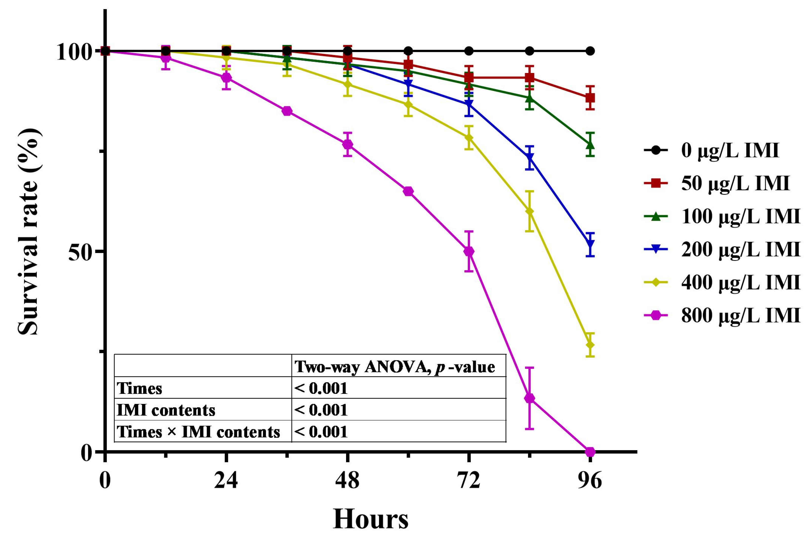
3.1. A Preliminary Experiment of Acute Toxicity Pre-Experiment

As shown in Figure 1, the survival rate of P. clarkii at nodes with different concentrations of IMI and at different times significantly decreased with the increase in exposure time to IMI (ANOVA, p < 0.05), and the survival rate significantly decreased with the increase in IMI concentration (ANOVA, p < 0.05). Moreover, when IMI was 800 μg/L, the survival rate of P. clarkii significantly decreased with the increase in IMI concentration (ANOVA, p < 0.05). The survival rate was 0 after 96 h of continuous exposure. As shown in Figure 2, through the analysis of primary and quadratic regression equations for the survival rate under different exposure times and different IMI, it was found that the 96 h semi-lethal concentration of IMI against P. clarkii was 207.62–317.07 μg/L, and the medium value was 262.36 μg/L as its 96h-LC50.

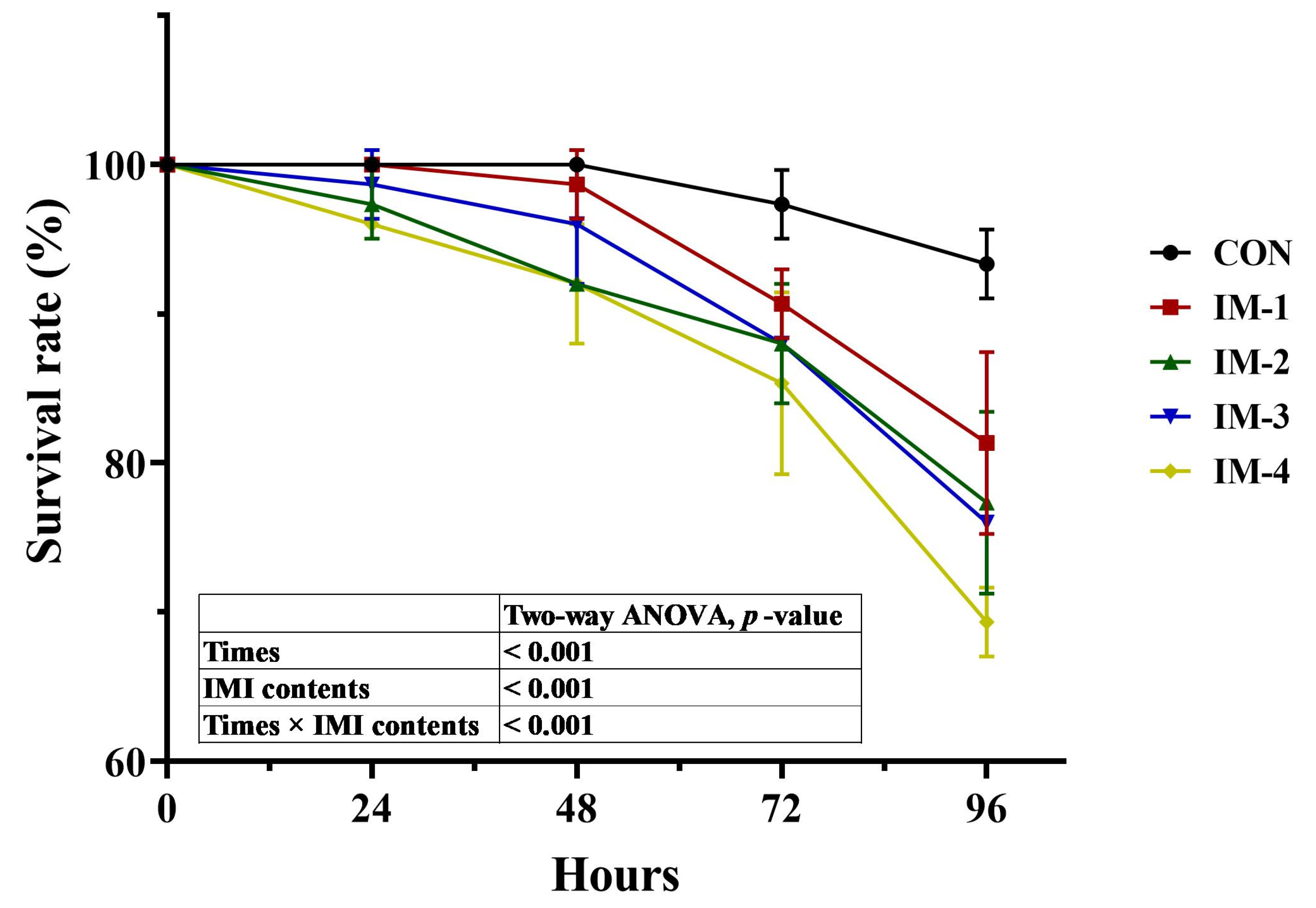
3.2. Acute Toxicity Formal Experiment

According to the results of the preliminary experiment, acute induction experiments were conducted with different concentrations of 0, 1/24 LC50, 1/12 LC50, 1/6 LC50, and 1/3 LC50, respectively, and the concentrations were 0, 10.93, 21.86, 43.73, and 87.45 μg/L, respectively. The survival rates were recorded for 24, 48, 72, and 96 h. As shown in Figure 3, with the increase in concentration and exposure time, the survival rate of P. clarkii showed a decreasing trend, and the survival rate was less than 70% when the concentration was 87.45 μg/L at 96 h and significantly decreased between 72 h and 96 h (ANOVA, p < 0.05).

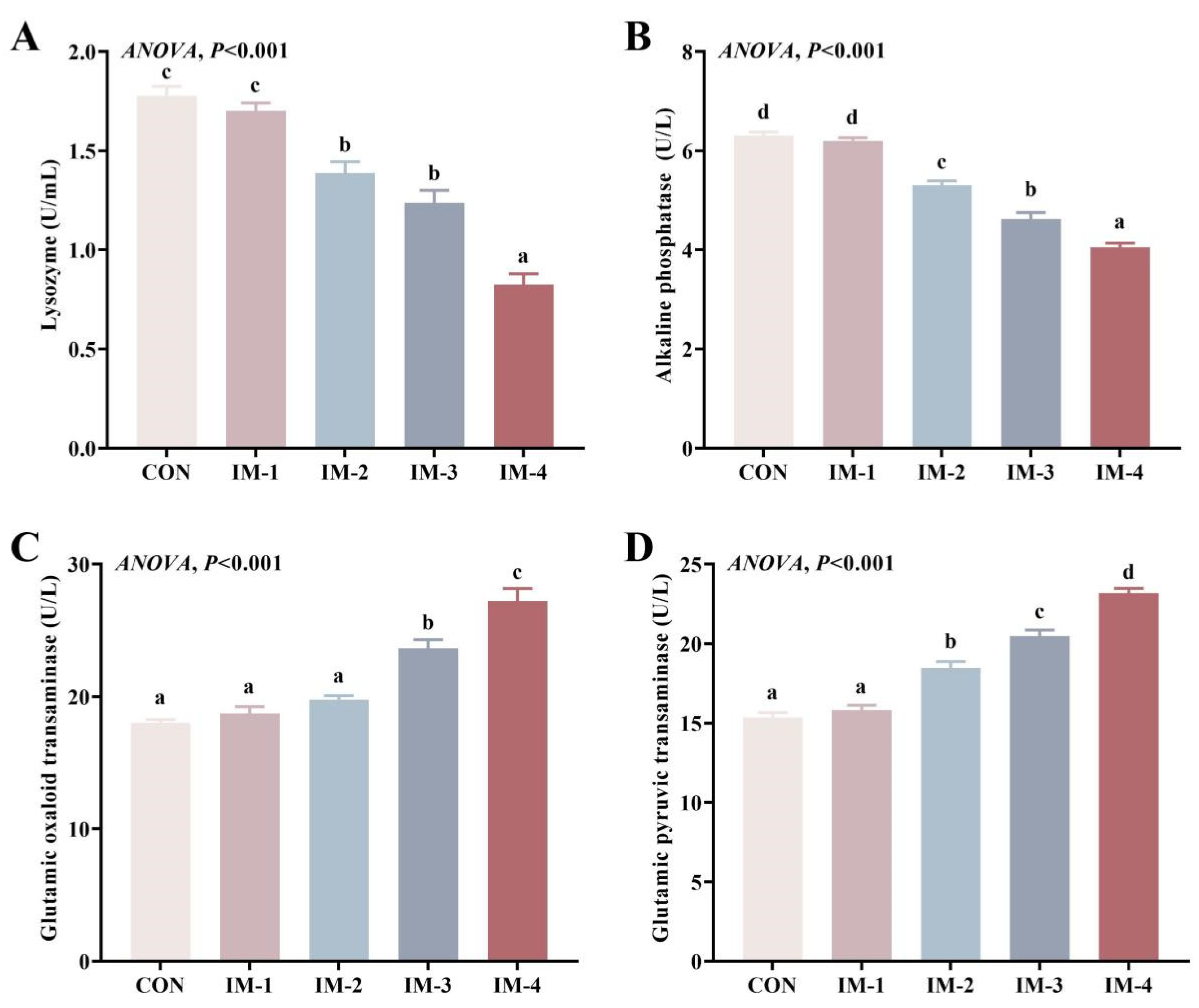
3.3. Hemolymph Biochemical and Immune Indexes

Hemolymph LZM and AKP contents were significantly decreased at medium and high concentrations (ANOVA, p < 0.05), while hemolymph GOT and GPT activities were significantly increased under medium- and high-concentration exposure conditions (ANOVA, p < 0.05) (Figure 4).

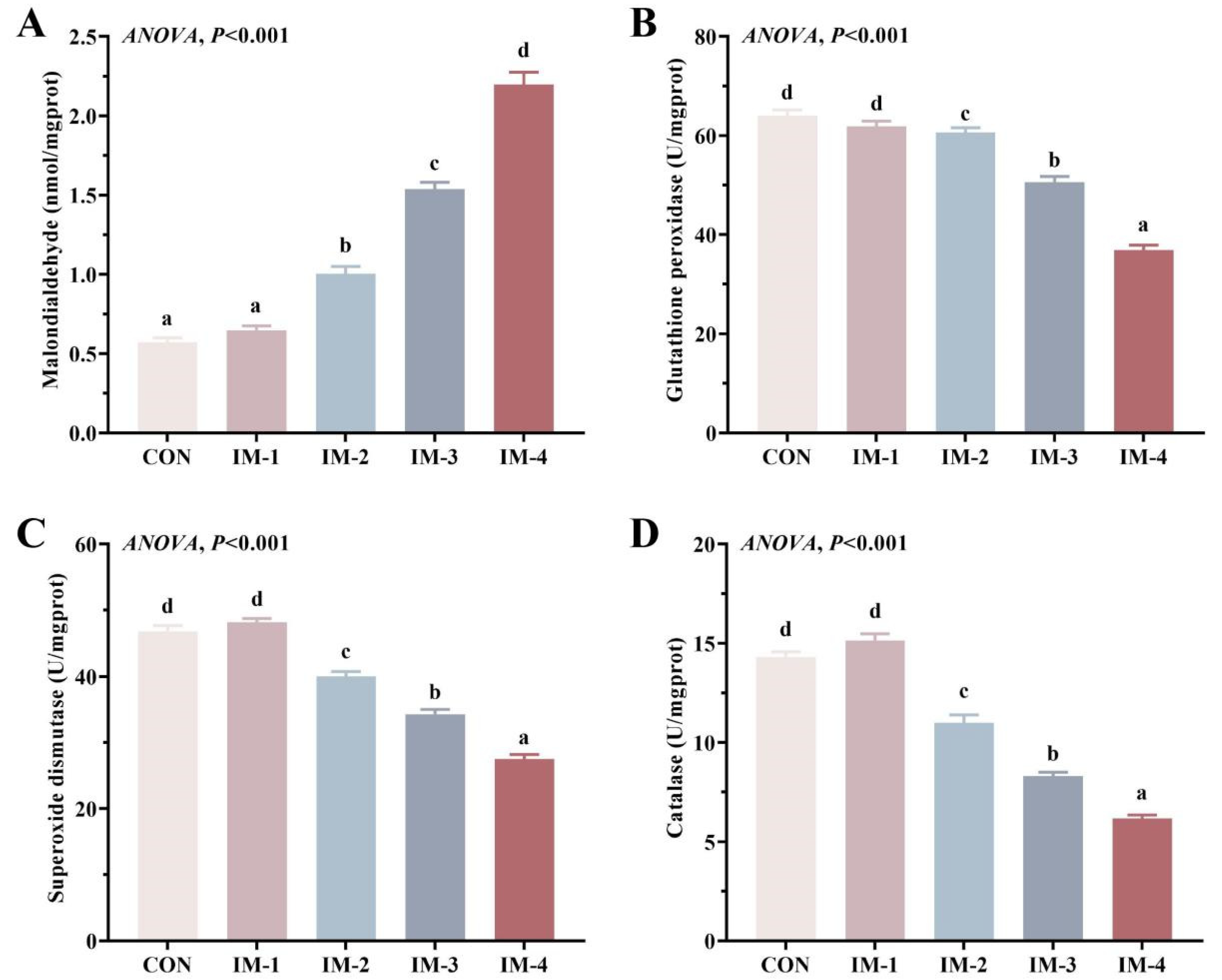
3.4. Intestinal Antioxidant Enzyme Activity

To investigate the presence of enzymes associated with oxidative stress in the gut, we examined the activity of three typical antioxidant enzymes and MDA levels. There were no significant differences in SOD, CAT, and GPx activities and MDA levels between CON and low-concentration groups (ANOVA, p > 0.05). Compared with the CON group, the activities of SOD, CAT, and GPX in the gut of the medium- and high-concentration groups were significantly decreased, and the level of MDA was significantly increased (ANOVA, p < 0.05) (Figure 5).

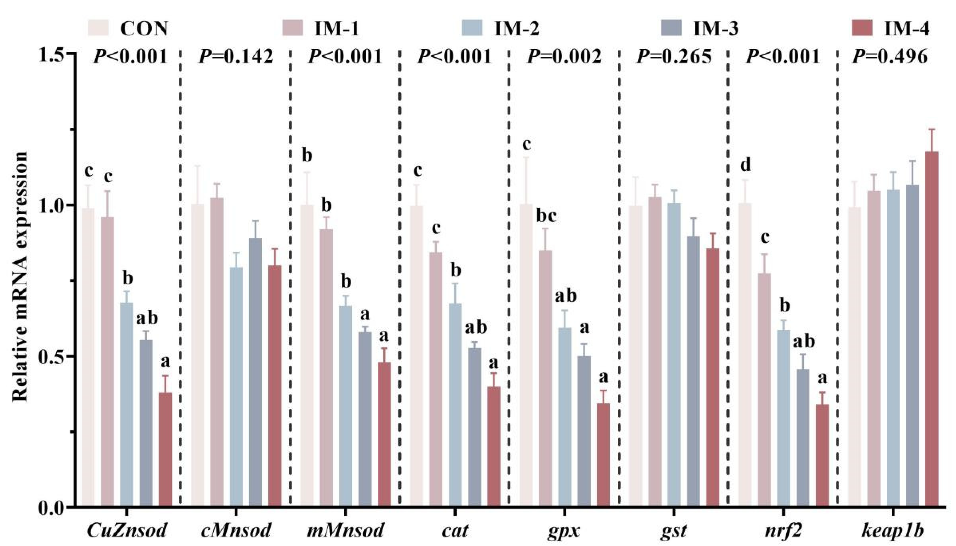
3.5. Expression of Intestinal Antioxidant-Related Genes

As shown in Figure 6, intestinal nrf2 gene expression of P. clarkii in all groups was significantly down-regulated under exposure to IMI (ANOVA, p > 0.05). Compared with the CON group, intestinal CuZnsod, mMnsod, cat, and gpx gene expressions in the low-concentration group were not significantly different (ANOVA, p > 0.05) but were significantly down-regulated in the medium- and high-concentration groups (ANOVA, p < 0.05). There were no significant differences in the expressions of cMnsod, gst, and keap1b genes among all groups (ANOVA, p > 0.05).

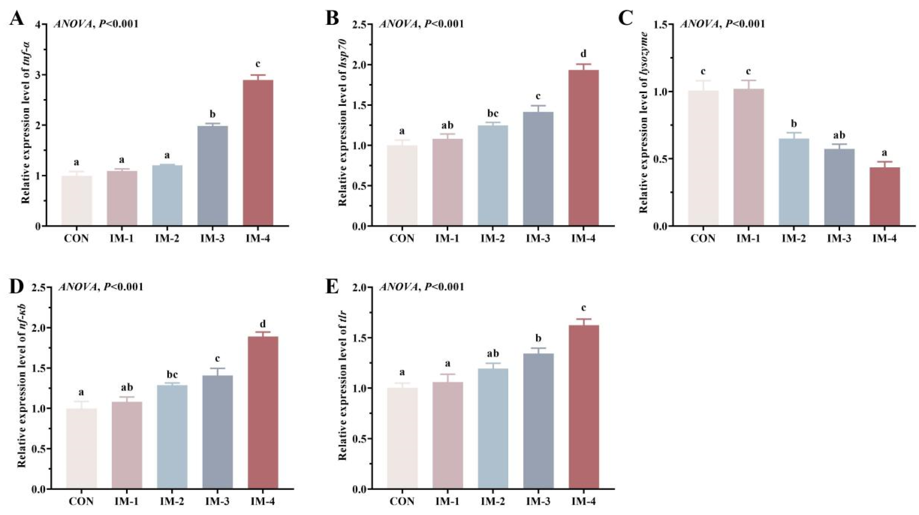
3.6. Expression of Intestinal Immune-Related Genes

As shown in Figure 7, compared with the CON group, intestinal expressions of tnf-α, hsp70, lysozyme, nf-κb, and tlr genes in the low-concentration group had no significant differences (ANOVA, p > 0.05), while expressions of hsp70 and nf-κb genes in the medium- and high-concentration groups were significantly up-regulated, and the expression of lysozyme gene was significantly down-regulated (ANOVA, p < 0.05). The expressions of tnf-α and tlr genes in the intestinal tract were significantly up-regulated under high IMI exposure (ANOVA, p < 0.05).

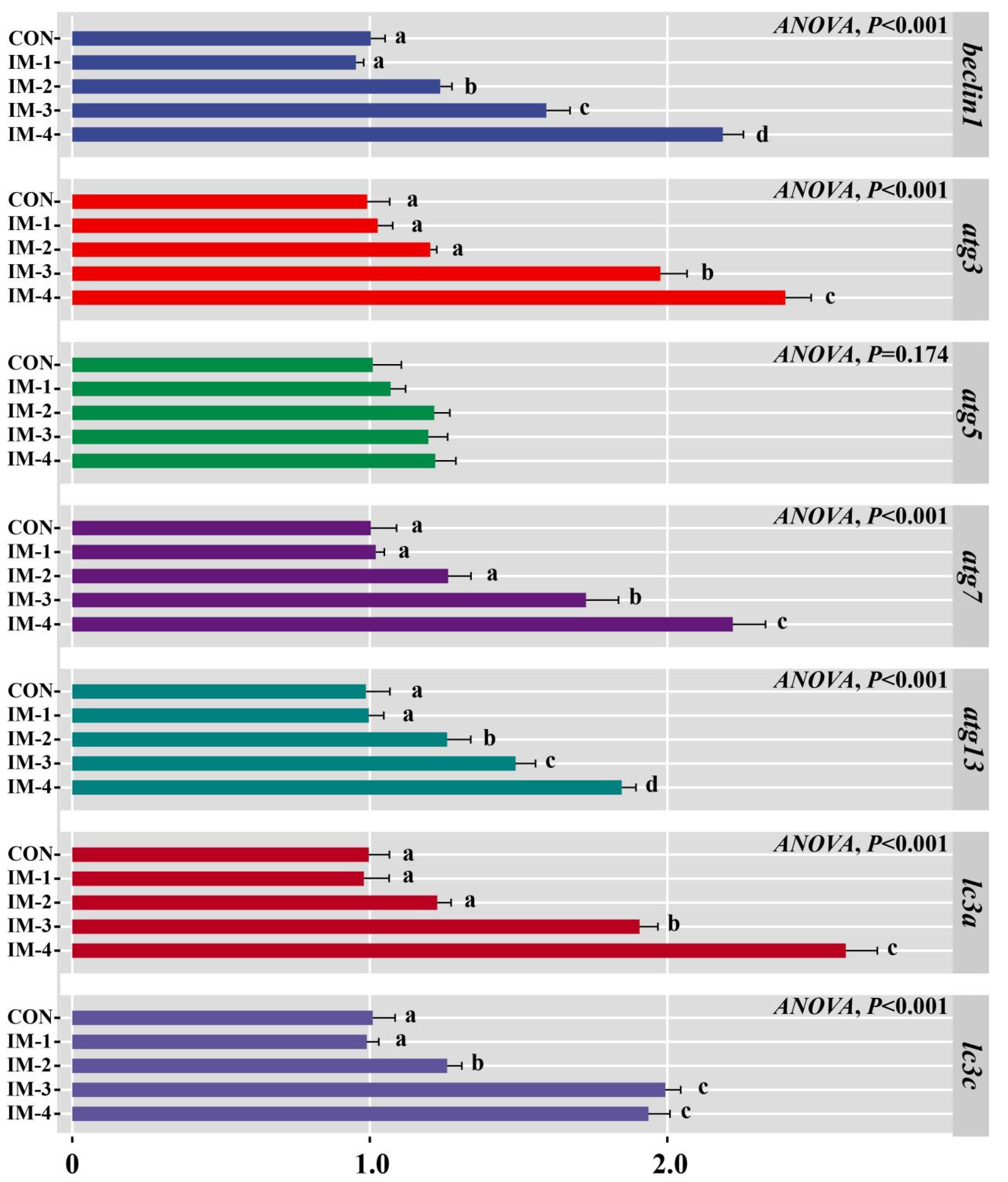
3.7. Expression of Intestinal Autophagy-Related Genes

As shown in Figure 8, compared with the CON group, intestinal beclin1, atg3, atg7, atg13, lc3a, and lc3c gene expressions in the low-concentration group had no significant differences (ANOVA, p > 0.05). The expressions of beclin1, atg5, atg13, and lc3c genes in the medium- and high-concentration groups were significantly up-regulated (ANOVA, p < 0.05). There were no significant differences in atg5 gene expression among all groups (ANOVA, p > 0.05).

4. Discussion

The present study is critical for understanding the heightened vulnerability of crustaceans to imidacloprid (IMI) exposure. Crustaceans are particularly susceptible due to factors such as the high water solubility and environmental stability of IMI, its unique damage mechanisms, the limited capacity for metabolic detoxification, and the presence of an open circulatory system [20,21,22]. Environmental monitoring studies worldwide have detected IMI concentrations ranging from 0.001 to 320 μg/L [3], indicating that aquatic organisms are routinely exposed to potentially harmful levels of this insecticide. In our preliminary experiments, we determined the 96 h LC₅₀ of IMI for P. clarkii to be 262.36 μg/L, a value that underscores the potential for environmental IMI residues to adversely affect survival. Comparative studies have reported 96 h LC₅₀ values of 352.66 μg/L for red claw crayfish (Cherax quadricarinatus) [22], 24,970 μg/L for Chinese mitten crab (Eriocheir sinensis) [23], and varying LC₅₀ values for different developmental stages of shrimp species, including 162.43 μg/L for Pacific white shrimp (Litopenaeus vannamei) [24] and 0.009, 5.0234, and 42.611 mg/L for post-larvae, juveniles, and adults of giant freshwater prawn (Macrobrachium rosenbergii), respectively [25]. These differences in tolerance among species and developmental stages highlight the complexity of IMI toxicity in crustaceans. By focusing on P. clarkii, our study not only fills a gap in current toxicological research but also provides valuable insights into the mechanisms underlying IMI-induced damage in crustaceans. This research is essential for informing risk assessments and developing environmental management strategies aimed at protecting these ecologically and economically important organisms.
Survival rate is the most intuitive index for observing the health status of aquatic animals [26]. In the formal experiment set up according to a preliminary experiment, the survival rate of P. clarkii decreased with the increase in IMI concentration and the increase in exposure time, and the survival rate of P. clarkii was less than 70% after 96 h of exposure at a high concentration (87.45 μg/L). Studies have shown that hemolymph biochemical indicators can indicate the health status of crustaceans [27]. In this study, hemolymph GOT and GPT activities were significantly increased under medium- and high-concentration exposure conditions in IMI. This is consistent with the findings in adult loach (Misgurnus anguillicaudatus) and P. clarkii [28,29]. GOT and GPT are enzymes that are typically abundant in the hepatopancreas and other tissues. The hepatopancreas is analogous to the liver in vertebrates and is crucial for detoxification and metabolism [30]. The reasons for the results of this study may be that medium and high levels of IMI can cause damage to the hepatopancreas, leading to the release of these enzymes into the hemolymph.
Oxidative stress is a condition characterized by an imbalance between the production of reactive oxygen species (ROS) and the organism’s antioxidant defenses [31]. In crustacean organisms, oxidative stress can damage proteins, lipids, and DNA, leading to impaired physiological functions [32]. Studies have shown that IMI can induce oxidative stress, leading to cellular damage in various aquatic species. For instance, research on grass carp (Ctenopharyngodon idella), neotropical fish (Prochilodus lineatus), common carp (Cyprinus carpio L.), and zebrafish has demonstrated that IMI exposure results in elevated ROS levels and lipid peroxidation, causing significant oxidative damage [16,33,34,35]. In this experiment, the activities of SOD, CAT, and GPX were significantly decreased, the contents of MDA were significantly increased, and the gene expressions of CuZnsod, mMnsod, cat, and gpx in the gut were significantly down-regulated after exposure to medium-high IMI, suggesting that medium-high concentrations of IMI caused damage to intestinal oxidative damage and reduced the antioxidant capacity of P. clarkii.
Both hemolymph LZM and AKP are important components of the immune system in crustaceans [36]. LZM acts as an antibacterial agent by breaking down bacterial cell walls, and AKP plays a role in dephosphorylation processes that are crucial for immune responses [37,38]. In this study, hemolymph LZM and AKP contents were significantly decreased at medium and high concentrations. This finding is consistent with that of Fu et al., who confirmed that IMI significantly inhibited AKP activities in Pacific white shrimp, which suggests that high levels of IMI can inhibit the activity of these enzymes, leading to reduced immune capabilities [24]. It was also found in the results of intestinal immune-related gene expression in this experiment that the expressions of nf-κb, hsp70, tnf-α, and tlr genes in the intestinal tract were significantly up-regulated, and the expression of lysozyme gene was significantly down-regulated under high IMI exposure. NF-κB is a transcription factor that plays a key role in regulating the immune response to infection and is regulated by upstream tlr signaling molecules [39]. Tnf-α is a pro-inflammatory cytokine involved in systemic inflammation and is part of the acute phase reaction [40]. Previous studies in grass carp have shown that IMI promotes inflammation through the NF-κB/Tlr pathway [35]. Studies have shown that hsp70 is involved in modulating immune responses [41]. Its down-regulation can weaken the immune system, making P. clarkii more susceptible to infections and other diseases, compounding the stress caused by imidacloprid exposure [42].
Autophagy is a vital process in maintaining cellular homeostasis and immune function, and it regulates apoptosis [43,44]. beclin1 is a core component of the autophagy initiation complex. It interacts with various proteins to promote the formation of autophagosomes [45]. Atg3, atg5, atg7, and atg13 play important roles in regulating the formation, extension, and activation of autophagosomes [46], and lc3a and lc3c mainly regulate the biogenesis and maturation of autophagy and promote the autophagy process [47]. In this acute toxicity test, the expressions of beclin1, atg5, atg13, and lc3c genes in the medium- and high-concentration groups were significantly up-regulated. These results indicated that autophagy-related genes were up-regulated by exposure to medium-high IMI, and autophagy was enhanced to degrade and recycle damaged proteins and organelles to reduce the damage. This is similar to the findings in grass carp, where exposure to IMI activates the JNK/BNIP3 pathway and promotes autophagy [35]. However, in some studies, exposure to IMI has been found to lead to inhibition of the autophagy pathway, thereby enhancing its toxic effects [24,47,48,49]. This may be related to exposure time and species.

5. Conclusions

In summary, this study elucidates that medium-high levels of IMI exposure impair intestinal immune function in P. clarkii through mechanisms involving oxidative stress, inflammatory response, and autophagy. The above results provide a basis for careful management and regulation of imidacloprid usage to protect aquatic ecosystems and the species that inhabit them.

Author Contributions

Validation, Y.S.; formal analysis, Z.L. and Y.S.; investigation, Z.L., Y.S., K.X., L.Z. and K.C.; data curation, Z.L.; writing—original draft, Z.L. and Y.S.; writing—review & editing, Y.H. and K.C.; supervision, Y.H.; project administration, Y.H. and K.C. All authors have read and agreed to the published version of the manuscript.

Funding

This study was supported by the Hunan Key Research and Development Program (No. 2022NK2030).

Institutional Review Board Statement

The animal subjects used in the experiment were fish. All the fish were handled in accordance with EU regulations on the protection of laboratory animals and approved by Animal Welfare of Hunan Agricultural University (protocol code 2023046, 11 May 2023).

Informed Consent Statement

Informed consent was obtained from all subjects involved in the study.

Data Availability Statement

The data that support the findings of this study are available upon reasonable request to the corresponding author.

Conflicts of Interest

The authors declare no conflicts of interest.

References

  1. Oficialdegui, F.J.; Clavero, M.; Sánchez, M.I.; Green, A.J.; Boyero, L.; Michot, T.C.; Klose, K.; Kawai, T.; Lejeusne, C. Unravelling the global invasion routes of a worldwide invader, the red swamp crayfish (Procambarus clarkii). Freshw. Biol. 2019, 64, 1382–1400. [Google Scholar] [CrossRef]
  2. Zhang, Y.; Li, Z.; Kholodkevich, S.; Sharov, A.; Chen, C.; Feng, Y.; Ren, N.; Sun, K. Effects of cadmium on intestinal histology and microbiota in freshwater crayfish (Procambarus clarkii). Chemosphere 2020, 242, 125105. [Google Scholar] [CrossRef] [PubMed]
  3. Morrissey, C.A.; Mineau, P.; Devries, J.H.; Sanchez-Bayo, F.; Liess, M.; Cavallaro, M.C.; Liber, K. Neonicotinoid contamination of global surface waters and associated risk to aquatic invertebrates: A review. Environ. Int. 2015, 74, 291–303. [Google Scholar] [CrossRef]
  4. Jeschke, P.; Nauen, R.; Schindler, M.; Elbert, A. Overview of the status and global strategy for neonicotinoids. J. Agric. Food Chem. 2011, 59, 2897–2908. [Google Scholar] [CrossRef]
  5. Tomizawa, M.; Zhang, N.; Durkin, K.A.; Olmstead, M.M.; Casida, J.E. The neonicotinoid electronegative pharmacophore plays the crucial role in the high affinity and selectivity for the Drosophila nicotinic receptor: An anomaly for the nicotinoid cation—π interaction model. Biochemistry 2003, 42, 7819–7827. [Google Scholar] [CrossRef]
  6. Kagabu, S. Studies on the synthesis and insecticidal activity of neonicotinoid compounds. J. Pestic. Sci. 1996, 21, 231–239. [Google Scholar] [CrossRef]
  7. Yamamoto, I.; Yabuta, G.; Tomizawa, M. Molecular mechanism for selective toxicity of nicotinoids and neonicotinoids. J. Pestic. Sci. 1995, 20, 33–40. [Google Scholar] [CrossRef]
  8. Shao, X.; Liu, Z.; Xu, X.; Li, Z.; Qian, X. Overall status of neonicotinoid insecticides in China: Production, application and innovation. J. Pestic. Sci. 2013, 38, 1–9. [Google Scholar] [CrossRef]
  9. Martelli, F.; Zhongyuan, Z.; Wang, J.; Wong, C.O.; Karagas, N.E.; Roessner, U.; Roessner, U.; Rupasinghe, T.; Venkatachalam, K.; Perry, T.; et al. Low doses of the neonicotinoid insecticide imidacloprid induce ROS triggering neurological and metabolic impairments in Drosophila. Proc. Natl. Acad. Sci. USA 2020, 117, 25840–25850. [Google Scholar] [CrossRef]
  10. Tišler, T.; Jemec, A.; Mozetič, B.; Trebše, P. Hazard identification of imidacloprid to aquatic environment. Chemosphere 2009, 76, 907–914. [Google Scholar] [CrossRef]
  11. Li, S.Q.; Yang, Q.H.; He, X.G.; Liu, Y.T.; Dong, J.; Yang, Y.B.; Xu, N.; Ai, X.H.; Liu, S.C. Enrichment and residual elimination of imidacloprid in integrated rice and Procambarus clarkii breeding model. J. Huazhong Agric. Univ. 2021, 40, 105–111. [Google Scholar]
  12. Xia, J.; Jin, C.; Pan, Z.; Sun, L.; Fu, Z.; Jin, Y. Chronic exposure to low concentrations of lead induces metabolic disorder and dysbiosis of the gut microbiota in mice. Sci. Total Environ. 2018, 631, 439–448. [Google Scholar] [CrossRef] [PubMed]
  13. Zhou, L.; Limbu, S.M.; Shen, M.; Zhai, W.; Qiao, F.; He, A.; Du, Z.; Zhang, M. Environmental concentrations of antibiotics impair zebrafish gut health. Environ. Pollut. 2018, 235, 245–254. [Google Scholar] [CrossRef]
  14. Jin, Y.; Wu, S.; Zeng, Z.; Fu, Z. Effects of environmental pollutants on gut microbiota. Environ. Pollut. 2017, 222, 1–9. [Google Scholar] [CrossRef]
  15. Shan, Y.; Yan, S.; Hong, X.; Zha, J.; Qin, J. Effect of imidacloprid on the behavior, antioxidant system, multixenobiotic resistance, and histopathology of Asian freshwater clams (Corbicula fluminea). Aquat. Toxicol. 2020, 218, 105333. [Google Scholar] [CrossRef]
  16. Luo, T.; Wang, X.; Jin, Y. Low concentrations of imidacloprid exposure induced gut toxicity in adult zebrafish (Danio rerio). Comp. Biochem. Physiol. Part C Toxicol. Pharmacol. 2021, 241, 108972. [Google Scholar] [CrossRef]
  17. Shi, Y.; Liu, Y.; Xie, K.; Zhang, J.; Wang, Y.; Hu, Y.; Zhong, L. Sanguinarine improves intestinal health in grass carp fed high-fat diets: Involvement of antioxidant, physical and immune barrier, and intestinal microbiota. Antioxidants 2023, 12, 1366. [Google Scholar] [CrossRef]
  18. Shi, Y.; Zhong, L.; Zhong, H.; Zhang, J.; Che, C.; Fu, G.; Hu, Y.; Mai, K. Taurine supplements in high-fat diets improve survival of juvenile Monopterus albus by reducing lipid deposition and intestinal damage. Aquaculture 2022, 547, 737431. [Google Scholar] [CrossRef]
  19. Shi, Y.; Zhong, L.; Liu, Y.; Xu, S.; Dai, J.; Zhang, Y.; Hu, Y. Dietary sanguinarine supplementation recovers the decrease in muscle quality and nutrient composition induced by high-fat diets of grass carp (Ctenopharyngodon idella). Anim. Nutr. 2024, 17, 208–219. [Google Scholar] [CrossRef]
  20. Sánchez-Bayo, F. Insecticides mode of action in relation to their toxicity to non-target organisms. J. Environ. Anal. Toxicol. 2012, 4, S4-002. [Google Scholar]
  21. Wang, X.; Li, E.; Xiong, Z.; Chen, K.; Yu, N.; Du, Z.; Chen, L. Low salinity decreases the tolerance to two pesticides, beta-cypermethrin and acephate, of white-leg shrimp, Litopenaeus vannamei. J. Aquac. Res. Dev. 2013, 4, 2. [Google Scholar] [CrossRef]
  22. Lu, Y.P.; Liu, J.H.; Zhang, X.X.; Xu, C.; Zheng, P.H.; Li, J.T.; Li, J.J.; Wang, D.M.; Xian, J.A.; Zhang, Z.L. Integration of transcriptome, gut microbiota, and physiology reveals toxic responses of the red claw crayfish (Cherax quadricarinatus) to imidacloprid. J. Hazard. Mater. 2024, 470, 134293. [Google Scholar] [CrossRef] [PubMed]
  23. Hong, Y.; Huang, Y.; Wu, S.; Yang, X.; Dong, Y.; Xu, D.; Huang, Z. Effects of imidacloprid on the oxidative stress, detoxification and gut microbiota of Chinese mitten crab, Eriocheir sinensis. Sci. Total Environ. 2020, 729, 138276. [Google Scholar] [CrossRef] [PubMed]
  24. Fu, Z.; Han, F.; Huang, K.; Zhang, J.; Qin, J.G.; Chen, L.; Li, E. Impact of imidacloprid exposure on the biochemical responses, transcriptome, gut microbiota and growth performance of the Pacific white shrimp Litopenaeus vannamei. J. Hazard. Mater. 2022, 424, 127513. [Google Scholar] [CrossRef]
  25. Rose, K.V.S.; Joseph, A. Acute toxicity of imidacloprid to various life stages of the giant freshwater prawn Macrobrachium rosenbergii, de Man, 1879. Saudi J. Life Sci. 2020, 5, 298–308. [Google Scholar] [CrossRef]
  26. Stoner, A.W. Assessing stress and predicting mortality in economically significant crustaceans. Rev. Fish. Sci. 2012, 20, 111–135. [Google Scholar] [CrossRef]
  27. Sladkova, S.V.; Kholodkevich, S.V. Total protein in hemolymph of crawfish Pontastacus leptodactylus as a parameter of the functional state of animals and a biomarker of quality of habitat. J. Evol. Biochem. Physiol. 2011, 47, 160–167. [Google Scholar] [CrossRef]
  28. Xia, X.; Xia, X.; Huo, W.; Dong, H.; Zhang, L.; Chang, Z. Toxic effects of imidacloprid on adult loach (Misgurnus anguillicaudatus). Environ. Toxicol. Pharmacol. 2016, 45, 132–139. [Google Scholar] [CrossRef]
  29. Yoloğlu, E.; Uçkun, M.; Alkan Uçkun, A.; Barım Öz, Ö. Biochemical responses of crayfish exposed to imidacloprid: Assessment of oxidative stress, osmoregulatory response and neurotoxicity. Chem. Ecol. 2025, 41, 50–68. [Google Scholar] [CrossRef]
  30. Yu, P.; Liu, Z.; Wu, D.; Chen, M.; Lv, W.; Zhao, Y. Accumulation of polystyrene microplastics in juvenile Eriocheir sinensis and oxidative stress effects in the liver. Aquat. Toxicol. 2018, 200, 28–36. [Google Scholar] [CrossRef]
  31. Bal, A.; Panda, F.; Pati, S.G.; Das, K.; Agrawal, P.K.; Paital, B. Modulation of physiological oxidative stress and antioxidant status by abiotic factors especially salinity in aquatic organisms. Comp. Biochem. Physiol. Part C Toxicol. Pharmacol. 2021, 241, 108971. [Google Scholar] [CrossRef] [PubMed]
  32. Frías-Espericueta, M.G.; Bautista-Covarrubias, J.C.; Osuna-Martínez, C.C.; Delgado-Alvarez, C.; Bojórquez, C.; Aguilar-Juárez, M.; Roos-Muñoz, S.; Osuna-López, I.; Páez-Osuna, F. Metals and oxidative stress in aquatic decapod crustaceans: A review with special reference to shrimp and crabs. Aquat. Toxicol. 2022, 242, 106024. [Google Scholar] [CrossRef] [PubMed]
  33. Vieira, C.E.D.; Pérez, M.R.; Acayaba, R.D.A.; Raimundo, C.C.M.; Martinez, C.B. DNA damage and oxidative stress induced by imidacloprid exposure in different tissues of the Neotropical fish Prochilodus lineatus. Chemosphere 2018, 195, 125–134. [Google Scholar] [CrossRef]
  34. Özdemir, S.; Altun, S.; Arslan, H. Imidacloprid exposure cause the histopathological changes, activation of TNF-α, iNOS, 8-OHdG biomarkers, and alteration of caspase 3, iNOS, CYP1A, MT1 gene expression levels in common carp (Cyprinus carpio L.). Toxicol. Rep. 2018, 5, 125–133. [Google Scholar] [CrossRef]
  35. Miao, Z.; Miao, Z.; Wang, S.; Wu, H.; Xu, S. Exposure to imidacloprid induce oxidative stress, mitochondrial dysfunction, inflammation, apoptosis and mitophagy via NF-kappaB/JNK pathway in grass carp hepatocytes. Fish Shellfish. Immunol. 2022, 120, 674–685. [Google Scholar] [CrossRef]
  36. Bao, J.; Li, X.; Xing, Y.; Feng, C.; Jiang, H. Effects of hypoxia on immune responses and carbohydrate metabolism in the Chinese mitten crab, Eriocheir sinensis. Aquac. Res. 2020, 51, 2735–2744. [Google Scholar] [CrossRef]
  37. Zhang, J.; Li, Y.; Zhang, X.; Zhou, B.; Sun, T.; Li, X. Physicochemical properties and antibacterial mechanism of TP microcapsules/LZM-PVA gradual sustained-release composite coatings. Prog. Org. Coat. 2020, 146, 105740. [Google Scholar] [CrossRef]
  38. Zhao, L.; Liang, J.; Liu, H.; Gong, C.; Huang, X.; Hu, Y.; Liu, Q.; He, Z.; Zhang Xi Yang, S.; Rahimnejad, S. Yinchenhao Decoction ameliorates the high-carbohydrate diet induced suppression of immune response in largemouth bass (Micropterus salmoides). Fish Shellfish. Immunol. 2022, 125, 141–151. [Google Scholar] [CrossRef]
  39. Carmody, R.J.; Chen, Y.H. Nuclear factor-kappaB: Activation and regulation during toll-like receptor signaling. Cell. Mol. Immunol. 2007, 4, 31–41. [Google Scholar]
  40. Ceciliani, F.; Giordano, A.; Spagnolo, V. The systemic reaction during inflammation: The acute-phase proteins. Protein Pept. Lett. 2002, 9, 211–223. [Google Scholar] [CrossRef]
  41. Borges, T.J.; Wieten, L.; Herwijnen, M.J.; Broere, F.; Zee, R.; Bonorino, C.; Eden, W. The anti-inflammatory mechanisms of Hsp70. Front. Immunol. 2012, 3, 95. [Google Scholar] [CrossRef] [PubMed]
  42. Hassan, F.U.; Nawaz, A.; Rehman, M.S.; Ali, M.A.; Dilshad, S.M.; Yang, C. Prospects of HSP70 as a genetic marker for thermo-tolerance and immuno-modulation in animals under climate change scenario. Anim. Nutr. 2019, 5, 340–350. [Google Scholar] [CrossRef] [PubMed]
  43. Opferman, J.T.; Korsmeyer, S.J. Apoptosis in the development and maintenance of the immune system. Nat. Immunol. 2003, 4, 410–415. [Google Scholar] [CrossRef] [PubMed]
  44. Clarke, A.J.; Simon, A.K. Autophagy in the renewal, differentiation and homeostasis of immune cells. Nat. Rev. Immunol. 2019, 19, 170–183. [Google Scholar] [CrossRef]
  45. Kang, R.; Zeh, H.J.; Lotze, M.T.; Tang, D.J.C.D. The Beclin 1 network regulates autophagy and apoptosis. Cell Death Differ. 2011, 18, 571–580. [Google Scholar] [CrossRef]
  46. Noda, N.N.; Inagaki, F. Mechanisms of autophagy. Annu. Rev. Biophys. 2015, 44, 101–122. [Google Scholar] [CrossRef]
  47. Schaaf, M.B.; Keulers, T.G.; Vooijs, M.A.; Rouschop, K.M. LC3/GABARAP family proteins: Autophagy-(un) related functions. FASEB J. 2016, 30, 3961–3978. [Google Scholar] [CrossRef]
  48. Li, X.; Yao, Y.; Wang, J.; Shen, Z.; Jiang, Z.; Xu, S. Eucalyptol relieves imidacloprid-induced autophagy through the miR-451/Cab39/AMPK axis in Ctenopharyngodon idellus kidney cells. Aquat. Toxicol. 2022, 249, 106204. [Google Scholar] [CrossRef]
  49. Eriksson, I.; Ward, L.J.; Vainikka, L.; Sultana, N.; Leanderson, P.; Flodin, U.; Li, W.; Yuan, X.M. Imidacloprid induces lysosomal dysfunction and cell death in human astrocytes and fibroblasts—Environmental implication of a clinical case report. Cells 2023, 12, 2772. [Google Scholar] [CrossRef]
Figure 1. The survival rate of P. clarkii in a preliminary experiment (mean ± SE, n = 3). Imidacloprid was administered at concentrations of 0, 50, 100, 200, 400, and 800 μg/L with three replicates per concentration and 20 P. clarkii per replicate. Survival rates were recorded at 12, 24, 36, 48, 60, 72, 84, and 96 h.
Fishes 10 00131 g001
Figure 2. Analysis of primary and quadratic regression equations for the mortality of P. clarkii in a preliminary experiment (mean ± SE, n = 3). (A) Quadratic regression equations; (B) Primary quadratic regression equations.
Fishes 10 00131 g002
Figure 3. Effects of imidacloprid (IMI) on survival rate of P. clarkii (mean ± SE, n = 3). Imidacloprid (IMI) was administered at concentrations of 0 (CON), 1/24 LC50 (IM-1), 1/12 LC50 (IM-2), 1/6 LC50 (IM-3), and 1/3 LC50 (IM-4), with three replicates per treatment group. Survival rates of P. clarkii were recorded at 24, 48, 72, and 96 h post-exposure.
Fishes 10 00131 g003
Figure 4. Effects of imidacloprid (IMI) on hemolymph biochemical and immune indexes of P. clarkii. (A) lysozyme (LZM, U/mL); (B) alkaline phosphatase (AKP, U/L); (C) glutamic oxalic aminotransferase (GOT, U/L); (D) glutamic pyruvic aminotransferase (GPT, U/L). CON, IM-1, IM-2, IM-3, and IM-4 represent imidacloprid (IMI) concentrations of 0, 1/24 LC₅₀, 1/12 LC₅₀, 1/6 LC₅₀, and 1/3 LC₅₀, respectively. Values (mean ± SE, n = 3) in the same row with different superscripts are significantly different (ANOVA, p < 0.05).
Fishes 10 00131 g004
Figure 5. Effects of imidacloprid (IMI) on intestinal antioxidant enzyme activity of P. clarkii. (A) malondialdehyde (MDA, nmol/mgprot); (B) glutathione peroxidase (GPx, U/mgprot); (C) superoxide dismutase (SOD, U/mgprot); (D) catalase (CAT, U/mgprot). CON, IM-1, IM-2, IM-3, and IM-4 represent imidacloprid (IMI) concentrations of 0, 1/24 LC₅₀, 1/12 LC₅₀, 1/6 LC₅₀, and 1/3 LC₅₀, respectively. Values (mean ± SE, n = 3) in the same row with different superscripts are significantly different (ANOVA, p < 0.05).
Fishes 10 00131 g005
Figure 6. Effects of imidacloprid (IMI) on intestinal antioxidation-related genes in P. clarkii. CON, IM-1, IM-2, IM-3, and IM-4 represent imidacloprid (IMI) concentrations of 0, 1/24 LC₅₀, 1/12 LC₅₀, 1/6 LC₅₀, and 1/3 LC₅₀, respectively. Values (mean ± SE, n = 3) in the same row with different superscripts are significantly different (ANOVA, p < 0.05).
Fishes 10 00131 g006
Figure 7. Effects of imidacloprid (IMI) on intestinal immune-related genes in P. clarkii. (A) tnf-α; (B) hsp70; (C) lysozyme; (D) nf-κb; (E) tlr. CON, IM-1, IM-2, IM-3, and IM-4 represent imidacloprid (IMI) concentrations of 0, 1/24 LC₅₀, 1/12 LC₅₀, 1/6 LC₅₀, and 1/3 LC₅₀, respectively. Values (mean ± SE, n = 3) in the same row with different superscripts are significantly different (ANOVA, p < 0.05).
Fishes 10 00131 g007
Figure 8. Effects of IMI on autophagy-related genes in intestinal cells of P. clarkii. CON, IM-1, IM-2, IM-3, and IM-4 represent imidacloprid (IMI) concentrations of 0, 1/24 LC₅₀, 1/12 LC₅₀, 1/6 LC₅₀, and 1/3 LC₅₀, respectively. Values (mean ± SE, n = 3) in the same row with different superscripts are significantly different (ANOVA, p < 0.05).
Fishes 10 00131 g008
Table 1. Sequences of primers.
GeneForward Primer Sequence (5′→3′)Reverse Primer Sequence (5′→3′)
CuZnsodACGGTGTATGGGCTGACTCCCGATGTAAGACTGGGACG
cMnsodCTGCAGCCAGTGTTGGAGTGAAAAGGGAATCAGACCGTGAGTGATC
mMnsodCCTGACCTGCCTTATGATTACATCTCCTCCATCTGGTGACAAA
catTTTCCCGTCTTTCATTCACACAAAATAAGAAAGTGGCTTGGTGT
gpxCCGCTCTTCACCTTCTTGGCGAGTGTATGGCTTACC
gstACCTGCCATATTACATTGACCCTTATCTTCTCTTGCTCTG
nrf2CATCTTCCAAAGCCTCCGAGGGGTCGTCTTTGCCTCTA
keap1bCCCCAAGAACTAAACCTCGGGCGAATAGCCTTGGGAGC
tnf-αCACCTTTCATCCCCTTCCATAATGCAGATGATAAAGCCCG
hsp70GGTGTTGGTGGGAGGGTCTAGGCTCGCTCTCCCTCATACAC
lysozymeGGACGTCCTCAGGAAAGGTGTTGTTAGTAGCGGCCGTGTT
nf-κbTAGTGCGTGATGATGGGTCTTGCTGATTATGGAGGCAGAAAA
beclin1AATCTGGCTCACTGTAAAGGAAACAACCAATCAGGAGGAAACT
atg3TGTTGTGAGGAGCGTCTGCTTGCCCTTGACCGTATTGA
atg5ACTGGATAGTGCCATTTTAGAGGGGCAGAAGATCGGGAGATTTTAC
atg7CTGCTTGGGTGTTACTTCACATCAGCGTCATTCAGG
atg13GTCAGCAAACATTCGGAGGGAGTGGGGCGACAACTGGTAC
lc3aAGGCACCTCCCTCTTCTGGCTGGAGACGCCGCCTTAT
lc3cGAGCGATACGCCAAAGAAACGGATGGTGACGAACTGGGAC
β-actinGAGGTTGCTGCCCTGGTTTAGCGGGAGTGTTGAAAG
Table 2. Real-time fluorescence quantitative PCR reaction system.
ReagentVolumeFinal Concentration
SYBR Green qPCR Mix10 μL
Forward primer sequence (10 μM)0.4 μL0.2 μM
Reverse primer sequence (10 μM)0.4 μL0.2 μM
DNA template× μL10~200 ng/20 μL
Nuclease-free waterTo 20 μL
Disclaimer/Publisher’s Note: The statements, opinions and data contained in all publications are solely those of the individual author(s) and contributor(s) and not of MDPI and/or the editor(s). MDPI and/or the editor(s) disclaim responsibility for any injury to people or property resulting from any ideas, methods, instructions or products referred to in the content.

