Article

Comparative Transcriptomic Analysis of Male and Female Gonads in the Zig-Zag Eel (Mastacembelus armatus)

1
Key Laboratory of Prevention and Control for Aquatic Invasive Alien Species, Ministry of Agriculture and Rural Affairs, Guangdong Engineering and Technology Research Center of Modern Recreational Fishery, Pearl River Fisheries Research Institute, Chinese Academy of Fishery Sciences, Guangzhou 510380, China
2
College of Fisheries and Life Science, Shanghai Ocean University, Shanghai 201306, China
3
Agricultural and Rural Bureau of Zengcheng District, Guangzhou 511300, China
4
Agro-Tech Extension Center of Guangdong Province, Guangzhou 510520, China
*
Authors to whom correspondence should be addressed.
Fishes 2025, 10(3), 117; https://doi.org/10.3390/fishes10030117
Submission received: 26 December 2024 / Revised: 4 March 2025 / Accepted: 4 March 2025 / Published: 6 March 2025
(This article belongs to the Special Issue Genetics and Breeding in Aquaculture)

Abstract

The zig-zag eel (Mastacembelus armatus) is a unique economic fish species in China and exhibits significant dimorphism of male and female phenotypes. Cultivating all-male seedlings can significantly improve production efficiency. To investigate sex differentiation and gonadal development in M. armatus, high-throughput sequencing technology was used to analyze the transcriptomes of male and female gonads at different developmental stages, both before and after sex differentiation. We identified key genes involved in sex differentiation, male-specific differentially expressed genes (DEGs), including dmrt1, amh, sox9, gsdf, and dmrt2b, and female-biased DEGs, including foxl2, rspo1, gdf9, bmp15, and wnt4. GO and KEGG enrichment analyses revealed that signaling pathways such as MAPK, Wnt, and TGF-β play significant roles in sex differentiation in M. armatus. The expression levels of 13 sex-related genes, including dmrt1, sox9, amh, foxl2, rspo1, and wnt4, were determined by RT–qPCR in addition to RNA sequencing. RT-qPCR validation results were consistent with the transcriptomic data, confirming the reliability of our findings. This research provides valuable insights into the mechanisms of sex differentiation in M. armatus and lays a foundation for developing all-male populations in aquaculture.
Key Contribution: This study identifies key genes involved in sex differentiation in M. armatus and highlights the critical signaling pathways regulating this process. The findings deepen our understanding of the molecular mechanisms underlying sex differentiation and provide a basis for the development of all-male populations in aquaculture.

1. Introduction

Sexual dimorphism refers to the differences between males and females of the same species, including traits such as body size, morphology, coloration, physiology, and behavior [1]. Such differences are common across numerous species and are particularly pronounced in fishes, where sexual size dimorphism (SSD) is frequently notable [2]. For instance, in species such as Nile tilapia (Oreochromis niloticus) [3], yellow catfish (Tachysurus fulvidraco) [4], and northern snakehead (Channa argus) [5], males generally grow faster than females. In contrast, females exhibit faster growth in species like common carp (Cyprinus carpio) [6], turbot (Scophthalmus maximus) [7], and Chinese tongue sole (Cynoglossus semilaevis) [8]. Modifying the sex ratio or producing monosex populations in aquaculture can significantly enhance growth rates and improve profitability, making these approaches valuable for optimizing production.
Transcriptome sequencing is a powerful technique that generates comprehensive sequences of mRNA transcripts from specific tissues or organs under controlled conditions, facilitating the identification of functional genes. This approach is particularly effective for investigating the molecular mechanisms underlying sex differentiation and gonadal development in fishes [9]. In fishes, gonadal development is regulated by various sex-related genes and signaling pathways. The transcriptomic analysis of gonads offers a comprehensive understanding of sex-specific gene expression profiles, enabling the identification of differentially expressed genes (DEGs) closely associated with sex differentiation. This analysis provides foundational insights into the regulatory networks involved in sex differentiation in gonadal development. For example, Tao et al. [10] conducted transcriptome sequencing on the gonads of Nile tilapia (Oreochromis niloticus) at various developmental stages from 5 to 180 days post-hatching (dph), revealing the relationship between gene expression patterns and the processes of sex differentiation and gonadal development. Their study found minimal differences in the transcriptomes from 5 to 20 dph, but significant differential gene expression emerged from 90 to 180 dph. Based on these results, key genes closely related to sex differentiation, such as cyp19a1a, foxl2, and amh, were identified. Similarly, Ribas et al. [11] carried out transcriptome analysis on 3-month-old turbot (Scophthalmus maximus), identifying 56 DEGs related to sex differentiation, including 44 genes associated with ovarian differentiation (e.g., cd98, gpd1, cry2) and 12 genes related to testicular differentiation (e.g., ace, capn8, nxph1). Fan et al. [12] performed transcriptome sequencing of the ovaries and testes of Chinese longsnout catfish (Leiocassis longirostris), screening 71 sex-related candidate genes. Among these genes, 50 were highly expressed in the testes (e.g., dmrt1, cyp17a1, samd7, wnt6, wt1) and 21 in the ovaries (e.g., foxl2, gdf9, zp3, zp1, figla, bmp15). Additionally, they identified 16 signaling pathways involved in gonadal development, including ovarian steroidogenesis, the TGF-β signaling pathway, and the GnRH signaling pathway.
Mastacembelus armatus, belonging to the order Synbranchiformes, family Mastacembelidae, and genus Mastacembelus [13], is a bottom-dwelling freshwater fish adapted to warm waters. It is mainly distributed in major river systems across Southeast Asia, particularly in tropical and subtropical regions [14]. The species has an elongated, eel-like body that is laterally compressed with a flattened tail. Its pointed snout features a tubular rostrum at the tip; although the oral cavity is small, the pharynx is wide, and the sharp teeth allow it to prey on small benthic aquatic animals such as shrimp and worms [15]. Owing to its tender meat and delicious taste, M. armatus is highly favored by consumers. However, in recent years, environmental pollution and overfishing have significantly reduced the population size of wild M. armatus. Consequently, in China, provinces such as Guangdong and Fujian have designated M. armatus as a protected wild aquatic species [16]. Moreover, large-scale aquaculture of M. armatus has not yet been successfully established. M. armatus has a male heterogametic sex determination system of XX/XY [17]. M. armatus exhibits notable sexual dimorphism, with males growing significantly faster than females [18]. This advantage makes males particularly valuable for aquaculture production, suggesting that cultivating all-male populations could further enhance production yields. However, in current aquaculture practices, the high female-to-male ratio hampers the development of the M. armatus industry. Additionally, the mechanisms of sex determination and differentiation are still not fully understood.
Therefore, this study utilized high-throughput sequencing technology to analyze the transcriptomic expression profiles of the ovaries and testes of M. armatus before and after gonadal differentiation, identifying differentially expressed genes related to sex differentiation. Additionally, several signaling pathways potentially involved in the sex differentiation of M. armatus were uncovered. In the future, gene editing (CRISPR/Cas9) technology can be further used to verify the function of key genes and explore methods of sex control. These findings provide valuable data for advancing our understanding of sex differentiation mechanisms and offer a basis for the targeted cultivation of all-male M. armatus populations in the future.

2. Materials and Methods

2.1. Sample Collection and Handling

The gonadal samples of M. armatus used in this experiment were sourced from Guangzhou Heshenghui Agricultural Technology Co., Ltd. (Guangzhou, China). All samples were collected from caudal fin tissue for DNA extraction and sex identification. PCR amplification was performed using sex-linked markers developed by Qin et al. to identify the genetic sex of M. armatus [19]. Pre-differentiation samples were derived from whole fish. At the pre-differentiation stage, due to the extremely small size of the gonads and the difficulty of avoiding contamination from surrounding tissues, we collected whole fish samples at 10 days post-hatch (dph) to ensure sufficient RNA for transcriptome sequencing and to capture transcriptomic information related to sex differentiation as comprehensively as possible. At the post-sex differentiation stage, three male and three female individuals, each three months old, were selected, and their gonadal tissues (testes and ovaries) were carefully dissected and collected. The fish were anesthetized with MS-222 (Sigma-Aldrich, St. Louis, MO, USA) before rapid dissection to remove the gonads. Each gonad was divided into two portions: one was immediately frozen in liquid nitrogen and stored at −80 °C for subsequent RNA extraction, and the other was fixed in 4% paraformaldehyde (Servicebio, Wuhan, China) for paraffin embedding and histological analysis.

2.2. Paraffin Sectioning of M. armatus Gonads

The gonadal tissues were kept in 4% paraformaldehyde for 24 h before being processed into paraffin sections for further examination. The paraffin sections underwent ethanol gradient dehydration, xylene transparency treatment, wax infiltration, and embedding. Continuous 5–6 μm sections were prepared and stained with hematoxylin–eosin (HE) (Servicebio, Wuhan, China). Finally, the sections were mounted with neutral resin, and the gonadal morphology was observed and photographed under a microscope.

2.3. Total RNA Extraction and Library Construction for Sequencing

Total RNA was extracted using the TRIzol reagent (Life Technologies, Foster City, CA, USA) according to the manufacturer’s protocol. RNA concentration and purity were assessed with a NanoDrop 2000 spectrophotometer (Thermo Fisher Scientific, Wilmington, DE, USA). This study employed Illumina RNA-seq technology for transcriptome sequencing, and the library construction was performed following the method described by Mortazavi et al. [20]. mRNA was isolated using poly-T oligo-conjugated magnetic beads, followed by cDNA synthesis using the Hieff NGS® Ultima Dual-mode mRNA Library Prep Kit for Illumina® (Yeasen Biotechnology, Shanghai, China) according to the manufacturer’s instructions. The products were then amplified by PCR and evaluated with an Agilent Bioanalyzer 2100 system (Agilent Technologies, Santa Clara, CA, USA) to assess their quality and integrity.

2.4. Transcriptome Sequencing and Analysis

The prepared libraries were sequenced on the Illumina NovaSeq platform (Yeasen Biotechnology, Shanghai, China), generating 150 bp paired-end reads. Raw reads were filtered with SOAPnuke (version 2.1.0) to remove low-quality sequences and adapters [21]. Data quality was evaluated based on Q20, Q30, and GC content. Hisat2 software (version 2.2.1) (Johns Hopkins University, Baltimore, MD, USA) was used to map with the reference genome. Differential gene expression between male and female gonads was analyzed using DESeq2 (version 1.36.0) [22], with gene expression levels quantified as FPKM (fragments per kilobase of exon model per million mapped reads) and fold change calculated as FPKMtestes/FPKMovaries. Genes with |log2 fold change| ≥ 1 and FDR (false discovery rate) < 0.05 were considered differentially expressed.
Gene Ontology (GO) enrichment analysis and KEGG pathway enrichment analysis of DEGs (differentially expressed genes) were performed using the clusterProfiler package in R (version 4.4.4) [23].

2.5. Real-Time Quantitative PCR (RT-qPCR) Validation

RT-qPCR was used to validate sex-related DEGs. Specific primers were designed using Primer Premier 5 (Premier, Vancouver, BC, Canada). All primers were synthesized by TianyiHuayu Gene Technology Co., Ltd. (Wuhan, China) (Table 1). Total RNA (1 μg) from male and female gonads was reverse-transcribed into cDNA using the PrimeScript RT Master Mix Kit (Takara, Shiga, Japan). The qPCR reactions were conducted on a 7500 Real-Time PCR system (ABI StepOnePlus, San Diego, CA, USA) using TB Green Fast qPCR Mix (Takara, Shiga, Japan). Each 20 μL reaction contained 10 μL of 2× TB Green Fast qPCR Mix, 0.4 μL of ROX Reference Dye II (50×), 0.8 μL each of the forward and reverse primers (10 µmol/L), 1 μL of cDNA template, and 7 μL of sterile water. The qPCR cycling conditions were as follows: 95 °C for 2 min, followed by 40 cycles of 95 °C for 15 s and 60 °C for 60 s. Each sample was tested in triplicate. Gene expression levels were calculated using the 2−ΔΔCt method for relative quantification. Statistical analyses were conducted using GraphPad Prism 9.5 (GraphPad Software, San Diego, CA, USA), with results presented as mean ± standard deviation (x ± SD).

3. Results and Analysis

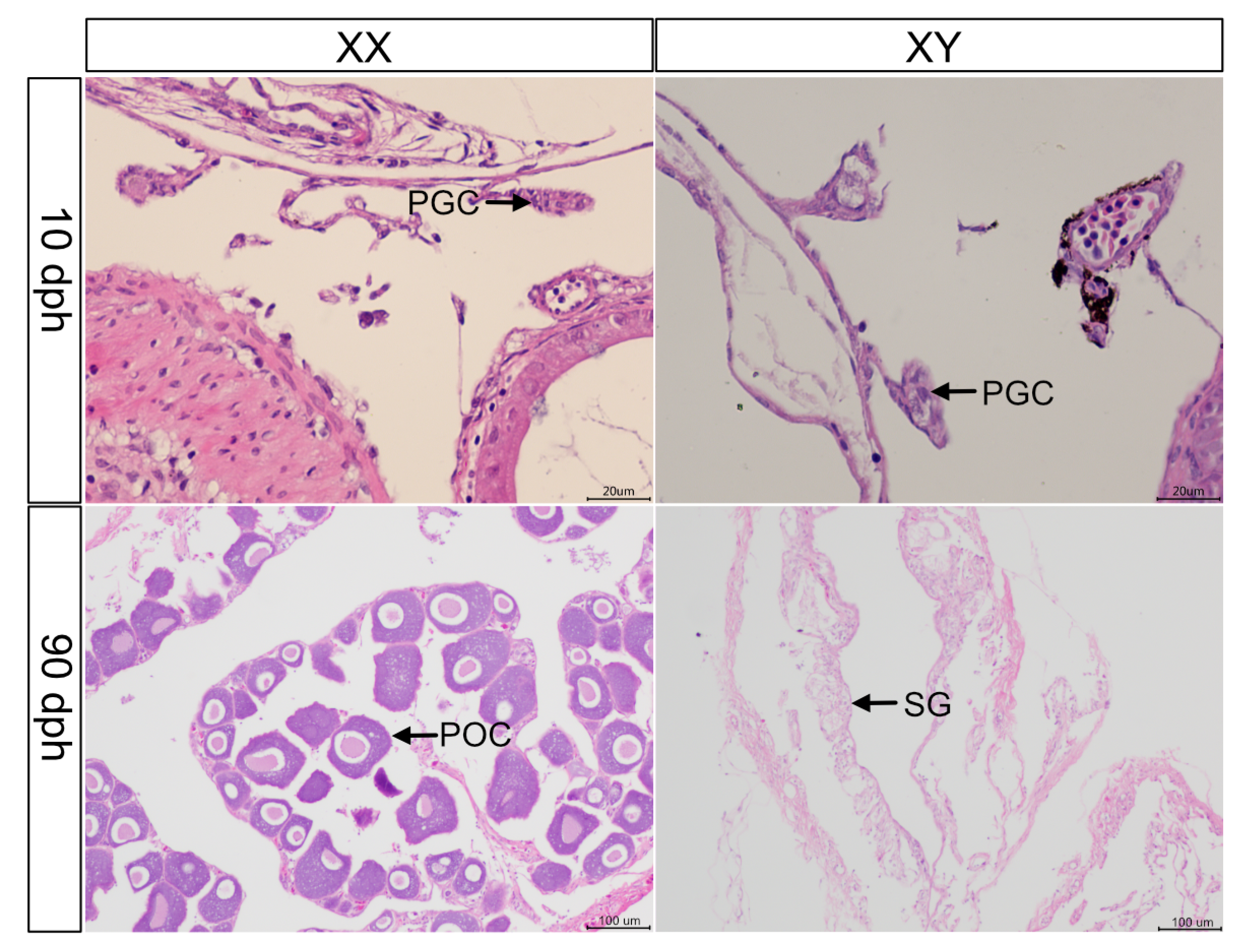
3.1. Histological Observation

Histological observations indicated that the gonads of the fish 10 days post-hatch (dph) were undifferentiated, presenting as primordial gonads containing primordial germ cells. Histological observations indicated that the gonads of three-month-old M. armatus contained differentiated cells (e.g., spermatogonia in males or primary oocyte in females), suggesting that sex differentiation had been completed (Figure 1). At 90 dph, male gonads were in developmental stage II, with elongated and branched seminiferous tubules containing spermatogonia. The spermatogonia nuclei were darkly stained, while the cytoplasm appeared more transparent. Similarly, female gonads were also in developmental stage II at 90 dph, primarily containing primary oocytes with large, centrally, or near centrally positioned nuclei that were darkly stained with prominent nucleoli. Follicular cells closely surrounded the irregularly shaped oocytes.

3.2. Transcriptome Sequencing Results

High-throughput sequencing was performed on 12 samples (3 male and 3 female samples before and after sex differentiation), yielding a total of 80.81 Gb of clean data. The clean data for each sample exceeded 6.14 Gb, with a Q20 base percentage above 98.60%, a Q30 base percentage above 96.00%, and GC content ranging from 44.79% to 48.59%, indicating a high level of data integrity (Table S1).
Clean reads from 12 samples were mapped to the reference genome (accession number: GCA_019455525.1) [17]. The overall mapping rates for all samples exceeded 84.40%, and the unique mapping rates were all above 70.54% (Table S2). These high mapping rates support accurate gene annotation for M. armatus gonadal samples.

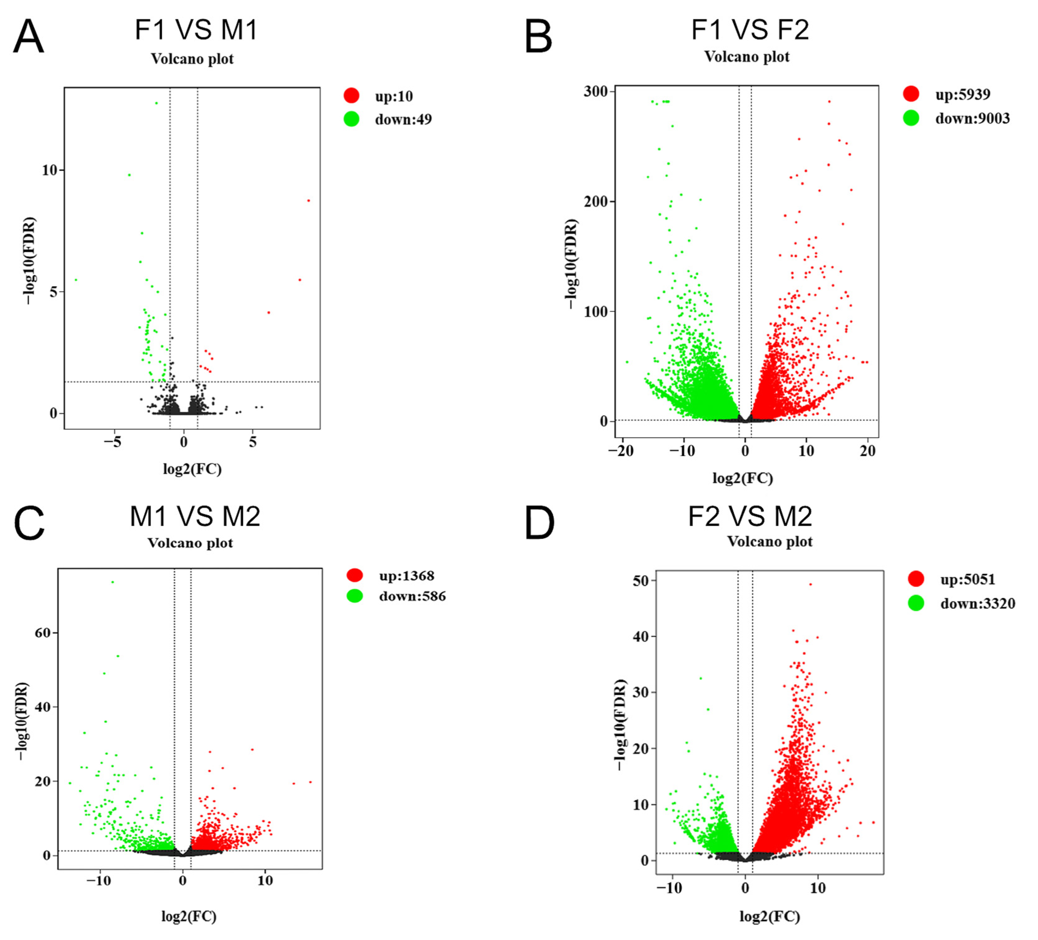
3.3. Differential Gene Expression Analysis

In the gonadal transcriptome of M. armatus, differential expression analysis was based on a threshold of |log2 fold change| ≥ 1 and FDR ≤ 0.05. M1/F1 represents males/females before sex differentiation, while M2/F2 represents males/females after sex differentiation. Compared to the F1 group, 10 genes were upregulated and 49 genes were downregulated in the M1 group (Figure 2A). Compared to the F1 group, 5939 genes were upregulated and 9003 genes were downregulated in the F2 group (Figure 2B). Compared to the M1 group, 1368 genes were upregulated and 586 genes were downregulated in the M2 group (Figure 2C). Lastly, the comparison of F2 vs. M2 showed that 5051 genes were upregulated and 3320 genes were downregulated in the M2 group (Figure 2D).

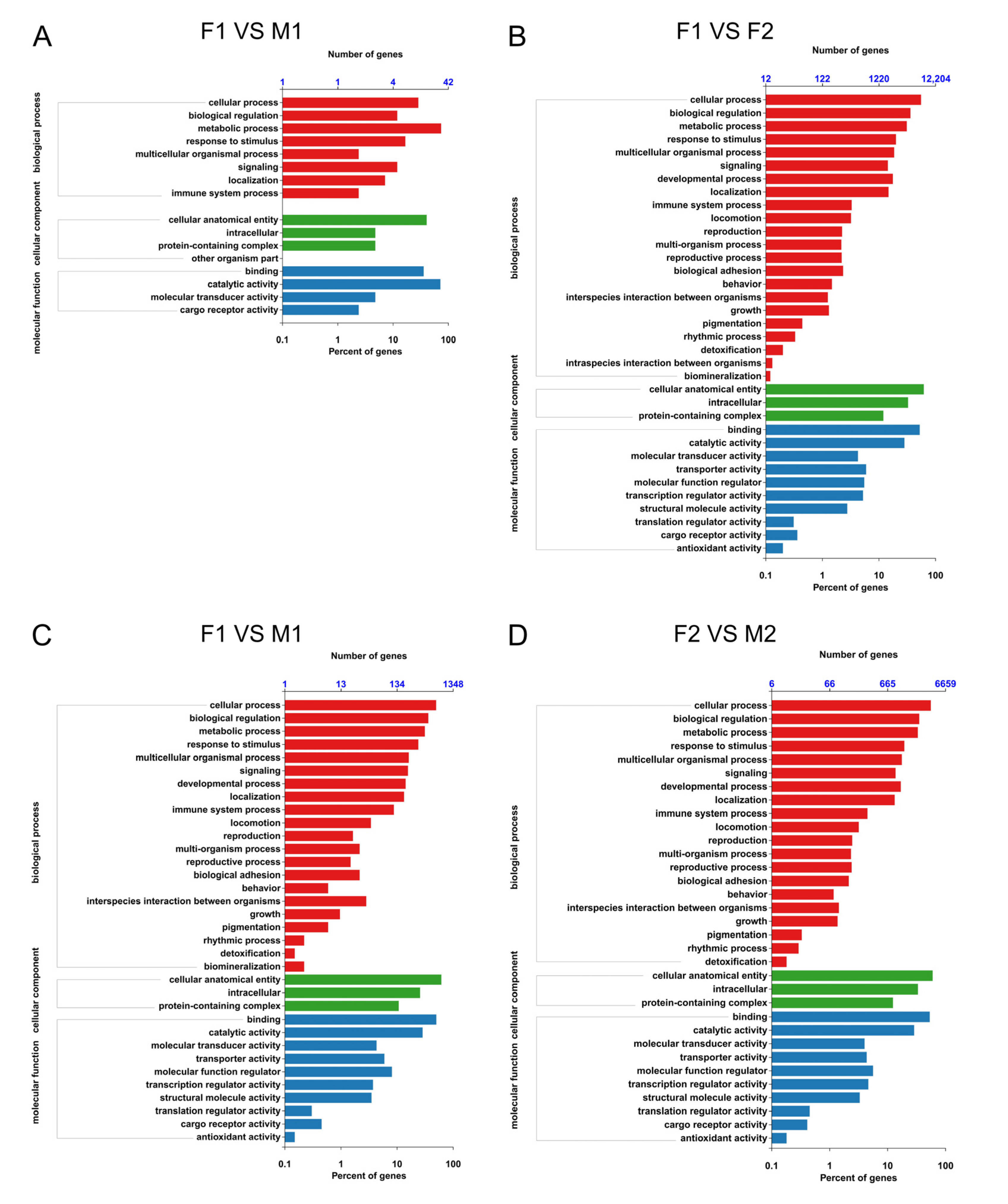
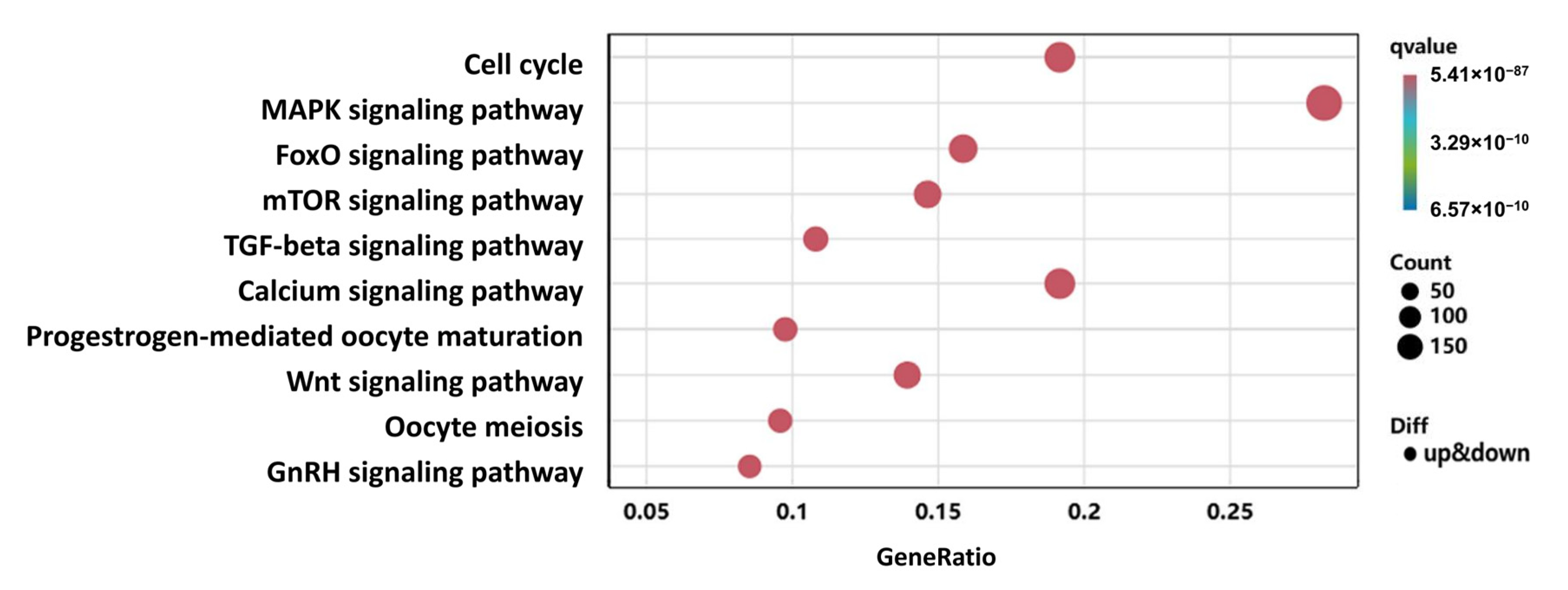
3.4. GO and KEGG Enrichment Analysis

The DEGs were mapped to the GO and KEGG databases for enrichment analyses. The results of the GO enrichment analyses are shown in Figure 3. Comparison of male and female samples prior to differentiation showed significant enrichment of the expression of genes involved in biological processes, such as cellular processes, biological regulation, and metabolic processes. Molecular functions such as binding and catalytic activity were prominently represented, highlighting key differences in transcriptional regulation and molecular interactions during the pre-differentiation phase (Figure 3A). A comparison of samples before and after female differentiation showed a notable upregulation of genes associated with developmental processes, multicellular organismal processes, and reproductive processes. Molecular functions related to protein binding, receptor activity, and transcription factor activity were particularly enriched, emphasizing molecular changes during ovarian differentiation (Figure 3B). A comparison of samples before and after male differentiation revealed significant changes in biological processes like reproductive processes, immune system processes, and cellular communication. Molecular functions such as catalytic activity, structural molecule activity, and transporter activity were enriched, reflecting the transcriptional shifts involved in testicular differentiation (Figure 3C). A comparison of male and female samples after differentiation showed prominent enrichment of the expression of genes involved in processes such as growth, developmental processes, and cellular processes. Molecular functions like signal transduction, transcription regulator activity, and antioxidant activity were differentially expressed, underlining the functional specialization of male and female gonads after differentiation (Figure 3D).
The results of the KEGG enrichment analyses are shown in Figure 4. A comparison of samples before and after male differentiation showed that the main pathways were categorized under metabolism, such as glycolysis/gluconeogenesis, fatty acid metabolism, and protein processing in the endoplasmic reticulum, along with signaling pathways such as the NOD-like receptor signaling pathway (Figure 4A). A comparison of male and female samples prior to differentiation showed that the main pathways were categorized under cellular processes, such as endocytosis, the regulation of the actin cytoskeleton, and the cell cycle, as well as apoptosis-related pathways like the autophagy–animal and p53 signaling pathway (Figure 4B). A comparison of samples before and after female differentiation showed that the main pathways were categorized under environmental information processing, including the MAPK signaling pathway, neuroactive ligand-receptor interaction, and calcium signaling pathway, as well as metabolic pathways like purine metabolism and pathways related to viral infection such as Herpes Simplex Virus 1 Infection (Figure 4C). A comparison of male and female samples after differentiation showed that the main pathways spanned multiple categories, including cellular processes such as focal adhesion and tight junction, environmental information processing like the MAPK signaling pathway, metabolic pathways such as amino acid metabolism, and disease-related pathways such as Herpes Simplex Virus 1 Infection (Figure 4D).
Among all of the identified pathways, 10 were found to be potentially related to sex differentiation and gonadal development, with 519 DEGs mapped to these pathways (Figure 5). The MAPK signaling pathway showed the highest enrichment, both in terms of DEG count and the enrichment ratio. Other sex-related pathways included the cell cycle, FoxO signaling, mTOR signaling, TGF-β signaling, Wnt signaling, oocyte meiosis, progesterone-mediated oocyte maturation, calcium signaling, and the GnRH signaling pathways.

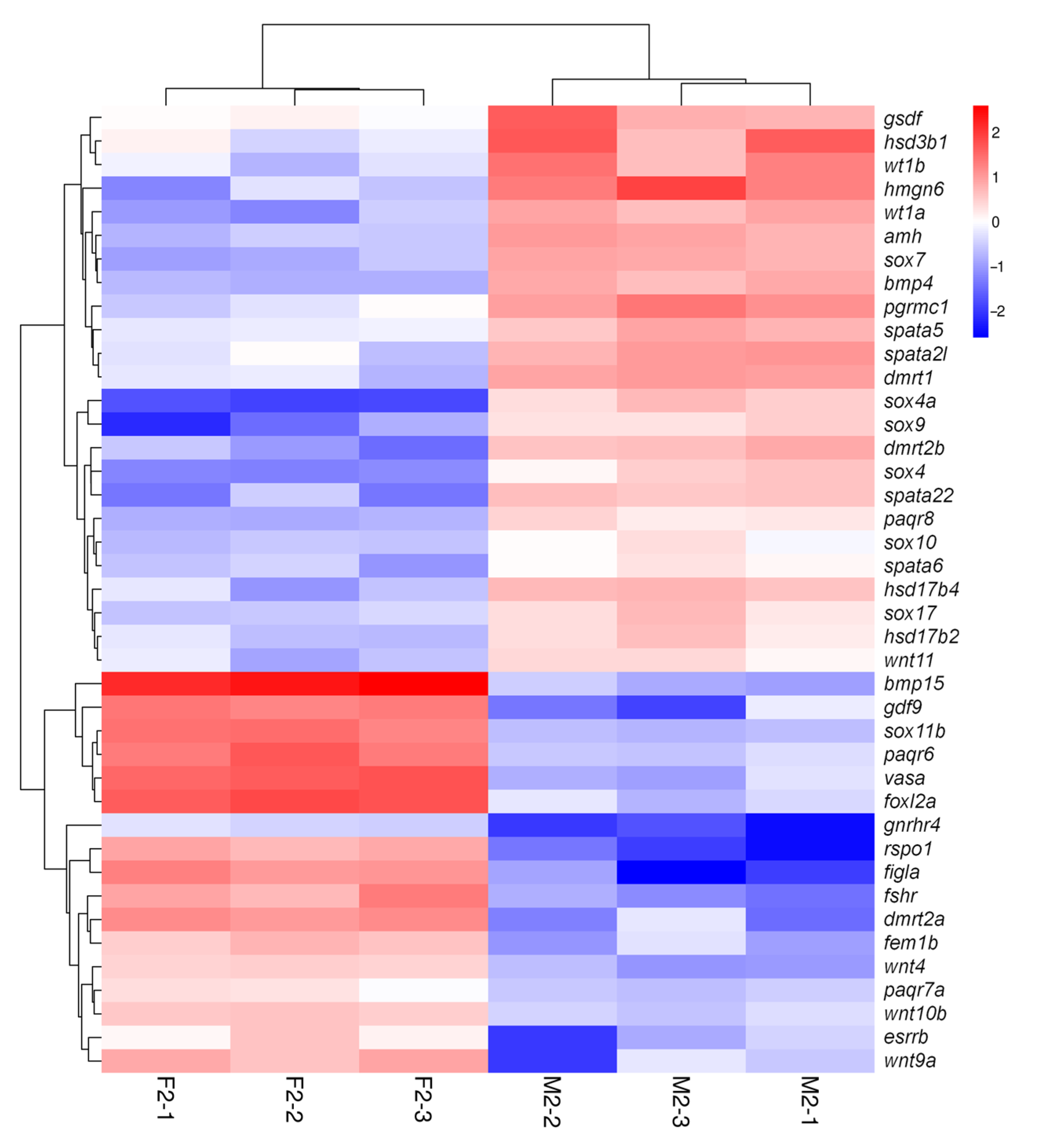
3.5. Screening and Analysis of Key Sex-Related DEGs

Through GO and KEGG enrichment analyses, we identified 41 key sex-related genes. These included classic sex-regulating gene families such as the DMRT gene family (dmrt1, dmrt2a, dmrt2b), the SOX gene family (sox4, sox11b, sox7, sox9a, sox10, sox17, sox4b), and the TGF-β superfamily (amh, gsdf, gdf9, bmp4, bmp15), as well as the 17β-hydroxysteroid dehydrogenase gene family (hsd3b1, hsd17b4, hsd17b12a). Additionally, genes related to reproduction and regulation were identified, including sex hormone receptors (fshr, pgrmc1, gnrhr4, paqr6, paqr8, paqr7a, esrrb), hormone regulatory factors (figla, fem1b, wt1a, wt1b), the Rspo1/Wnt/β-catenin signaling pathway (rspo1, wnt4, wnt9a, wnt10b, wnt11), the DEAD-box gene family (ddx4-vasa), genes involved in spermatogenesis (spata2l, spata5, spata6, spata22), and transcription factors such as foxl2.
The gene expression analysis of the aforementioned sex-related genes identified 24 genes highly expressed in the testes and 17 genes highly expressed in the ovaries; genes such as dmrt1, sox9, amh, and wt1 were predominantly expressed in the testes, whereas foxl2, rspo1, wnt4, gdf9, and bmp15 were significantly expressed in the ovaries (Figure 6).

3.6. RT-qPCR Validation

To validate the transcriptome sequencing results, 14 key genes related to sex differentiation were selected from the previously mentioned gene families and pathways for RT-qPCR analysis. The results showed that dmrt1, sox9, amh, wt1, bmp4, sox4, dmrt2b, and gsdf were highly expressed in the testes, while foxl2, rspo1, wnt4, gdf9, bmp15, and dmrt2a were highly expressed in the ovaries. These findings were consistent with the RNA-Seq data (Figure 7), confirming the reliability of the sequencing data and the accuracy of the DEG screening results.

4. Discussion

In this study, we conducted an in-depth analysis of the transcriptomes of male and female gonads in M. armatus before and after sex differentiation using high-throughput sequencing technology. Since the pre-differentiation samples were derived from whole fish tissues, the DEG analysis inevitably reflected overall gene expression differences. Therefore, we first manually filtered the DEGs to identify candidate sex-related genes and further compared them with the literature to select key genes with strong evidence supporting their roles in sex determination and gonadal development. Finally, we identified key sex-related genes and pathways involved in the sex differentiation of M. armatus.

4.1. Sex-Related DEGs in M. armatus

In fishes, gonadal development is regulated by complex, interconnected gene regulatory networks [24]. It is well known that dmrt1, sox9 and amh play pivotal roles in male gonadal development [25]. Dmrt1, a member of the highly conserved DMRT family of sex-regulating factors, is essential for initiating testicular differentiation [26], partly by promoting the expression of sox9 [27,28]. During this process, dmrt1 is highly expressed, activating sox9 and triggering downstream testicular development [29]. In females, sox9 induces amh expression, which suppresses the development of reproductive structures while activating genes involved in testicular differentiation and inhibiting ovarian differentiation pathways involving β-catenin [30]. Amh, a TGF-β superfamily gene, prevents Müllerian duct development and indirectly promotes male differentiation [31]. Mustapha’s study indicates that genes related to male gonadal development in spotted scat, Scatophagus argus, such as dmrt1, gsdf, and amh, are significantly more highly expressed in the testes [32], which is consistent with the findings of our study. In this study, prior to sex differentiation, although the male gonads had not fully differentiated morphologically, transcriptomic analysis revealed weak expression of male marker genes such as dmrt1 and sox9. As differentiation progressed, dmrt1, sox9, amh, and wt1 were predominantly expressed in the testes of M. armatus, playing critical roles in promoting testicular development. Additionally, in common carp (Cyprinus carpio), dmrt1 and sox9 not only promote male gonadal development but also inhibit the expression of female-specific genes such as foxl2, thereby facilitating male differentiation [33].However, further functional studies are needed to confirm the specific roles of these sex-related genes in the gonadal development and sex differentiation of M. armatus.
During female gonadal development, foxl2 is a key regulatory factor, functioning to suppress the expression of male-related genes while promoting ovary-specific gene expression [34,35]. Research on marble goby, Oxyeleotris marmorata, indicates that foxl2a was highly expressed during the female gonadal differentiation stage. It is suggested that these genes may play a role in oocyte polarity establishment, early oogenesis, and ovarian differentiation [36], which is consistent with the findings of this study. rspo1 and wnt4 are essential for ovarian differentiation, and the absence of the expression of either gene can result in sex reversal in Japanese rice fish (Oryzias latipes) [37]. In male medaka embryos, the overexpression of rspo1 can induce a transformation to female characteristics [38]. foxl2 upregulates rspo1 and wnt4, thereby activating the Wnt-β-catenin signaling pathway, which plays a key role in ovarian differentiation [39]. Wnt4 is vital for normal ovarian development. Once β-catenin is activated, it translocates to the nucleus, where it regulates genes involved in folliculogenesis and granulosa cell development [40,41,42]. Through the activation of the Rspo1-Wnt4 regulatory axis, foxl2 effectively prevents masculinization and supports normal ovarian development. In addition, foxl2 supports follicle development by regulating gdf9 and bmp15, which are important factors secreted by oocytes, to maintain ovarian function by regulating granulosa cell differentiation and follicle maturation [43,44]. In this study, prior to the onset of sex differentiation, although the morphological differentiation of female gonads was incomplete, transcriptomic analysis detected low-level expression of key female-specific genes, including foxl2, rspo1, and gdf9. As differentiation advanced, the expression levels of foxl2, rspo1, and wnt4 were markedly elevated in the ovaries, with bmp15 and gdf9 playing pivotal roles in regulating and promoting ovarian development. Nevertheless, further functional investigations are required to elucidate the precise roles of these sex-related genes in gonadal development and sex differentiation in M. armatus.

4.2. Role and Interaction of Signaling Pathways in Gonadal Development

KEGG enrichment analysis of DEGs related to sex differentiation and gonadal development in male and female gonads of M. armatus revealed several pathways involved in gonadal development. The MAPK signaling pathway is widely involved in cell proliferation and differentiation [45]. A previous study suggests that inhibiting the activity of p38 MAPKs in Brachymystax lenok leads to abnormal levels of gonadotropin-releasing hormone (GnRH), follicle-stimulating hormone (FSH), and estradiol, thereby affecting follicular development [46]. Further studies have also suggested that the MAPK pathway may be involved in regulating the development of female nuclei and the meiotic process of diploid crucian carp, regulating nuclear replication and chromosome doubling, thus playing a role in the process of gametogenesis [47]. The TGF-β signaling pathway regulates germ cell differentiation by activating Smad proteins and plays a role in sex differentiation during early gonadal development by promoting extracellular matrix remodeling and germ cell migration [48]. This pathway also interacts with other signaling pathways, such as the Wnt and MAPK pathways, to coordinate gonadal development. For example, cross-regulation between TGF-β and Wnt signaling is critical for ovarian development in mice, where these pathways collaborate to inhibit masculinization and support female gonadal differentiation [49].The GnRH (gonadotropin-releasing hormone) signaling pathway indirectly affects gonadal development by regulating hormone secretion within the hypothalamic–pituitary–gonadal axis [50]. GnRH stimulates the pituitary to release luteinizing hormone (LH) and follicle-stimulating hormone (FSH), which subsequently regulate sex hormone secretion, influencing processes such as follicle development, ovulation, and spermatogenesis [51]. The GnRH pathway also interacts with the progesterone-mediated oocyte maturation pathway to promote oocyte maturation and the ovulation process [52].
These signaling pathways involved in gonadal development interact closely by regulating cell proliferation, differentiation, and metabolism. Gaining insight into how these pathways interact is crucial for understanding gonadal development and sex differentiation mechanisms in M. armatus.
This study offers important resources for investigating sex differentiation mechanisms in M. armatus and lays the groundwork for developing all-male breeding populations. The findings also provide valuable insights for the conservation of M. armatus populations and have important implications for aquaculture practices aimed at improving breeding efficiency.

5. Conclusions

In this study, transcriptomic analysis was performed on male and female gonads before and after M. armatus differentiation. Key genes such as dmrt1, sox9, and amh were found to be essential for testicular development, while foxl2, rspo1, and wnt4 were identified as crucial for ovarian development. Furthermore, this study also identified that the MAPK, Wnt, and TGF-β signaling pathways are associated with sex differentiation. These results suggest that during sex differentiation, the activation of sex-specific genes and signaling pathways plays a crucial role in gonadal development. After sex differentiation, the aforementioned genes were significantly expressed in the different sexes, suggesting their crucial roles in the sex differentiation of M. armatus. RT-qPCR validation supported the RNA-seq findings, deepening our understanding of how these sex-related genes contribute to gonadal development and sex differentiation in M. armatus.

Supplementary Materials

The following supporting information can be downloaded at https://www.mdpi.com/article/10.3390/fishes10030117/s1: Table S1: Transcriptome sequencing data information; Table S2: Alignment results of male and female gonadal transcriptome data of M. armatus with the reference genome.

Author Contributions

Conceptualization, X.M. and Y.L.; methodology, F.C. and Y.W. (Yuanyuan Wang); software, Y.W. (Yuanyuan Wang); validation, F.C., C.L. and Y.W. (Yuanyuan Wang); formal analysis, F.C. and Y.W. (Yuanyuan Wang); investigation, F.C., Y.Y., Y.W. (Yuli Wu) and Z.J.; resources, Y.W. (Yuanyuan Wang); data curation, F.C. and J.S.; writing—original draft preparation, F.C.; writing—review and editing, F.C., Y.W. (Yuanyuan Wang), H.L., Y.Y., Z.J., J.S., C.L., Y.W. (Yuli Wu), X.M. and Y.L.; visualization, F.C. and Y.W. (Yuanyuan Wang); supervision, Y.L; project administration, X.M. and Y.W. (Yuanyuan Wang); funding acquisition, X.M. All authors have read and agreed to the published version of the manuscript.

Funding

This research was funded by the Central Public-Interest Scientific Institution Basal Research Fund, CAFS (NO.2023SJHX6), the Guangdong Agricultural Technology Service—Major Agricultural Technology Rural Promotion Project (2130106), China-ASEAN Maritime Cooperation Fund (CAMC-2018F), the Rural Revitalization Strategy Special Provincial Organization and Imple-mentation Project Funds (2023SBH00001), and the National Freshwater Genetic Resource Center (FGRC18537).

Institutional Review Board Statement

This study was conducted in accordance with ethical guidelines and received approval from the Laboratory Animal Ethics Committee of the Pearl River Fisheries Research Institute (code: LAEC-PRFRI-2024-02-01; date: 26 February 2024).

Informed Consent Statement

Not applicable.

Data Availability Statement

The data presented in this study are openly available in NCBI at UTL, reference number BioProject PRJNA1177591.

Conflicts of Interest

The authors declare no conflicts of interest.

References

  1. Mei, J.; Gui, J.F. Genetic basis and biotechnological manipulation of sexual dimorphism and sex determination in fish. Sci. China Life Sci. 2015, 58, 124–136. [Google Scholar] [CrossRef]
  2. Wang, H.P.; Gao, Z.X.; Rapp, D.; O’Bryant, P.; Yao, H.; Cao, X.J. Effects of temperature and genotype on sex determination and sexual size dimorphism of bluegill sunfish Lepomis macrochirus. Aquaculture 2014, 420, S64–S71. [Google Scholar] [CrossRef]
  3. Tao, W.J.; Zhu, X.; Cao, J.M.; Xiao, H.S.; Dong, J.J.; Kocher, T.D.; Lu, M.X.; Wang, D.S. Screening and characterization of sex-linked DNA markers in Mozambique tilapia (Oreochromis mossambicus). Aquaculture 2022, 557, 738331. [Google Scholar] [CrossRef]
  4. Wang, L.Y.; Qi, P.P.; Chen, M.; Yuan, Y.C.; Shen, Z.G.; Fan, Q.X. Effects of sex steroid hormones on sexual size dimorphism in yellow catfish (Tachysurus fulvidraco). Acta Hydrobiol. Sin. 2020, 44, 379–388. [Google Scholar] [CrossRef]
  5. Dai, S.; Chen, M.; Zheng, S.; Su, J.; Wu, J.; Han, L.; Zhou, C.; Zou, Y.; Wang, D.; Li, M. Sex-linked DNA marker screening and characterization in albino northern snakehead (Channa argus var.) via third-generation sequencing and pool resequencing. Aquaculture 2025, 594, 741449. [Google Scholar] [CrossRef]
  6. Zhai, G.; Shu, T.; Chen, K.; Lou, Q.; Jia, J.; Huang, J.; Shi, C.; Jin, X.; He, J.; Jiang, D. Successful production of an all-female common carp (Cyprinus carpio L.) population using cyp17a1-deficient neomale carp. Engineering 2021, 8, 181–189. [Google Scholar] [CrossRef]
  7. Li, X.Y.; Mei, J.; Ge, C.T.; Liu, X.L.; Gui, J.F. Sex determination mechanisms and sex control approaches in aquaculture animals. Sci. China Life Sci. 2022, 65, 1091–1122. [Google Scholar] [CrossRef]
  8. Wang, N.; Wang, R.; Wang, R.; Chen, S. Transcriptomics analysis revealing candidate networks and genes for the body size sexual dimorphism of Chinese tongue sole (Cynoglossus semilaevis). Funct. Integr. Genom. 2018, 18, 327–339. [Google Scholar] [CrossRef]
  9. Hrdlickova, R.; Toloue, M.; Tian, B. RNA-Seq methods for transcriptome analysis. Wiley Interdiscip. Rev. RNA 2017, 8, e1364. [Google Scholar] [CrossRef]
  10. Tao, W.; Chen, J.; Tan, D.; Yang, J.; Sun, L.; Wei, J.; Conte, M.A.; Kocher, T.D.; Wang, D. Transcriptome display during tilapia sex determination and differentiation as revealed by RNA-Seq analysis. BMC Genom. 2018, 19, 363. [Google Scholar] [CrossRef]
  11. Ribas, L.; Robledo, D.; Gómez-Tato, A.; Viñas, A.; Martínez, P.; Piferrer, F. Comprehensive transcriptomic analysis of the process of gonadal sex differentiation in the turbot (Scophthalmus maximus). Mol. Cell. Endocrinol. 2016, 422, 132–149. [Google Scholar] [CrossRef] [PubMed]
  12. Fan, J.H.; Ye, H.; Song, X.H.; Yue, H.M.; Huang, L.; Ruan, R.; Gao, W.H.; Li, C.J. Comparative transcriptomic analysis of male and female gonads of the Chinese longsnout catfish (Leiocassis longirostris). J. Fish. Sci. China 2024, 31, 129–143. [Google Scholar] [CrossRef]
  13. Nelson, J.S.; Grande, T.C.; Wilson, M.V. Fishes of the World; John Wiley & Sons: Hoboken, NJ, USA, 2016. [Google Scholar]
  14. Talwar, P.K.; Jhingran, A.G. Inland Fishes of India and Adjacent Countries; CRC Press: Boca Raton, FL, USA, 1991. [Google Scholar]
  15. Anand, S.; Gedam, A.; Sonawane, S. Structures associated with feeding in Mastacembelus armatus (Lacepede, 1800) from Kaigaon Toka (MS). J. Exp. Sci 2012, 3, 17–20. [Google Scholar] [CrossRef]
  16. Xue, L. Observation on the embryonic development of Mastacembelue armatus. Freshw. Fish. 2016, 44, 101–104. [Google Scholar] [CrossRef]
  17. Xue, L.; Gao, Y.; Wu, M.; Tian, T.; Fan, H.; Huang, Y.; Huang, Z.; Li, D.; Xu, L. Telomere-to-telomere assembly of a fish Y chromosome reveals the origin of a young sex chromosome pair. Genome Biol. 2021, 22, 203. [Google Scholar] [CrossRef]
  18. Zhou, H.Q.; Li, F.; Shu, H.; Zhong, D.M.; He, P.Y.; Huang, X.Q.; Chen, Z.K. Analysis on morphological indexes and discrimination of male and female Mastacembelus armatus. J. Guangdong Ocean Univ. 2019, 39, 1–6. [Google Scholar] [CrossRef]
  19. Qin, W.; Han, C.; Yang, J.; Yu, Z.; Feng, Y.; Wu, Y.; Lu, B.; Cui, M.; Shu, H. Development of genetic sex markers of zig-zag eel (Mastacembelus armatus) by a NGS method. Aquaculture 2023, 571, 739498. [Google Scholar] [CrossRef]
  20. Mortazavi, A.; Williams, B.A.; McCue, K.; Schaeffer, L.; Wold, B. Mapping and quantifying mammalian transcriptomes by RNA-Seq. Nat. Methods 2008, 5, 621–628. [Google Scholar] [CrossRef]
  21. Chen, Y.; Chen, Y.; Shi, C.; Huang, Z.; Zhang, Y.; Li, S.; Li, Y.; Ye, J.; Yu, C.; Li, Z. SOAPnuke: A MapReduce acceleration-supported software for integrated quality control and preprocessing of high-throughput sequencing data. Gigascience 2018, 7, gix120. [Google Scholar] [CrossRef]
  22. Niedziela, G.; Szabelska-Beręsewicz, A.; Zyprych-Walczak, J.; Graczyk, M. Application of edgeR and DESeq2 methods in plant experiments based on RNA-seq technology. Biom. Lett. 2022, 59, 127–139. [Google Scholar] [CrossRef]
  23. Young, M.D.; Wakefield, M.J.; Smyth, G.K.; Oshlack, A. Gene ontology analysis for RNA-seq: Accounting for selection bias. Genome Biol. 2010, 11, R14. [Google Scholar] [CrossRef] [PubMed]
  24. Guiguen, Y.; Fostier, A.; Herpin, A. Sex Determination and Differentiation in Fish: Genetic, Genomic, and Endocrine Aspects; John Wiley & Sons: Hoboken, NJ, USA, 2018. [Google Scholar]
  25. Matson, C.K.; Zarkower, D. Sex and the singular DM domain: Insights into sexual regulation, evolution and plasticity. Nat. Rev. Genet. 2012, 13, 163–174. [Google Scholar] [CrossRef] [PubMed]
  26. Lavery, R.; Chassot, A.-A.; Pauper, E.; Gregoire, E.P.; Klopfenstein, M.; de Rooij, D.G.; Mark, M.; Schedl, A.; Ghyselinck, N.B.; Chaboissier, M.-C. Testicular differentiation occurs in absence of R-spondin1 and Sox9 in mouse sex reversals. PLoS Genet. 2012, 8, e1003170. [Google Scholar] [CrossRef] [PubMed]
  27. Zarkower, D.; Murphy, M.W. DMRT1: An ancient sexual regulator required for human gonadogenesis. Sex. Dev. 2022, 16, 112–125. [Google Scholar] [CrossRef]
  28. Minkina, A.; Matson, C.K.; Lindeman, R.E.; Ghyselinck, N.B.; Bardwell, V.J.; Zarkower, D. DMRT1 protects male gonadal cells from retinoid-dependent sexual transdifferentiation. Dev. Cell 2014, 29, 511–520. [Google Scholar] [CrossRef]
  29. Wagner, S.; Whiteley, S.L.; Castelli, M.; Patel, H.R.; Deveson, I.W.; Blackburn, J.; Holleley, C.E.; Marshall Graves, J.A.; Georges, A. Gene expression of male pathway genes sox9 and amh during early sex differentiation in a reptile departs from the classical amniote model. BMC Genom. 2023, 24, 243. [Google Scholar] [CrossRef]
  30. Vining, B.; Ming, Z.; Bagheri-Fam, S.; Harley, V. Diverse regulation but conserved function: SOX9 in vertebrate sex determination. Genes 2021, 12, 486. [Google Scholar] [CrossRef]
  31. Zhou, Y.; Sun, W.; Cai, H.; Bao, H.; Zhang, Y.; Qian, G.; Ge, C. The role of anti-Müllerian hormone in testis differentiation reveals the significance of the TGF-β pathway in reptilian sex determination. Genetics 2019, 213, 1317–1327. [Google Scholar] [CrossRef]
  32. Mustapha, U.F.; Peng, Y.-X.; Huang, Y.-Q.; Assan, D.; Zhi, F.; Shi, G.; Huang, Y.; Li, G.-L.; Jiang, D.-N. Comparative transcriptome analysis of the differentiating gonads in Scatophagus argus. Front. Mar. Sci. 2022, 9, 962534. [Google Scholar] [CrossRef]
  33. Wang, M.; Chen, L.; Zhou, Z.; Xiao, J.; Chen, B.; Huang, P.; Li, C.; Xue, Y.; Liu, R.; Bai, Y. Comparative transcriptome analysis of early sexual differentiation in the male and female gonads of common carp (Cyprinus carpio). Aquaculture 2023, 563, 738984. [Google Scholar] [CrossRef]
  34. Uhlenhaut, N.H.; Jakob, S.; Anlag, K.; Eisenberger, T.; Sekido, R.; Kress, J.; Treier, A.-C.; Klugmann, C.; Klasen, C.; Holter, N.I. Somatic sex reprogramming of adult ovaries to testes by FOXL2 ablation. Cell 2009, 139, 1130–1142. [Google Scholar] [CrossRef]
  35. Georges, A.; Auguste, A.; Bessiere, L.; Vanet, A.; Todeschini, A.-L.; Veitia, R.A. FOXL2: A central transcription factor of the ovary. J. Mol. Endocrinol. 2014, 52, R17–R33. [Google Scholar] [CrossRef] [PubMed]
  36. Liu, W.; Zhang, H.; Xiang, Y.; Jia, K.; Luo, M.; Yi, M. A novel germline and somatic cell expression of two sexual differentiation genes, Dmrt1 and Foxl2 in marbled goby (Oxyeleotris marmorata). Aquaculture 2020, 516, 734619. [Google Scholar] [CrossRef]
  37. Zhou, L.; Charkraborty, T.; Yu, X.; Wu, L.; Liu, G.; Mohapatra, S.; Wang, D.; Nagahama, Y. R-spondins are involved in the ovarian differentiation in a teleost, medaka (Oryzias latipes). BMC Dev. Biol. 2012, 12, 36. [Google Scholar] [CrossRef] [PubMed]
  38. Tomizuka, K.; Horikoshi, K.; Kitada, R.; Sugawara, Y.; Iba, Y.; Kojima, A.; Yoshitome, A.; Yamawaki, K.; Amagai, M.; Inoue, A. R-spondin1 plays an essential role in ovarian development through positively regulating Wnt-4 signaling. Hum. Mol. Genet. 2008, 17, 1278–1291. [Google Scholar] [CrossRef]
  39. Nicol, B.; Estermann, M.A.; Yao, H.H.; Mellouk, N. Becoming female: Ovarian differentiation from an evolutionary perspective. Front. Cell Dev. Biol. 2022, 10, 944776. [Google Scholar] [CrossRef]
  40. Kim, Y.; Kobayashi, A.; Sekido, R.; DiNapoli, L.; Brennan, J.; Chaboissier, M.-C.; Poulat, F.; Behringer, R.R.; Lovell-Badge, R.; Capel, B. Fgf9 and Wnt4 act as antagonistic signals to regulate mammalian sex determination. PLoS Biol. 2006, 4, e187. [Google Scholar] [CrossRef]
  41. Kocer, A.; Pinheiro, I.; Pannetier, M.; Renault, L.; Parma, P.; Radi, O.; Kim, K.-A.; Camerino, G.; Pailhoux, E. R-spondin1 and FOXL2 act into two distinct cellular types during goat ovarian differentiation. BMC Dev. Biol. 2008, 8, 36. [Google Scholar] [CrossRef]
  42. Cederroth, C.R.; Pitetti, J.L.; Papaioannou, M.D.; Nef, S. Genetic programs that regulate testicular and ovarian development. Mol. Cell. Endocrinol. 2007, 265, 3–9. [Google Scholar] [CrossRef]
  43. Chen, W.; Liu, L.; Ge, W. Expression analysis of growth differentiation factor 9 (Gdf9/gdf9), anti-Müllerian hormone (Amh/amh) and aromatase (Cyp19a1a/cyp19a1a) during gonadal differentiation of the zebrafish, Danio rerio. Biol. Reprod. 2017, 96, 401–413. [Google Scholar] [CrossRef]
  44. Halm, S.; Ibáñez, A.J.; Tyler, C.R.; Prat, F. Molecular characterisation of growth differentiation factor 9 (gdf9) and bone morphogenetic protein 15 (bmp15) and their patterns of gene expression during the ovarian reproductive cycle in the European sea bass. Mol. Cell. Endocrinol. 2008, 291, 95–103. [Google Scholar] [CrossRef] [PubMed]
  45. Lu, C. Molecular Regulation Mechanism of MKPs Toward MAPKs in the MAPK Pathway; Tsinghua University: Beijing, China, 2018. [Google Scholar]
  46. Huang, T.; Gu, W.; Liu, E.; Zhang, L.; Dong, F.; He, X.; Jiao, W.; Li, C.; Wang, B.; Xu, G. Screening and validation of p38 MAPK involved in ovarian development of Brachymystax lenok. Front. Vet. Sci. 2022, 9, 752521. [Google Scholar] [CrossRef] [PubMed]
  47. Wang, Z.L.; Luo, Y.R.; Luo, Z.W.; Wang, J.; Xiao, Y.M. Expression of MAPK in the Gonadal Tissue of Different Reproductive Characteristics of the Hybrid Crucian Carp. Life Sci. Res. 2016, 20, 95–101. [Google Scholar]
  48. Ten Dijke, P.; Goumans, M.J.; Itoh, F.; Itoh, S. Regulation of cell proliferation by Smad proteins. J. Cell. Physiol. 2002, 191, 1–16. [Google Scholar] [CrossRef]
  49. Shi, Y.; Massagué, J. Mechanisms of TGF-β signaling from cell membrane to the nucleus. Cell 2003, 113, 685–700. [Google Scholar] [CrossRef]
  50. Sower, S.A. Landmark discoveries in elucidating the origins of the hypothalamic-pituitary system from the perspective of a basal vertebrate, sea lamprey. Gen. Comp. Endocrinol. 2018, 264, 3–15. [Google Scholar] [CrossRef]
  51. Stamatiades, G.A.; Carroll, R.S.; Kaiser, U.B. GnRH—A key regulator of FSH. Endocrinology 2019, 160, 57–67. [Google Scholar] [CrossRef]
  52. Hu, K.L.; Chen, Z.M.; Li, X.X.; Cai, E.; Yang, H.Y.; Chen, Y.; Wang, C.Y.; Ju, L.P.; Deng, W.H.; Mu, L.S. Advances in clinical applications of kisspeptin-GnRH pathway in female reproduction. Reprod. Biol. Endocrinol. 2022, 20, 81. [Google Scholar] [CrossRef]
Figure 1. HE staining of paraffin sections from gonads of Mastacembelus armatus. PGC: primordial germ cell; POC: primary oocyte; SG: spermatogonia.
Fishes 10 00117 g001
Figure 2. Volcano plot of differentially expressed genes. (A) F1 vs. M1; (B) F1 vs. F2; (C) M1 vs. M2; (D) F2 vs. M2. F represents female; M represents male; 1/2 denotes before or after sex differentiation, respectively. The horizontal dashed line represents log10 (FDR = 0.05), and the vertical dashed line represents |log2 fold change| = 1. Black dots represent unchanged genes.
Fishes 10 00117 g002
Figure 3. GO enrichment analyses of differentially expressed genes in the gonads of M. armatus. (A) F1 vs. M1; (B) F1 vs. F2; (C) M1 vs. M2; (D) F2 vs. M2. F represents female; M represents male; 1/2 denotes before or after sex differentiation, respectively.
Fishes 10 00117 g003
Figure 4. KEGG enrichment analyses of differentially expressed genes in the gonads of M. armatus. (A) F1 vs. M1; (B) F1 vs. F2; (C) M1 vs. M2; (D) F2 vs. M2. F represents female; M represents male; 1/2 denotes before or after sex differentiation, respectively.
Fishes 10 00117 g004
Figure 5. KEGG pathways related to sex differentiation and gonadal development in M. armatus.
Fishes 10 00117 g005
Figure 6. Heatmap of results of clustering analysis of sex-related genes in the gonads of M. armatus. F and M represent female and male, respectively. Numbers (1-1/2/3) refer to individuals before differentiation, and (2-1/2/3) refer to individuals after differentiation.
Fishes 10 00117 g006
Figure 7. RT-qPCR validation of 14 key sex-related genes in M. armatus. Fold change = relative expression of male/relative expression of female.
Fishes 10 00117 g007
Table 1. Primer sequences used for real-time quantitative PCR.
GeneSequences From (5′-3′)GeneSequences From (5′-3′)
dmrt1F:CGGCCCAGGTTGCCTTGAGfoxl2F:TCCGTCCCAGAAACCACCGTAT
R:CCAGCTTCATTCTTCACCATCAR:CCTGATGCTGTTCTGCCAACCT
sox9F:GAAGGACGAGGACGATAAGTTrspo1F:AAAGGCTCACAATCTCGG
R:TGGCATAGGCACGAGGGTR:CCTCCTCTACTGCCATCC
amhF:TCTGGCACTCAGCTTATCCwnt4F:TGGGCAACATCATCAAGG
R:CCATCTCCTCCTCCCTTAR:TGGATCATAGTCGCAGAAA
wt1F:TGCGTTCACCGTCCACTTgdf9F:ATCTACACCTGCTCATCAA
R:CGACCGTGCTGTAACCTGR:ACTGACTACTGAACCCTGAT
bmp4F:GTATCGGCTACAGTCAGGGbmp15F:GCAGAAAGCGGACCAGAA
R:ATCTTCGGGAATGGTGCTR:CGAGGGAAGAGTGTCAAGC
sox4F:GGGACTTGGATTTGAACTTTGdmrt2aF:CGGGAATACAAAGAACGAGA
R:TCGCTCACTTCGGGCGTAR:CGCTGACATTGGAGGAGAT
dmrt2bF:AACCAGGGAGGATAAGGAβ-actinF:TCATGAGGTAGTCTGTGAGGTCCC
R:GCTGACGTGCTATTTGAGTR:GCCTCTGGTCGTACCACTGGTATT
gsdfF:TCCAAGGAAGAACCTGCAACCT
R:CAGGCATCCATGGCTCAGACTC
Disclaimer/Publisher’s Note: The statements, opinions and data contained in all publications are solely those of the individual author(s) and contributor(s) and not of MDPI and/or the editor(s). MDPI and/or the editor(s) disclaim responsibility for any injury to people or property resulting from any ideas, methods, instructions or products referred to in the content.

