Article

Heat Shock Factors in the European Eel: Gene Characterization and Expression Response to Different Environmental Conditions and to Induced Sexual Maturation

Grupo de Acuicultura y Biodiversidad, Instituto de Ciencia y Tecnología Animal, Universitat Politècnica de València, Camino de Vera s/n, 46022 Valencia, Spain
*
Author to whom correspondence should be addressed.
Fishes 2025, 10(2), 73; https://doi.org/10.3390/fishes10020073
Submission received: 6 January 2025 / Revised: 8 February 2025 / Accepted: 9 February 2025 / Published: 12 February 2025

Abstract

:
Heat shock factors (HSFs) are transcription factors that regulate responses to environmental changes and play roles in physiological mechanisms like spermatogenesis. This study analyzed the HSF gene family and their expression profiles in the European eel under different environmental conditions and during testis maturation. Six HSF genes were identified in the studied vertebrates, in which the eel presents two HSF1 paralogs (HSF1a and HSF1b), likely resulting from the teleost whole-genome duplication event, while only one paralog is present for the other HSF (HSF2, HSF4, and HSF5). All five HSF genes were highly expressed in the testis, but some were also detected in the brain, intestine, and gills. Our findings showed that HSF1 changed their expression in response to different temperature and salinity conditions, suggesting that these may support males in perceiving the temperature and salinity changes possibly found during reproductive migration. During hCGrec-induced spermatogenesis, HSF genes presented a decreasing expression profile throughout testis maturation (with significant differences in HSF1a and HSF4), except HSF5, which showed the highest levels after 4 weeks of hormonal treatment. Our study indicates that HSF genes are potentially implicated in the response to environmental changes perception and during gonadal maturation.
Key Contribution: Six HSF genes were identified in vertebrates, with five found in the European eel and characterized for the first time in a teleost species. The eel served as a valuable model for analyzing HSF expression changes across environmental conditions changes and spermatogenesis stages.

1. Introduction

Heat shock factors (HSFs) are a family of DNA-binding proteins that are widely conserved across an extensive range of organisms, from fungi to humans [1]. These genes play a crucial role in regulating gene expression, primarily at the level of transcription, and are able to recognize specific DNA sequences and activate genes (e.g., heat shock proteins) to ensure proteostasis and to modulate the cellular response to different stressors [2].
In eukaryotes, only one single HSF has been identified, while in vertebrates, five distinct genes have been described, including HSF1 and HSF2 [3], whereas HSF3 has been identified in mice, some birds, reptiles, and amphibians [4]. HSF4 is present in mammals, zebrafish (Danio rerio) [5], and most recently, it has been identified in the Chinese tongue sole (Cynoglossus semilaevis) [6], the channel catfish (Ictalurus punctatus) [7], and in the Nile tilapia (Oreochromis niloticus) [8]. The fifth identified HSF member, HSF5, has been characterized in zebrafish [9] and mice [10,11].
Although HSFs are traditionally associated with a heat response, they also respond to other stressors aside from temperature, such as changes in salinity [12] and oxidative stress [13]. Beyond the stress response, HSF genes participate in gametogenesis, including male gonad development and spermatogenesis [2]. In mammals, the first member discovered, HSF1, protects early spermatogonia cells from heat shock, but it also mediates physiological functions by acting as a proapoptotic factor to eliminate injured male germ cells under stress [14,15]. A similar pattern of expression was observed in HSF2, but it has been demonstrated that HSF2 expression begins in spermatogonia and is upregulated until the differentiation process [16]. Indeed, HSF2 knockdown has been correlated with reduced testis size, defective spermatogenesis and spermiogenesis [17]. Both HSF1 and HSF2 participate in chromatin organization during spermatogenesis under non-stressful conditions, indicating their complementary roles during spermatogenesis [18,19]. The disruption of both genes through double knockout in mice resulted in the reduction of dividing spermatogonia, arrest of spermatogenesis at the first meiotic division and apoptosis [20].
In the case of HSF3, it is known that its potential to induce heat shock protein expression is absent in mice, and it is considered a pseudogene in humans [21]. However, both HSF1 and HSF3 regulate heat shock response cooperatively in lizards [22]. Until now, the HSF4 role in mammalian eye development is the most documented function, although there is evidence of its interaction with non-hsp genes [23,24]. Meanwhile, the fifth discovered member, HSF5, was characterized using the zebrafish as a model and proved to be essential for spermatogenesis with predominant expression within the zebrafish [9] and mouse testis [11]. The males with HSF5 knockdown showed increased expression of apoptotic genes and downregulation of genes related to cell cycle and signal transduction in the zebrafish [9]. In HSF5 knockout in mice, males were sterile and failed to undergo the meiotic sex chromosome inactivation that is essential for crossing the pachytene checkpoint in the maintenance of normal germ cell development [11].
Although the roles of HSF genes in mammalian spermatogenesis are well established, in teleost species, particularly those reliant on environmental factors for reproduction, is less known. This could be of significant interest due to the potential implications for the understanding of reproductive biology across different species.
The European eel (Anguilla anguilla) serves as an excellent model for studying these aspects. This teleost fish undertakes one of the longest and most complex reproductive migrations among the anguillid species [25]. While the exact duration and route remain unclear, adult eels begin their spawning migration from European rivers and coasts, traveling in great depths of between 200 and 1000 m in low temperatures (0–12 °C) before reaching the higher temperature spawning areas (18–20 °C) [26]. In the wild, eels undergo the silvering process, which involves several morphological and physiological modifications that prepare eels for reproductive migration. However, gonadal maturation never occurs spontaneously in captivity, and long-term hormonal treatments with recombinant human chorionic gonadotropins (hCGrec) are necessary for mature male eels [27,28]. Moreover, several studies carried out by our research team showed that even in captivity with hormone injections, this species strongly relies on the environmental conditions to mature as temperature and salinity changes modulate the steroidogenesis, gonad maturation, and spermiation process [29,30]. At low temperatures, androgen synthesis is enhanced, leading to early spermatogonia proliferation and differentiation. Subsequently, the testis maturation is arrested until a temperature rises that triggers a shift in steroidogenesis from androgen to progestin synthesis, inducing spermiation [29]. Previous studies revealed through transcriptomic study that male eels maintained at 10 °C without hormonal treatment showed a significantly different transcriptome profile, with GO-term enriched for thermoception, histone modification and steroid production [31]. Besides, the phylogenetic position of the European eel, which branches at the base of the teleost group [32,33], makes this species ideal for studying the basic regulatory functions that control reproduction in vertebrates. Therefore, by gaining an understanding of the potential functionality of HSF genes in the European eel by varying their conditions, valuable insight may be provided into the broader role of these genes in reproductive physiology across vertebrates.
The aim of the study was to conduct evolutionary analyses of all HSF genes in vertebrates, including the European eel, and to evaluate these gene expression profiles in the testis of male eels under different temperature and salinity conditions and during spermatogenesis induced by hormonal treatment. This is the first study to provide a comprehensive understanding of the potential implication of HSF genes in the sexual maturation of male European eels.

2. Materials and Methods

2.1. Identification of HSF Sequences

2.1.1. Gene Search for HSF Sequences

HSF sequences from vertebrate species were obtained by BLAST analyses in the NCBI or Ensembl genome databases. BLAST searches in the NCBI and Ensembl Genome Browser were also performed to detect unannotated heat shock factor genes within the genomes of the studied species.

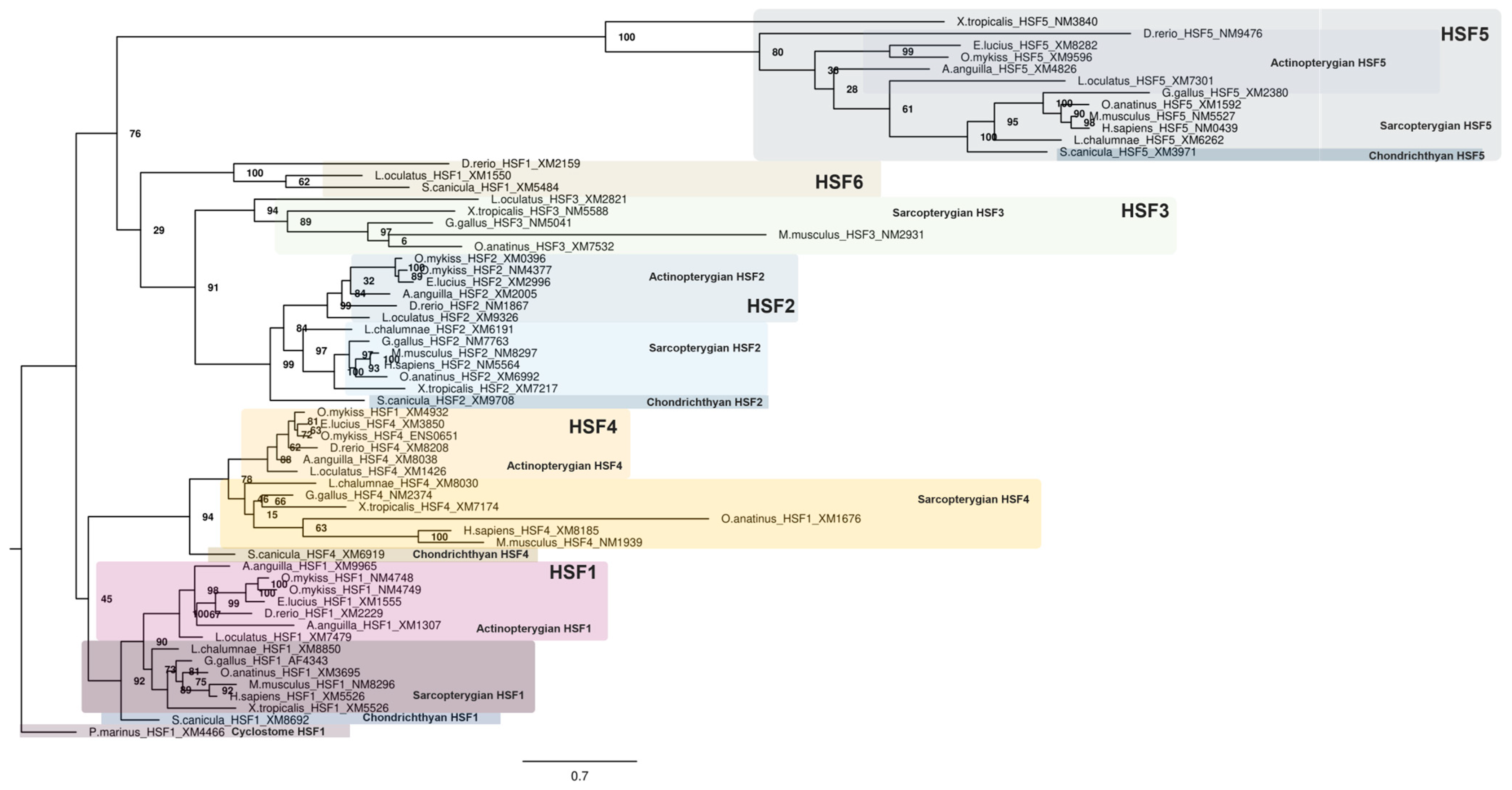
2.1.2. Phylogenetic Analyses of the HSF Family in Vertebrates

Phylogenetic analyses were conducted on various representative vertebrate species, including a cyclostome, the sea lamprey (Petromyzon marinus); a chondrichthyan, the spotted catshark (Scyliorhinus canicula); an early sarcopterygian, the coelacanth (Latimeria chalumnae); various tetrapods including sauropsids, the green anole (Anolis carolinensis) and chicken (Gallus gallus); an amphibian, the tropical clawed frog (Xenopus tropicalis); and mammals, the human (Homo sapiens), the platypus (Ornithorhynchus anatinus), and the house mouse (Mus musculus). Additionally, a non-teleost actinopterygian, the spotted gar (Lepisosteus oculatus), a representative of early diverging teleost, the European eel (Anguilla anguilla); and some other teleosts, the zebrafish (Danio rerio), the Northern pike (Esox lucius) and the rainbow trout (Onchorhynchus mykiss) were also included.
A rooted phylogenetic tree was constructed using the amino acid sequences of HSF (Table S1). The tree was rooted by the sea lamprey HSF sequence. The sequences were aligned using Clustal Omega [34] with Seaview version 5.0.5 software (http://doua.prabi.fr/software/seaview, accessed on 10 March 2024) and were manually adjusted. The phylogenetic tree for the HSF family was constructed using the RAXML program (Randomized Accelerated Maximum Likelihood; [35]) from the CIPRES Science Gateway [36] version 3.3 with 1000 bootstrap replicates, using the JTT (Jones, Taylor, and Thornton) protein substitution matrix obtained from the alignment [37] and the resulting tree was visualized using Figtree 1.4.4 (http://tree.bio.ed.ac.uk/, accessed on 10 March 2024).

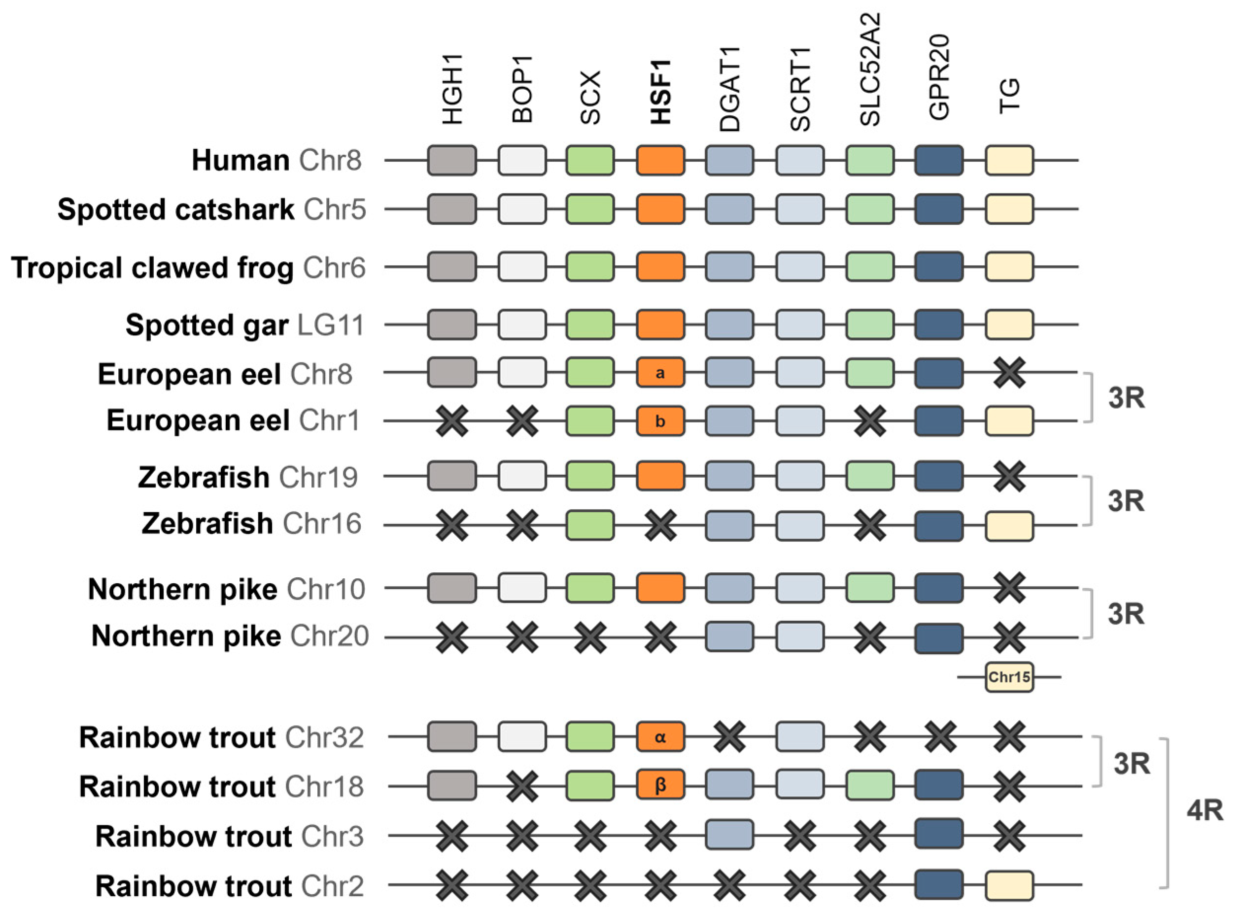
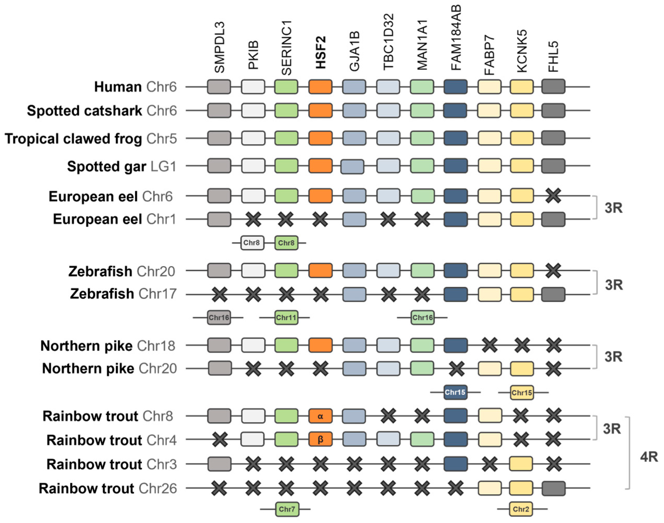
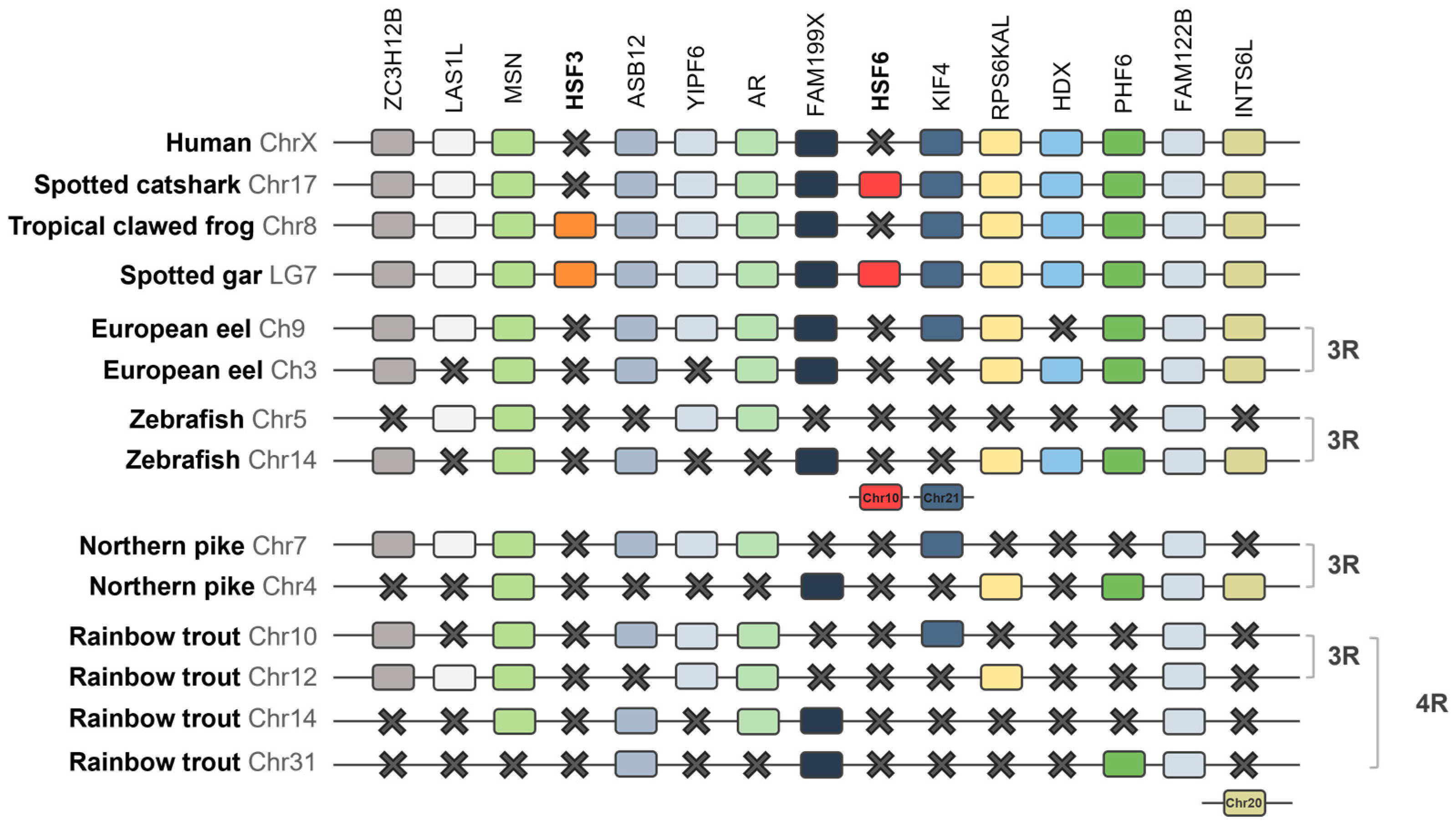
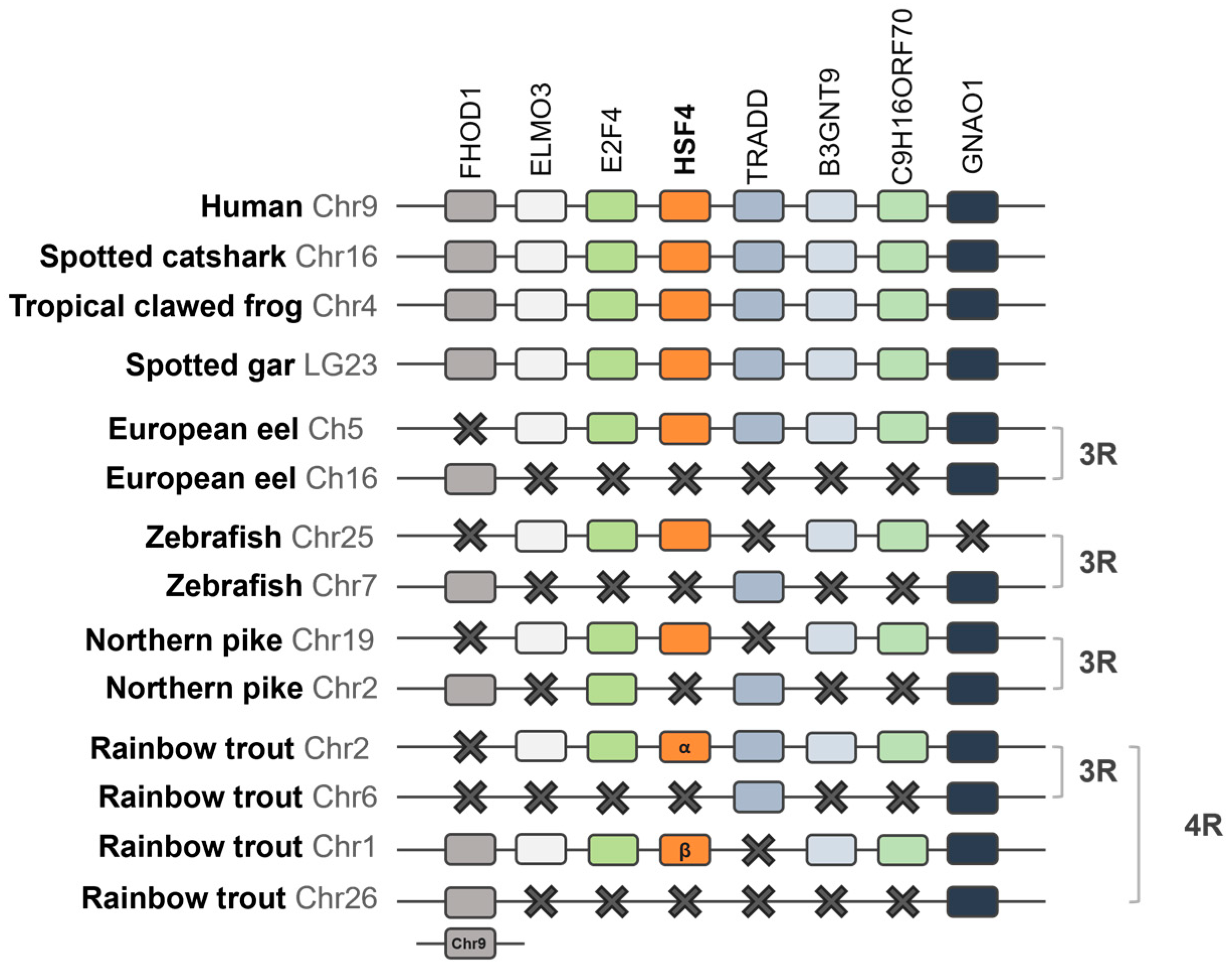
2.1.3. Synteny Analysis of the HSF Family in Vertebrates

Synteny analyses of HSF genomic regions were performed across representative vertebrate species, including one chondrichthyan (the spotted catshark), two sarcopterygians (the human and tropical clawed frog), and actinopterygians (the spotted gar and teleosts such as the European eel, zebrafish, northern pike, and rainbow trout). To carry out these analyses, the genes neighboring HSF were identified using Genomicus PhyloView of Genomicus v100.01, with either the human or the spotted catshark serving as the reference. BLAST searches were then conducted using the ENSEMBL and/or NCBI databases to identify potential paralogs among the neighboring genes or to uncover any unannotated genes in the genomes of the studied species. Since the European eel genome was not available in the Ensembl (https://www.ensembl.org/index.html, accessed on 8 February 2025) and Genomicus (https://www.genomicus.bio.ens.psl.eu/genomicus-110.01/cgi-bin/search.pl, accessed on 8 February 2025) databases, its genomic data (GCF_013347855.1, fAngAng1 genome, Future Genomics Technologies B.V., Leiden, The Netherlands) was analyzed using the NCBI database to characterize the genomic regions of HSF. A detailed list of HSF neighboring gene references and specific locations for each species can be found in Table S2.

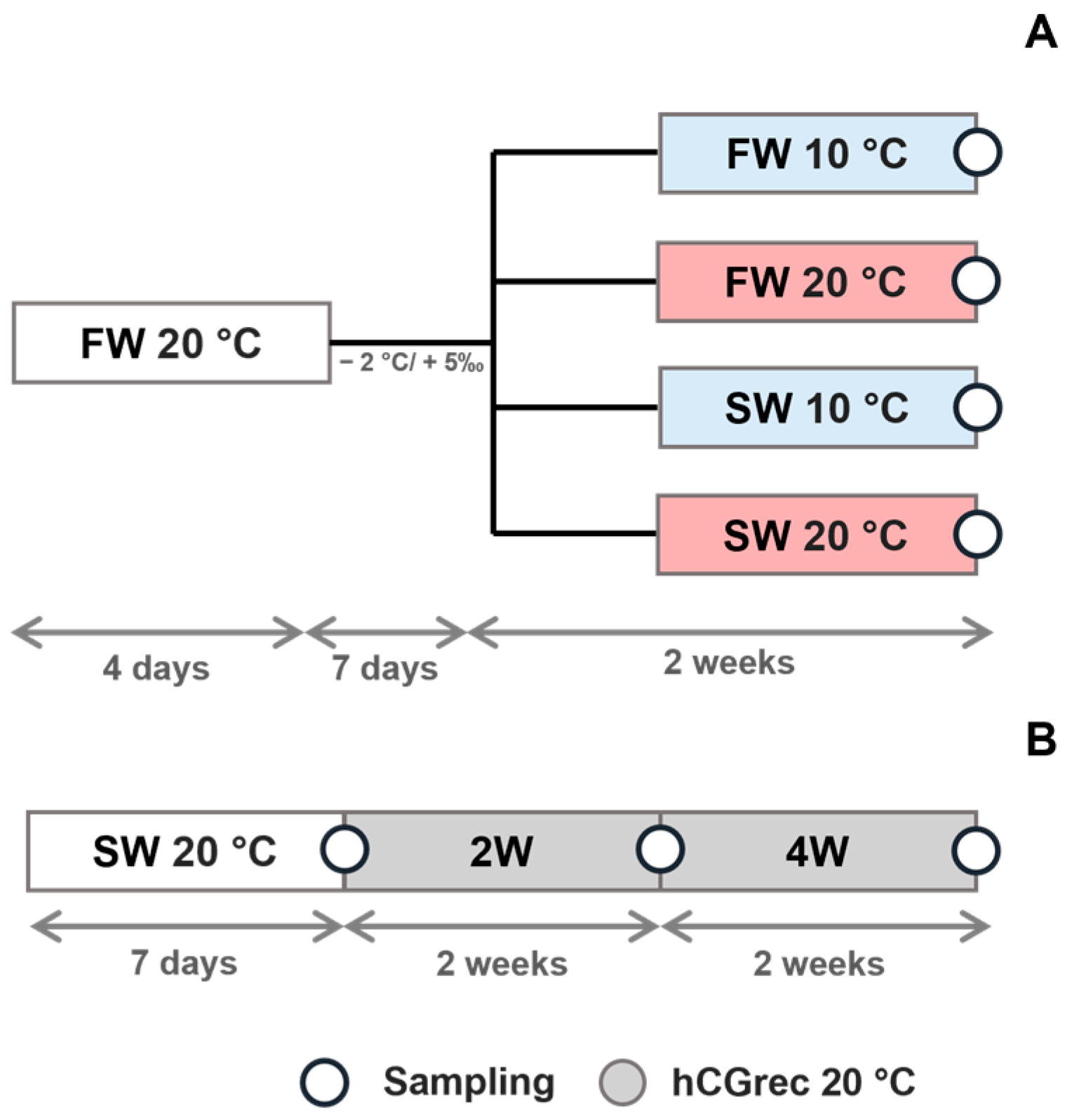
2.2. Experimental Design

2.2.1. Fish Maintenance and Handling

Sixty-four male eels maintained in freshwater at the local fish farm (Valencia, Spain) were moved to the Laboratory of Fish Reproduction at the Universitat Politècnica de València (UPV, Spain). On arrival, fish were distributed into 4 aquaria of 150 L (16 fish/aquaria) filled with freshwater (FW 20 °C) that simulated similar conditions to those used in the fish farm and to reduce stress for the fish. Each aquarium was fitted with an individual recirculation system and a temperature control setup (including both heaters and coolers). The eels were kept under these conditions for four days before being distributed to their respective experimental conditions, except for the male eels used to evaluate gene expression across various tissues.

2.2.2. Expression of HSF Genes in Different Tissues from the Male European Eel

To evaluate the expression of HSF genes in several tissues of the male European eels, 6 males (mean body weight = 89 ± 17 g) were sampled immediately upon arrival at the UPV facilities. Samples, including the pituitary, anterior, and posterior brain, skin, eye, testis, liver, muscle, spleen, intestine, and gills, were carefully collected and stored in RNAlater (Ambion Inc., Huntingdon, UK) at −20 °C until the total RNA extraction for gene expression analysis.

2.2.3. Expression of HSF Genes in the European Eel Testis Under Different Temperature and Salinity Conditions

To evaluate the effects of temperature and/or salinity on the expression of HSF genes (Figure 1A), thirty-two male eels (mean body weight = 95 ± 15 g) from freshwater conditions (FW 20 °C) were randomly distributed into 4 aquaria (8 fish/aquarium) to establish four groups with different combinations of temperature and salinity: freshwater at 10 °C (FW 10 °C) and at 20 °C (FW 20 °C), seawater (37 ± 0.3‰ salinity) at 10 °C (SW 10 °C) and 20 °C (SW 20 °C). To adjust these conditions, fish from each group were gradually acclimated over the course of one week by lowering 2 °C and/or raising the salinity by 5‰ salinity each day (with the exception of the male eels that were kept at FW 20 °C). Once the temperature and salinity values were achieved, fish were maintained for 2 weeks in their respective conditions before testis sampling for gene expression analysis.

2.2.4. Expression of HSF Genes in the European Eel Testis During Spermatogenesis Induced with Hormonal Maturation Treatment

To evaluate the HSF gene expression during spermatogenesis (Figure 1B), 24 eels (mean body weight = 91 ± 11 g) were gradually maintained in seawater at 20 °C (SW) for two weeks before hormonal maturation induction. Following this period, 8 eels were sampled to collect testis tissue for histology and gene expression analysis. The remaining eels were subsequently administered hCGrec treatment via an intraperitoneal injection (Ovitrelle®, Madrid, Spain, 1.5 IU/g fish) once a week, as described by Herranz-Jusdado et al. [27] Testis from eight eels were sampled after 2 (2 W hCGrec) and 4 (4 W hCGrec) weeks of hormonal maturation.

2.2.5. Samplings

Testis from all experiments were carefully collected from sacrificed male eels and were stored in 0.5 mL of RNAlater (Ambion Inc., Huntingdon, UK) at −20 °C until the total RNA extraction for gene expression analysis. Part of the testis samples from the hormonal maturation experiment were fixed in 4% glutaraldehyde diluted in phosphate buffer (pH adjusted to 7.4) for histological analysis.

2.3. Gene Expression Analyses by Quantitative Real-Time PCR

2.3.1. RNA Extraction and cDNA Synthesis

Total RNA was extracted from samples previously preserved in RNAlater using Trizol reagent (Life Technologies, Inc., Carlsbad, CA, USA), following the protocol outlined by Morini et al. [38] The extracted RNA was diluted in 20 µL of DEPC water. To confirm its suitability for cDNA synthesis, RNA quantity and purity were assessed using a NanoDrop 2000 C Spectrophotometer (Fisher Scientific SL; Valencia, Spain). cDNA synthesis was carried out using a QuantiTect Reverse Transcription Kit (Qiagen, Hilden, Germany) and 20 µL of cDNA from 500 ng of total RNA. Genomic DNA contamination was removed by the gDNA Wipeout Buffer included in the kit. The synthesized cDNA was then stored at −20 °C until further analysis.

2.3.2. Primer and Reference Genes

Quantitative real-time Polymerase Chain Reactions (qPCRs) were conducted using specific qPCR primers for each European eel HSF gene, and the acid ribosomal phosphoprotein (ARP) was used as a reference gene, as described previously by Morini et al. [38] Primers for qPCR (Table 1) were specifically to target European eel HSF genes, using their complete coding sequences as a reference.
The primer pairs were designed to span two different exons, preventing the amplification of potential genomic contamination. Primers were designed using Primer3 Software version 0.5 (Whitehead Institute/Massachusetts Institute of Technology, Boston, MA, USA) and acquired from Integrate DNA Technology, Inc. (IDT, Inc., Coralville, IA, USA).

2.3.3. Gene Expression Analysis

qPCR assays were performed using a StepOnePlusTM model (Applied Biosystems; Foster City, CA, USA) following the protocol outlined by Morini et al. [38] The cycling program began with an initial Taq polymerase activation at 95 °C for 10 min, followed by 40 cycles at 95 °C for 15 s and 60 °C for 30 s. Each reaction mixture included 5 μL of diluted DNA template (1:10), forward and reverse primers (250 nM each), 12 μL of SYBR Green/ROX Master Mix, and DEPC water to bring the total volume of 20 µL. Primer efficiencies were assessed using serial dilutions (1:20) of a cDNA pool derived from testis tissues, creating a five-point linear regression slope. A calibrator prepared from a testis tissue pool (1:20) was included in each run for the respective gene. Absolute mRNA expression levels for tissue screening were quantified using efficiency-corrected calculations [39]. For testis analysis, relative mRNA expression levels were determined using efficiency-corrected normalization based on the Ct (threshold cycles) values of reference genes [39]. Both target and reference genes were amplified in duplicate, and duplicate non-template control (cDNA replaced by water) reactions were also included for each primer pair.

2.4. Testis Histology

Testis samples from the hormonal maturation experiment (n = 8/group) were fixed in 10% neutral buffered formalin (Thermo Fisher Scientific, Waltham, MA, USA) for 24 h. Samples were then dehydrated in increasing percentages of ethanol and embedded in resin (Technovit 7100) in accordance with the instructions of the manufacturer. Sections of 5 µm thickness were cut with a Microm HM325 microtome and stained with hematoxylin and eosin. The slides were observed with a Nikon Eclipse E-400 microscope, and pictures were taken with a Nikon DS-5M camera attached to the microscope (Nikon, Tokyo, Japan). Cell types identified throughout the hormonal treatment were categorized as described for zebrafish [40] and for the European eel [31]. The undifferentiated spermatogonia type A cells (SPGAund) showed irregular nuclear membranes and were isolated. Differentiated SPG type A cells (SPGAdiff) formed groups of 2–8 cells within the Sertoli cell surroundings, with regular nuclear envelopes, one or more nucleolus and a cytoplasm darker than that found in SPGAund. SPG type B cells (SPGB) were found as smaller cells with lower amounts of cytoplasm and nuclei with large amounts of heterochromatin. Spermatocytes formed cysts with cells of a smaller size than spermatogonia, with a dense nucleus. Spermatids were also found in clusters, with a very small size and round formation. FIJI/ImageJ 2.0.0 software was used to count the number of each cell type from 5 microscope fields per sample within each experimental group. Cell distribution counts were transformed into percentage data (sum of each cell type found in each slide/sum of total cell counts ×100).

2.5. Statistical Analysis

Shapiro–Wilk and Levene’s tests were used to check the normality of data distribution and variance homogeneity, respectively. A one-way ANOVA (or Kruskal–Wallis tests for non-parametric data) followed by Student–Newman–Keuls post hoc tests were used to compare the absolute expression from different tissues and the relative expression from testis during hormonal maturation treatment. A two-way ANOVA test was used to study the main effects (and possible interactions between factors) of temperature and salinity conditions in the relative expression in the testis samples.
Results were presented as mean ± standard error (SEM). Significant differences were detected when p < 0.05. All statistical analyses were performed using the SPSS statistical package version 24.0 for Windows software (SPSS Inc., Chicago, IL, USA).

3. Results

3.1. Phylogenetic Analyses of the HSF Family in Vertebrates

The HSF phylogenetic tree (Figure 2) is composed of two main clades, one containing vertebrate HSF1 sequences while the other consists of the previously described HSF2, HSF3, HSF4, HSF5, and novel HSF6 sequences. All HSF clades are represented by sequences of representative species in accordance with their established phylogenetic positions, with the exception of the HSF5 clade. In the case of HSF1, the sister group of HSF2/3/4/5/6, one gene was found in the genome assembly of the cyclostome, the sea lamprey, and in the representative chondrichthyan, the spotted catshark, which clustered at the base of the sarcopterygian and actinopterygian HSF1 sequences. In the representative non-teleost actinopterygian, the spotted gar, the HSF1 sequence diverged basally to the teleost HSF1 sequences in accordance with their established phylogenetic positions. In the teleost clade, the studied species possess a single HSF1 gene, with the exception of the European eel and the rainbow trout, which exhibit two HSF1 genes. HSF4 represented a sister clade of the HSF2/3/5/6 monophyletic group. The HSF4 gene was not found in the sea lamprey but it is present as a single gene in the chondrichthyan and sarcopterygians investigated. In this clade, one sequence annotated as “HSF1” for the platypus was found, meaning that this gene has been incorrectly named in the NCBI database. In actinopterygians, a single HSF4 gene was found, with the exception of the rainbow trout, which exhibits two HSF4 paralogs.
One “HSF1” sequence for the rainbow trout was positioned in the HSF4 clade, meaning that these genes have been wrongly annotated. In a similar manner as the HSF4 clade, the HSF2 gene was not found for the cyclostome representative species, although one HSF2 sequence was present in the chondrichthyan, sarcopterygians, and actinopterygians, with the exception of the rainbow trout, which possesses two HSF2 genes. The BLAST searches revealed that HSF3 is not present in the cyclostome sea lamprey nor in the studied chondrichtian. It is only present as a single copy in tetrapods and in the representative species of the non-teleost actinopterygian, the spotted gar, but not in the studied teleosts. Our study revealed the existence of an undescribed HSF type vertebrate that we have named HSF6, which is only present in the chondrichtian spotted catshark, the sarcopterygian coelacanth, in the non-teleost actinopterygian spotted gar, and only one of the studied teleosts, the zebrafish. The HSF6 clade is a sister clade of the HSF2/3 clades. The HSF5 gene, which represents a sister clade of HSF2/3/6, is present as a single copy in all the gnathostomes studied (chondrichthyan, sarcopterygians, and actinopterygians).

3.2. Synteny Analysis of the HSF Family in Vertebrates

The human HSF1 genomic region was used as a template, and eight HSF1 neighboring genes were explored: HGH1, BOP1, SCX, DGAT1, SCTR1, SLC52A2, GPR20, and TG (Figure 3). In the teleosts, some of the HSF1 neighboring genes (SCX, DGAT1, SCTR1, and GPR20A) were presented as duplicated paralogs in the majority of the species studied. For some other neighboring genes (HGH1, BOP1, SLC52A2, and TG), only one single paralog gene was identified in all the teleosts. This was also the case for HSF1 itself, for which a single paralog was conserved. However, the European eel presented duplicated HSF1 paralogs, that were named HSF1a and HSF1b, corresponding to the nomenclature for the teleost 3R-paralog. In the rainbow trout, three copies of DGAT1 and GPR20 genes were identified. Two HSF1 paralogs were identified in this salmonid species-representative and nominated HSF1α and HSF1β, corresponding to the “α/β” nomenclature for salmonid 4R-paralogs.
In the case of HSF2, the human was used as a template, and the neighboring genes (SMPDL3, PKIB, SERINC1, GJA1B, TBC1D32, MAN1A1, FAM184AB, FABP7, KCNK5 and FNL5) (Figure 4) were retrieved. In the studied chondrichthyan, sarcopterygians, and the non-teleost actinopterygian species, only a single copy of HSF2 and the respective neighboring genes were found. In some teleost species, neighboring HSF2 genes (SMPDL3, GJA1B, FAM184AB, and KCNK5A) were present as duplicated paralogs. However, HSF2 itself and the remaining studied neighboring genes were found as single paralogs, including in the basal teleost, the European eel, suggesting the loss of the duplicated paralogs shortly after the teleost 3R. However, PKIB was present as a single copy in the zebrafish and in the northern pike; SERINC1 and FABP7 were only identified as one gene once again in the northern pike. In the rainbow trout, the three copies of FAM184AB, FABP7, and KCNK5A were identified in the genomic region. The rainbow trout possess two HSF2 paralogs, which were nominated HSF2α and HSF2β, corresponding to the “α/β” nomenclature for salmonid 4R-paralogs.
Interestingly, HSF3 and HSF6 share the same chromosomal location, as shown in Figure 5. for a broad range of gnathostomes, including several teleosts (with the exception of the zebrafish, which presented the HSF6 gene in a different chromosome). The spotted gar HSF3 and HSF6 genomic region was used as a template as both genes were absent in humans. A single HSF3 has been found in the sarcopterygian tropical clawed frog and in the actinopterygian non-teleost species, the spotted gar, while in the teleosts, it was absent in all the studied species. Similarly, a single HSF6 has been found in the chondrichtian catshark and in the actinopterygian non-teleost species, the spotted gar, as well as in one of the studied teleosts, the zebrafish. Thirteen neighboring genes were investigated.
ZC3H12B, LAS1L, MSN, ASB12, YIPF6, AR, FA199X, KIF4, RPS6KAL, HDX, PHF6, FAM122B, and INTS6L. All neighboring genes were found as a single copy in the representative sarcopterygians, the chondrichthyan spotted catshark and the non-teleost actinopterygian, the spotted gar. Some of these neighboring genes were duplicated in the studied teleosts, which all display duplicated copies of MSN and FAM122B as a result of the 3R event. In the rainbow trout, four copies of the FAM122B gene were identified. Other neighboring genes (ZC3H12B, YIPF6, and FAM199X) were identified as duplicates; several (MSN, ASB12, and AR) exhibited three paralogs, while others (LASL1, KIF4, RPS6KAL, PHF6, and INTS6L) were found as single copies.
The human HSF4 genomic region was used as a template, and seven neighboring genes were found: FHDOD1, ELMO3, E2F4, TRADD, B3GNT9, C9H16ORF70, and GNAO. In the sarcopterygian species, the representative chondrichthyan and non-teleost actinopterygian species possess a single copy of HSF4 and respective neighboring genes (Figure 6). In the case of the studied teleost species, only a single copy of HSF4 and some neighboring genes (FHDOD1, E2F4, ELMO3, TRADD, B3GNT9, and C9H16ORF70) were found. The only neighboring gene that presented a duplicated gene was GNAO1. However, there were some exceptions in certain species: E2F4 was duplicated in the northern pike, while the zebrafish exhibited only one single GNAO1 paralog. In the salmonid species, the rainbow trout, some neighboring genes were duplicated, and FHDOD1 and GNAO1 genes were found as three and four copies, respectively. Two HSF4 paralogs were present, which we propose to name HSF4alpha and HSF4beta, corresponding to the nomenclature for 4R-paralogs.
Once again, for HSF5 synteny, the human was used as a template and seven neighboring genes were retrieved: YWHAG, SUPT4H1, RNF43, MTMR4, SEPTIN4, DHX40, and FLOT2, all of which were found to be located in the HSF5 genomic region, with the exception of YWHAG in the human genome. The HSF5 gene and the neighboring genes were present in the studied sarcopterygians, the chondrichthyan, and the non-teleost actinopterygian species as a single copy (Figure 7). In the teleost species considered in this study, the HSF5 and some neighboring genes (SUPT4H1, RNF43, MTMR4, SEPTIN4, and DHX40) were found as a single paralog. Some of these genes were found duplicated, such as YWHAG and FLOT2, as a result of the 3R event. There were a few exceptions: the northern pike presented only one paralog YWHAG, but SEPTIN4 was found duplicated. In the rainbow trout, only a single SUPT4H1 gene was identified, while the remaining neighboring genes were found duplicated, except for FLOT2, which presented four gene copies.

3.3. Expression of HSF Genes in Different Tissues of the Male European Eel

The distribution of HSF genes revealed a differential expression between tissues (Figure 8). In the case of HSF1a and HSF1b (Figure 8A and Figure 8B, respectively), both were detected in all tissues but showed different expression patterns.
The HSF1a expression was higher in the testis than in the peripherical tissues (e.g., skin, liver, and skin), while both brain parts showed the highest HSF1b expression. In the case of HSF2 (Figure 8C), the highest expression was detected in the testis, but it was also detected in the intestine and the gills. The HSF4 expression was higher in the testis and in the intestine than in other tissues (Figure 8D). Similarly, HSF5 was highly expressed in the testis and in the anterior brain (Figure 8E).

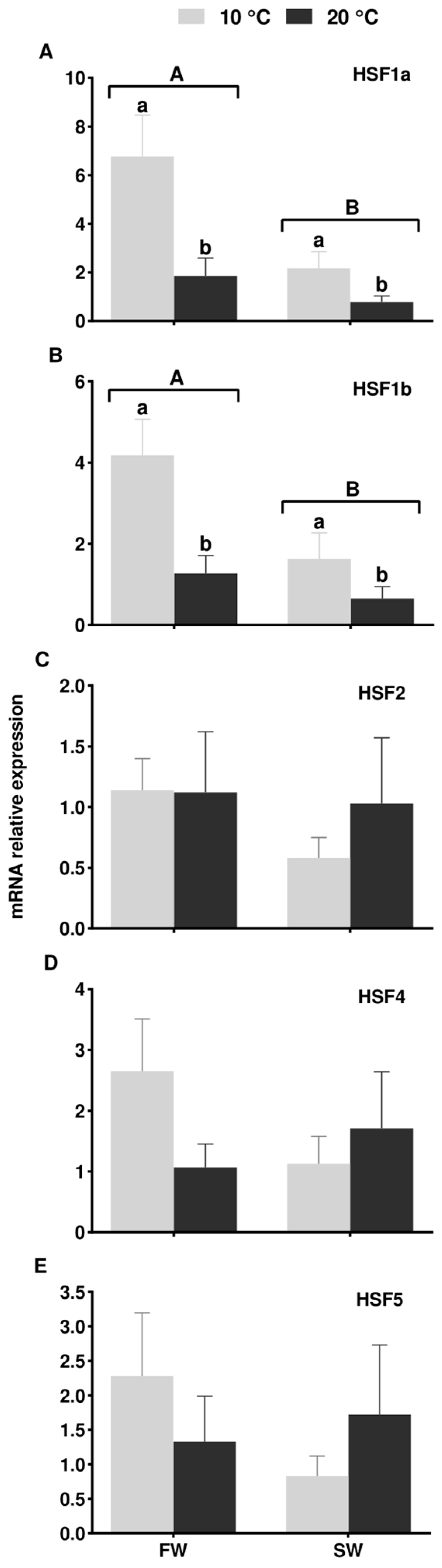
3.4. Expression of HSF Genes in the European Eel Testis Under Different Temperature and Salinity Conditions

Different heat shock factor expression profiles were observed in the testis of eels submitted to different temperature and salinity combinations (Figure 9).
The temperature and salinity effects were significant in the expression levels of HSF1a and HSF1b (Figure 9A and Figure 9B, respectively) as the increase of temperature and salinity downregulated the expression levels of both HSF1 genes in the eel testis. Regarding HSF2, HSF3, and HSF4 (Figure 9C, Figure 9D, and Figure 9E, respectively), neither the temperature nor salinity significantly affected their expression levels as these remained stable independent of the condition changes.

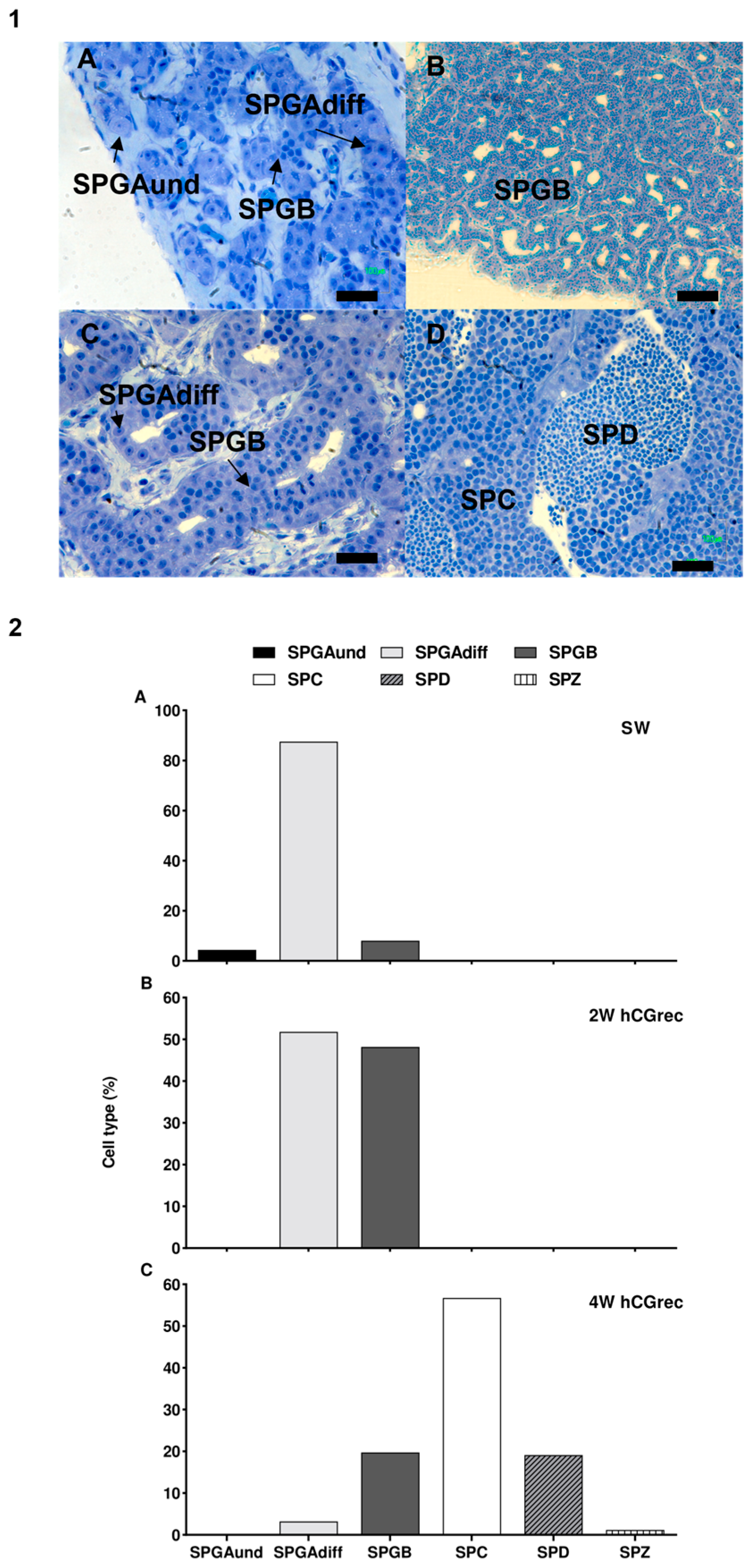
3.5. Expression of HSF Genes in the European Eel Testis During Spermatogenesis Induced by Hormonal Maturation

Different heat shock factor expression profiles were observed in the testis of eels under hormonal maturation (Figure 10).
The HSF1a mRNA levels in males maintained in SW (without the hCGrec treatment) and after 2 weeks of the hCGrec treatment were higher than those found after 4 weeks of hormone injections (Figure 10A). The HSF1b and HSF2 (Figure 10B,C) expression decreased, but no significant differences were observed. The HSF4 expression levels were significantly higher in males in SW in comparison with those treated with a hormonal treatment (Figure 10D). Finally, the expression of HSF5 increased significantly after 4 weeks of hormonal treatment, and the expression level was 7-fold higher in 4 W hCGrec males than in SW and 2 W hCGrec males (Figure 10E).

3.6. Testis Histology

Different types of cells were observed as a result of testis development induced by hormonal maturation (Figure 11(1A–D)).
The testis of eels maintained for two weeks in seawater (without the hCGrec treatment) showed testis containing mostly SPGAdiff cells (87.53%), but some SPGAund and SPGB (4.41 and 8.07%, respectively) were also found (Figure 11(2A)). Male eels from the 2 W hCGrec group showed similar proportions of SPGAdiff and SPGB cells (Figure 11(2B)). After 4 weeks of hormonal maturation, eels from the 4 W hCGrec still contained SPGAdiff and SPGB cells, but the presence of other cell types evidenced differentiation of the cells (Figure 11(2C)). SPC cells were predominant (56.75%), although other cell types, such as SPD and even SPZ, were also detected.

4. Discussion

4.1. Evolution of the HSF Family and Discovery of HSF6

The phylogenetic and synteny analysis of HSF genes offers valuable insight into the diversity of HSF types found in vertebrates, especially in teleosts. These species experienced an additional genome duplication beyond the two rounds that occurred in vertebrates. This specific evolutionary event, known as teleost-specific whole genome duplication (3R WGD), led to the loss or retention of paralogs [41]. In the present study, the phylogenetic tree allowed us to observe the evolution of the HSF family members in the studied vertebrates and conclude that the sequences of HSF1 are more closely related to HSF4 than those of HSF2 or HSF3, while HSF2 has a closer relation to those of HSF3. It was suggested that an ancestral gene of HSF1 and HSF4, as well as one of HSF2 and HSF3, was generated by the first WDG event, and then four genes came from the second event [3]. HSF3, HSF5, and HSF6 might also include sequences diverged at a faster rate during evolution, while those of HSF1 and HSF2 were more conserved, which is in accordance with previous results [21]. It seems that the sequences of HSF1 were highly conserved during vertebrate evolution, while HSF3 changed rapidly. As such, the accumulation of mutations might be greatly different among stress-related transcription factor family genes with the same function [22] in the studied vertebrates such as the chondrichthyan spotted shark, the coelacanth, the tropical clawed frog, the green anole, the non-teleost actinopterygian spotted gar and all the teleost species, HSF1, HSF2, and HSF4 genes were conserved. In the case of HSF3, it was conserved in some of the studied mammalians, such as the mouse and the platypus, and in birds like the chicken, and also the tropical clawed frog. Similarly, it was maintained in the coelacanth and the non-teleost actinopterygian spotted gar but was lost in all the other studied species. Interestingly, our study revealed the existence of a previously undescribed vertebrate HSF type that we named HSF6, although, in the genome databases, it has been named HSF1. This gene has only been found in some studied vertebrates, such as the chondrichthyan spotted catshark, the sarcopterygian coelacanth, the non-teleost actinopterygian spotted gar, and the zebrafish. According to our syntenic analysis, HSF3 and HSF6 are in the same genomic region and may have arisen from a specific local duplication in at least an osteichthyan ancestor. Finally, the HSF5 clade grouped the studied vertebrate amino acid sequences as a different branch and clustered as a sister clade of the HSF2/3/6 clade. This amino acid sequence is present in all the studied vertebrates and in accordance with that previously observed [9].
Our phylogenetic and syntenic analyses allowed the identification of duplicated HSF1 paralogs in the European eel genome for the first time. Synteny analyses showed that the HSF1 genomic region has been duplicated in the teleost studied, suggesting that the two paralogs of HSF1 found in the European eel resulted from the teleost-specific WGD (3R) and have been conserved after WGD. They were named HSF1a and HSF1b. One of the paralogs was lost in the other teleost species studied. This approach confirmed that the European eel retains more duplicated genes from the WGD event compared to other teleost species, as reported in previous studies [38]. In the rainbow trout, two HSF1 paralogs were identified and nominated HSF1α and HSF1β, corresponding to the “α/β” nomenclature for salmonid 4R-paralogs [42]. This evidenced the additional genomic duplication that occurred in the salmonids and followed the previous cloning of two HSF1 genes in the gonadal cell lines of this salmonid [43]. In contrast, a single HSF1 was reported in humans and in goldfish [44] and later in the tropical clawed frog [4]. Our study confirmed the presence of a single HSF1 paralog in these species, as well as in the chondrichthyan spotted catshark and in several actinopterygians. Only one single copy of HSF2, HSF4, or HSF5 was found in teleosts, suggesting that their duplicate was lost shortly after the 3R event. In the salmonid studied, the rainbow trout, two paralogs of HSF2 and HSF4 were identified, suggesting that a single paralog of these genes was inherited by the salmonid lineage and duplicated by 4R WGD [42].
The presence of several HSF genes in the chondrichthyan, actinopterygian, and sarcopterygian members suggests that all family members were generated early in the vertebrate evolution. According to previous studies, this may exert a wide range of effects on gene expression and epigenetic status on the whole genome [1]. The emergence of different HSFs might imply complementary roles to several physiological responses to various stimuli, and therefore, it is important to further study these genes.

4.2. Expression of HSF Genes in Different Tissues from the Male European Eel

In the present study, different expression patterns were found for all five HSF in tissues from the male European eel. It is worth noting that our results may have been affected by the maturational phase of the males, as male eels used for this experiment were probably at the beginning of the sexual maturation phase, also referred to as the “silvering stage”. Nevertheless, HSF genes showed differential expression profiles among tissues compared with the corresponding genes in other vertebrate species. Previously, it was shown that HSF1a and HSF1b isoforms were detected in several unspecified rainbow trout tissues [44]. Contrarily, HSF1 was isolated in the zebrafish and showed that both HSF1 isoforms were highly expressed in the gonads, but one isoform was expressed less in the liver than in the gills [45]. Our results in the European eel males contrast with those obtained in the trout, as HSF1a was highly expressed in the testis and HSF1b expression in the liver was lower than in the gills, while it was predominant in the brain, suggesting a species-specific expression.
In European eel males, the highest HSF2 expression was in the testis but also in the intestine and the gills. In mammals, HSF2 is present at different stages of spermatogenesis, from spermatogonia to spermatids [46] and is found in the cytoplasmic bridges that connect germ cells with the same spermatogonia [47]. It has also been shown to play a role in the shift from endo to exotrophic feeding during pikeperch development [48]. Our study found that European eel males had the highest expression of the HSF4 gene in the testis. Typically, in vertebrates, HSF4 is known for its role in the development of sensory organs, particularly the eye [19]. However, HSF4, when coordinating its expression with HSF1, plays a role in the proliferation and differentiation of epithelial cells, but it is also expressed in the testis [23]. Our preliminary results indicate that HSF4 may work in combination with HSF1, which could explain the similar high expression levels in the eel testis, suggesting a potential implication of HSF4 in the testis development as previously suggested [23]. In the case of HSF5, European eel males showed the highest expression in the gonads and anterior brain, coinciding with results observed previously in adult zebrafish [9]. In the zebrafish, HSF5 expression was detected in the brain, but the expression levels were higher in the testis than in other tissues. This previous study, together with our own results, suggests that HSF5 might have an important implication in teleost spermatogenesis development. Thus, in male eels, the HSF gene family was primarily expressed in the testis but also in non-reproductive tissues, such as the intestine and the gills. More research is needed to understand the function of HSF4 and HSF5 genes in the tissues of the European eel and other fish and to support these hypotheses.

4.3. Expression of HSF Genes in the European Eel Testis Under Different Temperature and Salinity Conditions

The purpose of these experimental conditions was to replicate the biological environment that European eels experience during changes in temperature and salinity, being the first in fish to explore these genes despite the logical limitations in the experimental background and previous knowledge for discussion. In mammals, HSF genes have been extensively studied regarding their role in thermotolerance and the activation of gene expression in response to environmental conditions. However, little is known about the HSF gene response in teleost [45]. In the present study, the expression levels of the HSF genes in European eel testis were evaluated under different environmental conditions probably faced by this species during its migration. Our study revealed that of the five HSF genes identified in the eel, only HSF1a and HSF1b exhibited varying expression levels under different temperature and salinity conditions. Both gene expression levels decreased with temperature, and salinity increased, downregulating their expression. These results suggest that these genes might play an important role in eel perception of changing temperature, as observed previously with the SODs [49]. Indeed, HSF1 is the only HSF that can bind to heat shock DNA-binding domain in a temperature-dependent manner in mammals [50]. Indeed, an early study of HSF1 expression in male germ cells isolated from the rainbow trout testis showed that low temperatures activate HSF1 [51]. This result suggests that the increase of HSF1 expression levels at low temperatures in the European eel may be due to specific HSF1 thermosensitivity to specific protein components such as heat shock proteins. Additionally, it has been shown that testosterone inhibits HSF1 expression in Sertoli cells from mammals, revealing that this androgen modulates HSF1 expression [52]. Previous studies in male eels confirmed that low temperature increases androgen levels [29,31], contrasting with the previous findings in mammals and suggesting that the HSF1 gene response to temperature might differ among vertebrate species as an adaptive feature to temperature. Moreover, our study also revealed that higher salinity reduced the expression of HSF1 genes in male testis kept at low temperatures. This supports the previously proposed idea that the European eel may have conserved both HSF1 paralog genes to face changes in both temperature and salinity. Previously, a similar hypothesis was proposed for the rainbow trout, another migratory fish with two 4R-HSF1 duplicated genes [43].
Our results showed that HSF2, HSF4, and HSF5 expression levels in the eel testis remained stable despite changes in temperature or salinity. The consistency of HSF2 and HSF4 upon environmental changes concurs with previous findings in mammals, where these genes were considered developmental factors [50]. Moreover, the potential role of HSF5 was evaluated in heat shock response by studying HSF5 knockdown males in zebrafish testis and no differences were found between wild fish types, suggesting that this gene may not be crucial in responding to environmental changes [9]. These results support our findings in the eels.

4.4. Expression of HSF Genes in the European Eel Testis During Hormonal Maturation

The aim of this experiment was to replicate the sexual maturation of the European eels and explore the HSF gene variations, thus being a pioneer in fish despite the logical limitations in the experimental background and previous knowledge for discussion. While HSF genes are primarily associated with responding to physiological and environmental cues, several studies in vertebrates have demonstrated that certain HSF genes also play a significant role in male gonad development and spermatogenesis [10,50]. This study is the first to report and compare the mRNA expression of five HSF throughout spermatogenesis in fish, having the European eel as a teleost model. Both HSF1a and HSF1b showed a similar decrease in expression during hCGrec-induced maturation. Additionally, HSF1a was significantly higher in eel testis sampled before and after two weeks of hormonal treatment, when SPGAdiff and SPGB were predominant, compared with those from eels following 4 weeks of hormonal treatment when spermatocyte was the predominant stage. Our findings indicate that in male eels, HSF1 downregulation coincides with the early spermatogenesis stages. These results contrast with those in mammals, as HSF1 is mainly found in spermatocyte and spermatid cells, suggesting differences in HSF1 expression between species during spermatogenesis [19]. Additionally, testosterone suppresses HSF1 expression in Sertoli cells by allowing the binding of the androgen receptor to the HSF1 promoter [52]. It is possible that the weekly hCGrec treatment used to induce eel spermatogenesis might have influenced the HSF1 mRNA levels, although further studies are needed to confirm this hypothesis.
In our study, a decreasing pattern in HSF2 expression levels during spermatogenesis (although without reaching significant differences) was registered, similar to the profile in HSF1 genes. In mammals, HSF1 and HSF2 share genomic-binding sites, and both factors cooperate to regulate the transcription of genes involved in spermatogenesis, including the encoding chaperones and co-chaperones that facilitate protein folding [50]. This cooperation between both genes might explain the similar expression profiles observed in male eels in our study.
Surprisingly, HSF4 expression was modulated by hCGrec-induced spermatogenesis, showing significantly higher levels of mRNA transcripts in eel testis without any hormonal treatment when SPGAdiff cells were predominant in comparison with those treated. In mammals, HSF4 expression was detected in the testis and showed that HSF4 knockdown individuals did not alter other genes. However, HSF1 and HSF4 regulate the expression of growth factor genes, which are essential for the proliferation and differentiation of epithelial cells [23]. Although further studies are needed, our results from eel testis might indicate a potential HSF4 role in the spermatogenesis process, supported by a similar HSF1 expression profile.
Finally, our findings showed that HSF5 levels were significantly higher after 4 weeks of hCGrec treatment when eel testis was mainly composed of spermatocytes. These results concur with those previously demonstrated in the zebrafish testis, which confirmed that HSF5 appears to be required for the correct progression of meiosis during spermatogenesis [10]. Thus, it is possible that a high expression of HSF5 in the eel testis at the later spermatogenesis stage might play a similar role.

5. Conclusions

In summary, our study characterized all five HSF genes found in the European eel. Through phylogenetic and syntenic analyses of HSF genes in vertebrates, we inferred their evolutive origin in the eel. Notably, the eel retained duplicates of HSF1 (HSF1a and HSF1b) after 3R, while HSF2, 4, and 5 genes lost their 3R duplicated paralogs. Phylogenetic and syntenic analysis revealed a new HSF6 gene in vertebrates. All five HSF genes exhibited high expression levels in the eel testis and showed differential responses to temperature and salinity conditions, suggesting a possible adaptation in gene expression to these two critical factors during migration. Moreover, HSF genes showed differential expression patterns during hormonally induced spermatogenesis. Specifically, both HSF1, HSF2, and HSF4 transcripts showed the highest levels during early spermatogenesis, whereas HSF5 expression was higher, coinciding with the presence of spermatocytes. Given that this is the first study to compare and analyze HSF gene expression in a teleost, future research expanding on these findings could have significant implications. Such studies could enhance our understanding of environmental adaptation and reproductive strategies in vertebrates, particularly in relation to aquaculture and conservation efforts. Moreover, further studies are needed to uncover the specific roles of HSF in teleosts, particularly in eel spermatogenesis, as this knowledge could be valuable in anticipating responses to emerging challenges like climate change.

Supplementary Materials

The following supporting information can be downloaded at: https://www.mdpi.com/article/10.3390/fishes10020073/s1, Table S1: Accession numbers for the phylogenic analysis of the HSF gene family; Table S2: Accession numbers for the synteny analysis of the HSF gene family.

Author Contributions

Conceptualization, L.F. and M.M.; methodology, L.F., M.M. and L.P.; formal analysis, L.F., M.M. and L.P.; investigation, L.F. and M.M.; data Curation, L.F.; writing—original draft preparation, L.F.; writing—review and editing, M.M. and J.F.A.; supervision, M.M. and J.F.A.; funding acquisition, J.F.A. All authors have read and agreed to the published version of the manuscript.

Funding

This work was supported by the Spanish MICIU (Project EELGONIA; RTI2018-096413-B-I00). L. Ferrão had a predoctoral contract funded by the Generalitat Valenciana, Programa Grisolía (GRISOLIAP/2020/063). The postdoctoral contract of M. Morini was supported by the Spanish Ministry of Science and Innovation (MCIN) with funding from the European Union NextGenerationEU (PRTRC17.I1) and the Generalitat Valenciana to SEASPERM (THINKINAZUL/2021/012).

Institutional Review Board Statement

This study was conducted in full compliance with the guidelines outlined in the Guide for the Care and Use of Laboratory Animals of the Spanish Royal Decree 53/2013 on the protection of animals used for scientific purposes (BOE 2013). The protocol received approval from the Experimental Animal Ethics Committee of the Universitat Politècnica de València (UPV) with the final authorization granted by the local government (Generalitat Valenciana, Permit Number: 2019/VSC/PEA/0073, approval date: 25 March 2019). Every effort was made to minimize suffering, and fish were euthanized using a benzocaine overdose followed by decapitation.

Informed Consent Statement

Not applicable.

Data Availability Statement

The original contributions presented in this study are included within the article/Supplementary Materials. Further inquiries can be directed at the corresponding author.

Conflicts of Interest

The authors declare that they have no known competing financial interests or personal relationships that could have appeared to influence the work reported in this paper.

References

  1. Nakai, A. (Ed.) Proteostasis and adaptation to high temperature stress. In Heat Shock Factor; Springer: Tokyo, Japan, 2016; pp. 3–30. [Google Scholar]
  2. Gomez-Pastor, R.; Burchfiel, E.T.; Thiele, D.J. Regulation of heat shock transcription factors and their roles in physiology and disease. Nat. Rev. Mol. Cell Biol. 2018, 19, 4–19. [Google Scholar] [CrossRef]
  3. Kovács, D.; Kovács, M.; Ahmed, S.; Barna, J. Functional diversification of heat shock factors. Biol. Futur. 2022, 73, 427–439. [Google Scholar] [CrossRef]
  4. Takii, R.; Fujimoto, M. Structure and function of the HSF family members. In Heat Shock Factor; Nakai, A., Ed.; Springer: Tokyo, Japan, 2016; pp. 31–50. [Google Scholar]
  5. Swan, C.L.; Evans, T.G.; Sylvain, N.; Krone, P.H. Zebrafish HSF4: A novel protein that shares features of both HSF1 and HSF4 of mammals. Cell Stress Chaperones 2012, 17, 623–637. [Google Scholar] [CrossRef] [PubMed]
  6. Wang, Q.; Hao, X.; Liu, K.; Feng, B.; Zhang, Z.; Tang, L.; Mhaboob, S.; Shao, C. Early response to heat stress in Chinese tongue sole (Cynoglossus semilaevis): Performance of different sexes, candidate genes and networks. BMC Genom. 2020, 21, 745. [Google Scholar] [CrossRef]
  7. Wang, W.; Yang, Y.; Tan, S.; Zhou, T.; Liu, Y.; Tian, C.; Bao, L.; Xing, D.; Su, B.; Wang, J.; et al. Genomic imprinting-like monoallelic paternal expression determines sex of channel catfish. Sci. Adv. 2022, 8, eadc8786. [Google Scholar] [CrossRef]
  8. Liu, X.; Zhou, L.; Li, W.; Wu, J.; Wang, D. Gata2a mutation causes progressive microphthalmia and blindness in Nile tilapia. Int. J. Mol. Sci. 2023, 24, 3567. [Google Scholar] [CrossRef] [PubMed]
  9. Saju, J.; Hossain, M.; Liew, W.; Pradhan, A.; Thevasagayam, N.; Tan, L.; Anand, A.; Olsson, P.; Orban, L. Heat shock factor 5 is essential for spermatogenesis in zebrafish. Cell Rep. 2018, 25, 3252–3261.e4. [Google Scholar] [CrossRef] [PubMed]
  10. Hemati, A.; Modarressi, M.H.; Kolivand, S.; Azarnia, M. Heat shock factor 5 is essential for spermatogenesis in mice: Detected by a new monoclonal antibody. Iran. J. Basic Med. Sci. 2020, 23, 293–297. [Google Scholar] [PubMed]
  11. Barutc, A.R.; Frit, A.J.; McCor, R.P.; Nick, J.A.; Asla, M. Heat shock factor 5 establishes the male germ-line meiotic sex chromosome inactivation through regulation of Smarca4. Heliyon 2023, 9, e15194. [Google Scholar] [CrossRef]
  12. Deng, Z.; Liu, H.; He, C.; Shou, C.; Han, Z. Heat shock protein 70 (Hsp70) and heat shock transcription factor (HSF) gene families in Cynoglossus semilaevis: Genome-wide identification and correlation analysis in response to low salinity stress. Mar. Freshw. Res. 2021, 72, 1132–1141. [Google Scholar] [CrossRef]
  13. Himanen, S.V.; Puustinen, M.C.; Da Silva, A.J.; Vihervaara, A.; Sistonen, L. HSFs drive transcription of distinct genes and enhancers during oxidative stress and heat shock. Nucleic Acids Res. 2022, 50, 6102–6115. [Google Scholar] [CrossRef] [PubMed]
  14. Izu, H.; Inouye, S.; Fujimoto, M.; Shiraishi, K.; Naito, K.; Nakai, A. Heat shock transcription factor 1 is involved in quality-control mechanisms in male germ cells. Biol. Reprod. 2004, 70, 18–24. [Google Scholar] [CrossRef] [PubMed]
  15. Hayashida, N.; Inouye, S.; Fujimoto, M.; Tanaka, Y.; Izu, H.; Takaki, E.; Ichikawa, H.; Rho, J.; Nakai, A. A novel HSF1-mediated death pathway that is suppressed by heat shock proteins. EMBO J. 2006, 25, 4773–4783. [Google Scholar] [CrossRef] [PubMed]
  16. Korfanty, J.; Stokowy, T.; Widlak, P.; Gogler-Piglowska, A.; Handschuh, L.; Podkowiński, J.; Vydra, N.; Naumowicz, A.; Toma-Jonik, A.; Widlak, W. Crosstalk between HSF1 and HSF2 during the heat shock response in mouse testes. Int. J. Biochem. Cell Biol. 2014, 57, 76–83. [Google Scholar] [CrossRef] [PubMed]
  17. Wang, G.; Zhang, J.; Moskophidis, D.; Mivechi, N.F. Targeted disruption of the heat shock transcription factor (HSF)-2 gene results in increased embryonic lethality, neuronal defects, and reduced spermatogenesis. Genesis 2003, 36, 48–61. [Google Scholar] [CrossRef]
  18. Åkerfelt, M.; Trouillet, D.; Mezger, V.; Sistonen, L. Heat shock factors at a crossroad between stress and development. Ann. N. Y. Acad. Sci. 2008, 1113, 15–27. [Google Scholar] [CrossRef] [PubMed]
  19. Åkerfelt, M.; Morimoto, R.I.; Sistonen, L. Heat shock factors: Integrators of cell stress, development and lifespan. Nat. Rev. Mol. Cell. Biol. 2010, 11, 545–555. [Google Scholar] [CrossRef] [PubMed]
  20. Wang, G.; Ying, Z.; Jin, X.; Tu, N.; Zhang, Y.; Phillips, M.; Moskophidis, D.; Mivechi, N.F. Essential requirement for both HSF1 and HSF2 transcriptional activity in spermatogenesis and male fertility. Genesis 2004, 38, 66–80. [Google Scholar] [CrossRef]
  21. Fujimoto, M.; Hayashida, N.; Katoh, T.; Oshima, K.; Shinkawa, T.; Prakasama, R.; Tan, K.; Inouye, S.; Takii, R.; Nakai, A. A novel mouse HSF3 has the potential to activate nonclassical heat-shock genes during heat shock. Mol. Biol. Cell 2010, 21, 106–116. [Google Scholar] [CrossRef]
  22. Takii, R.; Fujimoto, M.; Matsuura, Y.; Wu, F.; Oshibe, N.; Takaki, E.; Katiyar, A.; Akashi, H.; Makino, T.; Kawata, M.; et al. HSF1 and HSF3 cooperatively regulate the heat shock response in lizards. PLoS ONE 2017, 12, e0180776. [Google Scholar] [CrossRef]
  23. Fujimoto, M.; Izu, H.; Seki, K.; Fukuda, K.; Nishida, T.; Yamada, S.; Kato, K.; Yonemura, S.; Inouye, S.; Nakai, A. HSF4 is required for normal cell growth and differentiation during mouse lens development. EMBO J. 2004, 23, 4297–4306. [Google Scholar] [CrossRef] [PubMed]
  24. Fujimoto, M.; Nakai, A. The heat shock factor family and adaptation to proteotoxic stress. FEBS J. 2010, 277, 4112–4125. [Google Scholar] [CrossRef]
  25. Wright, R.; Piper, A.; Aarestrup, K.; Azevedo, J.; Cowan, G.; Don, A.; Gollock, M.; Ramallo, S.; Velterop, R.; Walker, A.; et al. First direct evidence of adult European eels migrating to their breeding place in the Sargasso Sea. Sci. Rep. 2022, 12, 15362. [Google Scholar]
  26. Righton, D.; Westerberg, H.; Feunteun, E.; Økland, F.; Gargan, P.; Amilhat, E.; Metcalfe, J.; Lobon-Cervia, J.; Sjöberg, N.; Simon, J.; et al. Empirical observations of the spawning migration of European eels: The long and dangerous road to the Sargasso Sea. Sci. Adv. 2016, 2, e1501694. [Google Scholar] [CrossRef]
  27. Herranz-Jusdado, J.G.; Kása, E.; Kollár, T.; Gallego, V.; Peñaranda, D.S.; Rozenfeld, C.; Pérez, L.; Horváth, Á.; Asturiano, J.F. Handling and treatment of male European eels (Anguilla anguilla) for hormonal maturation and sperm cryopreservation. JoVE 2018, 131, e56835. [Google Scholar] [CrossRef]
  28. Asturiano, J.F. Improvements on the reproductive control of the European eel. In Reproduction in Aquatic Animals: From Basic Biology to Aquaculture Technology; Yoshida, M., Asturiano, J.F., Eds.; Springer Nature: Singapore, 2020; pp. 293–320. [Google Scholar]
  29. Peñaranda, D.S.; Morini, M.; Tveiten, H.; Vílchez, M.C.; Gallego, V.; Dirks, R.P.; van der Thillart, G.E.; Pérez, L.; Asturiano, J.F. Temperature modulates testis steroidogenesis in European eel. Comp. Biochem. Physiol. A Mol. Integr. Physiol. 2016, 197, 58–67. [Google Scholar] [CrossRef] [PubMed]
  30. Mazzeo, I.; Peñaranda, D.S.; Gallego, V.; Baloche, S.; Nourizadeh-Lillabadi, R.; Tveiten, H.; Dufour, S.; Asturiano, J.F.; Weltzien, F.A.; Pérez, L. Temperature modulates the vitellogenesis progression in European eel. Aquaculture 2014, 434, 38–47. [Google Scholar] [CrossRef]
  31. Rozenfeld, C.; García-Carpintero, V.; Pérez, L.; Gallego, V.; Herranz-Jusdado, J.G.; Tveiten, H.; Johnsen, H.K.; Fontaine, R.; Weltzien, F.A.; Cañizares, J.; et al. Cold seawater induces early sexual developmental stages in the BPG axis of European eel males. BMC Genom. 2019, 20, 597. [Google Scholar] [CrossRef]
  32. Henkel, C.V.; Burgerhout, E.; de Wijze, D.L.; Dirks, R.P.; Minegishi, Y.; Jansen, H.J.; Spaink, H.P.; Dufour, S.; Weltzien, F.A.; Tsukamoto, K.; et al. Primitive duplicate Hox clusters in the European eel’s genome. PLoS ONE 2012, 7, e32231. [Google Scholar] [CrossRef] [PubMed]
  33. Henkel, C.V.; Dirks, R.P.; de Wijze, D.L.; Minegishi, Y.; Aoyama, J.; Jansen, H.J.; Turner, B.; Knudsen, B.; Bundgaard, M.; Hvam, K.L.; et al. First draft genome sequence of the Japanese eel, Anguilla japonica. Gene 2012, 511, 195–201. [Google Scholar] [CrossRef]
  34. Sievers, F.; Wilm, A.; Dineen, D.; Gibson, T.J.; Karplus, K.; Li, W.; Lopez, R.; McWilliams, H.; Remmert, M.; Sodling, J.; et al. Fast, scalable generation of high-quality protein multiple sequence alignments using Clustal Omega. Mol. Syst. Biol. 2011, 539, 7. [Google Scholar] [CrossRef] [PubMed]
  35. Jansen, H.J.; Liem, M.; Jong-Raadsen, A.; Dufour, S.; Weltzien, F.; Swinkels, W.; Koelewijn, A.; Palstra, A.P.; Pelster, B.; Spaink, H.P.; et al. Rapid de novo assembly of the European eel genome from nanopore sequencing reads. Sci. Rep. 2017, 7, 7213. [Google Scholar] [CrossRef]
  36. Miller, M.A.; Pfeiffer, W.; Schwartz, T. Creating the CIPRES Science Gateway for inference of large phylogenetic trees. In Proceedings of the Gateway Computing Environments Workshop (GCE), New Orleans, LA, USA, 14 November 2010; pp. 1–8. [Google Scholar]
  37. Abascal, F.; Zardoya, R.; Posada, D. ProtTest: Selection of best-fit models of protein evolution. Bioinformatics 2005, 21, 2104–2105. [Google Scholar] [CrossRef] [PubMed]
  38. Morini, M.; Peñaranda, D.S.; Vílchez, M.C.; Nourizadeh-Lillabadi, R.; Lafont, A.; Dufour, S.; Asturiano, J.F.; Weltzien, F.A.; Pérez, L. Nuclear and membrane progestin receptors in the European eel: Characterization and expression in vivo through spermatogenesis. Comp. Biochem. Physiol. A Mol. Integr. Physiol. 2017, 207, 79–92. [Google Scholar] [CrossRef]
  39. Pflaffl, M.W. A new mathematical model for relative quantification in real-time RT-PCR. Nucleic Acids Res. 2001, 29, e45. [Google Scholar] [CrossRef] [PubMed]
  40. Leal, M.C.; Cardoso, E.R.; Nóbrega, R.H.; Batlouni, S.R.; Bogerd, J.; França, L.R.; Schulz, R.W. Histological and stereological evaluation of zebrafish (Danio rerio) spermatogenesis with an emphasis on spermatogonial generations. Biol. Reprod. 2009, 81, 177–187. [Google Scholar] [CrossRef] [PubMed]
  41. Pasquier, J.; Braasch, I.; Batzel, P.; Cabau, C.; Montfort, J.; Nguyen, T.; Jouanno, E.; Berthelot, C.; Klopp, C.; Journot, L.; et al. Evolution of gene expression after whole-genome duplication: New insights from the spotted gar genome. J. Exp. Zool. B Mol. Dev. Evol. 2017, 328, 709–721. [Google Scholar] [CrossRef] [PubMed]
  42. Robertson, F.M.; Gundappa, M.K.; Grammes, F.; Hvidsten, T.R.; Redmond, A.K.; Lien, S.; Martin, S. Lineage-specific rediploidization is a mechanism to explain time-lags between genome duplication and evolutionary diversification. Genome Biol. 2017, 18, 111. [Google Scholar] [CrossRef] [PubMed]
  43. Ojima, N.; Yamashita, M. Cloning and characterization of two distinct isoforms of rainbow trout heat shock factor 1. Eur. J. Biochem. 2004, 271, 703–712. [Google Scholar] [CrossRef]
  44. Kim, S.; Chang, Z.; Park, J. Identification, tissue distribution and characterization of two heat shock factors (HSFs) in goldfish (Carassius auratus). Shellfish Immunol. 2015, 43, 375–386. [Google Scholar] [CrossRef]
  45. Råbergh, C.M.; Airaksinen, S.; Soitamo, A.; Björklund, H.V.; Johansson, T.; Nikinmaa, M.; Sistonen, L. Tissue-specific expression of zebrafish (Danio rerio) heat shock factor 1 mRNAs in response to heat stress. J. Exp. Biol. 2000, 203, 1817–1824. [Google Scholar] [CrossRef] [PubMed]
  46. Björk, J.K.; Sandqvist, A.; Elsing, A.N.; Kotaja, N.; Sistonen, L. miR-18, a member of Oncomir-1, targets heat shock transcription factor 2 in spermatogenesis. Development 2010, 137, 3177–3184. [Google Scholar] [CrossRef] [PubMed]
  47. Abane, R.; Mezger, V. Roles of heat shock factors in gametogenesis and development. FEBS J. 2010, 277, 4150–4172. [Google Scholar] [CrossRef] [PubMed]
  48. Schäfer, N.; Kaya, Y.; Rebl, H.; Stüeken, M.; Rebl, A.; Nguinkal, J.A.; Franz, G.P.; Brunner, R.M.; Goldammer, T.; Grunow, B.; et al. Insights into early ontogenesis: Characterization of stress and development key genes of pikeperch (Sander lucioperca) in vivo and in vitro. Fish Physiol. Biochem. 2021, 47, 515–532. [Google Scholar] [CrossRef] [PubMed]
  49. Ferrão, L.; Blanes-García, M.; Pérez, L.; Asturiano, J.F.; Morini, M. Superoxidase dismutases (SODs) in the European eel: Gene characterization, expression response to temperature combined with hormonal maturation and possible migratory implications. Comp. Biochem. Physiol. A Mol. Integr. Physiol. 2024, 290, 111590. [Google Scholar] [CrossRef] [PubMed]
  50. Widlak, W.; Vydra, N. The role of heat shock factors in mammalian spermatogenesis. In The Role of Heat Shock Proteins in Reproductive System Development and Function; MacPhee, D., Ed.; Springer: Cham, Switzerland, 2017. [Google Scholar]
  51. Le Goff, P.; Michel, D. HSF1 activation occurs at different temperatures in somatic and male germ cells in the poikilotherm rainbow trout. Biochem. Biophys. Res. Commun. 1999, 259, 15–20. [Google Scholar] [CrossRef] [PubMed]
  52. Yang, L.; Wang, Y.; Zhang, Q.; Lai, Y.; Li, C.; Zhang, Q.; Huang, W.; Duan, Y.; Jiang, Z.; Li, X.; et al. Identification of HSF1 as a novel androgen receptor-regulated gene in mouse Sertoli cells. Mol. Reprod. Dev. 2014, 81, 514–523. [Google Scholar] [CrossRef]
Figure 1. Representation of experimental designs for expression study of HSF gene in the European eel testis. Eels were first moved to aquaria with freshwater at approximately 20 °C for 4 days (FW 20 °C, n = 56), the same conditions as the aquaculture facility. (A) Experimental design for temperature and salinity effect on HSF expression. Eels were divided into four groups (8 fish/aquaria) and gradually conditioned for one week to different temperature and salinity combined conditions: freshwater at 10 °C (FW 10 °C) and/or at 20 °C (FW 20 °C), seawater at 10 °C (SW 10 °C) and/or at 20 °C (FW 20 °C). Fish were maintained in these conditions for 2 weeks after testis sampling. (B) Experimental design for hormonal maturation treatment. Eels were maintained in seawater at 20 °C (SW, n = 24) for two weeks. Testis were sampled after this period, and remaining eels started receiving recombinant human chorionic gonadotropin (hCGrec). Testis samples were recollected after 2 weeks (2 W hCGrec, n = 8) and after 4 weeks (4 W hCGrec, n = 8) of hormonal treatment under seawater conditions.
Fishes 10 00073 g001
Figure 2. Phylogenetic tree of vertebrate HSF family. The phylogeny tree was structured based on the amino acid sequences of the HSF family using the Maximum Likelihood method with 1000 bootstrap replicates. The percentage bootstrap values are displayed at each branch node for reference (%). The accession numbers for phylogeny of the HSF gene family are provided in Supplementary Table S1.
Fishes 10 00073 g002
Figure 3. Syntenic genes identified in vertebrate HSF1. Genomic synteny blocks showing the alignment of HSF1 and its neighboring genes across different species, including chondrichthyan (spotted catshark, Scyliorhinus canicula), tetrapods (human, Homo sapiens; and tropical clawed frog, Xenopus tropicalis), non-teleost actinopterygian (spotted gar, Lepisosteus oculatus), and teleosts (European eel, Anguilla anguilla; zebrafish, Danio rerio; northern pike, Esox lucius; and rainbow trout, Oncorhynchus mykiss). Orthologous genes are highlighted in a single color and arranged in corresponding columns across species. Missing genes are indicated as black cross. Details on the specific locations of neighboring genes are provided in Table S2.
Fishes 10 00073 g003
Figure 4. Syntenic genes identified in vertebrate HSF2. Genomic synteny blocks showing the alignment of HSF2 and its neighboring genes across different species, including chondrichthyan (spotted catshark, Scyliorhinus canicula), tetrapods (human, Homo sapiens; and tropical clawed frog, Xenopus tropicalis), non-teleost actinopterygian (spotted gar, Lepisosteus oculatus), and teleosts (European eel, Anguilla anguilla; zebrafish, Danio rerio; northern pike, Esox lucius; and rainbow trout, Oncorhynchus mykiss). Orthologous genes are highlighted in a single color and arranged in corresponding columns across species. Missing genes are indicated as black cross. Details on the specific locations of neighboring genes are provided in Table S2.
Fishes 10 00073 g004
Figure 5. Syntenic genes identified in vertebrates HSF3 and HSF6. Genomic synteny blocks showing the alignment of HSF3, HSF6 and its neighboring genes across different species, including chondrichthyan (spotted catshark, Scyliorhinus canicula), tetrapods (human, Homo sapiens; and tropical clawed frog, Xenopus tropicalis), non-teleost actinopterygian (spotted gar, Lepisosteus oculatus), and teleosts (European eel, Anguilla anguilla; zebrafish, Danio rerio; northern pike, Esox lucius; and rainbow trout, Oncorhynchus mykiss). Orthologous genes are highlighted in a single color and arranged in corresponding columns across species. Missing genes are indicated as black cross. Details on the specific locations of neighboring genes are provided in Table S2.
Fishes 10 00073 g005
Figure 6. Syntenic genes identified in vertebrate HSF4. Genomic synteny blocks showing the alignment of HSF4 and its neighboring genes across different species, including chondrichthyan (spotted catshark, Scyliorhinus canicula), tetrapods (human, Homo sapiens; and tropical clawed frog, Xenopus tropicalis), non-teleost actinopterygian (spotted gar, Lepisosteus oculatus), and teleosts (European eel, Anguilla anguilla; zebrafish, Danio rerio; northern pike, Esox lucius; and rainbow trout, Oncorhynchus mykiss). Orthologous genes are highlighted in a single color and arranged in corresponding columns across species. Missing genes are indicated as black cross. Details on the specific locations of neighboring genes are provided in Table S2.
Fishes 10 00073 g006
Figure 7. Syntenic genes identified in vertebrate HSF5. Genomic synteny blocks showing the alignment of HSF5 and its neighboring genes across different species, including chondrichthyan (spotted catshark, Scyliorhinus canicula), tetrapods (human, Homo sapiens; and tropical clawed frog, Xenopus tropicalis), non-teleost actinopterygian (spotted gar, Lepisosteus oculatus), and teleosts (European eel, Anguilla anguilla; zebrafish, Danio rerio; northern pike, Esox lucius; and rainbow trout, Oncorhynchus mykiss). Orthologous genes are highlighted in a single color and arranged in corresponding columns across species. Missing genes are indicated as black cross. Details on the specific locations of neighboring genes are provided in Table S2.
Fishes 10 00073 g007
Figure 8. Expression of HSF genes in different tissues from male European eel. Absolute mRNA expression in males (n = 6) of (A) HSF1a, (B) HSF1b, (C) HSF2, (D) HSF4, and (E) HSF5. Values are presented as means ± SEM. Ant. Brain: anterior brain, Post. Brain: posterior brain. Lowercase letters indicate values significantly different between each tissue. One-way ANOVA, post hoc Tukey’s p < 0.05.
Fishes 10 00073 g008
Figure 9. Expression of HSF genes in the European eel testis at different temperature (10 °C; 20 °C) and salinity (freshwater, FW; seawater, SW) conditions. Relative mRNA expression (A) HSF1a, (B) HSF1b, (C) HSF2, (D) HSF4, and (E) HSF5. Values are presented as means ± SEM (n = 8) and normalized to European eel ARP expression. Uppercase letters indicate significant differences between salinity conditions. Lowercase letters indicate values significantly different between temperatures. Two-way ANOVA p < 0.05.
Fishes 10 00073 g009
Figure 10. Expression of HSF genes in the European eel testis during hormonal maturation in seawater (SW) (without hCGrec treatment) and after 2 (2 W hCGrec) and 4 weeks (4 W hCGrec) of hCGrec injections. Relative mRNA expression of (A) HSF1a, (B) HSF1b, (C) HSF2, (D) HSF4, and (E) HSF5. Values are presented as means ± SEM (n = 8) and normalized to European eel ARP expression. Lowercase letters indicate values significantly different between hormonal maturation conditions. One-way ANOVA, post hoc Tukey’s p < 0.05.
Fishes 10 00073 g010
Figure 11. Testis histology results during hormonal maturation. (1) Histological sections at different development stages with several cell types. (A) SPGAund (spermatogonia A undifferentiated), SPGAdiff (spermatogonia A differentiated), SPGB (spermatogonia B); (B) SPGB; (C) SPGAdiff and SPGB; (D) SPC (spermatocytes) and spermatids (SPD). Scale bar = 40 μm. (2) Relative cell type percentages in eel testis (n = 8) at different developmental stages (SPGAund, spermatogonia A undifferentiated; SPGAdiff, spermatogonia A differentiated; SPGB, spermatogonia; SPC, spermatocytes; SPD, spermatids; SPZ, spermatozoids) in seawater (SW) (no hCGrec treatment) and after 2 weeks (2 W hCGrec) and 4 weeks (4 W hCGrec) of hormonal maturation.
Fishes 10 00073 g011
Table 1. Quantitative PCR primer sequences for HSF genes in the European eel.
NameSequence (5′–3′)OrientationEfficiency
HSF1aGGC CAG TTT TGT CCG ACA GC
CGA GCC TGA CGT CCT CAT GT
Forward
Reverse
1.92
HSF1bCCC CGA GTT CCC CAC CTT AC
GGG CGG GGC ATT CTT TTC TG
Forward
Reverse
2.05
HSF2AAC ATG GCC AGC TTT GTC AG
ATT CCA CTG GTC CAT CAC GT
Forward
Reverse
2.00
HSF4TGG ACC CCT TCA ACC CCA AC
GCC ACC GTA GAC CTC TGC AT
Forward
Reverse
1.96
HSF5TTG GGA TCA CAC TGG CCA GG
ACC GGG ACT CAG CTC TAC CT
Forward
Reverse
2.03
ARPGTG CCA GCT CAG AAC ACT G
ACA TCG CTC AAG ACT TCA ATG G
Forward
Reverse
2.07
Disclaimer/Publisher’s Note: The statements, opinions and data contained in all publications are solely those of the individual author(s) and contributor(s) and not of MDPI and/or the editor(s). MDPI and/or the editor(s) disclaim responsibility for any injury to people or property resulting from any ideas, methods, instructions or products referred to in the content.

