Article

Identification and Characterization of Myxobolus pronini (Cnidaria: Myxozoa) from Gibel Carp Carassius auratus gibelio and Goldfish C. auratus: New Fish Host, Infection Site, and Geographic Distribution in China

1
School of Marine Science and Engineering, Qingdao Agricultural University, Qingdao 266237, China
2
Institute of Evolution & Marine Biodiversity, Ocean University of China, Qingdao 266003, China
*
Author to whom correspondence should be addressed.
Fishes 2025, 10(2), 61; https://doi.org/10.3390/fishes10020061
Submission received: 2 December 2024 / Revised: 26 January 2025 / Accepted: 30 January 2025 / Published: 2 February 2025
(This article belongs to the Special Issue Fish Diseases Diagnostics and Prevention in Aquaculture)

Abstract

:
Myxozoans represent a significant group of parasitic pathogens affecting both wild and farmed fish. For accurate and comprehensive early detection, the composition of myxozoan pathogens in fish has consistently been a primary focus for parasitologists. In China, we undertook an investigation into myxozoan infection in fish from Shandong Province, China, successfully isolating a myxozoan species from gibel carp Carassius auratus gibelio Bloch, 1782 and goldfish C. auratus Linnaeus, 1758. In the present study, this myxozoan species was identified by detailed morphological and molecular analysis. This parasite is characterized by the presence of visible plasmodia in various organs (intestine, liver, gallbladder, and abdominal cavity) of gibel carp as well as the abdominal cavities of goldfish. In comparison to all documented myxozoan species, this myxozoan species exhibits morphological identity with Myxobolus pronini Liu, Batueva, Zhao, Zhang, Zhang, Li, Li, 2016, in terms of spore morphology. Molecular sequence analysis, including sequence similarity, variable site, and the secondary structure of SSU rRNA, indicates that the present myxozoan species closely resembles M. pronini. Furthermore, phylogenetic analyses revealed that all isolates collected from different organs and hosts in this study clustered with M. pronini, forming a distinct branch within the Carassius-infecting clade. Consequently, the present myxozoan species can be confidently identified as M. pronini. Compared with the previous reports on M. pronini, this study documents, for the first time, goldfish as a host, intestines and gallbladders as the sites of infection, and Shandong Province as the geographic distribution of this myxozoan species.
Key Contribution: Firstly, Myxobolus pronini was found to infect different organs of gibel carp and goldfish; secondly, different M. pronini isolates exhibited both morphometric and molecular variation; thirdly, a new host, infection site, and geographic distribution of M. pronini were provided.

1. Introduction

Myxozoans are a group of economically and ecologically important endoparasitic cnidarian [1,2]. A high species diversity of myxozoans has been documented, with more than 2600 myxozoan species recorded over the world [3]. Most myxozoans primarily infect fish, with a lower incidence of infection in amphibians, reptiles, birds, and mammals [4]. Although the majority of myxozoans do not pose a significant threat to fish hosts, some are important parasitic pathogens that cause the mass mortality of wild and farmed fish or significant economic losses [4]. Among these, the well-known species are Myxobolus cerebralis Hofer, 1903, which causes whirling disease in salmonid fish [5], Ceratonova shasta Noble, 1950, known for its high virulence and mortality rates nearing 100% in susceptible fish hosts [6], and M. honghuensis Liu, Whipps, Gu, Zeng, Huang, 2012, which infects pharynx of gibel carp Carassius auratus gibelio Bloch, 1782, and results in significant host mortality [7].
China is one of the countries with the richest myxozoan species diversity in the world, with approximately 700 myxozoan species documented [8,9,10]. In 1998, a book on one of the series of Fauna Sinica, named “Myxozoa: Myxosporea”, was published, which summarized 575 myxozoan species infecting freshwater fish in China. Due to the historical and technical limitations, the myxozoan species summarized in this book were identified solely by spore morphology [8]. One of the consequences is that many myxozoan species with similar morphological characteristics from various organs of different fish hosts were identified as the same species, which leads to a number of homonyms and cryptic species [11,12]. In the last two decades, the important roles of host/organ/tissue specificity and molecular sequence analysis have been gradually recognized in myxozoan species identification [11,13,14,15]. Therefore, an integrated approach combining spore morphology, host/organ/tissue specificity, and molecular sequence characteristics has been widely accepted for myxozoan species identification [4]. In China, a number of novel myxozoan species have been identified and numerous instances of misidentification and confusion have been corrected using this integrated approach [10,11,16,17,18,19,20]. However, the majority of surveys on myxozoan species diversity have predominantly focused on fish in aquaculture water bodies, with comparatively less attention given to fish in natural water bodies. In addition, investigations on myxozoan species diversity in ornamental fish remain limited as well [18].
To establish baseline data on myxozoan species diversity, an investigation was undertaken to examine myxozoan infections in fish from natural water bodies and ornamental fish in China. In this study, a myxozoan species was concurrently isolated from these two sources, which was described based on morphological and molecular characteristics.

2. Materials and Methods

2.1. Fish Collection

Sixty gibel carp C. auratus gibelio (13.3–17.3 cm in total length) and twenty-two goldfish C. auratus Linnaeus, 1758 (6.6–12.5 cm in total length) were collected from the Lake Weishan Wetland and Chengyang Fish Market in Qingdao city, Shandong Province, China, respectively. All the fish specimens were transported live to the laboratory, where they were anesthetized with tricaine methanesulfonate (MS222, SIGMA, Kawasaki, Japan) prior to dissection. Subsequently, the various external and internal organs, including gills, skin, fins, muscle, liver, spleen, kidney, intestine, heart, gallbladder, and swim bladder, were examined for myxozoans with the naked eye and light microscopy. All the fish specimens were treated in accordance with the guidelines of the Laboratory Animal Administration Law of China, with the permit number SD2007695 approved by the Ethics Committee of the Laboratory Animal Administration of Shandong Province, China.

2.2. Morphological Examination

To examine the suspected myxozoan plasmodia, the plasmodia were ruptured on slides to release spores. The fresh spores were measured and photographed using an Olympus BX53 microscope (Olympus Optical Co., Ltd., Tokyo, Japan) fitted with an Olympus DP74 camera and differential interference contrast. Morphological and morphometric data were obtained from 30 randomly selected fresh spores according to Lom and Arthur [21]. The spore dimension presented by the mean ± standard deviation (SD) followed by the range in parentheses was given in micrometers (μm) unless stated otherwise. The spores were fixed with 10% formalin and deposited in the Aquatic Animal Parasitology Laboratory, School of Marine Science and Engineering, Qingdao Agricultural University.

2.3. DNA Extraction, Amplification and Sequencing

The isolated myxozoan plasmodia were fixed in absolute ethanol. Genomic DNA was subsequently extracted from the ethanol-fixed plasmodia using the Universal Genomic DNA Kit (CWBio, Shanghai, China) following the manufacturer’s protocol for animal tissue. The SSU rDNA sequences of the myxozoan species were amplified utilizing the primer pairs ERIB1/ERIB10 [22] and MyxospecF/MyxospecR [23]. The PCR reaction was carried out in a 25 μL reaction mixture, which contained 0.5 μL (10 μM) of each primer, 1 μL genomic DNA, 12.5 μL 2 × AceTaq® Master Mix (Vazyme, Nanjing, China), and 10.5 μL double-distilled water. The PCR cycle consisted of an initial denaturation at 95 °C for 5 min, followed by 35 cycles at 95 °C for 60 s, 55 °C for 60 s (ERIB1/ERIB10)/45 s (MyxospecF/MyxospecR), and 72 °C for 2 min, and a final extension at 72 °C for 7 min. The PCR products were separated using a 1% agarose gel and subsequently sequenced bidirectionally employing an ABI PRISM® 3730XL DNA sequencer (AppliedBiosystems Inc., Foster, CA, USA). The resulting contiguous sequences were assembled using the SeqMan™ module within the Lasergene software suite version 7.1.0 (DNAStar, Madison, WI, USA). The newly acquired SSU rDNA sequences were aligned with sequences from the GenBank database using MAFFT v.7 with the L–INS–i algorithm [24]. Final sequence comparisons were edited and visualized utilizing Bioedit v.7 [25] and TBtools [26].

2.4. Secondary Structure Prediction of SSU rRNA Sequence

Using the secondary structure of the SSU rRNA sequence of Spherospora truttae Fischer-Scherl, El-Matbouli, Hoffmann, 1986 (AM410773), as a reference model [27], the secondary structures of V4, V6, and V7 regions of the SSU rRNA sequences of Myxobolus pronini Liu, Batueva, Zhao, Zhang, Zhang, Li, Li, 2016, were predicted. A multiple sequence alignment of the SSU rRNA sequences of M. pronini was performed using MAFFT version 7.490 [24]. The aligned sequences were subsequently compared with the reference sequence with MEGA version 7.0 [28] to identify the variable regions. A secondary structure prediction of the variable regions was conducted utilizing RNA Structure version 5.2, adhering to the principle of minimum free energy, with all parameters maintained at their default settings [29,30]. The predicted secondary structures were manually adjusted using RNAViz version 2.0 [31].

2.5. Phylogenetic Analyses

To assess the phylogenetic relationship of the newly identified myxozoan species with existing taxa, eight newly obtained sequences and forty-three myxozoan sequences from the Myxobolus clade were subject to phylogenetic analyses. Myxidium cuneiforme Fujita, 1924, and Zschokkella candia Kalatzis, Kokkari, Katharios, 2015, were chosen as outgroup taxa. A sequence alignment was performed using MAFFT version 7.490 [24] using default parameters and automatic selection of the appropriate alignment strategy, with a final alignment length of 1257 bp. Maximum likelihood (ML) analysis was conducted using IQ–TREE version 2.0 [32] with 10,000 ultrafast bootstrap replicates, employing the GTR + I + G model as determined by jModeltest 2 [33] under the Akaike Information Criterion (AIC). A Bayesian inference (BI) analysis was performed using MrBayes version 3.2.7 [34] with the GTR + I + G model selected via the AIC. Markov Chain Monte Carlo (MCMC) simulations were executed for 2 × 10⁶ generations, with sampling every 100 generations and a burn-in of 5000 trees. The remaining trees were utilized to compute posterior probabilities employing a 50% majority rule consensus approach. Phylogenetic trees were visualized using MEGA version 7 [28] or Figtree version 1.4.3 (http://tree.bio.ed.ac.uk/software/figtree/ (accessed on 13 January 2025)).

3. Results

3.1. Plasmodia of Myxobolus pronini

In the present study, M. pronini was found to infect various organs of gibel carp and goldfish. In gibel carp, the presence of round or irregularly shaped, white plasmodia of different sizes (0.2–6.4 mm in diameter) was predominantly noted in the intestine (Figure 1a–d) and liver (Figure 1e–g). In addition, plasmodia were observed on the outer wall of the gallbladder (Figure 1h) and freely within the abdominal cavity of a small number of gibel carp. Occasionally, plasmodia were concurrently detected in the intestine, liver, and gallbladder of the same gibel carp (Figure 1j). In goldfish, a singular round and white plasmodium (8.6 mm in diameter) was found free within the abdominal cavity (Figure 1i).

3.2. Spores of Myxobolus pronini

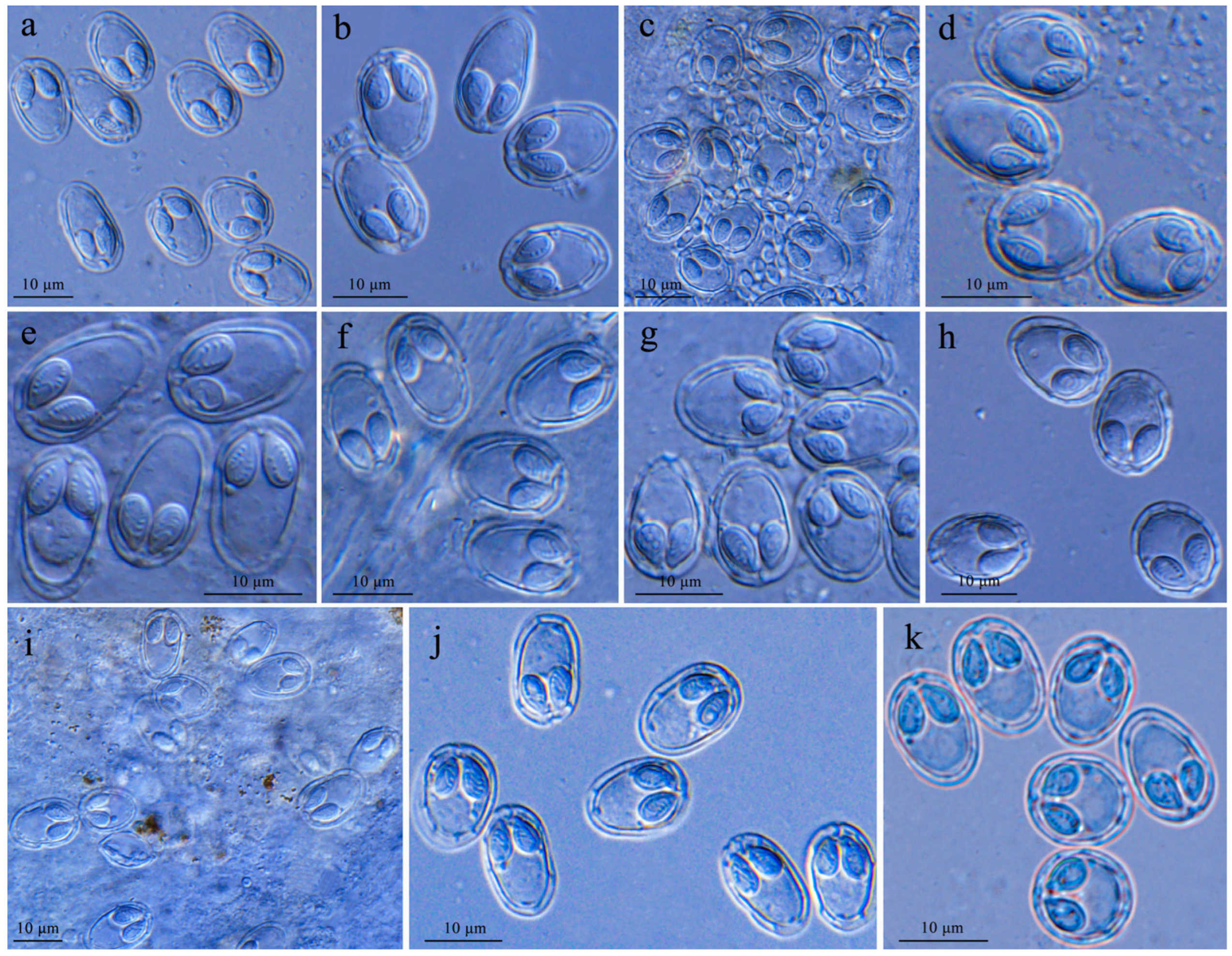
Spores from different organs and hosts were identical in shape (Figure 2). The spores were elongated obovate in frontal view with the blunt anterior ends wider than the posterior ends. Two pyriform polar capsules were equal in size. A polar filament was coiled with five to seven turns. In addition, a small number of abnormal spores with caudal appendages were observed in plasmodia from various organs of gibel carp (Figure 3). Morphometric analysis showed that spores of different isolates were generally of similar size. Although some indicators of spores showed obvious differences, there was an overlap in the morphometric data of different isolates. All the morphometric data of spores are given in Table 1.

3.3. Sequence and Phylogenetic Analyses

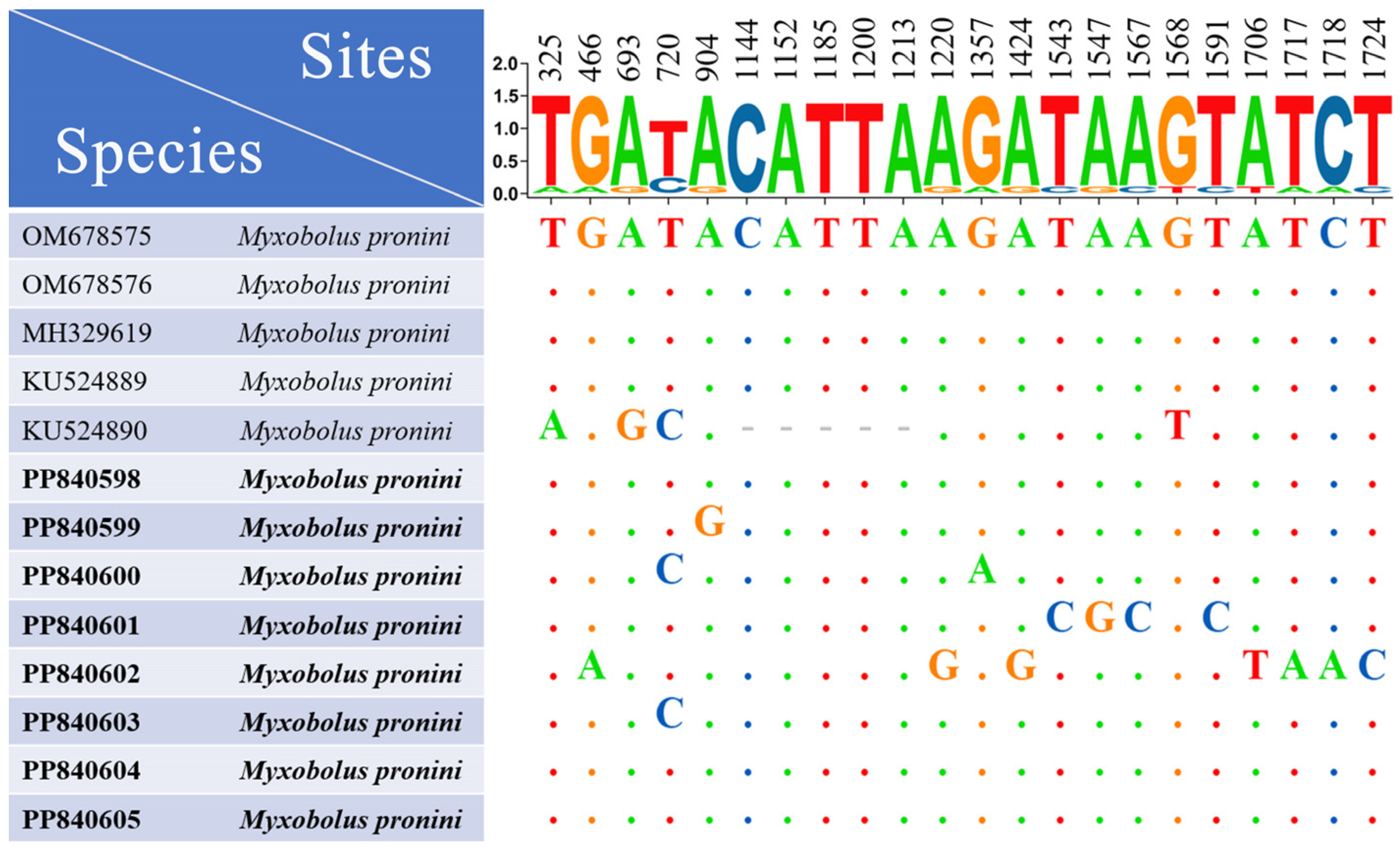
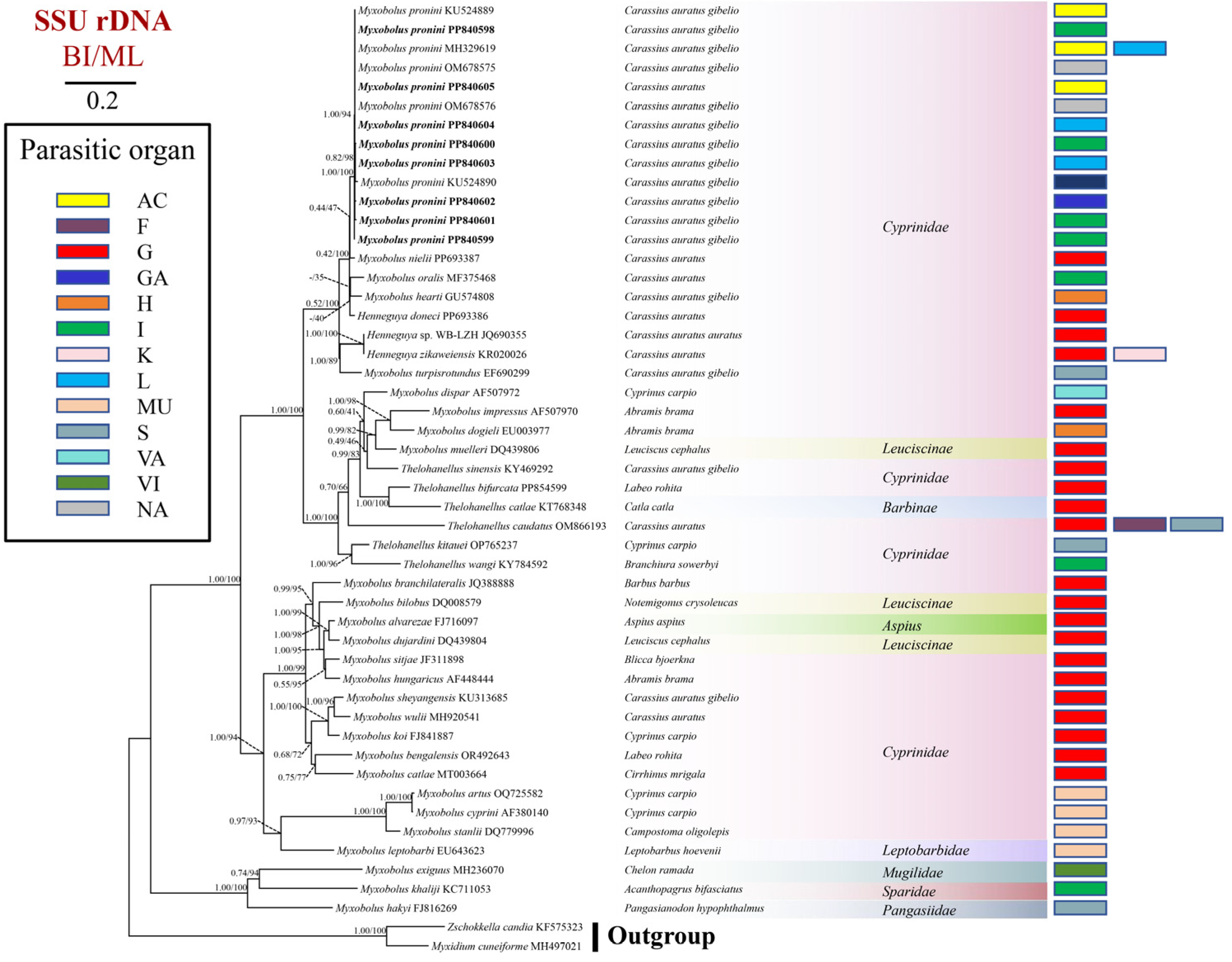
In the current study, seven SSU rDNA sequences were obtained from specimens collected from the intestine (length/accession number: 1949 bp/PP840598, 1992 bp/PP840599, 2074 bp/PP840600, 2104 bp/PP840601), liver (length/accession number: 1994 bp/PP840603, 1959 bp/PP840604), and gallbladder (length/accession number: 1610 bp/PP840602) of gibel carp. Additionally, a sequence with 1952 bp (accession number: PP840605) was acquired from specimens collected from the abdominal cavity of goldfish. BLAST analysis revealed that these SSU rDNA sequences in the present study exhibited 99.19–100% identity with M. pronini (MH329619, OM678575, OM678576, KU524890, KU524889). To assess intraspecific sequence variations, all available SSU rDNA sequences of M. pronini were subjected to sequence analysis, resulting in the identification of 22 variable sites distributed across 6 M. pronini isolates (Figure 4). The secondary structure analysis of SSU rRNA sequences showed that the V6 region’s structure was conserved across various M. pronini isolates (Figure 5). In contrast, structural variations were detected in the E23–3 section of the V4 region and the E43–2 section of the V7 region. Specifically, although all isolates exhibited one lateral process and one internal ring in the E23–3 section of the V4 region, the positions of the lateral process of three isolates (KU524890, PP840600, and PP840604) are different from that of other isolates. Furthermore, in the E43–2 section of the V7 region, most isolates possessed a large internal loop of 10 bases and a small internal loop of 8 bases, while an isolate from the intestine (PP840601) had two internal loops, both comprising 8 bases and an isolate. (KU524890) from Liu et al. (2016) had a larger internal loop of 12 bases. The ML analysis confirmed the clustering patterns of the Bayesian analysis in several cases with different bootstrap values. Phylogenetic analyses indicated that all M. pronini isolates clustered together, forming a distinct branch within the Carassius-infecting clade (Figure 6).
  • Taxonomic summary:
  • Host: Gibel carp Carassius auratus gibelio, goldfish C. auratus.
  • Host size: 13.3–17.3 cm in total length (gibel carp), 6.6–12.5 cm in total length (goldfish).
  • Site of infection: Intestine (gibel carp), liver (gibel carp), gallbladder (gibel carp), abdominal cavity (gibel carp, goldfish).
  • Prevalence: 33.3% (20/60, gibel carp), 4.5% (1/22, goldfish).
  • Locality: Lake Weishan Wetland (gibel carp), Chengyang Fish Market in Qingdao City (goldfish), Shandong Province, China.
  • Sample preservation: Accession no. MTR202205201, MTR202303121.

4. Discussion

In order to identify the present Myxobolus species, it was compared with all previously reported Myxobolus species. The findings revealed that this myxozoan species was morphologically indistinguishable from M. pronini based on spore morphology, as detailed in Table 1. Although morphometric variations were noted among different isolates of M. pronini, there was a significant overlap in the morphometric data of these isolates. This overlap suggests that the observed variations are likely intraspecific in nature. Indeed, the intraspecific morphometric variations of M. pronini spores were previously documented in the initial research by Liu et al. [35]. In addition, intraspecific morphometric variations of myxospores have been documented in a number of myxozoan species [37,38,39]. Early research primarily relied on spore morphology for the identification of myxozoans, and these intraspecific morphometric variations have posed significant challenges for accurate species identification, often leading to the establishment of numerous synonyms [8,16].
To further characterize the present myxozoan species, sequence analysis of the SSU rDNA was conducted in this study. The results indicated that the SSU rDNA sequences of the myxozoan species exhibited a remarkably high degree of similarity (99.19–100%) with M. pronini, which falls within the range of intraspecific sequence variations previously documented for myxozoan species [40,41]. In addition, variable sites within the SSU rDNA sequences of different isolates of M. pronini were identified in this study. This analysis revealed a total of 22 variable sites, with a maximum of fifteen variable sites observed between any two sequences. These results challenge the previously held assumption that the number of base variations within intraspecies of the genus Myxobolus is less than 10 bases [42]. Recently, a secondary structure of the SSU rRNA sequence has been identified as a potent tool for distinguishing myxozoan species with closely related molecular sequences [43]. In the current study, the structure of the V6 region was observed to be conserved across various isolates of M. pronini, whereas structural variations were detected in the E23–3 section of the V4 region and the E43–2 section of the V7 region. These findings indicated that the evolution rate of secondary structures in different regions of SSU rRNA may be different. Phylogenetic analyses revealed that all M. pronini isolates clustered together, forming a distinct branch placed in the Carassius-infecting clade, which is consistent with previous research [35,36]. Based on the comprehensive morphological and molecular analysis, the myxozoan species collected from various organs and hosts in the present study can be confidently identified as M. pronini, despite the observed morphological and molecular sequence variations among different isolates.
In the initial description by Liu et al. [35], M. pronini isolates were obtained from gibel carp from China and Russia. More recently, this parasite was again collected from gibel carp in China [36]. In the present study, in addition to gibel carp, M. pronini was first found to infect goldfish. Notably, several myxozoan species, such as M. honghuensis [44], M. ampullicapsulatus Zhao, Sun, Kent, Deng, Whipps, 2008 [36], M. wulii Landsberg, Lom, 1991 [36], M. pyramidis Chen, 1958 [36], have been documented to parasitize both gibel carp and goldfish, possibly due to their close genetic relationship. However, the limited attention given to myxozoans in goldfish has resulted in the identification of only approximately 10 myxozoan species in goldfish [10], as opposed to the over 50 myxozoan species reported to infect gibel carp [45]. We hypothesize that with increased research into myxozoan diversity in goldfish, a greater number of myxozoan species infecting both gibel carp and goldfish will be identified in the future. In China, M. pronini was initially collected from a natural water body, Lake Taibai, in Hubei Province [35]. Recently, this parasite was reported in aquaculture ponds in Hubei and Liaoning Provinces [36]. The present study has identified an expanded geographical distribution of this parasite, encompassing additional natural and aquaculture water bodies in Shandong Province. These findings suggest that M. pronini is a widely distributed parasite across China, thereby elevating its potential threat to both wild and farmed fish. In the initial description, M. pronini was reported to infect the abdominal cavities and visceral serous membranes of the livers of gibel carp [35]. Subsequently, free spores of M. pronini were observed in the gallbladders of gibel carp [36]. In addition to these known infection sites, the current study identified the intestine and gallbladder wall as additional target organs for M. pronini. The presence of multiple infection sites suggests the necessity of examining the various internal organs of a host when detecting M. pronini in the future.

5. Conclusions

This study identified the myxozoan species infecting various organs of gibel carp and goldfish as M. pronini. Isolates of this parasite from different organs and hosts exhibited both morphometric and molecular variations. In comparison to previous reports on M. pronini, the present research offers novel insights into its fish host, infection site, and geographic distribution.

Author Contributions

Methodology, F.Z., P.D. and R.S.; investigation, F.Z., X.Z., P.D. and R.S.; writing—original draft, F.Z.; writing—review and editing, X.Z. and Y.L.; resources, Z.W.; project administration, Z.W.; supervision, Y.L.; funding acquisition, Y.L. All authors have read and agreed to the published version of the manuscript.

Funding

The present work was financially supported by the Nature Science Foundation of China (grant number 32073012), the First Class Fishery Discipline ((2020)3), and a top talent plan “One Thing One Decision (Yishi Yiyi) ((2018)27)” in Shandong Province.

Institutional Review Board Statement

This study was approved by the Ethics Committee of the Laboratory Animal Administration of Shandong Province, China permit number SD2007695.

Informed Consent Statement

Not applicable.

Data Availability Statement

Data for this research article were available from the corresponding authors by reasonable request.

Conflicts of Interest

The authors declare no conflicts of interest.

References

  1. Holzer, A.S.; Bartošová–Sojková, P.; Born–Torrijos, A.; Lövy, A.; Hartigan, A.; Fiala, I. The joint evolution of the Myxozoa and their alternate hosts: A cnidarian recipe for success and vast biodiversity. Mol. Ecol. 2018, 27, 1651–1666. [Google Scholar] [CrossRef]
  2. Kayal, E.; Bentlage, B.; Sabrina Pankey, M.; Ohdera, A.H.; Medina, M.; Plachetzki, D.C.; Collins, A.G.; Ryan, J.F. Phylogenomics provides a robust topology of the major cnidarian lineages and insights on the origins of key organismal traits. BMC Evol. Biol. 2018, 18, 68. [Google Scholar] [CrossRef]
  3. Okamura, B.; Hartigan, A.; Naldoni, J. Extensive uncharted biodiversity: The parasite dimension. Integr. Comp. Biol. 2018, 58, 1132–1145. [Google Scholar] [CrossRef]
  4. Okamura, B.; Gruhl, A.; Bartholomew, J.L. Myxozoan Evolution, Ecology and Development; Springer International Publishing: Cham, Switzerland, 2015. [Google Scholar]
  5. Hoffman, G.L. Myxobolus cerebralis, a worldwide cause of salmonid whirling disease. J. Aquat. Anim. Health 1990, 2, 30–37. [Google Scholar] [CrossRef]
  6. Hallett, S.L.; Bartholomew, J.L. Myxobolus cerebralis and Ceratomyxa shasta. In Fish Parasites: Pathobiology and Protection; CAB International: Oxfordshire, UK, 2012; pp. 141–172. [Google Scholar]
  7. Liu, Y.; Whipps, C.M.; Gu, Z.M.; Zeng, C.; Huang, M.J. Myxobolus honghuensis n. sp. (Myxosporea: Bivalvulida) parasitizing the pharynx of allogynogenetic gibel carp Carassius auratus gibelio (Bloch) from Honghu Lake, China. Parasitol. Res. 2012, 110, 1331–1336. [Google Scholar] [CrossRef]
  8. Chen, Q.L.; Ma, C.L. Fauna Sinica. Myxozoa. Myxosporea; Science Press: Beijing, China, 1998. [Google Scholar]
  9. Liu, Y.; Whipps, C.M.; Nie, P.; Gu, Z.M. Myxobolus oralis sp. n. (Myxosporea: Bivalvulida) infecting the palate in the mouth of gibel carp Carassius auratus gibelio (Cypriniformes: Cyprinidae). Folia Parasitol. 2014, 61, 505–511. [Google Scholar] [CrossRef]
  10. Zhang, X.Y.; Yao, X.; Zhou, F.; Yang, C.Z.; Liu, Y. Identification of Thelohanellus pseudonikolskii n. sp. and Myxobolus koi Kudo, 1920 from goldfish Carassius auratus. Aquacult. Rep. 2022, 24, 101167. [Google Scholar] [CrossRef]
  11. Liu, Y.; Gu, Z.M.; Luo, Y.L. Some additional data to the occurrence, morphology and validity of Myxobolus turpisrotundus Zhang, 2009 (Myxozoa: Myxosporea). Parasitol. Res. 2010, 107, 67–73. [Google Scholar] [CrossRef]
  12. Batueva, M.D.D.; Pan, X.Y.; Zhang, J.Y.; Liu, X.H.; Wei, W.; Liu, Y. Morphological, histological and molecular characterization of Myxidium cf. rhodei infecting the kidney of Rutilus rutilus. Dis. Aquat. Organ. 2020, 141, 39–46. [Google Scholar] [CrossRef]
  13. Molnár, K.; Székely, C.; Hallett, S.L.; Atkinson, S.D. Some remarks on the occurrence, host–specificity and validity of Myxobolus rotundus Nemeczek, 1911 (Myxozoa: Myxosporea). Syst. Parasitol. 2009, 72, 71–79. [Google Scholar] [CrossRef]
  14. Huang, M.J.; Liu, Y.; Jia, L.; Zhai, Y.H.; Deng, Q.; Gu, Z.M. Supplementary studies on Myxobolus tsangwuensis Chen, 1954 (Myxozoa: Myxosporea) infecting the gills of common carp Cyprinus carpio (L.): Molecular and histological data. Acta Parasitol. 2014, 59, 653–660. [Google Scholar] [CrossRef] [PubMed]
  15. Baiko, D.; Lisnerová, M.; Bartošová–Sojková, P.; Holzer, A.S.; Blabolil, P.; Schabuss, M.; Fiala, I. Solving the Myxidium rhodei (Myxozoa) puzzle: Insights into its phylogeny and host specificity in Cypriniformes. Parasite 2024, 31, 35. [Google Scholar] [CrossRef] [PubMed]
  16. Liu, Y.; Whipps, C.M.; Liu, W.S.; Zeng, L.B.; Gu, Z.M. Supplemental diagnosis of a myxozoan parasite from common carp Cyprinus carpio: Synonymy of Thelohanellus xinyangensis with Thelohanellus kitauei. Vet. Parasitol. 2011, 178, 355–359. [Google Scholar] [CrossRef]
  17. Liu, Y.; Whipps, C.M.; Gu, Z.M.; Huang, M.J.; He, C.; Yang, H.L.; Molnár, K. Myxobolus musseliusae (Myxozoa: Myxobolidae) from the gills of common carp Cyprinus carpio and revision of Myxobolus dispar recorded in China. Parasitol. Res. 2013, 112, 289–296. [Google Scholar] [CrossRef]
  18. Zhang, B.; Zhai, Y.H.; Liu, Y.; Gu, Z.M. Myxobolus pseudowulii sp. n. (Myxozoa: Myxosporea), a new skin parasite of yellow catfish Tachysurus fulvidraco (Richardson) and redescription of Myxobolus voremkhai (Akhmerov, 1960). Folia Parasitol. 2017, 64, 030. [Google Scholar] [CrossRef]
  19. Li, Z.Y.; Wang, J.T.; Zhou, M.; Sato, H.; Zhang, J.Y. Morphological and molecular characterization of a new freshwater Ceratomyxa species (Cnidaria: Myxozoa) from the yellow catfish, Trachysurus fulvidraco in China. Parasitol. Int. 2023, 97, 102778. [Google Scholar] [CrossRef]
  20. Zhao, X.J.; Xu, L.W.; Ren, S.S.; Wei, Q.; Yin, Q.; Yang, Y.J.; Zhang, J.Y.; Xiang, J.G.; Yu, J.B.; Li, D.L.; et al. Myxobolus dumerilii sp. n. (Myxozoa: Myxobolidae) infecting the brain of Chinese longsnout catfish Tachysurus dumerili (Bleeker) in China. Syst. Parasitol. 2023, 100, 715–723. [Google Scholar] [CrossRef]
  21. Lom, J.; Arthur, J.R. A guideline for preparation of species descriptions in Myxosporea. J. Fish. Dis. 1989, 12, 151–156. [Google Scholar] [CrossRef]
  22. Barta, J.R.; Martin, D.S.; Liberator, P.A.; Dashkevicz, M.; Anderson, J.W.; Feigner, S.D.; Elbrecht, A.; Perkins–Barrow, A.; Jenkins, M.C.; Danforth, H.D.; et al. Phylogenetic relationships among eight eimeria species infecting domestic fowl inferred using complete small subunit ribosomal DNA sequences. J. Parasitol. 1997, 83, 262–271. [Google Scholar] [CrossRef]
  23. Fiala, I. The phylogeny of Myxosporea (Myxozoa) based on small subunit ribosomal RNA gene analysis. Int. J. Parasitol. 2006, 36, 1521–1534. [Google Scholar] [CrossRef] [PubMed]
  24. Katoh, K.; Standley, D.M. MAFFT multiple sequence alignment software version 7: Improvements in performance and usability. Mol. Biol. Evol. 2013, 30, 772–780. [Google Scholar] [CrossRef]
  25. Hall, T.A. BioEdit: A user–friendly biological sequence alignment editor and analysis program for windows 95/98/NT. Nucleic Acids Symp. Ser. 1999, 41, 95–98. [Google Scholar]
  26. Chen, C.J.; Chen, H.; Zhang, Y.; Thomas, H.R.; Frank, M.H.; He, Y.H.; Xia, R. TBtools: An integrative toolkit developed for interactive analyses of big biological data. Mol. Plant 2020, 13, 1194–1202. [Google Scholar] [CrossRef]
  27. Holzer, A.S.; Wootten, R.; Sommerville, C. The secondary structure of the unusually long 18S ribosomal RNA of the myxozoan Sphaerospora truttae and structural evolutionary trends in the Myxozoa. Int. J. Parasitol. 2007, 37, 1281–1295. [Google Scholar] [CrossRef] [PubMed]
  28. Kumar, S.; Stecher, G.; Tamura, K. MEGA7: Molecular Evolutionary Genetics Analysis Version 7.0 for Bigger Datasets. Mol. Biol. Evol. 2016, 33, 1870–1874. [Google Scholar] [CrossRef]
  29. Gonzalez, P.; Labarère, J. Phylogenetic relationships of Pleurotus species according to the sequence and secondary structure of the mitochondrial small–subunit rRNA V4, V6 and V9 domains. Microbiology 2000, 146, 209–221. [Google Scholar] [CrossRef] [PubMed]
  30. Hong, S.G.; Jung, H.S. Phylogenetic analysis of Ganoderma based on nearly complete mitochondrial smallsubunit ribosomal DNA sequences. Mycologia 2004, 96, 742–755. [Google Scholar] [CrossRef] [PubMed]
  31. De Rijk, P.; Wuyts, J.; De Wachter, R. RnaViz 2: An improved representation of RNA secondary structure. Bioinformatics 2003, 19, 299–300. [Google Scholar] [CrossRef] [PubMed]
  32. Minh, B.Q.; Schmidt, H.A.; Chernomor, O.; Schrempf, D.; Woodhams, M.D.; von Haeseler, A.; Lanfear, R. IQ–TREE 2: New models and efficient methods for phylogenetic inference in the genomic era. Mol. Biol. Evol. 2020, 37, 1530–1534. [Google Scholar] [CrossRef]
  33. Nylander, J.A.; Ronquist, F.; Huelsenbeck, J.P.; Nieves–Aldrey, J.L. Bayesian phylogenetic analysis of combined data. Syst. Biol. 2004, 53, 47–67. [Google Scholar] [CrossRef]
  34. Ronquist, F.; Teslenko, M.; van der Mark, P.; Ayres, D.L.; Darling, A.; Höhna, S.; Larget, B.; Liu, L.; Suchard, M.A.; Huelsenbeck, J.P. MrBayes 3.2: Efficient Bayesian phylogenetic inference and model choice across a large model space. Syst. Biol. 2012, 61, 539–542. [Google Scholar] [CrossRef] [PubMed]
  35. Liu, X.H.; Batueva, M.D.; Zhao, Y.L.; Zhang, J.Y.; Zhang, Q.Q.; Li, T.T.; Li, A.H. Morphological and molecular characterisation of Myxobolus pronini n. sp. (Myxozoa: Myxobolidae) from the abdominal cavity and visceral serous membranes of the gibel carp Carassius auratus gibelio (Bloch) in Russia and China. Parasit. Vectors 2016, 9, 562. [Google Scholar] [CrossRef] [PubMed]
  36. Zhang, B. Studies on Integrative Taxonomy and Molecular Phylogeny of Some Species of Myxobolus. Ph.D. Thesis, Huazhong Agricultural University, Wuhan, China, 2023. [Google Scholar]
  37. Urawa, S.; Freeman, M.A.; Johnson, S.C.; Jones, S.R.M.; Yokoyama, H. Geographical variation in spore morphology, gene sequences, and host specificity of Myxobolus arcticus (Myxozoa) infecting salmonid nerve tissues. Dis. Aquat. Organ. 2011, 96, 229–237. [Google Scholar] [CrossRef]
  38. Guo, Q.X.; Huang, M.J.; Liu, Y.; Zhang, X.P.; Gu, Z.M. Morphological plasticity in Myxobolus Bütschli, 1882: A taxonomic dilemma case and renaming of a parasite species of the common carp. Parasit. Vectors 2018, 11, 399. [Google Scholar] [CrossRef] [PubMed]
  39. Liu, Y.; Yao, J.Y.; Yao, X.; Zhang, K.J.; Wang, Q.Y.; Pan, X.Y.; Zhang, J.Y. Intraspecific morphological variation in myxosporeans: High pleomorphic myxospores in the same plasmodium of Myxobolus drjagini (Akhmerov, 1954). Folia Parasitol. 2022, 69, 006. [Google Scholar] [CrossRef]
  40. Whipps, C.M.; Diggles, B.K. Kudoa alliaria in flesh of Argentinian Hoki Macruronus magellanicus (Gadiformes; Merlucciidae). Dis. Aquat. Organ. 2006, 69, 259–263. [Google Scholar] [CrossRef] [PubMed]
  41. Ferguson, J.A.; Atkinson, S.D.; Whipps, C.M.; Kent, M.L. Molecular and morphological analysis of Myxobolus spp. of salmonid fishes with the description of Myxobolus species. J. Parasitol. 2008, 94, 1322–1334. [Google Scholar] [CrossRef] [PubMed]
  42. Zhao, Y.J.; Li, N.N.; Tang, F.H.; Dong, J.L. Remarks on the validity of Myxobolus ampullicapsulatus and Myxobolus honghuensis (Myxozoa: Myxosporea) based on SSU rDNA sequences. Parasitol. Res. 2013, 112, 3817–3823. [Google Scholar] [CrossRef]
  43. Zhang, X.Y.; Ding, P.; Zhang, C.H.; Sun, R.H.; Yang, C.Z.; Liu, Y. Redescription of Myxobolus ampullicapsulatus and comparison with molecular marker of M. honghuensis. J. Fish. China 2024, 48, 182–192. [Google Scholar]
  44. Guo, R.; Zhang, S.P.; Jiang, X.B.; Wang, W.; Wang, H.C.; Cai, L.M.; Yang, X.Q. Molecular identification and phylogenetic analysis of Myxobolus honghuensis found in pharynx of goldfish Carassius auratus. J. Fish. Res. 2021, 43, 61–66. [Google Scholar]
  45. Li, D.; Zhai, Y.H.; Gu, Z.M.; Liu, Y. Development of a multiplex PCR method for the simultaneous detection of four myxosporeans infecting gibel carp Carassius auratus gibelio. Dis. Aquat. Organ. 2017, 124, 31–39. [Google Scholar] [CrossRef] [PubMed]
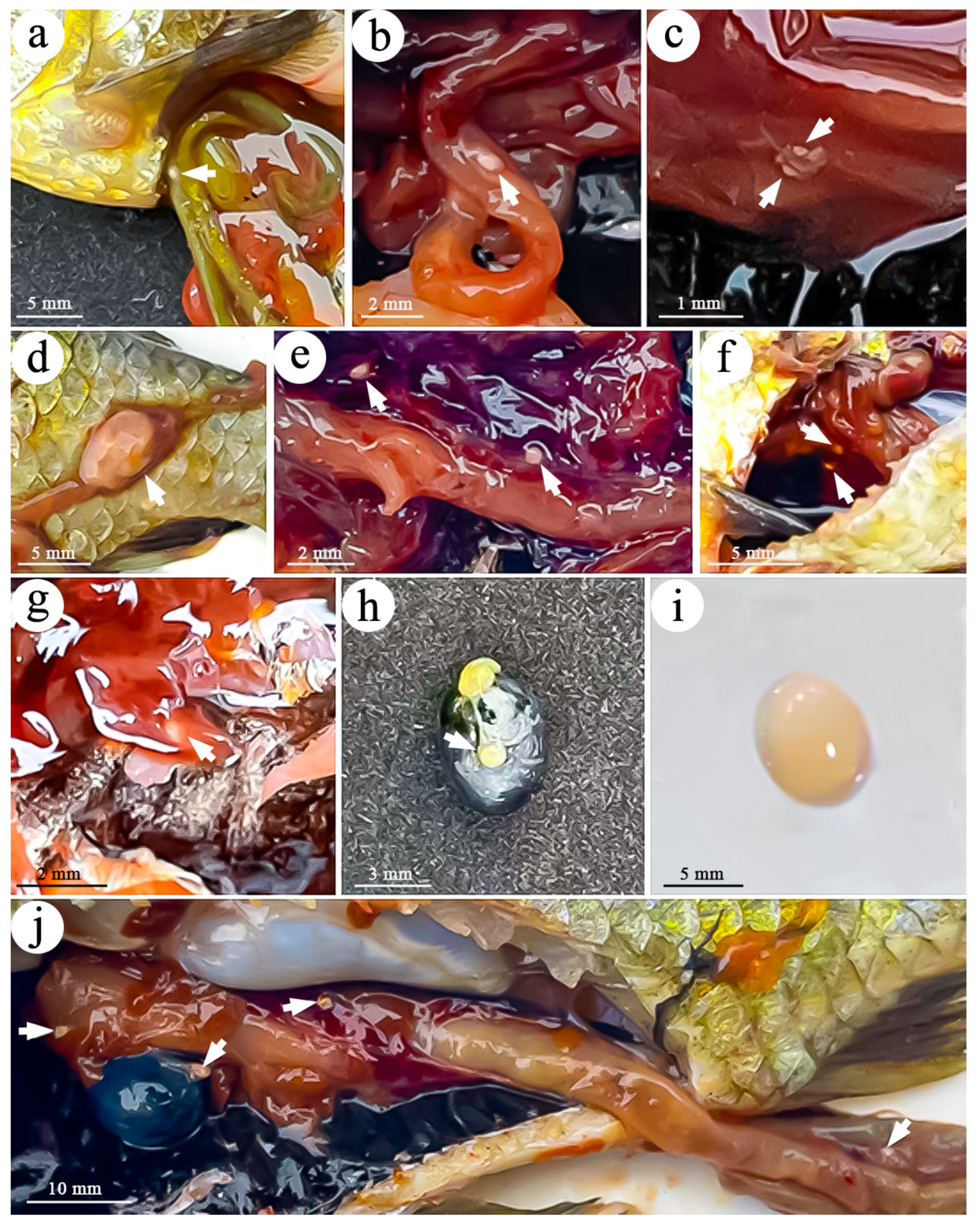
Figure 1. Plasmodia of Myxobolus pronini in various organs of gibel carp Carassius auratus gibelio and goldfish C. auratus. (ad) Plasmodia of M. pronini in intestine of gibel carp; (eg) plasmodia of M. pronini in liver of gibel carp; (h) plasmodium of M. pronini in gallbladder of gibel carp; (i) plasmodium of M. pronini in abdominal cavity of goldfish; (j) plasmodia of M. pronini in the intestine, liver, and gallbladder of the same gibel carp.
Fishes 10 00061 g001
Figure 2. Spores of Myxobolus pronini from various organs of gibel carp Carassius auratus gibelio and goldfish C. auratus. (ae) Spores of M. pronini from intestine of gibel carp; (fh) spores of M. pronini from liver of gibel carp; (i) spores of M. pronini from gallbladder of gibel carp; (j) spores of M. pronini from abdominal cavity of gibel carp; (k) spores of M. pronini from abdominal cavity of goldfish.
Fishes 10 00061 g002
Figure 3. Spores of Myxobolus pronini with caudal appendages from various organs of gibel carp Carassius auratus gibelio. (af) Spores of M. pronini with caudal appendages from intestine; (gj) spores of M. pronini with caudal appendages from liver; (k) spore of M. pronini with caudal appendages from gallbladder; (l) spore of M. pronini with caudal appendages from abdominal cavity.
Fishes 10 00061 g003
Figure 4. Nucleotide differences in SSU rDNA sequences among different isolates of Myxobolus pronini. The numbers in the header indicate the unmatched site positions.
Fishes 10 00061 g004
Figure 5. Secondary structures of V4, V6, and V7 regions of SSU rRNA sequences of Myxobolus pronini.
Fishes 10 00061 g005
Figure 6. Phylogenetic tree generated by SSU rDNA sequences of Myxobolus pronini and related species. Note: GenBank accession numbers are listed adjacent to species names. Support values at branching points are listed as Bayesian posterior probabilities/bootstrap values from maximum likelihood analysis. “-” represents the Bayesian posterior probability value that the branches of the BI tree do not match the branches of the ML tree. Abbreviations: AC—abdominal cavity; F—fins; G—gills; GA—gallbladder; H—heart; I—intestine; K—kidney; L—liver; MU—muscle; S—skin; VA—vascular; VI—visceral peritoneum; NA—not known.
Fishes 10 00061 g006
Table 1. Morphometric data of Myxobolus pronini collected in the present study with its other records (all measurements in μm). SL: spore length, SW: spore width, PCL: polar capsule length, PCW: polar capsule width, SI: site of infection. ”-”: not sequenced. The characteristics of spore shape (elongated obovate) and polar capsule shape (pyriform) are identical among all isolates.
SLSWPCLPCWSIHostSourceAccession Number
15.1 ± 0.3 (14.3–16.2)10.1 ± 0.1 (9.6–10.8)5.4 ± 0.6 (4.3–6.7)3.1 ± 0.1 (2.2–3.6)Abdominal cavityC. auratus gibelio[35]KU524889
14.7 ± 0.2 (13.8–15.6)9.6 ± 0.7 (9.0–13.3)5.3 ± 0.2 (4.8–5.6)3.0 ± 0.1 (2.9–3.4)Visceral serous membranes of liverC. auratus gibelio[35]KU524890
13.86 ± 0.8 (12.2–15.39)9.83 ± 0.5 (8.57–10.51)5.69 ± 0.42 (4.8–6.47)3.36 ± 0.21 (2.79–3.73)Abdominal cavity, gallbladderC. auratus gibelio[36]MH329619
16.8 ± 0.4 (16.0–17.9)9.5 ± 0.4 (8.7–10.2)6.0 ± 0.3 (5.6–6.8)3.4 ± 0.2 (3.1–3.8)IntestineC. auratus gibelioPresent researchPP840598
15.2 ± 0.5 (14.4–16.4)9.5 ± 0.5 (8.5–10.5)5.8 ± 0.4 (5.0–6.4)3.4 ± 0.2 (3.1–3.8)IntestineC. auratus gibelioPresent researchPP840599
16.5 ± 0.2 (16.1–16.9)10.6 ± 0.4 (9.9–11.2)5.8 ± 0.3 (5.3–6.3)3.6 ± 0.2 (3.3–3.9)IntestineC. auratus gibelioPresent researchPP840600
14.1 ± 0.4 (13.3–15.0)9.5 ± 0.5 (8.8–10.2)4.9 ± 0.4 (4.0–5.5)3.3 ± 0.3 (2.7–3.9)IntestineC. auratus gibelioPresent researchPP840601
13.0 ± 0.8 (11.4–14.3)9.5 ± 0.5 (8.3–10.6)5.3 ± 0.5 (4.4–6.3)3.1 ± 0.4 (2.3–3.9)IntestineC. auratus gibelioPresent research-
13.6 ± 0.5 (12.2–14.4)9.2 ± 0.5 (8.4–10.3)5.1 ± 0.3 (4.6–5.6)3.3 ± 0.2 (2.8–3.7)IntestineC. auratus gibelioPresent research-
15.0 ± 0.8 (13.6–16.5)9.4 ± 0.5 (8.0–10.4)5.8 ± 0.3 (5.0–6.3)3.2 ± 0.2 (2.9–3.5)Gallbladder wallC. auratus gibelioPresent researchPP840602
15.0 ± 0.3 (14.5–15.6)10.2 ± 0.3 (9.6–10.7)5.9 ± 0.2 (5.4–6.4)3.4 ± 0.2 (3.1–4.0)LiverC. auratus gibelioPresent researchPP840603
13.5 ± 0.4 (12.9–14.6)9.7 ± 0.4 (8.8–11.0)5.4 ± 0.3 (4.8–6.1)3.3 ± 0.2 (3.0–3.6)LiverC. auratus gibelioPresent researchPP840604
14.2 ± 0.8 (12.9–15.7)9.1 ± 0.3 (8.7–10.0)5.1 ± 0.4 (4.1–5.7)3.2 ± 0.2 (2.8–3.6)LiverC. auratus gibelioPresent research-
13.3 ± 0.8 (12.0–15.3)9.6 ± 0.6 (8.4–10.7)4.9 ± 0.4 (4.2–5.6)3.1 ± 0.3 (2.6–3.7)Abdominal cavityC. auratus gibelioPresent research-
12.8 ± 0.8 (11.2–14.3)9.5 ± 0.6 (8.4–10.5)4.8 ± 0.4 (4.2–5.6)2.9 ± 0.3 (2.3–3.5)Abdominal cavityC. auratusPresent researchPP840605
Disclaimer/Publisher’s Note: The statements, opinions and data contained in all publications are solely those of the individual author(s) and contributor(s) and not of MDPI and/or the editor(s). MDPI and/or the editor(s) disclaim responsibility for any injury to people or property resulting from any ideas, methods, instructions or products referred to in the content.