Article

Study on the Role and Pathological and Immune Responses of Silver Nanoparticles Against Two Aeromonas salmonicida subsp. salmonicida Strains at Different Virulence Levels in Rainbow Trout (Oncorhynchus mykiss)

by
Yunqiang Guo
1,†,
Chaoli Zheng
1,†,
Yingfei Wang
1,
Yongji Dang
1,
Ruiyuan Li
1,
Ye Tao
1,
Yucheng Yang
1,
Xiaofeng Sun
1,
Zekun Song
1,
Pengcheng Sun
1,
Qian Zhang
1,
Dandan Qian
1,
Wenhao Ren
1,
Xiyu Cao
1,
Bowen Wang
1,
Mengxi Xu
1,
Bingyang Jiang
1,
Yujing Li
1,
Qing Sun
1,
Jinye Wang
1,
Lei Zheng
2 and
Yanling Sun
1,*
add Show full author list remove Hide full author list
1
School of Marine Science and Engineering, Qingdao Agricultural University, Qingdao 266109, China
2
College of Marine Life Sciences, Xiamen Ocean Vocational College, Xiamen 361100, China
*
Author to whom correspondence should be addressed.
These authors contributed equally to this work.
Fishes 2025, 10(1), 29; https://doi.org/10.3390/fishes10010029
Submission received: 27 November 2024 / Revised: 7 January 2025 / Accepted: 7 January 2025 / Published: 13 January 2025
(This article belongs to the Special Issue Fish Diseases Diagnostics and Prevention in Aquaculture)

Abstract

:
Aeromonas species are among the main pathogens causing rainbow trout infections. Silver nanoparticles (AgNPs) have a broad spectrum of antimicrobial properties and are usually produced by various green-synthesis methods. However, the application of commercialized AgNPs has not fully been clarified. Thus, the objective of this study was to evaluate the antibacterial activities of commercialized AgNPs (range of sizes 10–12 nm) on two contrasting A. salmonicida strains (I-1 and I-4), isolated from rainbow trout; the antibacterial mechanism, histopathological alterations and the expression of immune-related genes were investigated. In vitro, the minimal inhibitory concentration (MIC) was 10 µg/mL for I-1, and lowered to 9.5 µg/mL for I-4, respectively. AgNPs were shown to disrupt both the cell wall and membrane of I-1 and I-4, resulting in cell lysis and degradation. In vivo, rainbow trout challenged by immersed or intraperitoneally injected infection, the 10 µg/mL AgNP-treated groups, both showed delayed deaths and lower mortalities compared to the control groups, without any clinical signs and pathological changes. Especially for the virulent I-4, the enhanced expressions of immune-related genes TNF-α, IL-1β, IL-10 and IL-11 were significantly reduced in the AgNP-treated group, indicating a lesser inflammation due to the application of AgNPs. This study would lay theoretical foundation for the wide application of silver nanoparticles in fish diseases.
Key Contribution: The commercialized AgNPs (range of sizes 10–12 nm) exhibited antibacterial activity against two contrasting A. salmonicida strains (I-1 and I-4) isolated from rainbow trout and the minimal inhibitory concentration was 10 µg/mL for I-1 and 9.5 µg/mL for I-4, respectively. Ultrastructural observation showed that 10 µg/mL AgNPs could disrupt the cell integrity of both I-1 and I-4, resulting in cell lysis and degradation. In vivo, the AgNP-treated groups showed lower mortality rates compared to the control groups, as well as normal clinical signs and no pathological changes, which were associated with the decreased expressions of inflammatory genes.

Graphical Abstract

1. Introduction

Aeromonas salmonicida, as a widely distributed pathogen, can induce a systemic furunculosis with high mortality, causing a great financial loss in the growing salmonid aquaculture worldwide [1,2]. A. salmonicida has previously been divided into five highly diverse subspecies: salmonicida, achromogenes, smithia, pectinolytica and masoucida [3], leading to the infection of salmonids [4,5] and non-salmonid species, such as multiple fishes in Petromyzonidae, Moronidae, Serranidae and Cyprinidae [6,7,8,9,10]. A. salmonicida can be spread in many ways, mainly including skin, blood, gills, mouth, and also from freshwater to the sea via a carrier fish harboring the bacterium without any signs of disease [11]. As a common pathogen, A. salmonicida has been identified to harbor numerous potential virulence factors, including flagella and pilli, outer-membrane proteins (OMPs), extracellular proteases, adhesins and functional secretion systems (T1SS, T2SS, T3SS, T6SS, etc.), and this pathogen induces typical furunculosis with the development of boils in the muscles in the chronic form and septicaemia in the acute form, leading to hemorrhagic lesions of skin in the anal region and the base of fins, as well as ascites and exophthalmia [1,3,12,13,14]. Thus, it is very difficult and complicated to prevent and control this pathogen due to its different subtypes, abundant virulence factors, various infected routes and fish species.
In the aquaculture industry, antibiotics were mainly prescribed to treat animals with Aeromonas diseases to avoid economic losses [11]. Also, many alternative strategies have been developed, which can avoid drug resistance and be a cure for Aeromonas diseases to improve fish health [1,15,16]. Various kinds of vaccines, including inactivated vaccine, subunit vaccine and adjuvants, polyvalent combination vaccine and phage application were constructed to defend against A. salmonicida. However, the effects of these vaccines are usually restricted to some subtypes or infected regions [1,6,7,16,17,18]. Thus, some other agents have also been used against A. salmonicida, including nanoparticles, probiotics and extracts of Chinese herbal medicines [14,15,19,20,21].
Nanoparticles, mostly as metal-based nanoparticles (NPs), including silver, copper, selenium, have unique properties and functions due to their small size, and have been paid increasing attention in a wide range of applications in recent years [22,23,24,25]. For instance, selenium nanoparticles have a protective effect against heat stress and promote the growth of rainbow trout as a supplementation alternative of fish diet [24,26]. Copper nanoparticles and vitamin C are also used as feed additives to elevate the growth performance, antioxidant capacity and disease resistance of rainbow trout against Yersinia ruckeri [23]. Silver nanoparticles (AgNPs) show antibacterial, antifungal and antiviral functions, potentially applied in aquaculture and agriculture [25,27,28,29,30,31,32]. As previously reported, AgNPs display antibacterial activity against Escherichia. coli, Vibrio harveyi and Enterococcus faecalis [27,33,34], and against fish pathogens such as A. salmonicida and Aphanomyces invadans [35], A. hydrophila and Flavobacterium aquidurense [36,37,38], and Pseudomonas aeruginosa and Vibrio species [39], and could potentially function as water disinfectants to inhibit the growth of pathogens and control water quality in fish farms. AgNPs have positive effects on rainbow trout against A. salmonicida and Pseudomonas species during egg incubation [25,30,32]. AgNPs have been reported to carry low toxicity to the hematopoietic system of rainbow trout (Oncorhynchus mykiss), despite that it can stimulate glycogenolysis in hepatocytes and decrease Na+ flux of gill cells when exposed to too-high levels of AgNPs [22,30,39,40,41,42]. In addition, AgNPs can be used as an effective antiseptic against foodborne pathogens, promising food protection [43].
Nowadays, AgNPs are produced by the green bio-synthesis or chemical reduction methods, and different sources of AgNPs can lead to different antibacterial effects [25,34,35,36,38,43,44,45]. So far, the knowledge about the application of AgNPs in fish diseases remains limited. Thus, the aim of this study is to test the antibacterial effects of commercialized AgNPs on rainbow trout against A. salmonicida strains. Here, using the two contrasting A. salmonicida strains with different virulence levels, the antibacterial roles of commercialized AgNPs were assessed in vitro and in vivo, and the antibacterial mechanism, histopathological alterations and immune-related gene responses were further investigated.

2. Methods and Materials

2.1. Fish, Bacteria and AgNPs

Juvenile rainbow trout, of an average body weight of 10 g, were obtained from the local farm in Weifang (Shandong, China) and maintained at 15 ± 1 °C in 1 m diameter plastic tanks within the aerated freshwater aquarium facility at the School of Marine Science and Engineering, Qingdao Agricultural University. Fish were fed using a commercial diet (2% body weight per day) at a photoperiod of 12 h: 12 h light and dark, and acclimatized for two weeks [2]. The current study was approved by the ethics committee of the School of Marine Science and Engineering, Qingdao Agricultural University.
A. salmonicida I-1 and I-4 were isolated from clinically infected rainbow trout from a previous furunculosis and identified as belonging to the subspecies salmonicida (unpublished), and preserved in equal volumes of their respective broths and of 30% glycerol in −80 °C at the fish disease laboratory of the School of Marine Science and Engineering, Qingdao Agricultural University. When used for bacterial treatment, they were inoculated in 5 mL Luria–Bertani nutrient broth (LB, MDBio, Qingdao, China) and incubated at 22 °C with shaking overnight as a starter culture. Then, 2 mL of the bacterial culture was transferred to 100 mL of LB and incubated overnight with shaking at 22 °C for use (OD600 at about 0.50, log phase of bacterial growth). As for the bacterial treatment, fish were anesthetized with 50 mg·L−1 MS-222 (Tricaine methanesulfonate, Sigma-Aldrich, St. Louis, MI, USA), and 50 μL of the I-1 or I-4 bacterial suspension (3.2 × 10⁷ CFU·mL−1) was intraperitoneally injected, respectively. Then, the cumulative mortality rates of fish were determined after 15–30 days. The bacteria isolated from dead fish had been identified as the same bacteria injected into fish bodies.
Liquid AgNPs (particle size: 10–15 nm, Aladdin, Shanghai, China) and solid superfine silver powder (particle size: 2 μm, Macklin, Shanghai, China) were purchased from the local agents and diluted by 1× phosphate buffered saline (PBS, pH = 7.4) solution to be prepared for use, respectively.

2.2. In Vitro Experiments of Bacterial Culture with AgNPs

I-4 bacterial solution was prepared as specified above (OD600 = ~0.50), and 100 μL of bacterial solution was spread on LB agar containing 10 μg/mL superfine silver powder and AgNPs, respectively. After 24 h, the bacterial counts were measured and analyzed.
In addition, 10 μL I-1 or I-4 was inoculated in 2 mL LB containing 0, 5, 5.5, 6, 6.5, 7, 7.5, 8, 8.5, 9, 9.5, 10 and 10.5 μg/mL AgNPs, respectively. They were cultured together at 22 °C with shaking at 150 rpm. After 24 h, the OD600 values of each treatment group were measured and the minimum inhibitory concentration (MIC) was calculated, respectively.

2.3. Transmission Electron Microscopy (TEM) of Bacterial Shapes

When I-1 and I-4 were grown in LB broth with shaking, as specified above, to the OD600 value of 0.2–0.3, 10 μg/mL AgNPs were added into LB and incubated together with shaking for 2 h. Then, the treated bacteria were collected and gently rinsed 2–3 times with 1× PBS. Subsequently, the bacteria samples were processed and embedded with copper wire meshes, examined and photographed with HT7700 TEM (Hitachi, Tokyo, Japan). In addition, 10 μL I-1 or I-4 were inoculated in 2 mL LB containing 10 μg/mL AgNPs, and after 12 h of incubation at 22 °C with shaking, the treated bacteria were collected and processed following the above procedures to be observed via TEM.

2.4. In Vivo Experiments of AgNPs in Fish with Bacterial Challenge

A total of 150 juvenile rainbow trout (10 ± 1.2 g) were divided into five groups (30 fish per group, placed in 50 L tanks): PBS, I-1, I-4, I-1+AgNPs and I-4+AgNPs groups. I-1 and I-4 bacterial solutions were prepared as specified above, collected by centrifugal force 5000× g and then washed for 2–3 times, and adjusted to the OD600 value of 0.55 using 1× PBS solution, respectively. As for the bacterial treatment, fish were anesthetized with 50 mg·L−1 MS-222 (Tricaine methanesulfonate, Sigma-Aldrich, St. Louis, MI, USA), and 50 μL of the I-1 or I-4 bacterial suspension (3.2 × 10⁷ CFU·mL−1) was intraperitoneally injected; as for the bacteria and AgNP treatment, 50 μL of I-1 + 10 μg/mL AgNPs or 50 μL of I-4 + 10 μg/mL AgNPs was intraperitoneally injected into fish body. As the control, the equal volume of PBS was intraperitoneally (i.p.) injected into fish bodies. At 6 and 24 hpi (hour after intraperitoneal injection), the head kidneys from the I-4 group and I-4+AgNPs group were sampled and grinded into tissue homogenates, and then diluted and spread in LB to count the bacterial loads. Samples were analyzed in triplicates from a pool of head kidneys of five fish. For one month, the clinical signs were observed and photographed, mortality was checked several times daily and dead fish were recorded and removed. The bacteria isolated from dead fish were identified as the same bacteria injected into fish bodies. Finally, the cumulative mortality rates of fish were determined after 15–30 days.
Similarly, the immersion experiments were performed under the same condition. A total of 150 fish were divided into five groups (30 fish per group, placed in 50 L tanks): PBS, I-1, I-4, I-1+AgNPs and I-4+AgNPs groups. I-1 and I-4 bacterial solutions were prepared and the fish were anesthetized as specified above. As for the bacterial treatment, the fish were immersed in 3.2 × 10⁸ CFU·ml−1 of I-1 and I-4 for 1 h, respectively; as for the bacteria and AgNP treatment, the immersed fish were then i.p. injected with 10 μg/mL AgNPs and transferred to 50 L tanks. The fish were i.p. injected with the equal volume of PBS as the control. The cumulative mortality rates of fish were determined after 15–30 days.

2.5. Histopathological Analyses

Fish tissues (gills, muscles, livers and intestines) were collected from the I-4 and I-4+AgNPs groups at 48 hpi, samples were fixed in 10% neutral buffered formalin, and those fixed samples were sent to the Servicebio® biotechnology Co., Ltd. (Wuhan, China) for sectioning and staining. They were processed (dehydration in an ascending series of ethanol, clearing in xylene, impregnation and embedding in melted paraffin), and then tissue sections (5 μm) were cut using a rotary microtome, and finally stained with the hematoxylin and eosin (HE) method, as described previously [3].

2.6. Immune Gene Transcription (RT-qPCR)

At 12 hpi, the head kidney and spleen tissues of fish from I-1, I-4, I-1+AgNPs and I-4+AgNPs groups were harvested, respectively. Total RNA was extracted using the FastPure® Cell/Tissue Total RNA Isolation Kit (Vazyme, Nanjing, China), with the concentration measured with a DS-11 Spectrophotometer (Denovix, Wilmington, DE, USA). A total of 100 ng RNA was reverse-transcribed into cDNA using the HiScript III 1st Strand cDNA Synthesis (Vazyme, Nanjing, China), and then the relative expressions of CK10, TNF-α, TRAF-1, IL-1β, IL-10 and IL-11 related to immune responses were quantified using Real-Time PCR (Thermofisher, Waltham, MA, USA) and SYBR Green qPCR Master Mix (ABM, Guangzhou, China), as described previously [3]. The expression of the target genes was corrected based on the endogenous control expression (EF-1α) according to the 2-ΔΔCT method as fold change [46]. Samples were analyzed in triplicates from a pool of each tissue of five fish. The primers used are listed in Table 1.

2.7. Statistical Analysis

The mortality was recorded and Kaplan–Meier survival curves were created by using GraphPad Prism (Version 6.0). For two comparisons, data were analyzed with one-way analysis of variance (ANOVA) using SPSS 17.0 (SPSS Inc., Chicago, IL, USA), and significant difference between two groups was shown by the asterisk (*, p < 0.05); For multiple comparisons, data were analyzed with two-way analysis of variance (ANOVA) using SPSS 17.0 (SPSS Inc., Chicago, IL, USA). All data were expressed as mean ± standard deviation (SD) and subjected to Duncan’s test. A p-value below 0.05 was considered statistically significant, as shown by the different lowercase letters, respectively.

3. Results

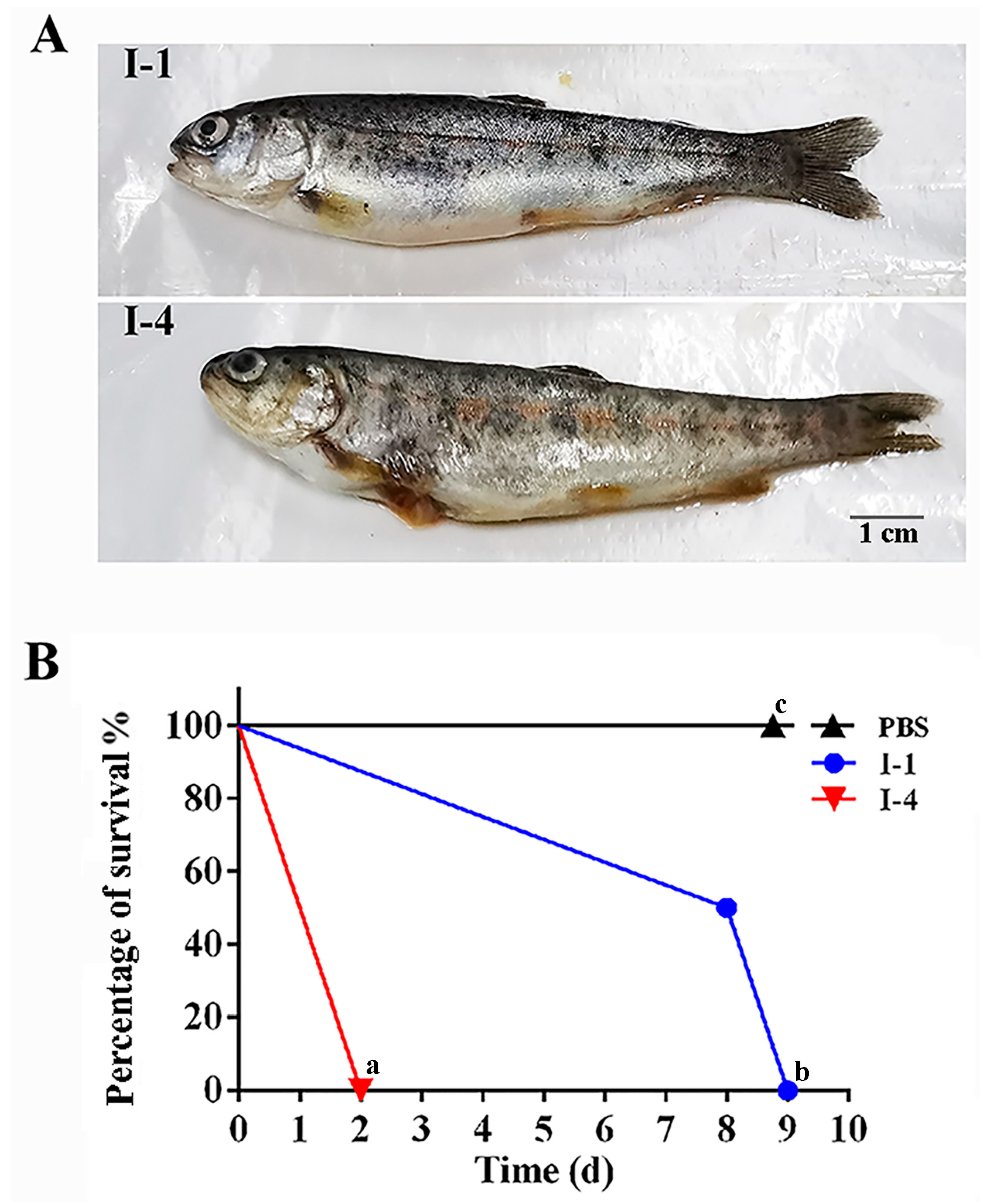
3.1. Characteristic of A. salmonicida I-1 and I-4

A. salmonicida I-1 and I-4 showed different virulence levels for rainbow trout. I-4 caused more severe clinical symptoms than I-1, including skin darkening, exophthalmia, hemorrhage and ascites. Evidently, when the fish were infected by I-4, the internal organs, muscle, the lateral line and anus were severely swollen and hemorrhagic. Specifically, the skin ulcer on the belly of the fish ruptured and the contents leaked out, and the caudal fin became rotted (Figure 1A). Moreover, the cumulative mortality rate of rainbow trout caused by I-4 (1 × 10⁹ CFU·ml−1) were up to 100% at 2 dpi (days after intraperitoneal injection), while the rainbow trout challenged by the equal amount of I-1 were all dead at 9 dpi (Figure 1B). Thus, I-1 and I-4 were named as the virulent strain and relatively low virulent strain, respectively.

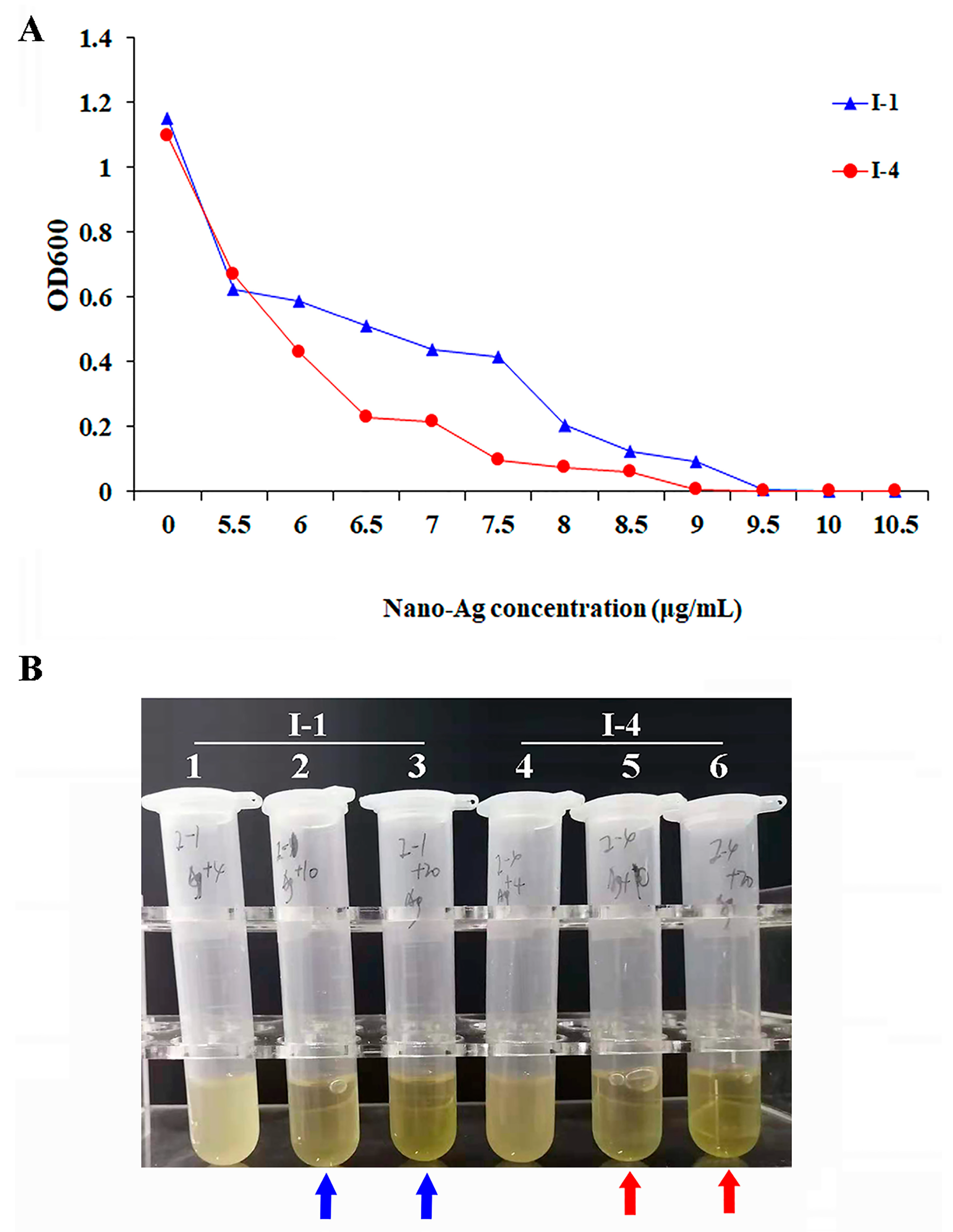
3.2. Antibacterial Effect of AgNPs

Here, AgNPs were chosen for the control of bacteria. Moreover, I-4 was used to check the effects of AgNPs and superfine silver powder. As showed in Figure 2A, when I-4 was spread in the LB containing 10 μg/mL AgNPs and superfine silver powder, after 16 h, the growth of I-4 was mostly inhibited by superfine silver powder, and evidently, it was totally inhibited by AgNPs and no bacteria appeared in the AgNP-containing LB. In addition, when I-4 (OD = 0.2–0.3) was inoculated in 2 mL LB containing 10 μg/mL AgNPs, no bacteria were measured after 0.5 or 2 h (Figure 2B). Thus, AgNPs had stronger antibacterial effect than superfine silver powder. Furthermore, AgNPs exhibited dose-dependent antibacterial activity, and the minimum inhibitory concentration (MIC) of I-1 was 10 μg/mL, while the MIC of I-4 was 9.5 μg/mL (Figure 3A). When I-1 or I-4 (OD = 0.2–0.3) was inoculated in 2 mL LB containing 5 and 10 μg/mL AgNPs, no bacteria were measured after 24 h (Figure 3B).
Importantly, in order to uncover the role of AgNPs, the shapes of I-1 and I-4 treated by AgNPs were examined via TEM. The result showed that the cell walls of I-1 (Figure 4A–C) and I-4 (Figure 4D–F) were destroyed after the treatment, and both ends of the bacteria also appeared broken (Figure 4B,E,H,L). Thus, AgNPs mainly disrupted the cell walls and membranes of bacteria.

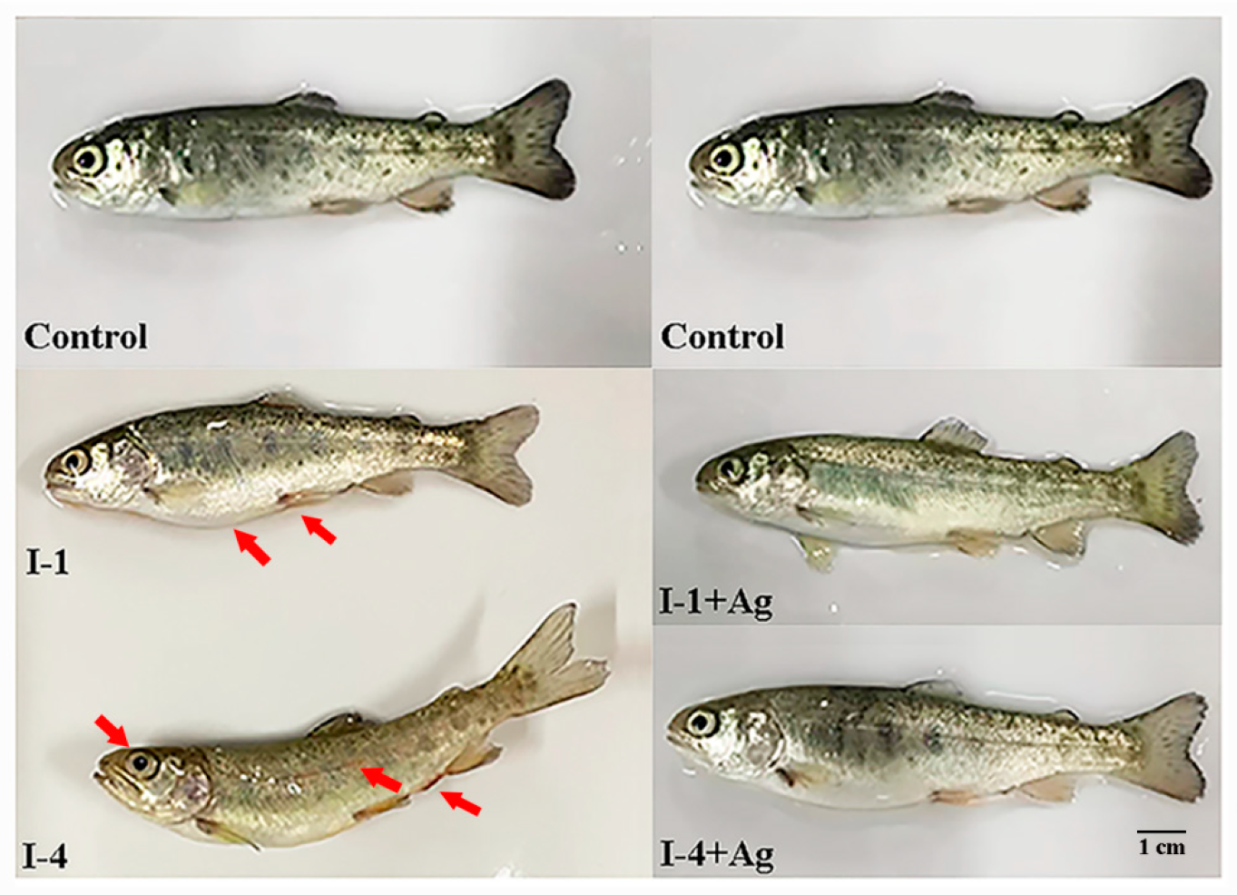
3.3. Influence of AgNPs on the Fish Against I-1 and I-4

Juvenile rainbow trout were challenged by A. salmonicida, which appeared to induce lethargy and decreased food intake, along with exophthalmia, hemorrhage, ascites, and multiple ulcerations on the skin which extended to the muscles underneath, while fish in both AgNP-treated groups did not show any clinical signs (Figure 5). Also, the bacterial loads of I-4 in the kidneys of the infected fish were examined, and it was found that the bacteria obviously propagated more in the kidney at 24 hpi than at 6 hpi in the I-4 group, but no bacteria were found in the kidney of the I-4+AgNPs group (Figure 6A). Furthermore, the immersed infection experiment showed that no mortalities were observed in the I-4+AgNPs group, while cumulative mortalities of 50.00% were found for the I-1+AgNPs group after 8 days, and no death had appeared since then. In contrast, fish in the I-1 and I-4 groups all died successively after 6 and 7 days (Figure 6B).
Moreover, in the injection challenge, dead fish both began to appear at 2 dpi and all died at 27 dpi in the I-1 group, while the inoculated fish in the AgNP-treated groups appeared to have delayed deaths (Figure 6C). After 23 dpi, no death appeared in the I-1+AgNPs group (Figure 6C). Evidently, the cumulative mortalities of the I-4 group were up to 100% at 7 dpi, but half of the inoculated fish died at 10 dpi, and no mortalities were observed in the I-4+AgNPs group (Figure 6D). Similarly, in goldfish, there were different virulence levels between I-1 and I-4, and the application of AgNPs induced delayed deaths (Figure S1).

3.4. Histopathological Changes

After being challenged by I-4 and I-4+AgNPs, the gill, muscle, liver and intestine isolated from the tested fish were examined at 48 dpi. Via histopathological observation, the fish in the I-4 group had bacterial aggregates, especially in the gill, liver and intestine (Figure 7). Evidently, some pathological changes were observed, including the swelled and rotted gill filaments, dissolved muscle fibers, nuclear pyknosis and hepatic congestion, necrotic mucosal epithelial and columnar cells in intestine, as well as shed intestinal villus (Figure 7A,C,E,G). In contrast, for the treated fish in the I-4+AgNPs group, these tissues had no similar lesions and maintained a healthy state (Figure 7B,D,F,H).

3.5. Gene Expression Analyses

Challenged by I-1 or I-4 with or without AgNPs, the expression of immune genes, including cytokeratin 10 (CK10), tumor necrosis factor-α (TNF-α), TNFR-associated factor (TRAF-1), interleukin-1β (IL-1β), IL-10 and IL-11, was detected in the kidney and spleen. As a result, in the kidney, the expression levels of these immune-related genes were all significantly higher in the I-1 or I-4 groups than in the control group at 12 hpi, especially for IL-1β and IL-11 in the I-4 group (Figure 8A,B). However, the immune-related genes were decreased in the I-4+AgNPs group, except CK10 and TNF-α (Figure 8B). In the I-1+AgNPs group, IL-1β was also downregulated, while other genes showed no obvious changes, and CK10, TRAF-1 and IL-10 were still upregulated compared to the control group (Figure 8A).
In the spleen, TNF-α, IL-10, IL-1β and IL-11 were obviously enhanced in the fish challenged by I-1 or I-4, but their expression levels became low in the fish from the I-1+AgNPs or I-4+AgNPs groups, although they were still higher than the control (Figure 8C,D). CK10 and TRAF-1 were increased in each group compared to the control at any point in time (Figure 8C,D).

4. Discussion

Silver nanoparticles (AgNPs) are widely recognized to have a broad spectrum of antimicrobial properties, associated with their tiny particle size, outsized surface area and biocompatibility. Significantly, various studies have reported on its application in the field of fish and shellfish diseases in recent years [25,31,35,37,38,44,45,47,48]. Using various bio-synthesis methods, AgNPs were produced and showed different antibacterial actions. However, the application of commercialized AgNPs is scarcely reported. Thus, this study was to investigate the antibacterial roles of commercialized AgNPs (range of sizes 10–12 nm) on two contrasting A. salmonicida strains (I-1 and I-4) in rainbow trout, and explore the mechanism of commercialized AgNPs against I-1 and I-4 via both in vitro and in vivo studies. As a result, in vitro, the minimal inhibitory concentration (MIC) of I-1 was at 10 µg/mL, while the MIC of I-4 was lowered to 9.5 µg/mL. AgNPs were observed attached to the cell wall and membrane of I-1 and I-4, leading to complete cell lysis and leakage of intracellular content. In vivo, the AgNP-treated groups both showed delayed deaths and lower mortalities than the control groups, and clinical signs or histopathological alterations similar to the control groups. Especially for the I-4+AgNP-treated group, the high expression of immune-related genes became obviously reduced after the application of AgNPs, indicating a relatively low immune response in the AgNP-treated group compared to the I-4 group.
The findings in the current study show that AgNPs have better inhibition for the virulent I-4 than the low virulent I-1. In vitro, 9.5 µg/mL AgNPs could completely inhibit the growth of I-4 and 10 µg/mL AgNPs could completely inhibit the growth of I-1. Ultrastructural observation showed that AgNPs attacked the cell walls and membranes of I-1 and I-4 strains and both ends or the outer periphery of the bacterial bodies were disrupted, resulting in the leakage of intracellular contents and complete cell lysis. These results are consistent with the previous studies on the antibacterial activity of the synthesized polyvinyl pyrrolidone (PVP)- or alginate-coated biogenic silver nanoparticles (Alg-AgNPs) against A. salmonicida and Pseudomonas strains [25,35,38,45]. Moreover, because of the multiple serotypes and variability of Aeromonas bacteria, various strategies, including antibiotics, Chinese herbal medicines, probiotics, vaccines and phage application, had previously been developed to prevent and control Aeromonas diseases in fish [1,6,7,14,15,16,17,18,19,20,21]. Back then, these strategies provided alternative solutions to control Aeromonas strains.
In vivo, in the immersed challenge, the survival rate of rainbow trout was up to 100% in the I-4+AgNPs group and 50% in the I-1+AgNPs group, while all the infected fish were dead after 7 days of treatment in the I-4 and I-1 groups. In contrast, the antibacterial effects of AgNPs in the injected challenge were not better than in the immersed challenge. In the intraperitoneal injected challenge, the survival rate of rainbow trout was up to 50% in the I-4+AgNPs group, and half of the fish suddenly died at 10 dpi, needing to be further investigated; In the I-1+AgNPs group, the survival rate of rainbow trout was unexpectedly 20%, and there was fluctuating death in the I-1 and I-1+AgNPs groups before 23 dpi, indicating the complexity of AgNPs applied in fish. Similarly, in goldfish, there were delayed deaths in the I-1+AgNPs group and I-4+AgNPs group, but the antibacterial effects on the whole was not useful. Probably, these deaths were mainly associated with the accumulation and cytotoxicity of AgNPs in fish. It was found that the fish remained healthy under the treatment of 10 µg/mL AgNPs, but began to die when the concentration of AgNPs reached up to 30 µg/mL. As previously reported, AgNPs in high concentrations were toxic to fish. For instance, 1.0 mg/L citrate-capped AgNPs and dialyzed citrate-capped AgNPs significantly inhibited unidirectional Na+ influx across the gills of juvenile rainbow trout [22], silver concentrations ranging from 0.1 mg/L to 10 mg/L had moderate cytotoxic effects in rainbow trout gill cells [40] and 10 g/mL AgNPs stimulated glycogenolysis in rainbow trout hepatocytes [41]. Mucus substance on the surface and atrophy of gill lamella, necrosis, degeneration and cell lysis were induced by the llex purpurea leaf-extract-capped AgNPs at 0.04 mg/L and 0.06 mg/L [42]. In zebrafish, when exposed to 10 μg/mL (9 nm size) AgNPs, the mortality rate was 75% and reached up to 100% mortality when doubling the concentration with the same size, while exposure to 30 nm-sized AgNPs resulted in only a 15% mortality rate at 10 μg/mL concentration [49]. Thus, the size and concentration of AgNPs are crucial factors influencing their potential toxicity. Previously, when rainbow trout were challenged by A. salmonicida, no death was shown in the jointly-treated groups intraperitoneally injected with 17 μg/mL PVP-capped AgNPs or exposed to 100 μg/L AgNPs by immersion for 3 h, based on the previous study [45]. In this study, 10 μg/mL AgNPs (10–11 nm size) were used and some death appeared in juvenile rainbow trout. Thus, the size, concentration and usage of AgNPs still need to be further modulated in rainbow trout against A. salmonicida. In Cyprinus carpio, the accumulation of AgNPs in gills and liver were observed and could induce oxidative stress in fish [50]. In rainbow trout, at 7 days post challenge with A. salmonicida, AgNPs were still found in the kidney, spleen and liver with no lesions, while at the 35th day post challenge, the AgNP-treated groups did not show any clinical signs, mortalities or histopathological alterations, except the liver necrosis that persisted [45]. Comparatively, in this study, the I-4+AgNPs group kept normal histological structures of gill, muscle, liver and intestine without any lesions in relation to the I-4 group at 48 hpi. Nevertheless, the long-time influence of AgNPs on the histopathological alterations in fish still needs to be checked.
To investigate the effects of AgNPs on the immune response of fish, the expressions of several immune-related genes were examined, including CK10, TRAF-1, TNF-α, IL-1β, IL-10 and IL-11. CK10 is a chemokine released from infected tissues to recruit diverse populations of leukocytes [51], and tumor necrosis factor receptor-associated factors (TRAFs) are critical intracellular signal adapters that activate downstream signals by binding directly to the cytoplasmic domain of tumor necrosis factor receptors (TNFR), which play an important role in regulating innate and adaptive immunity of aquatic organisms to confront various pathogens [52,53,54]. In this study, after bacterial challenge, CK10 and TRAF-1 were both upregulated compared to the control group. Contrastingly, in the I-4+AgNPs group, TRAF-1 was significantly decreased in the kidney, but CK10 was still increased, indicating its important role in the immune response to AgNPs at the early stage of rainbow trout infection. TNF-α and IL-1β are known as important indicators of phagocytic activity and the first cytokines produced in the early stages of cell-mediated inflammation in fish [55]. IL-10 is considered as a cytokine synthesis inhibitory factor that minimizes damage to target cells by suppressing the transcription of pro-inflammatory cytokines [56,57]. IL-11 is a multifunctional cytokine and has important immunomodulatory effects; it was markedly upregulated in rainbow trout with bacterial infection [58]. Remarkably, IL-1β, IL-10 and IL-11 were increased in the kidney and spleen of rainbow trout challenged by I-1 and I-4, but decreased in the AgNP-treated groups, indicating that the application of AgNPs reduced the inflammatory reactions caused by I-1 and I-4. Thus, it was inferred that AgNPs inhibited the growth of bacteria to reduce the inflammatory reactions in fish tissues. Correspondingly, histopathological observation in the I-4+AgNPs group showed that the gill, muscle, liver and intestine maintained normal states. Especially in I-1+AgNPs, the expression of IL-10 and IL-11 remained at high levels, which needs to be studied next. Moreover, via the miRNA-mRNA Seq, the gene regulatory network in rainbow trout response to AgNPs and bacterial infection may be uncovered and understood better in the future.

5. Conclusions

In summary, commercialized AgNPs (sizes 10–12 nm) completely inhibited the growth of the low virulent A. salmonicida I-1 at 9.5 µg/mL and the virulent I-4 at 10 µg/mL in vitro, and it was observed that AgNPs attached to the cell wall and membrane of bacteria, leading to cell lysis and degradation. In vivo, there were delayed deaths and lower mortalities in the AgNP-treated groups than the controls. Especially for the virulent I-4, the 10 µg/mL AgNP-treated group showed normal clinical signs and tissue structures, similarly to the control, associated with decreased expressions of the inflammatory genes IL-1β, IL-10 and IL-11. These results provide new valuable information on the application of silver nanoparticles against fish pathogens in aquaculture.

Supplementary Materials

The following supporting information can be downloaded at: https://www.mdpi.com/article/10.3390/fishes10010029/s1, Figure S1: The cumulative mortalities of the goldfishes challenged by I-1 and I-4 with or without silver nanoparticles.

Author Contributions

Y.G., C.Z., Y.D., R.L., Y.Y. and Y.S. performed the experiments. Y.G., X.S., Z.S., P.S., Q.Z., D.Q. and Y.S. analyzed the data, drew the figures and drafted the manuscript. Y.W., Y.T., W.R., X.C., B.W. and J.W. assisted in fish culture and sampling. M.X., B.J., Y.L., Q.S., L.Z. and Y.S. edited the manuscript. Y.S. supplied the chemical reagents and resources. All authors have read and agreed to the published version of the manuscript.

Funding

This work was supported by a research grant from the Natural Science Foundation of Shandong Province (ZR2023MC154), the Opening Fund for Key Laboratories of Xiamen’s smart fisheries (XMKLIF-OP-202406) and the National Natural Science Foundation of China (31902408).

Institutional Review Board Statement

The current study followed a standard working methodology approved by the Ethics Committee of the School of Marine Science and Engineering, Qingdao Agricultural University, (Protocol No. QAU2023-20) on 20 February 2023.

Informed Consent Statement

Not applicable.

Data Availability Statement

The raw data supporting the conclusions of this article will be made available by the authors upon request.

Conflicts of Interest

The authors declare no conflict of interest.

References

  1. Lund, V.; Mikkelsen, H.; Schrøder, M.B. Comparison of atypical furunculosis vaccines in spotted wolffish (Anarhicas minor O.) and Atlantic halibut (Hippoglossus hippoglossus L.). Vaccine 2008, 26, 2833–2840. [Google Scholar] [CrossRef] [PubMed]
  2. Ji, L.; Sun, G.; Li, X.; Liu, Y. Comparative transcriptome analysis reveals the mechanism of β-glucan in protecting rainbow trout (Oncorhynchus mykiss) from Aeromonas salmonicida infection. Fish. Shellfish Immunol. 2020, 98, 87–99. [Google Scholar] [CrossRef] [PubMed]
  3. Sun, X.N.; Wang, Q.; Wang, Y.F.; Tao, Y.; Zheng, C.L.; Wang, M.H.; Che, M.Y.; Cui, Z.H.; Li, X.L.; Zhang, Q.; et al. Isolation and identification of vapA-absent Aeromonas salmonicida in diseased snakehead Channa argus in China. Int. Microbiol. 2024, 27, 1137–1150. [Google Scholar] [CrossRef]
  4. Marana, M.H.; Jørgensen, L.V.; Skov, J.; Chettri, J.K.; Holm Mattsson, A.; Dalsgaard, I.; Kania, P.W.; Buchmann, K. Subunit vaccine candidates against Aeromonas salmonicida in rainbow trout Oncorhynchus mykiss. PLoS ONE 2017, 12, e0171944. [Google Scholar] [CrossRef]
  5. Marana, M.H.; Skov, J.; Chettri, J.K.; Krossøy, B.; Dalsgaard, I.; Kania, P.W.; Buchmann, K. Positive correlation between Aeromonas salmonicida vaccine antigen concentration and protection in vaccinated rainbow trout Oncorhynchus mykiss evaluated by a tail fin infection model. J. Fish Dis. 2017, 40, 507–516. [Google Scholar] [CrossRef]
  6. Diamanka, A.; Loch, T.P.; Cipriano, R.C.; Winters, A.D.; Faisal, M. Infection of sea lamprey with an unusual strain of Aeromonas salmonicida. J. Wildl. Dis. 2014, 50, 159. [Google Scholar] [CrossRef]
  7. Brietzke, A.; Korytář, T.; Jaros, J.; Köllner, B.; Goldammer, T.; Seyfert, H.M.; Rebl, A. Aeromonas salmonicida infection only moderately regulates expression of factors contributing to Toll-Like receptor signaling but massively activates the cellular and humoral branches of innate immunity in rainbow trout (Oncorhynchus mykiss). J. Immunol. Res. 2015, 2015, 901015. [Google Scholar] [CrossRef]
  8. Alghabshi, A.; Austin, B.; Crumlish, M. Aeromonas salmonicida isolated from wild and farmed fish and invertebrates in Oman. Int. Aquat. Res. 2018, 10, 145. [Google Scholar] [CrossRef]
  9. Bhat, R.A.H.; Thakuria, D.; Dubey, M.K.; Tandel, R.S.; Sharma, P.; Khangembam, V.C.; Dash, P.; Tripathi, G.; Sarman, D. Lethal dose and histopathological alterations induced by Aeromonas salmonicida in experimentally challenged common carp, Cyprinus carpio. Microb. Pathog. 2021, 158, 105110. [Google Scholar] [CrossRef]
  10. Al-Mokaddem, A.K.; Abdel-moneam, D.A.; Ibrahim, R.A.; Saleh, M.; Shaalan, M. Molecular identification, histopathological analysis and immunohistochemical characterization of non-pigmented Aeromonas salmonicida subsp. salmonicida in Mugil carinatus (Valenciennes, 1836). Aquac. Rep. 2022, 24, 101103. [Google Scholar] [CrossRef]
  11. Dalsgaard, I.; Madsen, L. Bacterial pathogens in rainbow trout, Oncorhynchus mykiss (Walbaum), reared at Danish freshwater farms. J. Fish Dis. 2000, 23, 199–209. [Google Scholar] [CrossRef]
  12. Austin, B.; Austin, D. Bacterial Fish Pathogens: Disease of Farmed and Wild Fish, 5th ed.; Springer: Dordrecht, The Netherlands, 2012. [Google Scholar]
  13. Vanden Bergh, P.; Frey, J. Aeromonas salmonicida subsp. salmonicida in the light of its type-three secretion system. Microb. Biotechnol. 2014, 7, 381–400. [Google Scholar] [CrossRef] [PubMed]
  14. Hayatgheib, N.; Fournel, C.; Calvez, S.; Pouliquen, H.; Moreau, E. In vitro antimicrobial effect of various commercial essential oils and their chemical constituents on Aeromonas salmonicida subsp. salmonicida. J. Appl. Microbiol. 2020, 129, 137–145. [Google Scholar] [CrossRef] [PubMed]
  15. Akhtar, M.S.; Tripathi, P.H.; Pandey, A.; Ciji, A. β-glucan modulates non-specific immune gene expression, thermal tolerance and elicits disease resistance in endangered Tor putitora fry challenged with Aeromonas salmonicida. Fish Shellfish Immunol. 2021, 119, 154–162. [Google Scholar] [CrossRef] [PubMed]
  16. Veenstra, K.A.; Wang, T.; Russell, K.S.; Tubbs, L.; Ben Arous, J.; Secombes, C.J. Montanide™ ISA 763A VG and ISA 761 VG induce different immune pathway responses in rainbow trout (Oncorhynchus mykiss) when used as adjuvant for an Aeromonas salmonicida bacterin. Fish Shellfish Immunol. 2021, 114, 171–183. [Google Scholar] [CrossRef]
  17. Vincent, A.T.; Paquet, V.E.; Bernatchez, A.; Tremblay, D.M.; Moineau, S.; Charette, S.J. Characterization and diversity of phages infecting Aeromonas salmonicida subsp. salmonicida. Sci. Rep. 2017, 7, 7054. [Google Scholar] [CrossRef]
  18. Lim, J.; Hong, S. Characterization of Aeromonas salmonicida and A. sobria isolated from cultured salmonid fish in Korea and development of a vaccine against furunculosis. J. Fish Dis. 2020, 43, 609–620. [Google Scholar] [CrossRef]
  19. Gao, X.Y.; Liu, Y.; Miao, L.L.; Li, E.W.; Sun, G.X.; Liu, Y.; Liu, Z.P. Characterization and mechanism of anti-Aeromonas salmonicida activity of a marine probiotic strain, Bacillus velezensis V4. Appl. Microbiol. Biotechnol. 2017, 101, 3759–3768. [Google Scholar] [CrossRef]
  20. Cornet, V.; Khuyen, T.D.; Mandiki, S.N.M.; Betoulle, S.; Bossier, P.; Reyes-López, F.E.; Tort, L.; Kestemont, P. GAS1: A new β-Glucan immunostimulant candidate to increase rainbow trout (Oncorhynchus mykiss) resistance to bacterial infections with Aeromonas salmonicida achromogenes. Front. Immunol. 2021, 12, 693613. [Google Scholar] [CrossRef]
  21. Yonar, M.E.; Mişe Yonar, S.; İspir, Ü.; Ural, M.Ş. Effects of curcumin on haematological values, immunity, antioxidant status and resistance of rainbow trout (Oncorhynchus mykiss) against Aeromonas salmonicida subsp. achromogenes. Fish Shellfish Immunol. 2019, 89, 83–90. [Google Scholar] [CrossRef]
  22. Schultz, A.G.; Ong, K.J.; MacCormack, T.; Ma, G.; Veinot, J.G.; Goss, G.G. Silver nanoparticles inhibit sodium uptake in juvenile rainbow trout (Oncorhynchus mykiss). Environ. Sci. Technol. 2012, 46, 10295–10301. [Google Scholar] [CrossRef] [PubMed]
  23. Delavari, N.M.; Gharaei, A.; Mirdar, H.J.; Davari, A.; Rastiannasab, A. Modulatory effect of dietary copper nanoparticles and vitamin C supplementations on growth performance, hematological and immune parameters, oxidative status, histology, and disease resistance against Yersinia ruckeri in rainbow trout (Oncorhynchus mykiss). Fish Physiol. Biochem. 2022, 48, 33–51. [Google Scholar] [CrossRef] [PubMed]
  24. Yanez-Lemus, F.; Moraga, R.; Smith, C.T.; Aguayo, P.; Sánchez-Alonzo, K.; García-Cancino, A.; Valenzuela, A.; Campos, V.L. Selenium Nanoparticle-Enriched and Potential Probiotic, Lactiplantibacillus plantarum S14 Strain, a Diet Supplement Beneficial for Rainbow Trout. Biology 2022, 11, 1523. [Google Scholar] [CrossRef] [PubMed]
  25. Kulikouskaya, V.; Nikalaichuk, V.; Hileuskaya, K.; Ladutska, A.; Grigoryan, K.; Kozerozhets, I.; Hovsepyan, V.; Sargsyan, M.; Sidarenka, A. Alginate coated biogenic silver nanoparticles for the treatment of Pseudomonas infections in rainbow trout. Int. J. Biol. Macromol. 2023, 251, 126302. [Google Scholar] [CrossRef]
  26. Sun, J.; Liu, Z.; Quan, J.; Li, L.; Zhao, G.; Lu, J. Protective effects of different concentrations of selenium nanoparticles on rainbow trout (Oncorhynchus mykiss) primary hepatocytes under heat stress. Ecotoxicol. Environ. Saf. 2022, 230, 113121. [Google Scholar] [CrossRef]
  27. Sondi, I.; Salopek-Sondi, B. Silver nanoparticles as antimicrobial agent: A case study on E. coli as a model for Gram-negative bacteria. J. Colloid Interface Sci. 2004, 275, 177–182. [Google Scholar]
  28. Suchomel, P.; Kvitek, L.; Panacek, A.; Prucek, R.; Hrbac, J.; Vecerova, R.; Zboril, R. Comparative study of antimicrobial activity of AgBr and Ag nanoparticles (NPs). PLoS ONE 2015, 10, e0119202. [Google Scholar] [CrossRef]
  29. Sarmast, M.K.; Salehi, H. Silver nanoparticles: An influential element in plant nanobiotechnology. Mol. Biotechnol. 2016, 58, 441–449. [Google Scholar] [CrossRef]
  30. Clark, N.J.; Shaw, B.J.; Handy, R.D. Low hazard of silver nanoparticles and silver nitrate to the haematopoietic system of rainbow trout. Ecotoxicol. Environ. Saf. 2018, 152, 121–131. [Google Scholar] [CrossRef]
  31. Saadh, M.J. Silver nanoparticles inhibit goatpox virus replication. Arch. Virol. 2023, 168, 32. [Google Scholar] [CrossRef]
  32. Kowalska-Góralska, M.; Senze, M.; Łuczyńska, J.; Czyż, K. Effects of the ionic and nanoparticle forms of Cu and Ag on these metals’ bioaccumulation in the eggs and fry of rainbow trout (Oncorhynchus mykiss W.). Int. J. Environ. Res. Public Health 2020, 17, 6392. [Google Scholar] [CrossRef] [PubMed]
  33. Vaseeharan, B.; Ramasamy, P.; Chen, J.C. Antibacterial activity of silver nanoparticles (AgNps) synthesized by tea leaf extracts against pathogenic Vibrio harveyi and its protective efficacy on juvenile Feneropenaeus indicus. Lett. Appl. Microbiol. 2010, 50, 352–356. [Google Scholar] [CrossRef] [PubMed]
  34. Chandran, N.; Ramesh, S.; Shanmugam, R. Synthesis of silver nanoparticles using Azadirachta indica and Syzygium aromaticum extract and its antibacterial action against Enterococcus faecalis: An in vitro study. Cureus 2024, 16, e65044. [Google Scholar] [CrossRef] [PubMed]
  35. Shaalan, M.I.; El-Mahdy, M.M.; Theiner, S.; El-Matbouli, M.; Saleh, M. In vitro assessment of the antimicrobial activity of silver and zinc oxide nanoparticles against fish pathogens. Acta Vet. Scand. 2017, 59, 49. [Google Scholar] [CrossRef]
  36. Antony, J.J.; Nivedheetha, M.; Siva, D.; Pradeepha, G.; Kokilavani, P.; Kalaiselvi, S.; Sankarganesh, A.; Balasundaram, A.; Masilamani, V.; Achiraman, S. Antimicrobial activity of Leucas aspera engineered silver nanoparticles against Aeromonas hydrophila in infected Catla catla. Colloids Surf. B Biointerfaces 2013, 109, 20–24. [Google Scholar] [CrossRef]
  37. Nemati, T.; Johari, S.A.; Sarkheil, M. Will the antimicrobial properties of ZnONPs turn it into a more suitable option than AgNPs for water filtration? Comparative study in the removal of fish pathogen, Aeromonas hydrophila from the culture of juvenile common carp (Cyprinus carpio). Environ. Sci. Pollut. Res. Int. 2019, 26, 30907–30920. [Google Scholar] [CrossRef]
  38. Ramya, J.R.; Ali, S.; K, T.A.; Vijayalakshmi, R.; Gajendiran, J.; Gnanam, S.; Ramachandran, K. Antimicrobial efficiency against fish pathogens on the green synthesized silver nanoparticles. Microb. Pathog. 2024, 193, 106725. [Google Scholar] [CrossRef]
  39. Saad, M.F.; Elsayed, M.M.; Khder, M.; Abdelaziz, A.S.; El-Demerdash, A.S. Biocontrol of multidrug resistant pathogens isolated from fish farms using silver nanoparticles combined with hydrogen peroxide insight to its modulatory effect. Sci. Rep. 2024, 14, 7971. [Google Scholar] [CrossRef]
  40. Farkas, J.; Christian, P.; Gallego-Urrea, J.A.; Roos, N.; Hassellöv, M.; Tollefsen, K.E.; Thomas, K.V. Uptake and effects of manufactured silver nanoparticles in rainbow trout (Oncorhynchus mykiss) gill cells. Aquat. Toxicol. 2011, 101, 117–125. [Google Scholar] [CrossRef]
  41. Massarsky, A.; Labarre, J.; Trudeau, V.L.; Moon, T.W. Silver nanoparticles stimulate glycogenolysis in rainbow trout (Oncorhynchus mykiss) hepatocytes. Aquat. Toxicol. 2014, 147, 68–75. [Google Scholar] [CrossRef]
  42. Ali, I.; Khan, S.; Shah, K.; Haroon; Kalimullah; Bian, L. Microscopic analysis of plant-mediated silver nanoparticle toxicity in rainbow trout fish (Oncorhynchus mykiss). Microsc. Res. Tech. 2021, 84, 2302–2310. [Google Scholar] [CrossRef] [PubMed]
  43. El-Sherbiny, M.M.; Devassy, R.P.; El-Hefnawy, M.E.; Al-Goul, S.T.; Orif, M.I.; El-Newehy, M.H. Facile synthesis, characterization, and antimicrobial assessment of a silver/montmorillonite nanocomposite as an effective antiseptic against foodborne pathogens for promising food protection. Molecules 2023, 28, 3699. [Google Scholar] [CrossRef] [PubMed]
  44. Thepbandit, W.; Papathoti, N.K.; Hoang, N.H.; Siriwong, S.; Sangpueak, R.; Saengchan, C.; Laemchiab, K.; Kiddeejing, D.; Tonpho, K.; Buensanteai, K. Bio-synthesis and characterization of silver nanoparticles from Trichoderma species against cassava root rot disease. Sci. Rep. 2024, 14, 12535. [Google Scholar] [CrossRef] [PubMed]
  45. Shaalan, M.; El-Mahdy, M.; Theiner, S.; Dinhopl, N.; El-Matbouli, M.; Saleh, M. Silver nanoparticles: Their role as antibacterial agent against Aeromonas salmonicida subsp. salmonicida in rainbow trout (Oncorhynchus mykiss). Res. Vet. Sci. 2018, 119, 196–204. [Google Scholar] [CrossRef]
  46. Livak, K.J.; Schmittgen, T.D. Analysis of relative gene expression data using real-time quantitative PCR and the 2(-Delta Delta C(T)) Method. Methods 2001, 25, 402–408. [Google Scholar] [CrossRef]
  47. Youssef, A.M.; Abdel-Aziz, M.S. Preparation of polystyrene nanocomposites based on silver nanoparticles using marine bacterium for packaging. Polym.-Plast. Technol. Eng. 2013, 52, 607–613. [Google Scholar] [CrossRef]
  48. Franci, G.; Falanga, A.; Galdiero, S.; Palomba, L.; Rai, M.; Morelli, G.; Galdiero, M. Silver nanoparticles as potential antibacterial agents. Molecules 2015, 20, 8856–8874. [Google Scholar] [CrossRef]
  49. Mosselhy, D.A.; He, W.; Li, D.; Meng, Y.; Feng, Q. Silver nanoparticles: In vivo toxicity in zebrafish embryos and a comparison to silver nitrate. J. Nanopart Res. 2016, 18, 222. [Google Scholar] [CrossRef]
  50. Liaqat, F.; Hanif, U.; Bahadur, S.; Faheem, M.; Rasool, S.; Gulzar, S.; Zaman, W.; Urooj, Z.; Shaheen, S.; Munir, M. Comparative evaluation of the toxicological effect of silver salt (AgNO3) and silver nanoparticles on Cyprinus carpio synthesized by chemicals and marine algae using scanning electron microscopy. Microsc. Res. Tech. 2021, 84, 1531–1541. [Google Scholar] [CrossRef]
  51. Bird, S.; Tafalla, C. Teleost Chemokines and Their Receptors. Biology 2015, 4, 756–784. [Google Scholar] [CrossRef]
  52. Syahputra, K.; Kania, P.W.; Al-Jubury, A.; Marnis, H.; Setyawan, A.C.; Buchmann, K. Differential immune gene response in gills, skin, and spleen of rainbow trout Oncorhynchus mykiss infected by Ichthyophthirius multifiliis. PLoS ONE 2019, 14, e0218630. [Google Scholar] [CrossRef] [PubMed]
  53. You, S.L.; Jiang, X.X.; Zhang, G.R.; Ji, W.; Ma, X.F.; Zhou, X.; Wei, K.J. Molecular characterization of nine TRAF genes in Yellow catfish (Pelteobagrus fulvidraco) and their expression profiling in response to Edwardsiella ictaluri infection. Int. J. Mol. Sci. 2023, 24, 8363. [Google Scholar] [CrossRef] [PubMed]
  54. Zou, Y.; Zhou, Y.; Chen, Z.; Zou, P.; Zhu, Y.; Zhang, J.; Zhang, Z.; Wang, Y. Members of the TRAF gene family in Octopus sinensis and their response to PGN, poly I:C, and Vibrio parahaemolyticus. Fish Shellfish Immunol. 2024, 154, 109905. [Google Scholar] [CrossRef] [PubMed]
  55. Skadberg, E.S.S. Inflammatory Effects in Zebrafish (Danio rerio) Exposed to Gold Nanoparticles. Master’s Thesis, University of Oslo, Oslo, Norway, 2015. [Google Scholar]
  56. Opal, S.M.; Wherry, J.C.; Grint, P. Interleukin-10: Potential benefits and possible risks in clinical infectious diseases. Clin. Infect. Dis. 1998, 271, 497–507. [Google Scholar] [CrossRef]
  57. Opal, S.M.; DePalo, V.A. Anti-inflammatory cytokines. Chest 2000, 117, 1162–1172. [Google Scholar] [CrossRef]
  58. Wang, T.; Holland, J.W.; Bols, N.; Secombes, C.J. Cloning and expression of the first nonmammalian interleukin-11 gene in rainbow trout Oncorhynchus mykiss. FEBS J. 2005, 272, 1136–1147. [Google Scholar] [CrossRef]
Figure 1. Clinical signs and mortality rates of the rainbow trout infected by A. salmonicida I-1 or I-4. (A) Juvenile rainbow trout were artificially infected by I-1 or I-4, and the fish showed more severe clinical signs infected by I-4 in relation to I-1. (B) The cumulative mortality rates of the fish challenged by I-4 were up to 100% at 2 dpi, while all the fish challenged by I-1 were all dead at 9 dpi, indicating the different virulence levels. The experiments were repeated 3 times, and significant differences at p < 0.05 are shown as the different lowercase letters, respectively. Bar = 1 cm.
Fishes 10 00029 g001
Figure 2. Antibacterial effects of AgNPs in vitro. (A) I-4 was spread in the LB containing 10 μg/mL superfine silver power and AgNPs for 12 h, respectively. The red box indicated that no bacteria grew in the LB media containing 10 μg/mL AgNPs. (B) First, 10 μL I-4 was inoculated in 2 mL LB containing 10 μg/mL AgNPs for 0.5 and 2 h, then spread in the LB and the number of bacteria was measured by the plate-counting method. The experiments were repeated 3 times, and significant differences at p < 0.05 are shown as the asterisk (*), respectively.
Fishes 10 00029 g002
Figure 3. Minimum inhibitory concentration (MIC) of AgNPs in vitro. (A) I-1 and I-4 were inoculated in 2 mL LB containing 0, 5, 5.5, 6, 6.5, 7, 7.5, 8, 8.5, 9, 9.5, 10 and 10.5 μg/mL AgNPs, and after 24 h, the growth pattern of I-1 and I-4 was constructed. (B) I-1 and I-4 were inoculated in 2 mL LB containing 0, 5 and 10 μg/mL AgNPs for 24 h. The blue and red arrows indicate the I-1 and I-4 treated by 5 and 10 μg/mL AgNPs, respectively.
Fishes 10 00029 g003
Figure 4. Roles of AgNPs against I-1 and I-4. (A,G) The untreated I-1; (B,H) The AgNP-treated I-1 for 2 h; (C,I) The AgNP-treated I-1 for 12 h. (D,J) The untreated I-4. (E,K) The AgNP-treated I-4 for 2 h; (F,L) The AgNP-treated I-4 for 12 h; Bar = 1 μm.
Fishes 10 00029 g004
Figure 5. The phenotypic response of fish to I-1 and I-4 with or without AgNPs. The fish in the I-1 and I-4 groups showed exophthalmia, hemorrhage, ascites, and multiple ulcerations on the skin which extended to the muscles underneath (the red arrows). In contrast, the fish in both AgNP-treated groups did not show any clinical signs, similarly to the control group. Bar = 1 cm.
Fishes 10 00029 g005
Figure 6. Bacterial loads and cumulative mortalities of the fish challenged by I-1 and I-4 with or without AgNPs. (A) The head kidneys of the fish challenged by I-4 were sampled at 6 and 24 hpi, and the bacterial loads were measured by the plate-counting method. (B) In the immersed experiment, the cumulative mortalities of the fish were recorded in the PBS, I-1, I-4, I-1+AgNPs and I-4+AgNPs groups for 15 days. In the injected experiment (C), the cumulative mortalities of the fish were recorded in the PBS, I-1 and I-1+AgNPs groups and (D) the cumulative mortalities of the fish were recorded in the PBS, I-4, and I-4+AgNPs groups for one month. The experiments were repeated 3 times, and significant differences at p < 0.05 are shown as the asterisk (*) or different lowercase letters, respectively.
Fishes 10 00029 g006
Figure 7. Microphotograph of HE-stained fish tissue sections of the fish challenged by I-4 with or without AgNPs. Following the challenged experiment at 48 hpi, in the I-4 group, (A) the gill filaments swelled and rotted (the green arrows); (C) the muscle fibers dissolved (the yellow arrows); (E) the liver showed hepatic congestion and nuclear pyknosis (the red arrows); (G) the intestine showing mucosal epithelial and columnar cells became necrotic, and intestinal villus fell off (the black arrows). In the I-4+AgNPs group, the gill (B), muscle (D), liver (F) and intestine (H) showed a normal state compared to the I-4 group. Bar = 100 μm.
Fishes 10 00029 g007
Figure 8. Relative expression levels of immune-related genes in the fish challenged by I-1 and I-4 with or without AgNPs. In the head kidney (A,B) and spleen (C,D), the expression levels of immune-related genes in the control, I-1, I-4, I-1+AgNPs and I-4+AgNPs groups were detected at 12 hpi by quantity real-time PCR, respectively. The data were shown as mean ± SD, and the different lowercase letters above the bars represent significant differences in gene expression levels at p < 0.05.
Fishes 10 00029 g008
Table 1. All primers used by the real-time PCR.
Gene NamesGenebank. NoSequences (5′-3’)
EF1α-FAF498320TCCTCTTGGTCGTTTCGCTG
EF1α-R ACCCGAGGGACATCCTGTG
CK10-FCA361535ATTGCCAAGATCCTCTTCTGTGTTC
CK10-R CCTGAGGCTGGTAACCTATGACAAC
TNF-α-FAJ277604AGCATGGAAGACCGTCAACGAT
TNF-α-R ACCCTCTAAATGGATGGCTGCTT
TRAF1-FXM021605353CCAGGTATGGATTCAAGGTGTG
TRAF1-R TTTGGAAGGATGAGGAGGTTAGAT
IL-10-FAB118099CGACTTTAAATCTCCCATCGAC
IL-10-R GCATTGGACGATCTCTTTCTTC
IL-1β-FAJ223954GGAGAGGTTAAAGGGTGGCGA
IL-1β-R TGCCGACTCCAACTCCAACA
IL-11-FAJ535687CCCTGCCACTAATGAACAACA
IL-11-R GCAGGGTGGTGGTGTTTCC
Disclaimer/Publisher’s Note: The statements, opinions and data contained in all publications are solely those of the individual author(s) and contributor(s) and not of MDPI and/or the editor(s). MDPI and/or the editor(s) disclaim responsibility for any injury to people or property resulting from any ideas, methods, instructions or products referred to in the content.

