Article

Effects of Astragalus Polysaccharide and Isatis indigotica Extract Synergy on the Antioxidant Status, Inflammation, Autophagy, Apoptosis, and Intestinal Health of Larimichthys crocea Juveniles

1
State Key Laboratory of Mariculture Breeding, Key Laboratory of Marine Biotechnology of Fujian Province, College of Marine Sciences, Fujian Agriculture and Forestry University, Fuzhou 350002, China
2
Fuzhou Haima Feed Co., Ltd., Fuzhou 350311, China
3
Beijing Engineering Technology Research Center for Traditional Chinese Veterinary Medicine, Beijing Centre Biology Co., Ltd., Beijing 101121, China
4
Fujian Key Laboratory of Functional Aquafeed and Culture Environment Control, Zhangzhou 363500, China
*
Authors to whom correspondence should be addressed.
These authors contributed equally to this work.
Fishes 2025, 10(11), 593; https://doi.org/10.3390/fishes10110593
Submission received: 24 October 2025 / Revised: 6 November 2025 / Accepted: 11 November 2025 / Published: 19 November 2025
(This article belongs to the Special Issue Advances in the Immunology of Aquatic Animals)

Abstract

This research aimed to examine the combined influence of Astragalus polysaccharide (APS) and Isatis indigotica extract (IIE) dietary supplements on oxidative-inflammatory status, cellular homeostasis, and intestinal integrity in large yellow croaker (Larimichthys crocea). Three replicates of experimental fish (n = 160) received one of five dietary regimens: a basal control (CON) diet, the CON diet containing 0.1% APS (AP), and the AP diet supplemented with 0.05%, 0.1%, and 0.15% IIE (AI1, AI2, and AI3) for 8 weeks. The combined supplements, particularly in the AI2 group, significantly improved intestinal morphology and enhanced the activities of key digestive enzymes. Gene expression analysis revealed that the APS-IIE combination consistently upregulated the intestinal mRNA abundance of major tight junction proteins (CLDN4, OCLN, ZO1, ZO2) compared to the CON or AP groups. Liver antioxidant capacity was strengthened (enhanced CAT capacity), as evidenced by a significant reduction in lipid peroxidation (MDA) levels. In the head kidney, the combination downregulated the expression of pro-inflammatory cytokines (IL8, TNF) and toll-like receptors (TLR1, TLR2, TLR5), and promoted the expression of anti-inflammatory cytokines (IL10, TGFB1). Furthermore, dietary supplementation modulated the crosstalk between autophagy and apoptosis, indicated by altered expression of key marker genes (e.g., increased MAP1LC3B and decreased CASP3/8/9). In conclusion, the simultaneous inclusion of APS and IIE in diets promotes intestinal health, strengthens antioxidant status, and alleviates inflammatory responses, with the 0.1% APS + 0.1% IIE (AI2) formulation demonstrating the most pronounced benefits.
Key Contribution: Our findings demonstrate that the APS-IIE synergy alleviates oxidative stress, suppresses pro-inflammatory cytokine, modulates autophagy–apoptosis crosstalk, and improves intestinal barrier integrity through upregulation of tight junction proteins and downregulation of mucins. These results elucidate mechanisms of traditional herbal medicines in disease prevention and health management.

Graphical Abstract

1. Introduction

The adoption of intensive breeding techniques have propelled aquaculture into becoming one of the most rapidly expanding food production sectors globally [1,2]. Nevertheless, the practice of high-density breeding has presented various challenges, including stress response, skin damage, and disease outbreaks, ultimately leading to elevated mortality rates among farmed fish and significant economic losses for the aquaculture industry [3]. Therefore, it is particularly urgent to find some safe and effective immunopotentiators that benefit the health and disease resistance of aquatic animals. In China, where the use of growth-promoting antibiotics in feed is prohibited, Chinese herbal medicines have emerged as a pivotal research topic for this purpose. Therapeutic supplementation of aquafeeds with specific Chinese herbal compounds or extracts enhances aquatic species’ immune function, promotes feeding activity and growth performance, and simultaneously lowers whole-body lipid deposition [4].
Astragalus polysaccharide (APS), a key bioactive component from Astragalus membranaceus, has been well-documented for its immunomodulatory and growth-promoting effects in fish. Our previous studies, along with others, have confirmed its efficacy in improving immunity and disease resistance in the large yellow croaker (Larimichthys crocea) [5,6,7]. Similar beneficial effects of Astragalus-based compounds, such as enhanced phagocytosis, serum lysozyme activity, and antioxidant capacity, have also been reported in other fish species like yellow catfish (Pelteobagrus fulvidraco) [8], tilapia (Oreochromis mossambica) [9], and mirror carp (Cyprinus carpio var. specularis) [10]. Concurrently, accumulating pharmacodynamic evidence has substantiated the therapeutic potential of Isatis indigotica extract (IIE), given its recognized anti-inflammatory, antiviral, and antioxidant properties in managing infectious diseases [11]. Positive applications of IIE in improving non-specific immunity and stress resistance have also been observed in several aquatic species [12,13,14]. However, there are few studies on the synergistic use and ratio of APS and IIE in the fish feed. The combination of different herbal medicines often aims to achieve synergistic effects, targeting multiple physiological pathways simultaneously. However, despite the individual merits of APS and IIE being established, to our knowledge, no study has systematically investigated their synergistic efficacy, optimal ratio, and underlying mechanisms in aquafeeds, particularly in large yellow croaker. This constitutes a significant knowledge gap in the application of compound herbal supplements in aquaculture.
Inflammatory response, autophagy, and apoptosis are intricately interconnected biological processes that regulate one another through sophisticated molecular mechanisms. Together, they maintain cellular and organismal homeostasis and play pivotal roles in the pathogenesis and progression of various diseases [15,16]. Caspase-3 activation, essential for apoptosis execution, modulates both autophagy-related 16-like 1 (ATG16L1) and Beclin-1 through critical regulatory functions [17,18,19]. These observations suggest coordinated regulation between apoptosis and autophagy, potentially functioning in concert. This mechanistic interplay gains additional validation from the autophagy-inhibiting action of anti-apoptotic proteins BCL2 and BCL2L1, which suppress autophagic flux via direct binding to Beclin-1 [20,21,22,23]. Therefore, it is important to understand the key regulators in controlling the balance between apoptosis and autophagy. The effects of Astragalus membranaceus and IIE on apoptosis an autophagy had been identified in multi mammal diseases [24,25,26,27], but the potential changes in the apoptosis and autophagy by the addition of APS and IIE compound merits further research.
The large yellow croaker is an economically important marine species in China, facing great challenges in achieving environmentally responsible and antibiotic-free aquaculture. Traditional Chinese herbal medicines boast advantages such as low toxicity and minimal side effects. Based on the distinct yet complementary bioactivities of APS and IIE, we postulated that their combination might act on complementary pathways, potentially leading to synergistic enhancements in host health. Therefore, this study aimed to investigate the combined influence of APS and IIE on the antioxidant status, inflammatory response, autophagy–apoptosis crosstalk, and intestinal health of large yellow croaker. We hypothesized that dietary co-supplementation of APS and IIE would produce synergistic effects, resulting in significantly enhanced antioxidant capacity, suppressed inflammatory response, modulated autophagy–apoptosis crosstalk, and improved intestinal barrier function compared to fish fed either supplement alone. The concentrations of APS used in this study were selected based on our previous studies [5,6,7], while the concentrations of IIE (0.05%, 0.1%, and 0.15%) were selected based on our internal preliminary trials, which established these levels as safe and physiologically relevant for large yellow croaker.

2. Materials and Methods

2.1. Diets and Fish Husbandry

The APS (Centre Technology, Beijing, China) and IIE (Centre Technology, Beijing, China) used in the present study are as follows: APS with a purity of 45%; IIE containing adenosine (C10H3N5O4) as a functional component at a concentration of 0.45 mg/g. As shown in Table 1, five isonitrogenous and isolipidic experimental diets were formulated: the control (CON) diet, the CON diet supplemented with 0.1% APS (AP), and the AP diet supplemented with 0.05%, 0.1%, and 0.15% IIE (AI1, AI2, and AI3). Based on our previous research methodology, the feed formulation for the AP group was supplemented with APS at a concentration of 0.1% [5,6]. Feed ingredients underwent comminution to pass a 320 μm sieve, followed by successive incorporation of oils and water to form stiff doughs. Utilizing an experimental single-screw feed mill, the formulated diets were pelletized into strands measuring 3 mm in diameter. Subsequently, the pellets underwent drying in a forced-air oven at 50 °C for 12 h. All experimental diets were maintained at −20 °C until required for use.
A school of 2400 large yellow croaker juveniles, averaging 20.26 g in initial body weight, was commercially sourced from Fufa Fishery Co., Ltd. in Ningde, China. Following a two-week acclimatization period in sea-based floating cages during which they received the CON diet, the fish were randomly distributed across 15 cages (2 m × 2 m × 1.5 m, L × W × H), 160 fish per cage. This setup constituted five experimental treatments, each with three replicate cages. Over the subsequent 8-week feeding trial, juveniles were provided feed ad libitum twice daily (8:00 and 18:00). Throughout the experimental duration, environmental parameters were maintained within the following ranges: water temperature, 19.4–23.1 °C; dissolved oxygen, 5.9–7.2 mg/L; and salinity, 25.4–29.8‰.

2.2. Sample Collection

Following eugenol-induced anesthesia (1:10,000; Macklin, Shanghai, China), 3 fish per tank were selected for the final sampling. Next to the alcohol lamp, tissue specimens (head kidney, liver, intestine) from all the 9 fish per treatments were processed as follows: intestinal segments were preserved in Bouin’s fixative for histological examination; liver and head kidney tissues were immediately frozen at −80 °C for subsequent biochemical assays and RNA extraction, respectively.

2.3. Intestinal Histology

Following 24 h fixation in Bouin’s solution, intestinal tissue samples (3 fish per tank, 1 section per fish) designated for morphological analysis were preserved in 70% ethanol. Subsequent processing involved dehydration of the samples, embedding in paraffin wax, and sectioning at a 5 μm thickness. These sections were then subjected to hematoxylin and eosin (H&E, G1120, Solarbio, Beijing, China) staining. Intestinal sections underwent microscopic examination using a Nikon ECLIPSE Si RS light microscope (Tokyo, Japan) integrated with a DST SYA-C20H digital camera (Tokyo, Japan) for image capture. Villus height measurements were subsequently quantified through Image-Pro Plus® software (Media Cybernetics, Rockville, MD, USA). For each fish, one well-oriented, transverse section of the intestine was selected for analysis. For each intestinal section, ten intact, well-oriented villi were randomly selected and measured. Villi that were sectioned obliquely or showed artifacts were excluded to ensure measurement accuracy.

2.4. Biochemical Analysis

Liver and intestinal tissues (3 fish per tank) underwent mechanical disruption in ice-cold isotonic saline (1:9, w/v) followed by centrifugation (2500× g, 15 min, 4 °C). The clarified supernatants were harvested as analyte sources for subsequent biochemical characterization. Total protein concentration was determined using a bicinchoninic acid assay kit (P0011, Beyotime, Shanghai, China). Intestinal digestive enzyme activities (amylase, lipase, trypsin) and hepatic oxidative stress markers including superoxide dismutase (SOD), catalase (CAT), malondialdehyde (MDA), along with reduced glutathione (GSH) content were spectrophotometrically assayed with commercial kits (C016-1-1, A054-1-1, A080-2-2, A001-3-2, A007-1-1, A003-1-2 & A006-2-1, Jiancheng Bioengineering Institute, Nanjing, China) under strict adherence to manufacturer protocols.

2.5. RNA Extraction and Quantitative Real-Time PCR (qRT-PCR)

Total RNA was isolated from head kidney, liver, and intestinal tissues (3 fish per tank) using VeZol Reagent (R411-01, Vazyme, Nanjing, China), a Trizol-like extraction solution. RNA integrity was verified through electrophoretic separation on 1% denaturing agarose gels, while quantification was performed employing a microvolume spectrophotometer (DS-500, Yipu, Shanghai, China). For cDNA synthesis, 1 μg of total RNA underwent reverse transcription with HiScript IV All-in-One Ultra RT SuperMix for qPCR (R433-01, Vazyme, Nanjing, China). Target gene expression levels were normalized against two constitutive reference genes (ACTB and GAPDH). Gene-specific primers, designed from genomic sequences predicted in database [29], were commercially synthesized by Tsingke Biotech (Xiamen, China) (Table 2). Quantitative real-time PCR amplifications were executed on an ABI PRISM 7500 System (Applied Biosystems, Carlsbad, CA, USA) with ChamQ Universal SYBR qPCR Master Mix (Q711-02, Vazyme, Nanjing, China). Relative expression values were calculated via the 2−ΔΔCT method [30].

2.6. Statistical Analysis

Statistical analyses were performed via one-way ANOVA using GraphPad Prism 8 software (San Diego, CA, USA), followed by Tukey’s multiple range test. Results are expressed as means ± SD (n = 3). Statistical significance (p < 0.05) between dietary groups is denoted by different letters.

3. Results

3.1. Intestinal Histology

Compared with the CON group, the intestine of the fish in the AP group showed a lighter infiltration of admixed leucocytes in the lamina propria and increased height of the intestinal villi. Compared with the AP group, adding 0.05% and 0.1% of IIE to the diet contributed to the histological phenotypes in terms of the significantly (p < 0.05) increased height of the intestinal villi. And the height of the intestinal villi in the AI3 group showed no difference (p > 0.05) with that in the AP group (Figure 1).

3.2. Activity of Intestinal Digestive Enzymes

Compared with the CON group, the activity of intestinal amylase was significantly (p < 0.05) lower in the AP group. However, the compound of APS and IIE (AI1, AI2, and AI3) showed a prominent (p < 0.05) induction on the amylase activity compared with the AP group. As for the intestinal lipase and trypsin activities, there is no remarkable (p > 0.05) difference between CON and AP. Lipase activity in the AI1, AI2 and AI3 group were not significantly (p > 0.05) higher than those in the CON and AP group. However, the trypsin activity in the AI2 group was significantly (p < 0.05) higher than those in the CON group, whereas those in the AI1 and AI2 group showed no significant (p > 0.05) difference with the CON group (Table 3).

3.3. The Antioxidant Parameters in the Liver of Large Yellow Croaker

Oxidative parameters (SOD, CAT, T-AOC, MDA) in liver exhibited no statistically significant differences (p > 0.05) between CON and AP groups, with T-AOC levels remaining unaffected by compound supplementation. Contrastingly, AI2 and AI3 groups demonstrated significantly suppressed SOD activity (p < 0.05) relative to both CON and AP controls. Concurrently, MDA content in AI3 group was markedly reduced (p < 0.05) versus control groups. CAT activity displayed divergent responses: AI1 and AI2 groups showed significantly elevated CAT levels (p < 0.05) compared to CON and AP groups, as detailed in Table 4.

3.4. Relative Gene Expression of Tight Junction and Mucin Proteins in the Intestine of Large Yellow Croaker

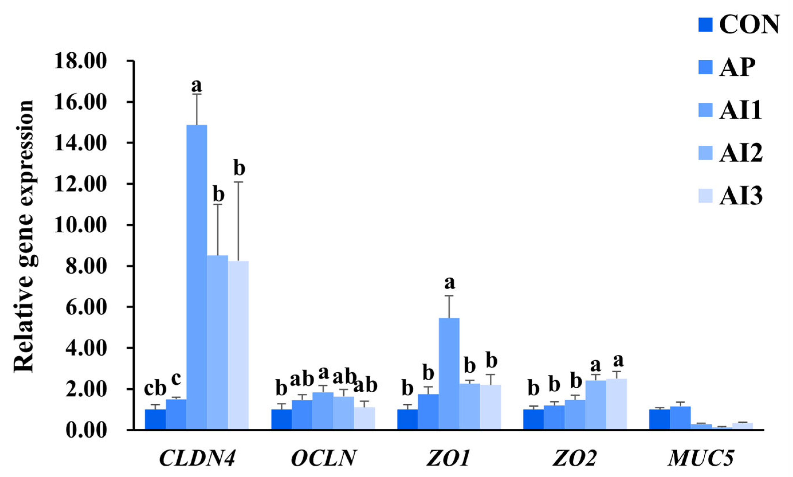
No statistically significant differential expression (p > 0.05) was observed for these genes between CON and AP groups. Dietary supplementation with 0.1% APS combined with graded IIE levels (0.05%, 0.1%, 0.15%) significantly upregulated claudin (CLDN4) transcription (p < 0.05) while concurrently downregulating mucin 5 (MUC5) expression (p < 0.05) relative to both CON and AP controls. Notably, AI1 group exhibited enhanced occluding (OCLN) mRNA abundance (p < 0.05) versus CON group. And the zonula occludens 1 (ZO1) gene expression in the intestine of AI1 group was significantly (p < 0.05) higher than those of the CON and AP group. As for the zonula occludens 2 (ZO2) gene expression in the intestine of large yellow croaker, the gene expression in the intestine of AI2 and AI3 group was significantly (p < 0.05) higher than those of the CON and AP group (Figure 2).

3.5. Antioxidant-Related Gene Expression in the Liver of Large Yellow Croaker

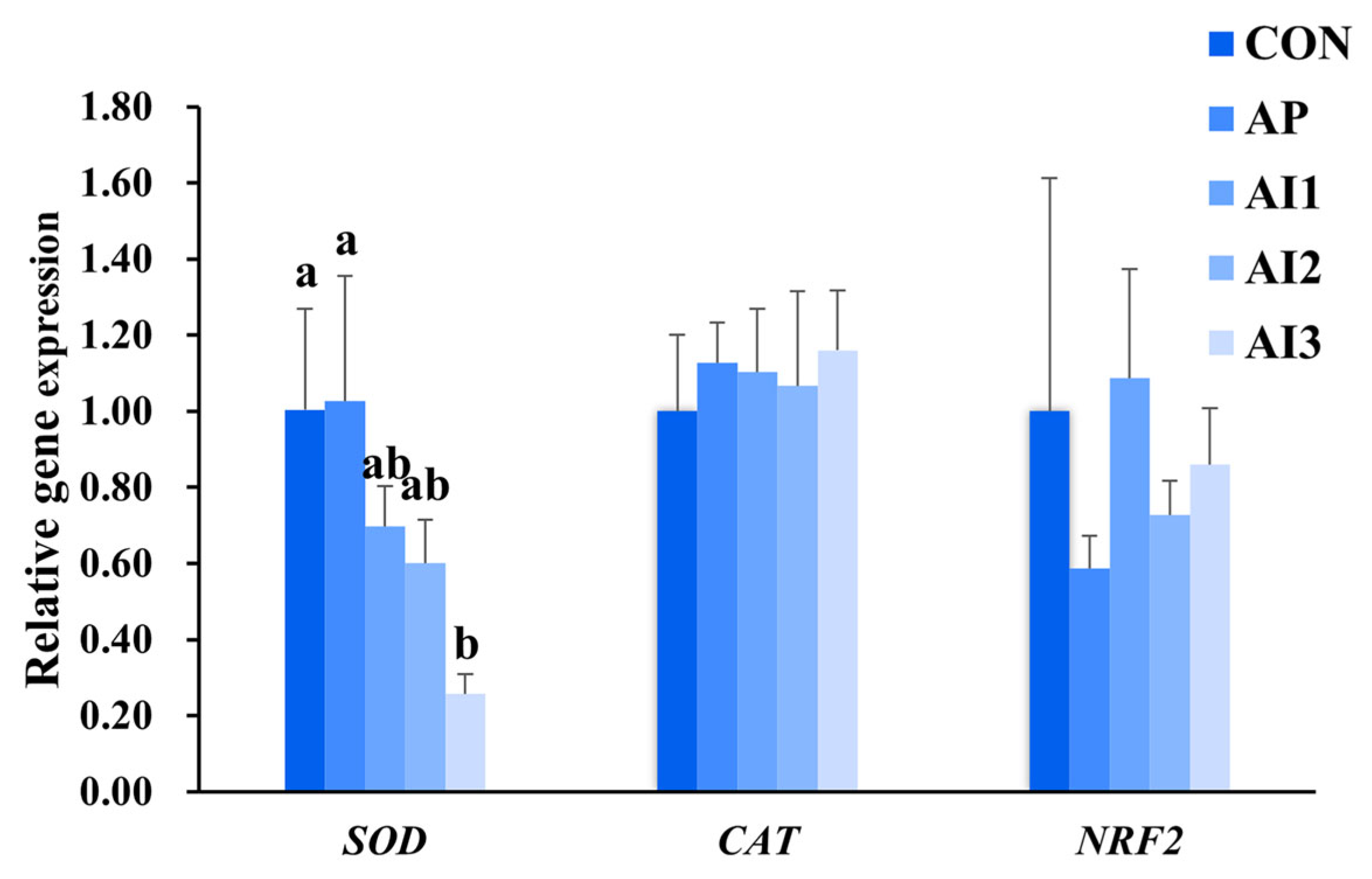
There is no significant (p > 0.05) difference in the gene expression of SOD among the CON, AP, AI1, and AI2 groups, but the one in the AI3 group was remarkably (p < 0.05) lower than those in the CON and AP groups. As for the CAT and nuclear factor erythroid 2-related factor 2 (NRF2) gene, the treatments seemed not (p > 0.05) influence the gene expression (Figure 3).

3.6. Inflammation-Related Gene Expression in the Head Kidney of Large Yellow Croaker

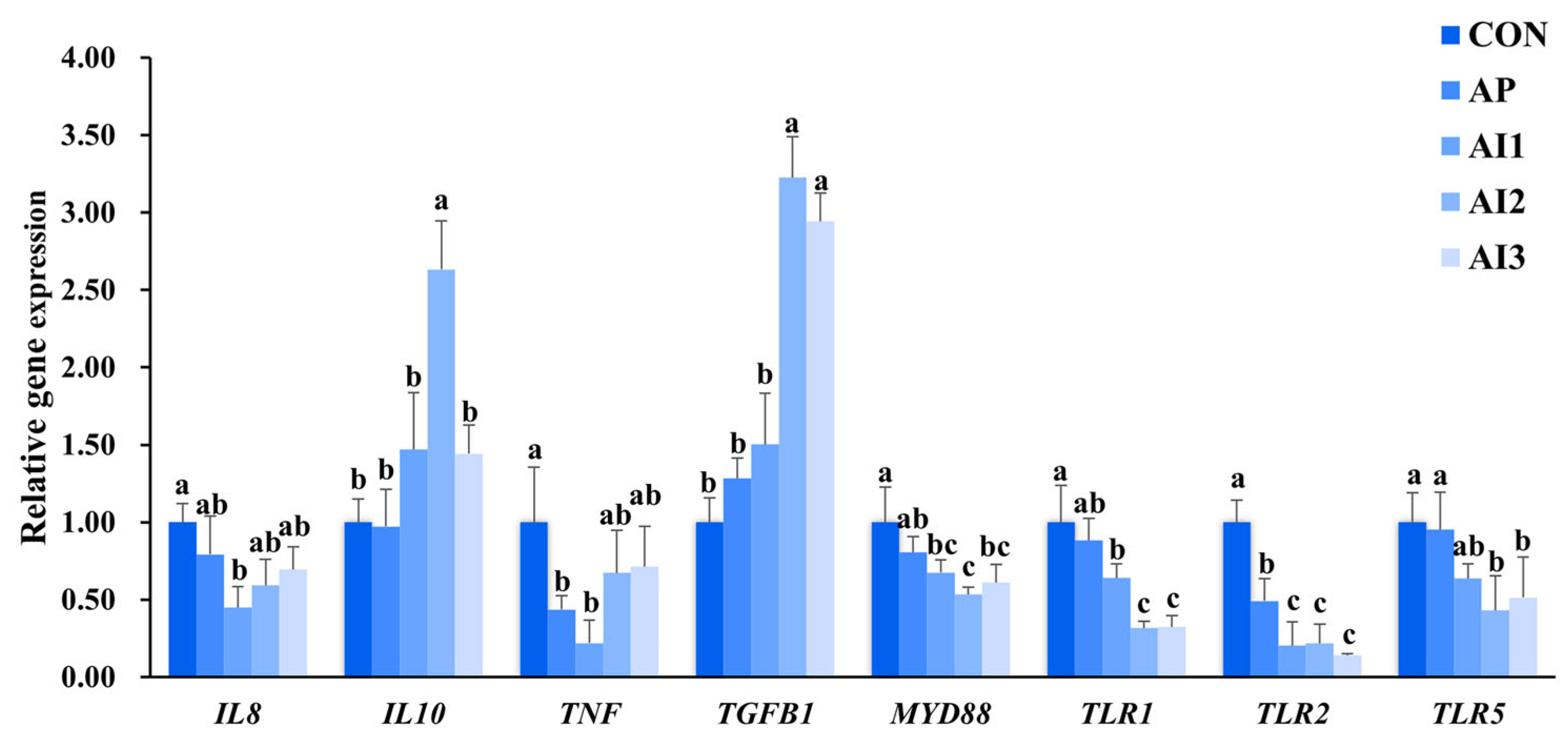
Expression levels of the pro-inflammatory cytokines interleukin 8 (IL8) and tumor necrosis factor (TNF)are presented in Figure 4. Analysis revealed a significant (p < 0.05) reduction in IL8 expression in the AI1 group compared to the CON group. However, expression levels among the CON, AP, AI2, and AI3 groups did not differ significantly (p > 0.05). Regarding TNF expression, both the AP and AI2 groups exhibited markedly (p < 0.05) lower levels than the CON group. Nevertheless, no statistically significant differences were observed between the CON, AI2, and AI3 groups (p > 0.05). This study also assessed the gene expression of the anti-inflammatory cytokines interleukin 10 (IL10) and transforming growth factor beta 1 (TGFB1) (Figure 4). The results indicated that IL10 and TGFB1 expression in the AP group was comparable to that in the CON group (p > 0.05). In contrast, the AI2 group displayed significantly (p < 0.05) elevated expression of both IL10 and TGFB1 relative to both the CON and AP groups. Furthermore, TGFB1 expression in the AI3 group was significantly (p < 0.05) higher than in the CON and AP groups.
As for the gene expression of toll-like receptors (TLRs), the results were shown in the Figure 4. Compared with the CON group, the gene expression of TLR1 and TLR5 showed no significant (p > 0.05) differences in the AP group, while the gene expression of TLR2 showed a significant (p < 0.05) decrease in the AP group. Compared with the AP group, the gene expressions of TLR1 and TLR5 showed no significant (p > 0.05) difference in the AI1 group, whereas the gene expression of TLR2 showed a significant (p < 0.05) decrease in the AI1 group. However, the gene expression of TLR1, TLR2, and TLR5 in the AI2 and AI3 group was prominently (p < 0.05) lower than that of AP group (Figure 4).

3.7. Autophagy and Apoptosis-Related Gene Expression in the Head Kidney of Large Yellow Croaker

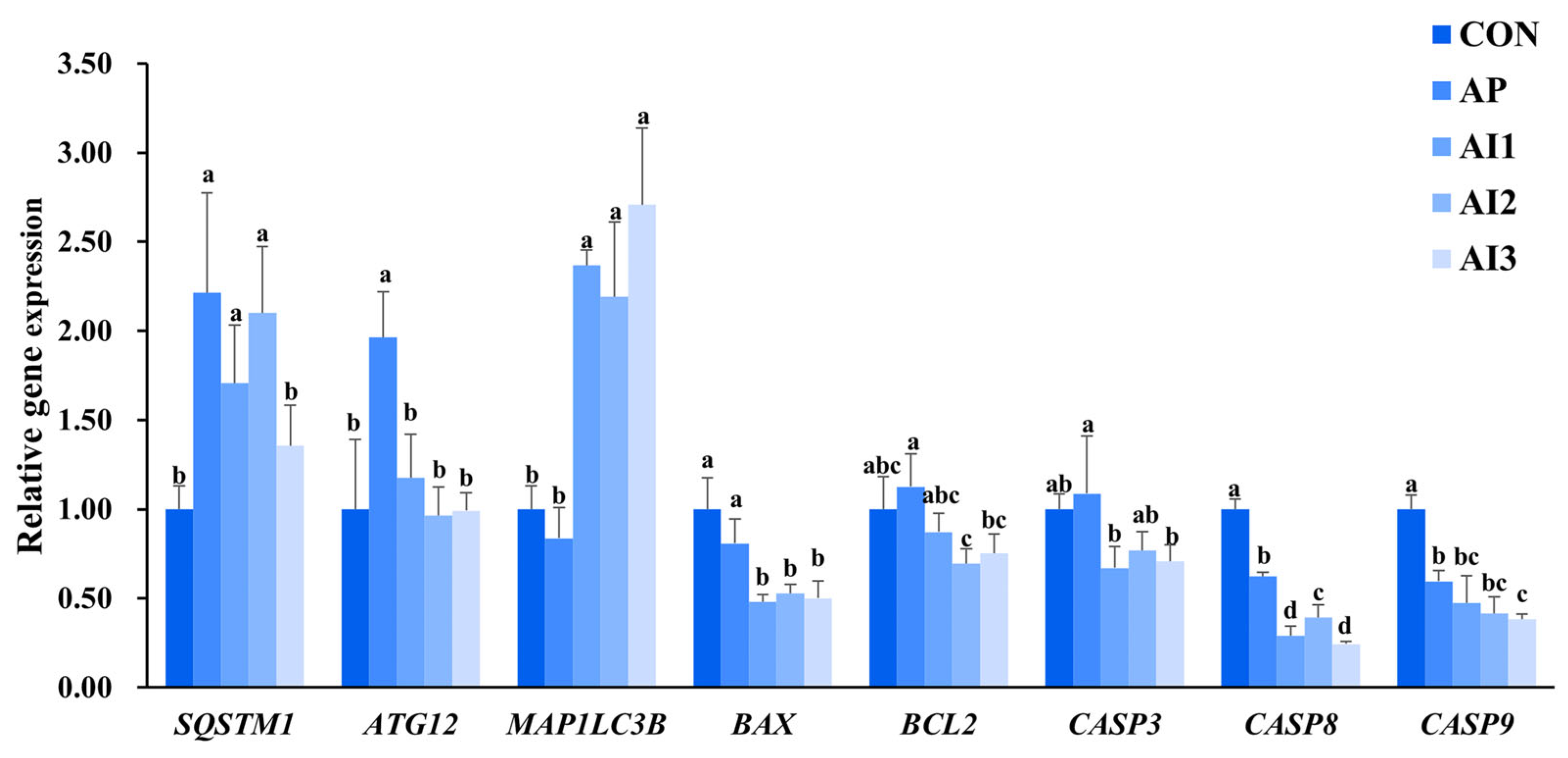
Regarding autophagy-related gene expression (Figure 5), statistically significant alterations were observed in the AP group relative to the CON group. Specifically, expression levels of sequestosome 1 (SQSTM1) (p < 0.05) and autophagy related 12 (ATG12) (p < 0.05) were markedly increased, whereas expression of microtubule-associated protein 1 light chain 3 beta (MAP1LC3B) (p < 0.05) were significantly reduced. Supplementation with IIE at concentrations of 0.05% and 0.1% (AI1, AI2) did not induce a significant change (p > 0.05) in SQSTM1 expression compared to the AP group alone. However, the highest IIE dose (0.15%, AI3) resulted in a significant decrease (p < 0.05). All IIE-supplemented groups (AI1, AI2, AI3) exhibited significantly lower ATG12 expression (p < 0.05) than the AP group, although levels remained comparable to the CON group (p > 0.05). Paradoxically, MAP1LC3B expression was significantly elevated (p < 0.05) in the AI groups compared to both CON and AP groups. Interestingly, while MAP1LC3B expression in the AI3 group was significantly reduced (p < 0.05) relative to the CON group, it did not differ significantly (p > 0.05) from the AP group.
Expression profiles of apoptosis-related genes are also presented in Figure 5. Relative to the CON group, the AP group exhibited comparable expression levels (p > 0.05) of bcl2-associated x protein (BAX), bcl2 apoptosis regulator (BCL2) and caspase 3 (CASP3). In contrast, expressions of both caspase 8 (CASP8) and caspase 9 (CASP9) was significantly (p < 0.05) elevated in the AP group. Administration of 0.05% IIE (AI1) did not induce significant (p > 0.05) alterations in BCL2 or CASP9 expression compared to the AP group. However, this treatment significantly decreased (p < 0.05) BAX, CASP3 and CASP8 expressions. Supplementation with 0.1% IIE (AI2) resulted in no significant (p > 0.05) changes in CASP3 or CASP9 levels versus the AP group, but significantly (p < 0.05) reduced BAX, BCL2, and CASP8 expression. Notably, dietary addition of 0.15% IIE (AI3) failed to significantly (p > 0.05) alter BCL2 expression relative to the AP group, yet significantly (p < 0.05) lowered the expression of BAX, CASP3, CASP8, and CASP9.

4. Discussion

The large yellow croaker is a key mariculture fish in China. Intensive mariculture’s growth and environmental degradation have led to increased diseases threatening the large yellow croaker industry [31]. As a naturally occurring and environmentally benign disease-resistant resource, Chinese herbal medicine has emerged as an increasingly significant player in the large yellow croaker aquaculture industry [28,32]. In the context of enhancing the disease resistance of large yellow croaker, Chinese herbal medicine is extensively utilized for the prevention and treatment of a multitude of diseases [33]. Our previous studies indicated the good application effect of APS in large yellow croaker farming [5,6,7]. Furthermore, the combined treatment of APS and various Chinese herbal medicines has an excellent promoting effect on the growth, immune status and intestinal health of large yellow croaker [28,32]. The current study extends these findings by systematically evaluating the synergistic effects of APS and IIE. We found that the APS-IIE combination significantly promoted intestinal health, antioxidant capacity, and suppressed inflammation and apoptosis, with the AI2 (0.1% APS + 0.1% IIE) diet being the most effective.
The antioxidant enzyme system resident in fish is fundamental for preserving physiological stability and bolstering the fish’s capacity to withstand stress. This system encompasses vital enzymes, including SOD, CAT, and GSH, which function to effectively neutralize free radicals and oxidative species, thereby protecting cellular structures and maintaining redox equilibrium [34]. In response to environmental challenges, this enzyme system demonstrates its importance by alleviating oxidative stress, reinforcing the immune system, supporting fish growth, and enhancing their ability to adapt to harsh environmental circumstances. Consequently, the antioxidant enzyme system is not merely the foundation for normal physiological operations in fish, it also represents a crucial line of defense against detrimental external influence [35]. In the present study, a key finding was the significant reduction in hepatic MDA content, a direct marker of lipid peroxidation [36], in the AI groups. This indicates a clear alleviation of oxidative stress. The concomitant decrease in SOD and CAT activities could be interpreted as an adaptive down-regulation in response to the reduced oxidative burden, as the need for high enzymatic activity diminished. While other interpretations, such as direct enzymatic inhibition, are possible, they are less consistent with the overall improvement in health status observed across multiple organs.
A profound correlation is evident between antioxidant capacity and inflammatory response in biological systems. Oxidative stress-induced free radicals within organisms have the capacity to activate inflammatory pathways, potentially leading to detrimental impacts on cellular structures [37,38]. Consequently, the maintenance of a dynamic equilibrium between antioxidant capacity and inflammatory response is indispensable for safeguarding the health status of organisms and mitigating disease progression. The results showed that significant changes were observed in the expression of inflammatory cytokines and immune-regulatory genes, indicating a robust inflammatory regulation by the AI diets. The pro-inflammatory cytokine (IL8 and TNF) expression was suppressed in certain groups, while the anti-inflammatory cytokine (IL10 and TGFB1) expression was enhanced. These findings suggest that APS and IIE compounds may have anti-inflammatory effects in the large yellow croaker, whose positive effects might be attributed to the antioxidant effect of the APS and IIE compound. Similar results were shown in the study on the atrazine treated Nile tilapia, dietary isatis alleviated the inflammatory cell infiltration in the intestinal mucosa with separation of lining epithelium [39]. The coordinated downregulation of TLRs and MyD88 provides a plausible mechanism for the suppression of the pro-inflammatory response, potentially through the inhibition of the downstream NF-κB signaling pathway. This transcription factor holds a central position in regulating the body’s response to external stimuli and plays an indispensable role in modulating inflammatory responses, thereby maintaining the intricate and delicate balance of immune homeostasis [40,41,42]. It is important to note that this interpretation is based on transcriptional data of upstream regulators, and future studies directly measuring NF-κB activation are warranted to confirm this mechanism.
Autophagy and apoptosis are essential regulatory mechanisms in fish, maintaining physiological homeostasis and environmental adaptation [43]. Autophagy inhibits the activation of inflammasome by degrading damaged organelles (such as mitochondria) or misfolded proteins and reducing the release of endogenous damage-associated molecular patterns such as reactive oxygen species and mitochondrial DNA [16,44]. Apoptosis, a caspase-mediated cell death pathway, eliminates genomically compromised cells and plays a critical role in fish immunity [45]. The antagonism between apoptosis and autophagy is an important mechanism for cells to determine survival or death under stress conditions. They achieve a dynamic balance through shared molecular regulatory networks (such as BCL2) and functional crosstalk in the inflammatory response [15]. In the present study, the gene expression of autophagy- and apoptosis-related genes was examined. Compared to the AP group, the AI group showed significant inductions in the expression of SQSTM1 and MAP1LC3B, indicating promotions in autophagy pathways. On the contrary, the expression of apoptosis-related genes (BCL2, BAX, CASP3, CASP8, and CASP9) was also affected by the dietary treatments. These results suggest that the combination of APS and IIE could suppress the apoptosis processes in the large yellow croaker. While these mRNA profiles suggest a coordinated modulation of autophagy and apoptosis, the role of key regulatory proteins like Beclin-1 also remains to be elucidated at the protein level. Previous studies have documented the potent anti-apoptotic effects of APS and IIE, achieved through inhibiting the expression of apoptotic factors while enhancing the expression of pro-autophagy factors [27,28,46,47]. Therefore, the strategic modulation of APS and IIE offers a promising avenue for balancing apoptosis and autophagy in fish inflammatory response. Further research is needed to investigate the potential mechanisms involved in the antagonism of autophagy and apoptosis influenced by the combination of APS and IIE.
The intestinal epithelium constitutes a dynamic barrier system, organized as a monolayer of columnar epithelial cells interconnected via tight junctions, adherens junctions, and desmosomes. These intercellular junctions not only provide mechanical anchorage but also modulate the equilibrium between selective nutrient absorption and barrier defense through precise regulation of paracellular permeability [48]. Furthermore, a viscoelastic mucus layer overlies the epithelial surface, predominantly comprising polymerized mucin glycoproteins secreted by goblet cells. This stratified organization facilitates commensal microbiota colonization in the outer layer while maintaining pathogen exclusion through the compact inner layer [49]. In the present study, the gene expression of tight junction proteins (CLDN4, OCLN, ZO1, and ZO2) and mucin protein (MUC5) was investigated to evaluate intestinal barrier function. No significant differences were observed between the CON and AP groups. However, dietary administration of APS combined with various concentrations of IIE significantly enhanced the gene expression of CLDN4, OCLN, ZO1, and ZO2. These changes suggest that the combination of APS and IIE may improve intestinal barrier function by modulating the expression of tight junction protein. Intestinal mucus secretion, usually boosted by dietary antigens or pathogenic challenges as a first line of defense [50], showed significantly reduced mucin production in the AI groups compared to the CON and AP groups, possibly due to the decrease in immunogenic stimuli. Regarding intestinal histology, the combinational addition of APS and IIE in the diet of large yellow croaker led to a lighter infiltration of admixed leucocytes in the lamina propria and increased height of the intestinal villi), indicating the better physical intestinal performance in the AI group. The functional competence of the intestine is intrinsically dependent on the structural integrity of its epithelial architecture [51]. Thus, the increased activities of intestinal digestive enzymes were also observed in the AI groups.

5. Conclusions

In summary, this study demonstrates that the APS-IIE synergy effectively enhances the overall health of large yellow croaker. The most significant improvements were observed with the 0.1% APS + 0.1% IIE formulation. The suppressed systemic inflammation in the large yellow croaker may be ascribed to the alleviated oxidative stress, suppressed apoptosis, and induced autophagy. The combined supplementation notably improved intestinal health, as evidenced by enhanced villus height, increased digestive enzyme activities, and strengthened barrier function through the upregulation of key tight junction proteins. However, it is important to acknowledge two main limitations of this study. First, the synergistic effects between APS and IIE were interpreted based on comparative treatment outcomes and were not statistically confirmed by a factorial analysis. Second, the evidence for autophagy and apoptosis was supported solely at the transcriptional level, and protein-level validation is lacking. Future studies should incorporate factorial designs and protein-level assays to further elucidate the mechanisms of action and confirm the synergistic potential of APS and IIE. Nevertheless, the current results strongly support the application of this herbal combination as a promising dietary strategy for improving intestinal health and overall resilience in sustainable aquaculture.

Author Contributions

Conceptualization, J.S. and X.C.; Data curation, Z.C. (Zhichu Chen), C.Z., A.W., H.W. and X.Z.; Formal analysis, Z.C. (Zhichu Chen), C.Z., A.W., H.W. and X.Z.; Investigation, Z.C. (Zhichu Chen), C.Z., A.W., H.W. and X.Z.; Methodology, J.S.; Writing—original draft preparation, Z.C. (Zhichu Chen); Writing—review and editing, Z.C. (Zhichu Chen), Z.C. (Zhengbang Chen), H.L., Q.Q., P.W., J.S. and X.C.; Funding acquisition, Z.C. (Zhichu Chen), J.S. and X.C.; Supervision., J.S. and X.C. All authors have read and agreed to the published version of the manuscript.

Funding

This work was supported by the grant from National Key Research and Develop-ment Program of China (Grant number [2022YFD2401001]), grants from National Natural Science Foundation of China (Grant numbers [32373148], [32303016], and [U23A20253]), grants from the Fujian Science and Technology Department (Grant numbers [2024J09027], [2024S0007], [2025J01607], and [2022NZ033021]), the fund of Fujian Key Laboratory of Functional Aquafeed and Culture Environment Control (Grant number [FACE20230009]), the grant from Fujian Agriculture and Forestry University (XJQ202109), and China Agriculture Research System (Grant number [CARS47-G19]).

Institutional Review Board Statement

The study was conducted in accordance with the animal ethics protocols of Fujian Agriculture and Forestry University (PZCAFAFU-CMS-23015). Approval date: 10 February 2023.

Informed Consent Statement

Not applicable.

Data Availability Statement

Upon receipt of a reasonable request, the corresponding author is able to provide access to all data presented in this article.

Acknowledgments

The authors have reviewed and edited the output and take full responsibility for the content of this publication.

Conflicts of Interest

Author Zhengbang Chen was employed by the Fuzhou Haima Feed Co., Ltd. Author Huiyuan Lv and Qiong Qi were employed by the Beijing Centre Biology Co., Ltd. The remaining authors declare that the research was conducted in the absence of any commercial or financial relationships that could be construed as a potential conflict of interest.

Abbreviations

The following abbreviations are used in this manuscript:
ACTBActin beta
AIAPS and IIE diet
APAPS diet
APSAstragalus polysaccharide
ATG12Autophagy Related 12
BAXBCL2-associated x protein
BCL2BCL2 apoptosis regulator
BCL2L1BCL2 Like 1
CASPCaspase
CATCatalase
CLDNClaudin
CONControl diet
GAPDHGlyceraldehyde-3-Phosphate Dehydrogenase
GSHGlutathione
IIEIsatis indigotica extract
ILInterleukin
MAP1LC3BMicrotubule-associated protein 1 light chain 3 beta
MDAMalondialdehyde
MUC5Mucin 5
MyD88Myeloid differentiation primary response 88
NF-κBNuclear factor kappa B
NRF2Nuclear factor erythroid 2-related factor 2
OCLNOccludin
SODSuperoxide dismutase
SQSTM1Sequestosome 1
T-AOCTotal antioxidant capacity
TGFB1transforming growth factor β
TLRToll-like receptor
TNFTumor necrosis factor
ZOZonula occludens

References

  1. Ahmad, A.; Abdullah, S.R.S.; Hasan, H.A.; Othman, A.R.; Ismail, N.I. Aquaculture industry: Supply and demand, best practices, effluent and its current issues and treatment technology. J. Environ. Manag. 2021, 287, 112271. [Google Scholar] [CrossRef]
  2. Gjedrem, T.; Robinson, N.; Rye, M. The importance of selective breeding in aquaculture to meet future demands for animal protein: A review. Aquaculture 2012, 350, 117–129. [Google Scholar] [CrossRef]
  3. Bergqvist, J.; Gunnarsson, S. Finfish aquaculture: Animal welfare, the environment, and ethical implications. J. Agric. Environ. Ethics 2013, 26, 75–99. [Google Scholar] [CrossRef]
  4. Pu, H.; Li, X.; Du, Q.; Cui, H.; Xu, Y. Research progress in the application of Chinese herbal medicines in aquaculture: A review. Engineering 2017, 3, 731–737. [Google Scholar] [CrossRef]
  5. Zhang, W.; Zhang, M.; Cheng, A.; Hao, E.; Huang, X.; Chen, X. Immunomodulatory and antioxidant effects of Astragalus polysaccharide liposome in large yellow croaker (Larimichthys crocea). Fish Shellfish Immunol. 2020, 100, 126–136. [Google Scholar] [CrossRef]
  6. Zhang, W.; Wang, Y.; Cheng, A.; Liu, R.; Kang, F.; Zhao, J.; Shao, J.; Huang, X.; Chen, X. Protective effects of dietary Astragalus polysaccharides on large yellow croaker (Larimichthys crocea) against Vibrio alginolyticus infection. Aquaculture 2024, 581, 740398. [Google Scholar] [CrossRef]
  7. Wang, Y.; Wu, Z.; Chen, H.; Liu, R.; Zhang, W.; Chen, X. Astragalus polysaccharides protect against inactivated Vibrio alginolyticus-induced inflammatory injury in macrophages of large yellow croaker. Fish Shellfish Immunol. 2022, 131, 95–104. [Google Scholar] [CrossRef]
  8. Zhang, Y.; Zheng, J.; Li, W.; Xiao, L. Effects of Chinese herbal medicines on growth and nonspecific immunity in yellow catfish Pelteobagrus fulvidraco. Fish. Sci. 2010, 29, 225–228. [Google Scholar]
  9. Wang, Q.; Zhao, H.; Lv, Z.; Chen, C.; Xing, K. Effects of dietary Angelica sinensis polysaccharideon nonspecific immunity of Epinephelus coioides. J. Anhui Agric. Sci. 2011, 39, 13857–13860. [Google Scholar]
  10. Meng, Z.; Chen, Y.; Liu, M. Effect of Chinese herbal compounds on nonspecific immunity of Cyprinus carpio var. specularis Juvenile fish. J. Northeast Agric. Univ. 2011, 42, 54–59. [Google Scholar]
  11. Chen, Q.; Lan, H.Y.; Peng, W.; Rahman, K.; Liu, Q.C.; Luan, X.; Zhang, H. Isatis indigotica: A review of phytochemistry, pharmacological activities and clinical applications. J. Pharm. Pharmacol. 2021, 73, 1137–1150. [Google Scholar] [CrossRef]
  12. Song, L.; Guo, Z.; Wang, A. Effects of Isatis root polysaccharide on non-specific immune responses and nutritive indices in obscure pufferfish, Takifugu obscurus. Aquac. Res. 2018, 49, 603–613. [Google Scholar] [CrossRef]
  13. Pan, T.; Yan, M.; Chen, S.; Wang, X. Effects of ten traditional Chinese herbs on immune response and disease resistance of Sciaenops ocellatus (Actinopterygii: Perciformes: Sciaenidae). Acta Ichthyol. Piscat. 2013, 43, 41–49. [Google Scholar] [CrossRef]
  14. Wang, E.; Chen, X.; Wang, K.; Wang, J.; Chen, D.; Geng, Y.; Lai, W.; Wei, X. Plant polysaccharides used as immunostimulants enhance innate immune response and disease resistance against Aeromonas hydrophila infection in fish. Fish Shellfish Immunol. 2016, 59, 196–202. [Google Scholar] [CrossRef]
  15. Elmore, S. Apoptosis: A review of programmed cell death. Toxicol. Pathol. 2007, 35, 495–516. [Google Scholar] [CrossRef]
  16. Levine, B.; Kroemer, G. Autophagy in the pathogenesis of disease. Cell 2008, 132, 27–42. [Google Scholar] [CrossRef]
  17. Murthy, A.; Li, Y.; Peng, I.; Reichelt, M.; Katakam, A.K.; Noubade, R.; Roose-Girma, M.; DeVoss, J.; Diehl, L.; Graham, R.R. A Crohn’s disease variant in Atg16l1 enhances its degradation by caspase 3. Nature 2014, 506, 456–462. [Google Scholar] [CrossRef] [PubMed]
  18. Rubinstein, A.D.; Kimchi, A. Life in the balance—A mechanistic view of the crosstalk between autophagy and apoptosis. J. Cell Sci. 2012, 125, 5259–5268. [Google Scholar] [CrossRef] [PubMed]
  19. Wirawan, E.; Vande Walle, L.; Kersse, K.; Cornelis, S.; Claerhout, S.; Vanoverberghe, I.; Roelandt, R.; De Rycke, R.; Verspurten, J.; Declercq, W. Caspase-mediated cleavage of Beclin-1 inactivates Beclin-1-induced autophagy and enhances apoptosis by promoting the release of proapoptotic factors from mitochondria. Cell Death Dis. 2010, 1, e18. [Google Scholar] [CrossRef]
  20. Luo, S.; Rubinsztein, D. Apoptosis blocks Beclin 1-dependent autophagosome synthesis: An effect rescued by Bcl-xL. Cell Death Differ. 2010, 17, 268–277. [Google Scholar] [CrossRef] [PubMed]
  21. Maiuri, M.C.; Criollo, A.; Kroemer, G. Crosstalk between apoptosis and autophagy within the Beclin 1 interactome. EMBO J. 2010, 29, 515–516. [Google Scholar] [CrossRef]
  22. Marino, G.; Niso-Santano, M.; Baehrecke, E.H.; Kroemer, G. Self-consumption: The interplay of autophagy and apoptosis. Nat. Rev. Mol. Cell Biol. 2014, 15, 81–94. [Google Scholar] [CrossRef]
  23. Salminen, A.; Kaarniranta, K.; Kauppinen, A. Beclin 1 interactome controls the crosstalk between apoptosis, autophagy and inflammasome activation: Impact on the aging process. Ageing Res. Rev. 2013, 12, 520–534. [Google Scholar] [CrossRef]
  24. Park, H.-J.; Park, S.-H. Induction of apoptosis by Ethyl Acetate Fraction of Astragalus membranaceus in human non-small cell lung cancer cells:-apoptosis induction by Astragalus membranaceus. J. Pharmacopunct. 2018, 21, 268. [Google Scholar] [CrossRef]
  25. Shan, H.; Zheng, X.; Li, M. The effects of Astragalus membranaceus active extracts on autophagy-related diseases. Int. J. Mol. Sci. 2019, 20, 1904. [Google Scholar] [CrossRef]
  26. Chang, C.-C.; Su, H.J.; You, H.L.; Kao, C.W.; Hung, I.L.; Huang, S.T. Isatis indigotica Inhibits Influenza A Virus (H1N1) Virulent Protein Production and Autophagosome Accumulation. J. Herb. Med. 2024, 43, 100827. [Google Scholar] [CrossRef]
  27. Chung, Y.C.; Tang, F.Y.; Liao, J.W.; Chung, C.H.; Jong, T.T.; Chen, S.S.; Tsai, C.H.; Chiang, E.P. Isatis indigotica induces hepatocellular cancer cell death via caspase-independent apoptosis-inducing factor translocation apoptotic pathway in vitro and in vivo. Integr. Cancer Ther. 2011, 10, 201–214. [Google Scholar] [CrossRef] [PubMed]
  28. Zhao, W.; Li, C.; Wu, X.; Zhang, J.; Wang, L.; Chen, X.; Shao, J. Synergistic effects of dietary Forsythia suspensa extract and Astragalus membranaceus polysaccharides on growth enhancement, antioxidant boost, and inflammation alleviation in juvenile Larimichthys crocea. Aquaculture 2025, 598, 742029. [Google Scholar] [CrossRef]
  29. Ao, J.; Mu, Y.; Xiang, L.X.; Fan, D.; Feng, M.; Zhang, S.; Shi, Q.; Zhu, L.Y.; Li, T.; Ding, Y. Genome sequencing of the perciform fish Larimichthys crocea provides insights into molecular and genetic mechanisms of stress adaptation. PLoS Genet. 2015, 11, e1005118. [Google Scholar] [CrossRef] [PubMed]
  30. Livak, K.J.; Schmittgen, T.D. Analysis of relative gene expression data using real-time quantitative PCR and the 2−ΔΔCT method. Methods 2001, 25, 402–408. [Google Scholar] [CrossRef]
  31. Qin, P.; Chen, X.; Lou, B.; Wu, T.; Yang, J.; Wang, W.; Huang, G.; Chen, X. Outbreak of trypanosomiasis in cage-cultured large yellow croaker in China. J. Fish Dis. 2024, 48, e13952. [Google Scholar] [CrossRef]
  32. Shao, J.; Wu, L.; Wu, X.; Zhang, J.; Wang, L.; Chen, X.; Zhao, W. Synergistic benefits of Eleutherococcus senticosus extract and Astragalus membranaceus polysaccharides in juvenile Larimichthys crocea: Enhanced growth, amplified antioxidant activity, and reduced inflammation. Aquaculture 2024, 598, 742028. [Google Scholar] [CrossRef]
  33. Zhang, W.; Zhao, J.; Ma, Y.; Li, J.; Chen, X. The effective components of herbal medicines used for prevention and control of fish diseases. Fish Shellfish Immunol. 2022, 126, 73–83. [Google Scholar] [CrossRef]
  34. Hoseinifar, S.H.; Yousefi, S.; Van Doan, H.; Ashouri, G.; Gioacchini, G.; Maradonna, F.; Carnevali, O. Oxidative stress and antioxidant defense in fish: The implications of probiotic, prebiotic, and synbiotics. Rev. Fish. Sci. Aquac. 2020, 29, 198–217. [Google Scholar] [CrossRef]
  35. Yu, G.; Zhao, S.; Ou, W.; Ai, Q.; Zhang, W.; Mai, K.; Zhang, Y. Host-associated Bacillus velezensis T20 improved disease resistance and intestinal health of juvenile turbot (Scophthalmus maximus). Aquac. Rep. 2024, 35, 101927. [Google Scholar] [CrossRef]
  36. Gaweł, S.; Wardas, M.; Niedworok, E.; Wardas, P. Malondialdehyde (MDA) as a lipid peroxidation marker. Wiad. Lek. 2004, 57, 453–455. [Google Scholar] [PubMed]
  37. Sadiq, I.Z. Free radicals and oxidative stress: Signaling mechanisms, redox basis for human diseases, and cell cycle regulation. Curr. Mol. Med. 2023, 23, 13–35. [Google Scholar] [CrossRef] [PubMed]
  38. Lugrin, J.; Rosenblatt-Velin, N.; Parapanov, R.; Liaudet, L. The role of oxidative stress during inflammatory processes. Biol. Chem. 2014, 395, 203–230. [Google Scholar] [CrossRef]
  39. Ali, M.F.; Soliman, A.A.; Gewaily, M.S.; Abdel-Kader, T.Y.; Amer, A.A.; Zaineldin, A.I.; Al-Asgah, N.A.; Younis, E.M.; Abdel-Warith, A.W.A.; Sewilam, H. Isatis phytogenic relieved atrazine induced growth retardation, hepato-renal dysfunction, and oxidative stress in Nile tilapia. Saudi J. Biol. Sci. 2022, 29, 190–196. [Google Scholar] [CrossRef]
  40. Chen, Z.; Ceballos-Francisco, D.; Guardiola, F.A.; Huang, D.; Esteban, M.Á. The alleviation of skin wound-induced intestinal barrier dysfunction via modulation of TLR signalling using arginine in gilthead seabream (Sparus aurata L). Fish Shellfish Immunol. 2020, 107, 519–528. [Google Scholar] [CrossRef] [PubMed]
  41. Chen, Z.; Huang, D.; Yongyut, P.; Li, G.; Esteban, M.Á.; Jintasataporn, O.; Deng, J.; Zhang, W.; Ai, Q.; Mai, K. Vitamin D3 deficiency induced intestinal inflammatory response of turbot through nuclear factor-κB/inflammasome pathway, accompanied by the mutually exclusive apoptosis and autophagy. Front. Immunol. 2022, 13, 986593. [Google Scholar] [CrossRef]
  42. Zhao, S.; Chen, Z.; Zheng, J.; Dai, J.; Ou, W.; Xu, W.; Ai, Q.; Zhang, W.; Niu, J.; Mai, K. Citric acid mitigates soybean meal induced inflammatory response and tight junction disruption by altering TLR signal transduction in the intestine of turbot, Scophthalmus maximus L. Fish Shellfish Immunol. 2019, 92, 181–187. [Google Scholar] [CrossRef]
  43. Rauta, P.R.; Nayak, B.; Das, S. Immune system and immune responses in fish and their role in comparative immunity study: A model for higher organisms. Immunol. Lett. 2012, 148, 23–33. [Google Scholar] [CrossRef]
  44. Mizushima, N.; Komatsu, M. Autophagy: Renovation of cells and tissues. Cell 2011, 147, 728–741. [Google Scholar] [CrossRef] [PubMed]
  45. Secombes, C.; Wang, T. The innate and adaptive immune system of fish. In Infectious Disease in Aquaculture; Elsevier: Amsterdam, The Netherlands, 2012; pp. 3–68. [Google Scholar]
  46. Li, X.; Qu, L.; Dong, Y.; Han, L.; Liu, E.; Fang, S.; Zhang, Y.; Wang, T. A review of recent research progress on the Astragalus genus. Molecules 2014, 19, 18850–18880. [Google Scholar] [CrossRef]
  47. Zhou, R.; Chen, H.; Chen, J.; Chen, X.; Wen, Y.; Xu, L. Extract from Astragalus membranaceus inhibit breast cancer cells proliferation via PI3K/AKT/mTOR signaling pathway. BMC Complement. Altern. Med. 2018, 18, 83. [Google Scholar] [CrossRef]
  48. Peterson, L.W.; Artis, D. Intestinal epithelial cells: Regulators of barrier function and immune homeostasis. Nat. Rev. Immunol. 2014, 14, 141–153. [Google Scholar] [CrossRef] [PubMed]
  49. Johansson, M.E.; Hansson, G.C. Immunological aspects of intestinal mucus and mucins. Nat. Rev. Immunol. 2016, 16, 639–649. [Google Scholar] [CrossRef] [PubMed]
  50. Schroeder, B.O. Fight them or feed them: How the intestinal mucus layer manages the gut microbiota. Gastroenterol. Rep. 2019, 7, 3–12. [Google Scholar] [CrossRef]
  51. Chelakkot, C.; Ghim, J.; Ryu, S.H. Mechanisms regulating intestinal barrier integrity and its pathological implications. Exp. Mol. Med. 2018, 50, 1–9. [Google Scholar] [CrossRef]
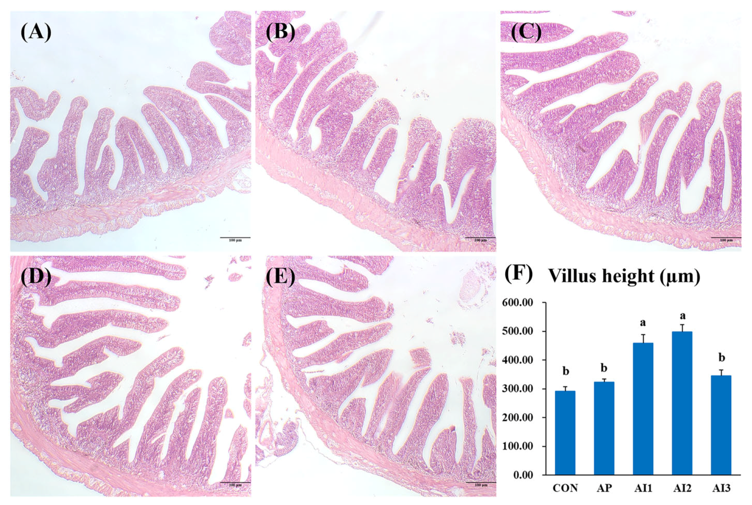
Figure 1. Effect of different experimental diets on the intestinal histology of the large yellow croaker. CON (A), AP (B), AI1 (C), AI2 (D), AI3 (E), Villus height (µm, (F)). Staining: H&E. Scale bar = 100 μm. Results are shown as means ± SD of 3 replicate cages. Different letters indicate significant differences (p < 0.05).
Fishes 10 00593 g001
Figure 2. Effect of different experimental diets on the mRNA level of tight junction proteins and mucin in the intestine of the large yellow croaker. Results are shown as means ± SD of 3 replicate cages. Different letters indicate significant differences (p < 0.05).
Fishes 10 00593 g002
Figure 3. Effect of different experimental diets on the mRNA level of antioxidant-related genes in the liver of the large yellow croaker. Results are shown as means ± SD of 3 replicate cages. Different letters indicate significant differences (p < 0.05).
Fishes 10 00593 g003
Figure 4. Effect of different experimental diets on the mRNA level of inflammation-related genes in the head kidney of the large yellow croaker. Results are shown as means ± SD of 3 replicate cages. Different letters indicate significant differences (p < 0.05).
Fishes 10 00593 g004
Figure 5. Effect of different experimental diets on the mRNA level of autophagy and apoptosis-related genes in the head kidney of the large yellow croaker. Results are shown as means ± SD of 3 replicate cages. Different letters indicate significant differences (p < 0.05).
Fishes 10 00593 g005
Table 1. Formulation and proximate composition of experimental diets (% dry matter).
IngredientsCONAPAI1AI2AI3
Fish meal 140.0040.0040.0040.0040.00
Soybean meal 130.0030.0030.0030.0030.00
Wheat flour 121.9021.8021.7521.7021.65
Fish oil2.602.602.602.602.60
Soybean lecithin1.001.001.001.001.00
Brewer’s yeast2.502.502.502.502.50
Vitamin premix 21.001.001.001.001.00
Mineral premix 21.001.001.001.001.00
Isatis indigotica extract0.000.000.050.100.15
Astragalus polysaccharides0.000.100.100.100.10
Proximate analysis
Moisture9.419.589.429.559.43
Crude protein43.2743.1943.4643.3643.58
Crude lipid9.429.289.359.329.35
Ash10.2610.1910.3010.1610.14
1 Fish meal, wheat gluten, and wheat flour were purchased from Dachang Feed Corporation (Fuzhou, China). Fish meal: crude protein: 67%, crude lipid: 9.5%; Wheat gluten: crude protein: 78.2%, crude lipid: 1.9%; Wheat flour: crude protein: 13%, crude lipid: 0.6%. 2 Vitamins premix and minerals premix were purchased from Dachang Feed Corporation (Fuzhou, China). And specific additional level of each component was referred to as [28].
Table 2. Primers used in quantitative real-time PCR (qRT-PCR).
Target GenesPrimer Sequence (5′-3′)Primer Amplification EfficienciesAccession Number
SODF-ATGGTGATCCACGAGAAGGC2.01NM_001303360.1
R-CTACCAGCGTTGCCAGTCTT
CATF-GACGCTCTACTGTTCCCGTC2.09XM_010735178.3
R-GCAAACCTCGATCGCTGAAC
NRF2F-GATGGAAATGGAGGTGATGC2.02XM_010737768.3
R-CATGTTCTTTCTGTCGGTGG
CLDN4F-CGTCCAGGATTTCTACAACCC2.08XM_010734071.3
R-CCCAGCTCTCTTTTGTGCAT
ZO1F-TGTCAAGTCCCGCAAAAATG1.97XM_019260744.2
R-CAACTTGCCCTTTGACCTCT
ZO2F-ACCCGACCTGTTTGTTATTG1.98XM_019272858.2
R-ATGCCGTGCTTGCTGTC
OCLNF-AGGCTACGGCAACAGTTATG1.92XM_010734512.3
R-GTGGGTCCACAAAGCAGTAA
MUC5ATAAATAAGCTCCAGTCAGCAC1.95XM_027285416.1
ACCACAGACTCTTTAGCCTT
IL8F-CCCTGCTTGTCGTCGTAACT2.01XM_010754327.3
R-CTCACCAGGCCAGCTATCAC
IL10F-AGTCGGTTACTTTCTGTGGTG2.07XM_010738826.3
R-TGTATGACGCAATATGGTCTG
TNFF-ACACCTCTCAGCCACAGGAT1.98XM_010745990
R-CCGTGTCCCACTCCATAGTT
TGFB1F-AGCAACCACCGTACATCCTG1.99XM_027280465.1
R-AGGTATCCCGTTGGCTTGTG
MYD88F-TCGGCCTTTGATTGATGAGGA2.03XM_010753272.3
R-GTGAGACCAACCCTGTCTCG
TLR5F-GGCACAGTGAGGAAAGGT2.06XM_019267725.2
R-TAGCAAGCGTCCACATAC
TLR2F-GTCCGACAACCTGCTGACTGA2.10KKF22682.1
R-CAGGTGGGTGAGTTTGGAGAG
TLR1F-CTTTGTCAAGAGCGAGTGGT 1.98KF318376.1
R-GGTTCATCATGGCCTTCAGC
SQSTM1F-GTGGAGGTCTGGCTTTTTTATG1.99XM_019265187.2
R-AGTGATCGTTGTGGCTATTG
ATG12F-CCTCACCGGATCAAGACGTA2.03XM_027286876.1
R-AAGAGAGCAGTTTAACCCCAG
MAP1LC3BF-TGGGTCAGAACCACCACAGAACT2.05XM_027285483.1
R-GCTACTACGTGGGCCTGCAATG
BAXF-GGTGGCTGGGAGGGTATTCGTT1.99XM_019258722.2
R-TCCCGTCCACATTTCCCTTCGT
BCL2F-TGCTTGCGGACACGGACTCA1.92XM_010729997.2
R-CACAGCACAGGCGTATCCACAA
CASP3F-GATGGCACTACAAAGATCCCTGTG1.95EU878546.1
R-GCTACTCTGTGGCTGAAGGTTAC
CASP8F-TCAGCGAAGACCACAACC2.06XM_010754584.3
R-CACCACAGTGAAGCCAAG
CASP9F-GCGCAACACAGACAAATCTCG1.95XM_010755472.3
R-TGATCCCGGCGCTCTTTATC
ACTBF-GACCTGACAGACTACCTCATG1.99GU584189
R-AGTTGAAGGTGGTCTCGTGGA
GAPDHF-CGGAGTCAACGGATTTGGTC2.10XM_010743420.3
R-AGCCTTCTCCATGGTCGTGA
Table 3. Effect of different experimental diets on the activity of intestinal digestive enzymes of large yellow croaker.
CONAPAI1AI2AI3
Amylase (U/mg protein)1.06 b ± 0.040.91 a ± 0.031.06 b ± 0.011.36 c ± 0.021.56 d ± 0.02
Lipase (U/g protein)2.62 ab ± 0.112.847 ab ± 0.222.02 a ± 0.453.48 b ± 0.763.36 b ± 0.25
Trypsin (U/mg protein)1183.39 b ± 234.331672.12 ab ± 303.711434.12 ab ± 57.501926.27 a ± 317.781476.98 ab ± 396.79
Results are shown as means ± SD of 3 replicate cages. Different superscript letters indicate significant differences (p < 0.05).
Table 4. Effect of different experimental diets on the activity of hepatic oxidative stress markers of large yellow croaker.
CONAPAI1AI2AI3
SOD (U/mg protein)11.72 a ± 0.9912.94 a ± 1.0712.06 ab ± 2.875.57 c ± 0.509.06 b ± 0.12
T-AOC (mmol/g protein)0.155 ± 0.0140.152 ± 0.010.123 ± 0.0260.146 ± 0.010.156 ± 0.019
MDA (nmol/mg protein)5.63 a ± 1.055.08 ab ± 0.654.84 ab ± 0.344.61 ab ± 0.364.14 b ± 0.37
CAT (nmol/mg protein)7.85 b ± 1.628.04 b ± 1.2013.59 a ± 1.2612.57 a ± 1.239.71 b ± 0.72
Results are shown as means ± SD of 3 replicate cages. Different superscript letters indicate significant differences (p < 0.05).
Disclaimer/Publisher’s Note: The statements, opinions and data contained in all publications are solely those of the individual author(s) and contributor(s) and not of MDPI and/or the editor(s). MDPI and/or the editor(s) disclaim responsibility for any injury to people or property resulting from any ideas, methods, instructions or products referred to in the content.