You are currently viewing a new version of our website. To view the old version click .
Fishes
  • Article
  • Open Access

13 November 2025

Gene Expression and Antioxidant Characteristics of Rainbow Trout (Oncorhynchus mykiss) Eggs Used for Meiotic Gynogenesis

,
,
,
,
,
and
1
Department of Marine Biology and Biotechnology, Faculty of Oceanography and Geography, University of Gdansk, Al. M. Piłsudskiego 46, 81-378 Gdynia, Poland
2
Department of Salmonid Research, National Inland Fisheries Research Institute in Olsztyn, Rutki, 83-330 Żukowo, Poland
*
Author to whom correspondence should be addressed.
Fishes2025, 10(11), 585;https://doi.org/10.3390/fishes10110585 
(registering DOI)
This article belongs to the Special Issue Genetics and Breeding of Fishes

Abstract

Gynogenesis is a reproductive mode where offspring inherit exclusively maternal chromosomes. Gynogenetic development in fish may be induced intentionally by activating eggs with the UV-irradiated, inactive spermatozoa. In the meiotic variant of gynogenesis, the resultant haploid gynogenetic zygote is then exposed to a physical shock to inhibit the release of the 2nd polar body and to reconstitute the diploid state of the embryo. Here, meiotic gynogenesis was induced in the rainbow trout eggs from different clutches to find any differences in terms of gene expression and antioxidant enzyme activity between eggs with high and low ability for gynogenetic development. The survival rates of the gynogenotes after hatching from the eggs originating from five females varied from 16.6 ± 4.3% to 53.8 ± 9.8%. Biochemical and molecular examination revealed that eggs with higher developmental potential for meiotic gynogenesis exhibited significantly greater glutathione peroxidase (GPx) activity than eggs with lower efficiency of gynogenesis. Moreover, eggs exhibiting the highest ability for gynogenetic development showed increased transcription of the keratin 8 gene and decreased abundance of keratin 18 and tubulin β mRNA transcripts. Since keratins protect oocytes from physical stress after ovulation, the high abundance of keratin 8 in the rainbow trout eggs may increase their resilience to the physical shock applied for the zygote diploidization during gynogenesis. On the other hand, a low level of tubulin-building microtubules may increase the efficiency of high hydrostatic pressure (HHP) shock used for diploidization of the gynogenetic zygotes.
Key Contribution:
Rainbow trout eggs with efficient antioxidant barrier and increased resistance to the physical stress after ovulation may have better potential for development after gynogenetic activation followed by the high hydrostatic pressure shock.

1. Introduction

Gynogenesis is a form of reproduction in which spermatozoa activate eggs to develop with no paternal chromosomes. Gynogenesis occurs naturally in several amphibian and fish species [,,,]; however, it can also be induced under control conditions []. Haploid gynogenetic zygotes are produced by activating eggs with spermatozoa inactivated by irradiation (UV, ionizing radiation). Exposure of gynogenetically activated eggs to chemical, thermal, or high hydrostatic pressure (HHP) shock shortly after insemination prevents extrusion of the 2nd polar body, which results in production of the diploid heterozygous gynogenotes (meiotic gynogenesis). In turn, application of shock around the prophase of the 1st mitosis prevents the first cell cleavage, leading to the duplication of maternal chromosomes and generation of fully homozygous gynogenetic Doubled Haploids (DHs) (mitotic gynogenesis) []. Meiotic gynogenesis has been applied to investigate the genetic basis of sex determination in fish [,,] and to produce all-female offspring []. In turn, gynogenetic DHs are utilized to study the phenotypic expression of the recessive alleles [] and to assess inbreeding effect on fish body deformities [,]. Fully homozygous DHs are also used in the fish-breeding programs [] and for genome-sequencing research []. Using eggs produced by gynogenetic DH females for meiotic gynogenesis results in fish clonal lines being produced [].
Nevertheless, increased mortality of the gynogenotes limits application of gynogenesis in aquaculture. The reduced heterozygosity and alterations in egg organelles caused by the physical shock mean that the hatching yields of the meiogynogenetic fish rarely exceed 60%, while the hatching rate of the homozygous gynogenotic DHs is usually below 20% [,]. Variations in the survival of gynogenotes developing in eggs from different females observed in several fish species [,,,] suggest that both the quality of eggs and maternal effects related to the egg origin need to be considered as factors that may in part explain the decreased survival of the gynogenetic fish []. Egg quality is defined as the ability to be fertilized and develop into the normal embryos [,] and is influenced by the environmental and rearing conditions (1), the heterozygosity level of the egg donors (2), and maternal gene expression (3) [,]. Additionally, delayed collection and prolonged storage of eggs after ovulation and before fertilization lead to the post-ovulatory oocyte aging, which impairs egg developmental competence [,]. Unfortunately, reliable techniques to discriminate high- from low-quality fish eggs before fertilization are available for only a very limited number of species and are usually population, or stock specific. For example, in the rainbow trout from the autumn-spawning strain cultured in France, an analysis of the maternal RNA deposited in the eggs during oogenesis that controls early embryonic development in fish revealed variation in the transcription of keratin, tubulin, cathepsin, and polyglutamylase genes between low- and high-quality eggs []. In turn, eggs with higher developmental potential originating from females from the spring-spawning broodstock aquacultured in Poland exhibited an increased level of antioxidant enzyme activity [], which supports the assumption that oxidative stress is linked to the post-ovulatory oocyte aging [].
Since egg quality can decline after ovulation, manipulations performed during induced gynogenesis may further reduce their developmental potential. Thus, using only high-quality eggs for gynogenesis might improve the efficiency of this process. In the present study, the activity of antioxidant enzymes and the expression of genes previously used to assess rainbow trout egg quality were evaluated to determine whether they can distinguish eggs with low and high competence for meiotic gynogenesis.

2. Material and Methods

2.1. Ethics

The study was carried out in strict accordance with the recommendations in the Polish ACT of 15 January 2015 on the Protection of Animals Used for Scientific or Educational Purposes, Journal of Laws 2015, item 266. The protocol was approved by the Local Ethical Committee for the Experiments on Animals in Bydgoszcz (resolution No. 14/2022).

2.2. Eggs and Sperm

The experiment was conducted on 15 November 2023. Rainbow trout gamete donors originated from the winter-spawning broodstock kept at the Department of Salmonid Research, Inland Fisheries Institute in Olsztyn, Rutki, Poland. Eggs were collected from six 4-year-old rainbow trout females and kept separately in the plastic bowls at 10 °C for no longer than 30 min until activation. In turn, spermatozoa were collected from three 4-year-old rainbow trout males and placed into the separate plastic containers. Motility of the spermatozoa was confirmed under the microscope. Sperm from different males was pooled and kept in +4 °C for the further use.

2.3. Irradiation of Sperm, Egg Activation and Diploidization

A standard protocol for UV inactivation of rainbow trout spermatozoa was used []. In short, a 0.375 mL portion of sperm was diluted in 15 mL of the rainbow trout seminal plasma (40×). The glass beaker (50 mm diameter, 30 mm height) with diluted sperm was placed on a magnetic stirrer and exposed to a UV-C light (Phillips TUW 30 Watt UV bulb; Philips, Eindhoven, The Netherlands) for 12 min. The distance between the surface of the magnetic stirrer and the UV lamp was 20 cm. During the irradiation, diluted sperm was mixed at 1400× g. UV intensity and dose were 2075 μW/cm2 and 1494 J/m2, respectively. Immediately after UV exposure, irradiated sperm was used for egg activation.
Portions of diluted and UV-irradiated sperm were added separately to batches of around 800–900 (Supplementary File S1) eggs from each female (~150,000 spermatozoids per egg) and covered with sperm-activating medium (SAM) (154 mM NaCl, 20 mM Tris, 30 mM glycine, 1 mM CaCl2, pH 9.0) [] to induce gynogenetic development. Five minutes after activation, the eggs were rinsed with hatchery water. To restore the diploid state in the gynogenetic embryos, eggs activated with the UV-irradiated spermatozoa were exposed to a high hydrostatic pressure (HHP) shock (9500 psi/3 min) exactly 35 min after insemination, using a TRC-APV electric/hydraulic device (TRC Hydraulics Inc. Dieppe, NB, Canada) []). Between activation and exposure for the HHP shock, the eggs were incubated in a water bath at 10 °C. To establish control groups (C), portions of approximately 300 eggs from each female were fertilized with non-irradiated sperm collected from the same rainbow trout males. Eggs from each female and experimental group were incubated in three separate replicates at 6–8 °C, using vertical egg tray incubators. Dead and live fish in all groups were counted at the eyed-egg, hatching, and swim-up stages to calculate survival rates (%) for each of these three stages.

2.4. mRNA Extraction

For gene expression analysis, several eggs from each female were preserved in RNAlater solution (Sigma-Aldrich, St. Louis, MO, USA) within 10 min after stripping. After placement in the RNAlater solution, eggs were incubated overnight at 4 °C and subsequently stored at −25 °C until RNA extraction. RNA was isolated from eight eggs (separate biological replicates) from each female using the Bead-Beat Total RNA Mini kit (A&A Biotechnology, Gdańsk, Poland) according to the manufacturer’s instructions. Residual DNA in the extracted RNA samples was removed using the Clean-Up Concentrator kit (A&A Biotechnology, Gdańsk, Poland). RNA concentration and purity were measured with a NanoDrop One spectrophotometer (Thermo Fisher Scientific, Waltham, MA, USA), and RNA integrity was assessed with 1% agarose gel electrophoresis. RNA samples showing A260/A280 ratios between 1.8 and 2.1, A260/A230 ratios above 2.0, and two sharp ribosomal RNA bands (28S and 18S) on agarose gel were considered of satisfactory quality and used for further processing. Due to the low RNA quality, samples from Female 2 were excluded from the analysis.
The cDNA synthesis was performed using the RevertAid First Strand cDNA Synthesis Kit (Thermo Fisher Scientific, Waltham, MA, USA) with RNA templates of satisfactory quality. The reaction mixtures were prepared in a total volume of 20 µL, comprising 1× Reaction Buffer, 5 μM of Random Hexamer primers, 1 μM of dNTP Mix, 20U RiboLock RNase Inhibitor, 20U RevertAid M-MuLV RT Reverse Transcriptase, and 100 ng of RNA template. Reverse-transcription reactions were carried out on a Mastercycler® X50a (Epppendorf, Hamburg, Germany). Samples were incubated for 10 min at 25 °C, followed by 120 min at 37 °C, and the reaction was terminated by heating at 85 °C for 5 min. The resulting cDNA samples were stored at −20 °C until further qPCR analysis.

2.5. Quantitative Real-Time PCR Analysis

Quantitative real-time PCR analysis included five selected mRNA transcripts encoding keratin 8 (K8), keratin 18 (K18), tubulin β (TUBB), cathepsin Z (CTSZ) and tubulin polyglutamylase complex subunit 2 (PSG2), using β-actin (Actb) and elongation factor 1-alpha (Elf1a) as the house-keeping genes. Primer sequences for all genes originated from previously published data (Table 1). qPCR was performed separately for target and housekeeping genes by a QuantStudio™ 3 Real-Time PCR (Thermo Fisher Scientific™, Walthman, MA, USA) with the PowerTrack SYBR Green Master Mix (Applied Biosystems, Foster City, CA, USA). Reaction efficiencies were estimated from standard curves generated from 10-fold serial cDNA dilutions starting from about 20 ng/μL, with optimal efficiencies ranging from 90 to 110%. qPCR reaction mixtures (10 μL) contained 1× PowerTrack SYBR Green Master Mix, 0.5–0.8 μM primers for target genes, and 0.15–0.5 μM primers for housekeeping genes, as well as 5 ng of cDNA template. Real-time PCRs were run in triplicate with the following thermal cycling conditions: an initial polymerase activation at 95 °C for 5 min, followed by 35 cycles of 95 °C for 30 s (denaturation), 60 °C for 20 s (primer annealing), and 72 °C for 15 s (elongation). During each run, negative controls using nuclease-free water and non-transcribed RNA were included to check for reagent or sample contamination. Melting curve analysis (60–95 °C) was carried out at the end of each run to complete the protocol. Fluorescence data were collected after the elongation step and at 0.1 °C increments during the melting curve analysis. Relative expression of target genes was calculated using the ΔCt method []. The Ct values for the reference genes were normalized with geNorm 3.4 software []. Each individual egg was analyzed separately and all qPCR reactions were performed in technical triplicates. The mean Ct value from the triplicates was used for each egg as a single biological data point in subsequent analyses.
Table 1. Forward (F) and reverse (R) primer sequences used for the real-time PCR analysis of selected genes in the present study.

2.6. Antioxidant Enzyme Activity Analysis

For antioxidant enzyme activity analysis, several eggs (3.2 g) from each female were collected, rinsed with phosphate-buffered saline (PBS, pH 7.4). Eight eggs from each batch (separate biological replicates) were homogenized in assay-specific buffers. The homogenates were centrifuged at 10,000× g for 15 min at 4 °C. During the sample preparation stage, lipids from the trout eggs form a separate phase that allows for the collection of the lipid-free supernatant. Next, the supernatants were collected and stored at –80 °C for no longer than one month prior to the enzymatic analysis. Enzymatic activity assays were conducted using colorimetric methods in 96-well microplates, with absorbance measured on a microplate reader (Molecular Devices, Sunnyvale, CA, USA). Recorded enzyme activities were expressed as units per milliliter (U mL−1) for superoxide dismutase (SOD) and as nanomoles per minute per milliliter (nmol·min−1·mL−1) for catalase (CAT) and glutathione peroxidase (GPx). For each female, eggs were analyzed separately and all enzymatic assays were performed in technical triplicates to ensure reproducibility and statistical reliability. The averaged values from triplicates represented one biological replicate (egg).
Superoxide dismutase (SOD) activity was determined using the Superoxide Dismutase Assay Kit (Cayman Chemical Co., Ann Arbor, MI, USA), which employs a tetrazolium salt to detect superoxide radicals generated by the xanthine oxidase/hypoxanthine system. A standard curve was constructed using a SOD standard and absorbance was measured at 450 nm.
Catalase (CAT) activity was measured using the Catalase Assay Kit (Cayman Chemical Co., Ann Arbor, MI, USA). The assay exploits CAT’s peroxidatic activity, in which methanol acts as an electron donor in the presence of hydrogen peroxide (H2O2) to produce formaldehyde. The resulting formaldehyde is quantified spectrophotometrically using 4-amino-3-hydrazino-5-mercapto-1,2,4-triazole (AHMT) as the chromogenic reagent with absorbance measured at 540 nm.
Glutathione peroxidase (GPx) activity was assessed using the Glutathione Peroxidase Assay Kit (Cayman Chemical Co., Ann Arbor, MI, USA). The assay is based on a coupled enzymatic reaction with glutathione reductase (GR), which recycles oxidized glutathione (GSSG) to its reduced form using NADPH. The oxidation of NADPH to NADP+ results in accompanied by a decrease in absorbance at 340 nm, which is directly proportional to the GPx activity in the sample.

2.7. Statistical Analysis

Statistical analyses of survival rates in gynogenetic and control fish at each developmental stage (eyed-egg, hatch and swim-up), as well as gene expression and antioxidant enzyme activities in eggs from each female, were performed using Statistica v.10.0 (StatSoft Inc., Tulsa, OK, USA) and RStudio (v.2024.12.1.563). Each egg was considered as independent biological replicate and all technical replicates were averaged prior to analysis. Before statistical test selection, data distribution and homogeneity of variance were assessed with the Shapiro–Wilk and Levene’s tests, respectively. For the survival data, the differences in the proportions of surviving fish between females within groups (control and gynogenesis) were compared using the chi-square (χ2) test of independence. In turn, comparisons between groups within females were performed using the Mann–Whitney U test. For the gene expression and enzyme activity analyses, comparisons were made among eggs from different females using one-way ANOVA followed by Tukey’s HSD test, or Kruskal–Wallis with Dunn’s post hoc test if normality was not met.
Significance was established at the p < 0.05 level.

3. Results

3.1. Survival of Embryos Developing in Eggs Fertilized with Non- and UV-Irradiated Sperm

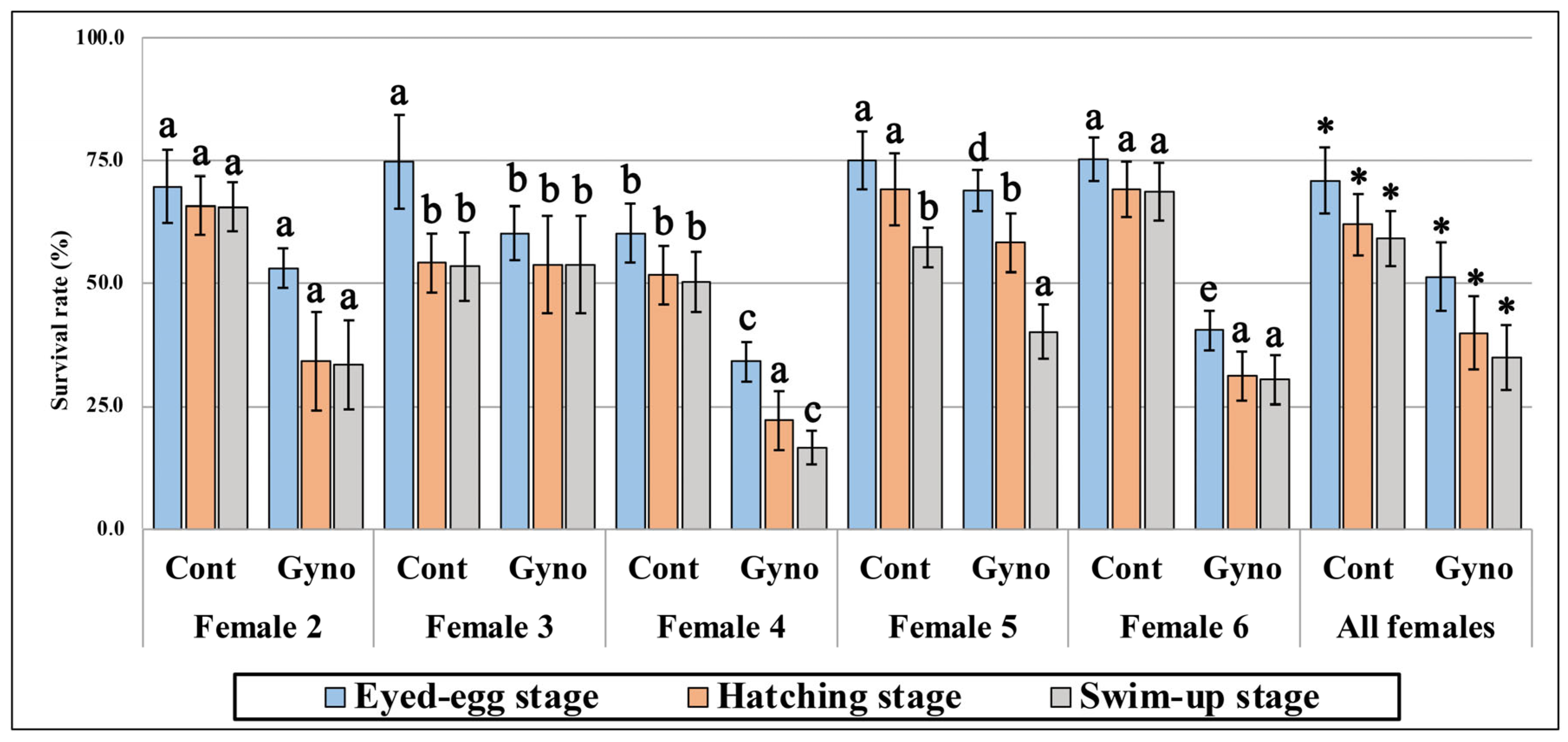
Eggs stripped from five out of six females were successfully fertilized and activated by either UV-irradiated or non-irradiated sperm, and embryonic development was not observed in eggs originated from Female 1 as the eggs failed to activate (Supplementary File S1). Survivability of embryos developing in eggs fertilized with UV-irradiated sperm (gynogenetic groups) was lower than in the control groups at all developmental stages. Moreover, significant differences (p < 0.05) were found between the control and gynogenetic groups at each developmental stage when data from all females were combined (Figure 1). Significant differences (p < 0.05) in the survival rates were also observed between females within control and gynogenetic variants (Figure 1). At the eyed-egg stage, survivability in the gynogenetic group ranged from 34.1 ± 7.0% (Female 4) to 68.9 ± 6.1% (Female 5), whereas in the control group it varied between 60.1 ± 7.7% (Female 4) and 75.2 ± 4.4% (Female 6). The recorded differences were even more pronounced at the hatching stage, where gynogenetic survivability dropped to between 22.1 ± 2.6% (Female 4) and 58.3 ± 5.9% (Female 5), when compared to 51.7 ± 9.9% (Female 4) and 69.2 ± 7.3% (Female 6) in the control group. In turn, at the swim-up stage, the recorded survival rates in the gynogenetic groups ranged from 16.6 ± 4.3% (Female 4) to 53.8 ± 9.8% (Female 3), while fish from the control group showed survival rates ranging from 50.3 ± 11.4% (Female 4) to 68.7 ± 5.9% (Female 6). Based on the final survival rates, we are able to define eggs from Female 3 as those with the highest potential for gynogenesis, while the eggs from Female 4 are those with the lowest ability to develop after gynogenetic activation. Eggs from other females showed intermediate potential for gynogenetic development.
Figure 1. Survival rates (%) of rainbow trout developing in eggs fertilized with non- (Cont) and UV-irradiated sperm (Gyno) recorded across all analyzed developmental stages. Different letters denote statistically significant differences (p < 0.05) in the recorded survival rate of eggs fertilized with non- (Cont) and UV-irradiated sperm (Gyno) among examined females. Asterisks indicate significant differences (p < 0.05) in the recorded survival rates averaged for all females between Cont and Gyno groups. Error bars represent 95% confidence intervals.

3.2. Maternal mRNA Transcription

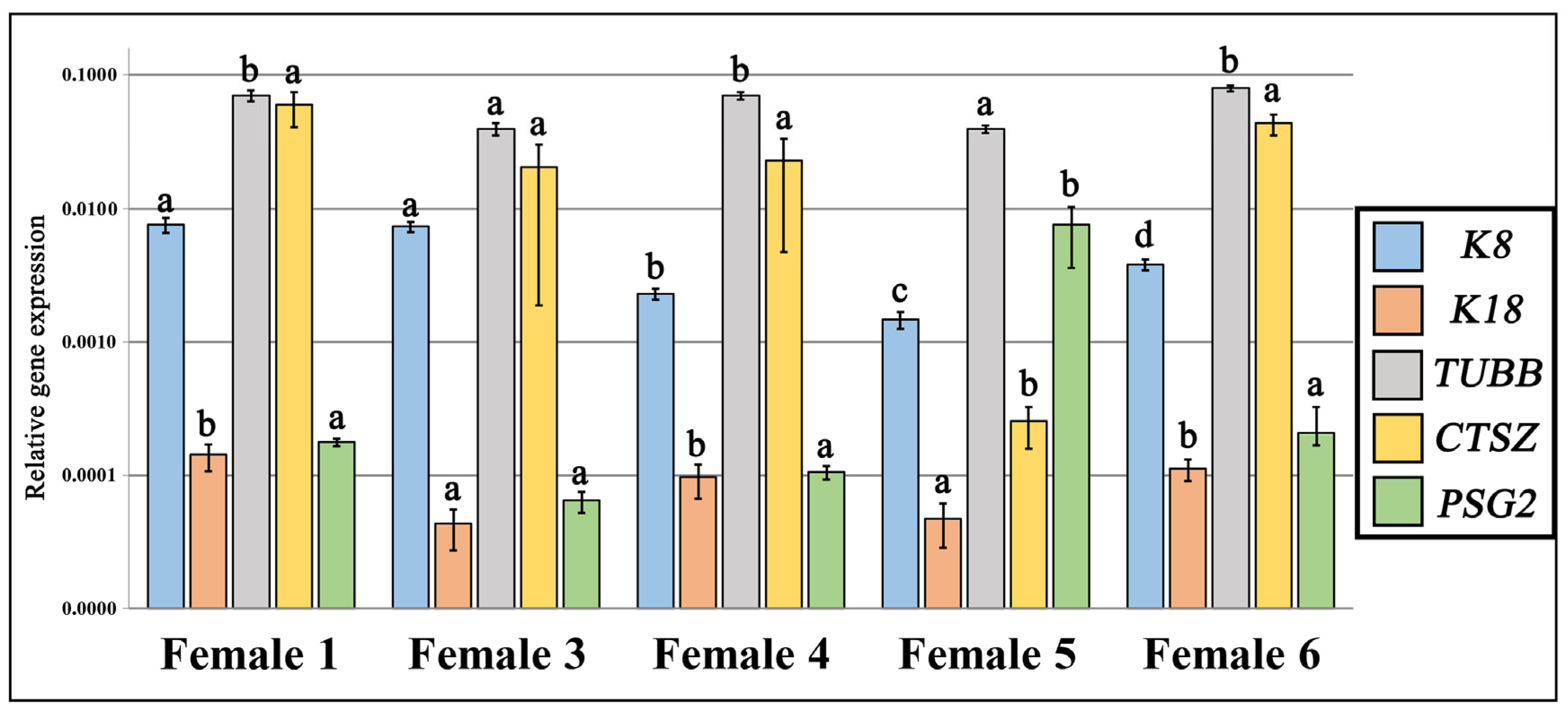
Real-time PCR was used to quantify the relative expression levels of five selected maternal mRNA transcripts (K8, K18, TUBB, CTSZ and PSG2) in eggs originating from the rainbow trout females examined in this study. The analysis revealed significant (p < 0.05) inter-individual variation in transcript levels across all examined mRNA transcripts (Figure 2). Expression of the K8 differed significantly among most of females, with the highest levels observed in Females 1 and 3, and the lowest in Female 5. Significant upregulation (p < 0.05) of K18 and TUBB mRNA transcripts was detected in Females 1, 4, and 6 compared to Females 3 and 5. Additionally, a significant (p < 0.05) downregulation of CTSZ and upregulation of PSG2 were observed exclusively in Female 5, with no significant differences among the other females (Figure 2).
Figure 2. Relative mRNA expression levels of six analyzed transcripts in eggs sampled from the rainbow trout females examined in this study. Distinct letters indicate statistically significant (p < 0.05) differences in relative expression levels among females within each transcript. Expression levels were normalized to the reference genes β-actin and elongation factor 1-alpha. Each bar represents the mean value along with variation ± calculated from eight biological replicates (individual eggs), with each sample analyzed in technical triplicate by qPCR. Variation is derived from the SEM of the recorded Ct values. Relative expression data were log10-transformed prior to visualization to improve readability and scale consistency across genes differing widely in expression magnitude.

3.3. Activity of Antioxidant Enzymes in Eggs

Significant (p < 0.05) inter-individual differences in the activity were detected in all analyzed antioxidant enzymes (Figure 3, Supplementary Table S1). Eggs originating from Female 4 exhibited the highest catalase (CAT) activity (~59 nmol·min−1·mL−1), significantly (p < 0.05) exceeding those recorded in the other individuals, whereas eggs from Female 1 showed the lowest activity (~25 nmol min−1 mL−1). Eggs from the remaining individuals displayed comparable CAT activity levels (~33–43 nmol min−1 mL−1) with relatively small variation.
Figure 3. Activity levels of catalase (CAT), superoxide dismutase (SOD), and glutathione peroxidase (GPx) recorded in eggs from six rainbow trout females. Different letters denote statistically significant differences (p < 0.05). Data is shown as mean ± standard deviation.
For superoxide dismutase (SOD), the highest activity (~47 U mL−1) with minimal variability was recorded in eggs from Female 5,while the lowest activity (~32 U mL−1) was observed in eggs from Female 6. Eggs from the remaining females exhibited intermediate SOD activity levels ranging from approximately 40 to 44 U mL−1.
The highest glutathione peroxidase (GPx) activity was found in eggs from Female 1, 3 and 5, with levels ranging from 65 to 68 nmol min−1 mL−1. Eggs from Female 4 showed slightly lower activity (~59 nmol min−1 mL−1), while eggs from Female 6 exhibited moderate activity (~60 nmol min−1 mL−1). The lowest GPx activity is observed in eggs from Female 2 (~52 nmol min−1 mL−1).

4. Discussion

Gynogenetic specimens produced by activation of eggs with UV-inactivated spermatozoa and HHP-induced inhibition of the meiosis retain some heterozygosity []. Although, meiogynogenotes usually show significantly lower survival rates than their siblings hatched from eggs activated with the non-irradiated sperm (Figure 1). The increased mortality of the gynogenetic individuals may be triggered as well by the poor quality eggs used for gynogenesis and the side effects of the physical shock [,]. Variation in survival was also observed between gynogenetic specimens developing in eggs stripped from different females (Figure 1). Although inter-clutch differences in survival of meiogynogenetic rainbow trout were significant, the limited number of egg donors used in this research means that we cannot confirm this (s). Nevertheless, based on the obtained results, we may at least assume that rainbow trout eggs from some females have increased potential for meiotic gynogenesis. In goldfish (Carrassius sp.), variation in egg sensitivity related to the physical shock has been proposed to explain such notable inter-clutch differences in the survival of gynogenotes []. In turn, our recent study proved that eggs with high and low potential for mitotic gynogenesis have dissimilar transcriptional profiles [,]. Here, rainbow trout eggs with the lowest and the highest developmental potential after activation with UV-treated spermatozoa also showed some differences in the antioxidant enzyme activity and the expression levels of the certain maternal genes.
Activity of the glutathione peroxidase (GPx) was high in the rainbow trout eggs, with increased developmental potential for meiotic gynogenesis, and significantly lower in eggs clutches characterized by low gynogenesis efficiency (Supplementary Table S1, Figure 3). The GPx is one of the most important antioxidant enzymes, regulating reactive oxygen species (ROS) homeostasis and protecting cells from oxidative damage. ROS are highly reactive molecules generated during normal aerobic metabolism and are involved in processes such as cell differentiation, regulation of cell proliferation and growth, intercellular signaling, and the synthesis of biologically important compounds, among others [,]. However, various environmental stresses can generate an excessive amount of ROS in cells, resulting in diminished redox homeostasis and oxidative stress (OS). Excessive levels of ROS causes protein degradation, spindle formation errors, DNA damage, lipid peroxidation, accelerated rate of the telomere shortening, and cancer progression []. OS may impair the quality of mammalian oocytes and deteriorate fertilization and embryo development [,]. In fish, OS is considered to be associated with post-ovulatory oocyte aging, which leads to a general decline in egg quality []. Recently, we found that high-quality ova from the spring-spawning rainbow trout showed an increased ability to develop after irradiation and fertilization with non-irradiated sperm (androgenesis) and exhibited high activities of GPx, SOD and CAT [].
The early development of fish embryos is governed by the maternal RNA deposited in the egg cytoplasm during oocyte maturation. Zygotic Genome Activation (ZGA) is initiated only during the mid-blastula transition (MBT), which occurs around the 10th cell cleavage, and an appropriate level of transcripts of the specific genes may ensure efficient fertilization and proper early embryogenesis. In several fish species, the quantity of maternal RNA has been analyzed to identify differentially expressed genes in eggs with varying quality [,,,,,]. The transcriptomic differences between low- and high-quality eggs have been observed in rainbow trout, where eggs of decreased quality due to oocyte aging were characterized by high levels of keratins 8 and 18, cathepsin Z, and prostaglandin synthase 2, while transcripts of Npm2, tubulin b, and IGF1 genes were less abundant in these eggs []. Here, we also observed differences in the transcription of the aforementioned genes between eggs with varied competence for gynogenesis. Ova with the highest developmental potential for gynogenesis (Female 3) were characterized by a high level of keratin 8 and decreased abundance of keratin 18 and tubulin b. In contrast, eggs with the lowest potential to develop after activation with UV-irradiated sperm (Female 4) exhibited increased transcript levels of keratin 18 and tubulin b, but decreased keratin 8 mRNA abundance (Figure 2). Keratins and tubulins are proteins engaging in cellular organization; however, they belong to different cytoskeletal networks. Keratins help to maintain cell integrity and are cytoskeletal components expressed in the oocytes and early developing embryos. Keratins protect oocytes from the physical stress after ovulation and play important roles during oocyte maturation and fertilization [,,]. Tubulin, in turn, is a major component of microtubules, building the mitotic spindle that is essential for chromosome segregation during cell divisions []. In the present study, we found that eggs with the highest potential for gynogenesis had an increased level of K18 mRNA and decreased tubulin expression. Higher expression of keratin genes may ensure cell integrity and the better protection of eggs after HHP shock. On the other hand, it can be assumed that the decreased level of tubulins makes microtubules of these eggs prone to depolymerization, which increases the efficiency of the HHP shock. Although a small number of egg donors used in the research does not allow us to draw far-reaching conclusions, the results obtained may indicate how best to approach searching for an explanation of the inter-clutch variation in gynogenesis success.

5. Conclusions

The results of our pilot study showed that rainbow trout eggs from different females may exhibited variation in their ability to develop after activation with UV-irradiated sperm. Biochemical examination revealed that eggs with higher developmental potential for meiotic gynogenesis exhibited significantly increased activity of glutathione peroxidase (GPx) than eggs with lower efficiency of gynogenesis. Moreover, eggs with the highest developmental potential for gynogenesis showed increased transcription of the keratin 8 gene and decreased abundance of keratin 18 and tubulin β mRNA transcripts.

Supplementary Materials

The following supporting information can be downloaded at: https://www.mdpi.com/article/10.3390/fishes10110585/s1, Supplementary File S1: Survival of rainbow trout embryos and larvae from gynogenetic and control groups; Table S1: Antioxidant enzymes activity (mean ± SD) in rainbow trout eggs.

Author Contributions

Conceptualization, K.O. Methodology, K.O., M.K., A.B. and S.D., Formal Analysis, K.O., M.K., K.P., L.P. and R.R.; Resources, K.O. and S.D.; Writing—Original Draft Preparation, K.O. and M.K. Writing—Review and Editing, K.O. Visualization, M.K. and L.P., Supervision, K.O.; Funding Acquisition, K.O. All authors have read and agreed to the published version of the manuscript.

Funding

This study has been financially supported by the National Science Centre, Poland, project number 2020/39/B/NZ9/00865.

Institutional Review Board Statement

The study was carried out in strict accordance with the recommendations in the Polish ACT of 15 January 2015 on the Protection of Animals Used for Scientific or Educational Purposes, Journal of Laws 2015, item 266. The protocol was approved by the Local Ethical Committee for the Experiments on Animals in Bydgoszcz (resolution No. 27/2022, issued on 20 April 2022).

Data Availability Statement

The original contributions presented in this study are included in the article/Supplementary Materials. Further inquiries can be directed to the corresponding author.

Conflicts of Interest

The authors declare no conflicts of interest.

References

  1. Morris, M.A.; Brandon, R.A. Gynogenesis and hybridization between Ambystoma platineum and Ambystoma texanum in Illinois. Copeia 1984, 1984, 324–337. [Google Scholar] [CrossRef]
  2. Gumm, J.M.; Thaker, M. Association preferences of unisexual Amazon mollies (Poecilia formosa): Differential response to swords based on sex of the bisexual parental species. Behaviour 2009, 146, 907–921. [Google Scholar] [CrossRef]
  3. Grismer, J.L.; Grismer, L.L. Who’s your mommy? Identifying maternal ancestors of asexual species of Leiolepis Cuvier, 1829 and the description of a new endemic species of asexual Leiolepis Cuvier, 1829 from Southern Vietnam. Zootaxa 2010, 2433, 47–61. [Google Scholar] [CrossRef]
  4. Neaves, W.B.; Baumann, P. Unisexual reproduction among vertebrates. Trends Genet. 2011, 27, 81–88. [Google Scholar] [CrossRef] [PubMed]
  5. Pandian, T.J.; Koteeswaran, R. Ploidy induction and sex control in fish. Hydrobiologia 1998, 384, 167–243. [Google Scholar] [CrossRef]
  6. Campos-Ramos, R.; Harvey, S.C.; McAndrew, B.J.; Penman, D.J. An investigation of sex determination in the Mozambique tilapia, Oreochromis mossambicus, using synaptonemal complex analysis, FISH, sex reversal and gynogenesis. Aquaculture 2003, 221, 125–140. [Google Scholar] [CrossRef]
  7. Chen, S.-L.; Tian, Y.-S.; Yang, J.-F.; Shao, C.-W.; Ji, X.-S.; Zhai, J.-M.; Liao, X.-L.; Zhuang, Z.-M.; Su, P.-Z.; Xu, J.-Y.; et al. Artificial gynogenesis and sex determination in half-smooth tongue sole (Cynoglossus semilaevis). Mar. Biotechnol. 2008, 11, 243–251. [Google Scholar] [CrossRef]
  8. Fopp-Bayat, D.; Hliwa, P.; Ocalewicz, K. Presence of gynogenetic males suggests a female heterogamety in sterlet Acipenser ruthenus L. Anim. Reprod. Sci. 2018, 189, 110–118. [Google Scholar] [CrossRef]
  9. Arai, K. Genetic improvement of aquaculture finfish species by chromosome manipulation techniques in Japan. Aquaculture 2001, 197, 205–228. [Google Scholar] [CrossRef]
  10. Harris, M.P.; Henke, K.; Hawkins, M.B.; Witten, P.E. Fish is Fish: The use of experimental model species to reveal causes of skeletal diversity in evolution and disease. J. Appl. Ichthyol. 2014, 30, 616–629. [Google Scholar] [CrossRef]
  11. Jagiełło, K.; Zalewski, T.; Dobosz, S.; Michalik, O.; Ocalewicz, K. High rate of deformed larvae among gynogenetic brown trout (Salmo trutta m. fario) doubled haploids. BioMed Res. Int. 2017, 2017, 2975187. [Google Scholar] [CrossRef]
  12. Zhang, X.; Mizukoshi, M.; Zhang, H.; Tan, E.; Igarashi, Y.; Suzuki, Y.; Mitsuyama, S.; Kinoshita, S.; Saito, K.; Watabe, S.; et al. Ultrahigh-density linkage map construction using low-coverage whole-genome sequencing of a doubled haploid population: Case study of Torafugu (Takifugu rubripes). Genes 2018, 9, 120. [Google Scholar] [CrossRef]
  13. Komen, H.; Thorgaard, G.H. Androgenesis, gynogenesis and the production of clones in fishes: A review. Aquaculture 2007, 269, 150–173. [Google Scholar] [CrossRef]
  14. Jagiełło, K.; Dobosz, S.; Zalewski, T.; Polonis, M.; Ocalewicz, K. Developmental competence of eggs produced by rainbow trout Doubled Haploids (DHs) and generation of the clonal lines. Reprod. Domest. Anim. 2018, 53, 1176–1183. [Google Scholar] [CrossRef] [PubMed]
  15. Naruse, K.; Ijiri, K.; Shima, A.; Egami, N. The production of cloned fish in the medaka (Oryzias latipes). J. Exp. Zool. 1985, 236, 335–341. [Google Scholar] [CrossRef] [PubMed]
  16. Komen, J.; Bongers, A.B.J.; Richter, C.J.J.; Van Muiswinkel, W.B.; Huisman, E.A. Gynogenesis in common carp (Cyprinus carpio L.): II. The production of homozygous gynogenetic clones and F1 hybrids. Aquaculture 1991, 92, 127–142. [Google Scholar] [CrossRef]
  17. Quillet, E.; Garcia, P.; Guyomard, R. Analysis of the production of all homozygous lines of rainbow trout by gynogenesis. J. Exp. Zool. 1991, 257, 367–374. [Google Scholar] [CrossRef]
  18. Ocalewicz, K. Quality of fish eggs and production of androgenetic and gynogenetic doubled haploids (DHs). Fish Physiol. Biochem. 2024, 50, 1947–1957. [Google Scholar] [CrossRef]
  19. Bobe, J.; Labbé, C. Egg and sperm quality in fish. Gen. Comp. Endocrinol. 2010, 165, 535–548. [Google Scholar] [CrossRef]
  20. Migaud, H.; Bell, G.; Cabrita, E.; McAndrew, B.; Davie, A.; Bobe, J.; Herráez, M.P.; Carrillo, M. Gamete quality and broodstock management in temperate fish. Rev. Aquac. 2013, 5, S194–S223. [Google Scholar] [CrossRef]
  21. Bobe, J. Egg quality in fish: Present and future challenges. Anim. Front. 2015, 5, 66–72. [Google Scholar] [CrossRef]
  22. Aegerter, S.; Jalabert, B.; Bobe, J. Large scale real-time PCR analysis of mRNA abundance in rainbow trout eggs in relationship with egg quality and post-ovulatory ageing. Mol. Reprod. Dev. 2005, 72, 377–385. [Google Scholar] [CrossRef] [PubMed]
  23. Bonnet, E.; Fostier, A.; Bobe, J. Characterization of rainbow trout egg quality: A case study using four different breeding protocols, with emphasis on the incidence of embryonic malformations. Theriogenology 2007, 67, 786–794. [Google Scholar] [CrossRef] [PubMed]
  24. Polonis, M.; Błaszczyk, A.; Jagiełło, K.; Panasiak, L.; Dobosz, S.; Ocalewicz, K. Inter-clutch egg differences and androgenesis in rainbow trout (Oncorhynchus mykiss, Walbaum 1792). Oceanol. Hydrobiol. Stud. 2021, 50, 160–168. [Google Scholar] [CrossRef]
  25. Samarin, A.M.; Sampels, S.; Policar, T.; Rodina, M.; Hematyar, N. mRNA abundance changes during in vitro oocyte ageing in African catfish Clarias gariepinus (Burchell, 1822). Aquac. Res. 2017, 49, 1037–1045. [Google Scholar] [CrossRef]
  26. Goryczko, K.; Dososz, S.; Mäkinen, T.; Tomasik, L. UV-irradiation of rainbow trout sperm as a practical method for induced gynogenesis. J. Appl. Ichthyol. 1991, 7, 136–146. [Google Scholar] [CrossRef]
  27. Billard, R. Utilisation d’un syst’eme tris-glycocolle pour tamponner le dilueurd’ins’emination pour truite. Bull. Français Piscic. 1997, 264, 102–112. [Google Scholar]
  28. Livak, K.J.; Schmittgen, T.D. Analysis of relative gene expression data using real-time quantitative PCR and the 2−ΔΔCT method. Methods 2001, 25, 402–408. [Google Scholar] [CrossRef]
  29. Vandesompele, J.; De Preter, K.; Pattyn, F.; Poppe, B.; Van Roy, N.; De Paepe, A.; Speleman, F. Accurate normalization of real-time quantitative RT-PCR data by geometric averaging of multiple internal control genes. Genome Biol. 2002, 3, RESEARCH0034. [Google Scholar] [CrossRef]
  30. Carrizo, V.; Valenzuela, C.A.; Zuloaga, R.; Aros, C.; Altamirano, C.; Valdés, J.A.; Molina, A. Effect of cortisol on the immune-like response of rainbow trout (Oncorhynchus mykiss) myotubes challenged with Piscirickettsia salmonis. Veter-Immunol. Immunopathol. 2021, 237, 110240. [Google Scholar] [CrossRef]
  31. Meiler, K.A.; Cleveland, B.; Radler, L.; Kumar, V. Oxidative stress-related gene expression in diploid and triploid rainbow trout (Oncorhynchus mykiss) fed diets with organic and inorganic zinc. Aquaculture 2021, 533, 736149. [Google Scholar] [CrossRef]
  32. Yamaha, E.; Otani, S.; Minami, A.; Arai, K. Dorso-ventral axis perturbation in goldfish embryos caused by heat- and pressure-shock treatments for chromosome set manipulation. Fish. Sci. 2002, 68, 313–319. [Google Scholar] [CrossRef]
  33. Ocalewicz, K.; Gurgul, A.; Polonis, M.; Dobosz, S. Preliminary identification of candidate genes related to survival of gynogenetic rainbow trout (Oncorhynchus mykiss) based on comparative transcriptome analysis. Animals 2020, 10, 1326. [Google Scholar] [CrossRef] [PubMed]
  34. Ocalewicz, K.; Gurgul, A.; Dobosz, S.; Jasielczuk, I.; Szmatoła, T.; Semik-Gurgul, E.; Kucharski, M.; Rożyński, R. Transcriptome Profiling Reveals Differences Between Rainbow Trout Eggs with High and Low Potential for Gynogenesis. Genes 2025, 16, 803. [Google Scholar] [CrossRef]
  35. Mantovani, G.; Macciò, A.; Madeddu, C.; Mura, L.; Gramignano, G.; Lusso, M.R.; Murgia, V.; Camboni, P.; Ferreli, L.; Mocci, M.; et al. The impact of different antioxidant agents alone or in combination on reactive oxygen species, antioxidant enzymes and cytokines in a series of advanced cancer patients at different sites: Correlation with disease progression. Free Radic. Res. 2003, 37, 213–223. [Google Scholar] [CrossRef]
  36. Goud, A.P.; Goud, P.T.; Diamond, M.P.; Gonik, B.; Abu-Soud, H.M. Reactive oxygen species and oocyte aging: Role of superoxide, hydrogen peroxide, and hypochlorous acid. Free. Radic. Biol. Med. 2008, 44, 1295–1304. [Google Scholar] [CrossRef]
  37. Sasaki, H.; Hamatani, T.; Kamijo, S.; Iwai, M.; Kobanawa, M.; Ogawa, S.; Miyado, K.; Tanaka, M. Impact of oxidative stress on age-associated decline in oocyte developmental competence. Front. Endocrinol. 2019, 10, 811. [Google Scholar] [CrossRef]
  38. Tarín, J.J.; Pérez-Albalá, S.; Cano, A. Consequences on offspring of abnormal function in ageing gametes. Hum. Reprod. Updat. 2000, 6, 532–549. [Google Scholar] [CrossRef]
  39. Prasad, S.; Tiwari, M.; Pandey, A.N.; Shrivastav, T.G.; Chaube, S.K. Impact of stress on oocyte quality and reproductive outcome. J. Biomed. Sci. 2016, 23, 36. [Google Scholar] [CrossRef]
  40. Samarin, A.M.; Østbye, T.-K.K.; Ruyter, B.; Sampels, S.; Burkina, V.; Blecha, M.; Gela, D.; Policar, T. Alteration of mRNA abundance, oxidation products and antioxidant enzyme activities during oocyte ageing in common carp Cyprinus carpio. PLoS ONE 2019, 14, e0212694. [Google Scholar] [CrossRef]
  41. Lanes, C.; Fernandes, J.; Kiron, V.; Babiak, I. Profiling of key apoptotic, stress, and immune-related transcripts during embryonic and postembryonic development of Atlantic cod (Gadus morhua L.). Theriogenology 2012, 78, 1583–1596.e2. [Google Scholar] [CrossRef] [PubMed]
  42. Chapman, J.R.; Waldenström, J. With reference to reference genes: A Systematic review of endogenous controls in gene expression studies. PLoS ONE 2015, 10, e0141853. [Google Scholar] [CrossRef] [PubMed]
  43. Ma, H.; Martin, K.; Dixon, D.; Hernandez, A.G.; Weber, G.M. Transcriptome analysis of egg viability in rainbow trout, Oncorhynchus mykiss. BMC Genom. 2019, 20, 319. [Google Scholar] [CrossRef] [PubMed]
  44. Weber, G.M.; Birkett, J.; Martin, K.; Dixon, D.; Gao, G.; Leeds, T.D.; Vallejo, R.L.; Ma, H. Comparisons among rainbow trout, Oncorhynchus mykiss, populations of maternal transcript profile associated with egg viability. BMC Genom. 2021, 22, 448. [Google Scholar] [CrossRef]
  45. Rise, M.L.; Nash, G.W.; Hall, J.R.; Booman, M.; Hori, T.S.; Trippel, E.A.; Gamperl, A.K. Variation in embryonic mortality and maternal transcript expression among Atlantic cod (Gadus morhua) broodstock: A functional genomics study. Mar. Genom. 2014, 18, 3–20. [Google Scholar] [CrossRef]
  46. Lim, H.Y.G.; Alvarez, Y.D.; Gasnier, M.; Wang, Y.; Tetlak, P.; Bissiere, S.; Wang, H.; Biro, M.; Plachta, N. Keratins are asymmetrically inherited fate determinants in the mammalian embryo. Nature 2020, 585, 404–409. [Google Scholar] [CrossRef]
  47. Plancha, C.E. Cytokeratin dynamics during oocyte maturation in the hamster requires reaching of metaphase I. Differentiation 1996, 60, 87–98. [Google Scholar] [CrossRef]
  48. Kabashima, K.; Matsuzaki, M.; Suzuki, H. Intermediate Filament Keratin Dynamics During Oocyte Maturation Requires Maturation/M-Phase Promoting Factor and Mitogen-Activated Protein Kinase Kinase Activities in the Hamster. Reprod. Domest. Anim. 2009, 45, e184–e188. [Google Scholar] [CrossRef]
  49. Janke, C.; Magiera, M.M. The tubulin code and its role in controlling microtubule properties and functions. Nat. Rev. Mol. Cell Biol. 2020, 21, 307–326. [Google Scholar] [CrossRef]
Disclaimer/Publisher’s Note: The statements, opinions and data contained in all publications are solely those of the individual author(s) and contributor(s) and not of MDPI and/or the editor(s). MDPI and/or the editor(s) disclaim responsibility for any injury to people or property resulting from any ideas, methods, instructions or products referred to in the content.

Article Metrics

Citations

Article Access Statistics

Multiple requests from the same IP address are counted as one view.