Abstract
Dissolved oxygen (DO) is a crucial determinant of aquatic organism health. This study demonstrates that hypoxia (at MH, 2.0 mg/L; SH, 0.5 mg/L) disrupts intestinal homeostasis in the blood clam, Anadara granosa. Exposure to hypoxia induced severe histopathological damage, including villus loss, inflammatory cell infiltration, and epithelial cell vacuolization. Immune-related gene expression analysis revealed coordinated regulation, with TLR4 and NF-κB significantly up-regulated by 4.5-fold and 5-fold, respectively, in the SH14 group, while HSP70 showed a remarkable 13-fold increase in the MH14 group. In contrast, TAK1 and TRAF6 exhibited substantial downregulation. High-throughput sequencing of the 16S rRNA gene revealed a significant reduction in gut microbiota diversity under hypoxic conditions, as evidenced by notable decreases of approximately 30% in the Chao1 index and 35% in the Shannon index in the SH group compared to the normoxic control (N group). Functional pathway analysis indicated alterations in pathways associated with xenobiotic biodegradation, lipid metabolism, and energy metabolism. These findings highlight a strong association between hypoxia and adverse intestinal health outcomes in A. granosa, underscoring the critical importance of maintaining adequate dissolved oxygen levels to support bivalve health. Future research should aim to develop strategies to mitigate hypoxia-induced stress and further elucidate the molecular mechanisms underlying hypoxia adaptation in bivalves.
Keywords:
Anadara granosa; hypoxia; intestinal microbiota; intestinal morphology; immune-related genes Key Contribution:
This study provides an integrative analysis and systematically demonstrates that hypoxia disrupts the “microbiota–intestine–immune” axis in the blood clam Anadara granosa, revealing a strong correlation between microbial dysbiosis, intestinal tissue damage, and modulated immune gene expression under low oxygen stress.
1. Introduction
Dissolved oxygen (DO) serves as a vital limiting factor in aquaculture, exerting a significant influence on the behavior, immune function, reproduction, growth, spatial distribution, and interspecific interactions of aquatic organisms []. Optimal oxygen concentrations in water typically range from 8 to 10 mg/L, whereas anoxic conditions are characterized by concentrations below 2 mg/L [,,]. Hypoxia, a prevalent and natural phenomenon in numerous marine environments, represents an extremely adverse condition frequently encountered by aquatic organisms within aquaculture systems.
Hypoxia has a profound impact on animal intestinal health, characterized by a complex interplay between physiological stressors and biological responses, which leads to significant alterations in intestinal morphology and function [,,,]. In vertebrates, neonatal mice exposed to hypoxic conditions exhibit marked structural abnormalities in the ileum and distal colon, including disruption of tight junctions and dysregulation of genes associated with intestinal barrier function []. Similarly, in fish species such as Micropterus salmoides, hypoxic stress has been shown to induce oxidative stress, shedding of intestinal villus epithelium, and increased apoptosis []. Hypoxia modulates the expression of immune-related genes. In newborn mice, hypoxia modifies immune gene activity and weakens the integrity of the intestinal barrier []. In Salmo salar, chronic exposure to low oxygen levels triggers chronic inflammation and modulates the expression of key cytokines, including IL-1β and IL-10 []. In Eriocheir sinensis, hypoxia induces the expression of immune-related genes, which is part of the organism’s adaptive response to hypoxic conditions []. Hypoxia can lead to notable shifts in immune-related gene expression among bivalves [,,]. In Mytilus chilensis, the expression of genes related to immune responses in the gills is significantly up-regulated, while in Ruditapes philippinarum, some immune defense genes such as IAP and HSP70 are significantly up-regulated.
The gut microbiota plays a critical role in shaping the host’s physiology, reproduction, development, energy balance, behavior, and life history [,]. It is essential for nutrition metabolism, immune system regulation, disease prevention, and maintaining ecological balance [,]. Hypoxia greatly diminishes the diversity and abundance of beneficial bacteria in the gut microbiota in Eriocheir sinensis, disrupting the microecological balance with limited recovery after reoxygenation []. In Mytilus chilensis, hypoxia causes an imbalance in gut microbiota reducing beneficial bacteria, increasing pathogenic bacteria, impairing metabolic pathways, and raising disease risk []. A stable and robust gut microbiota is key to bivalve health and growth.
Anadara granosa (synonym: Tegillarca granosa), commonly known as the blood clam, belongs to the phylum Mollusca, class Bivalvia, order Veneroida, family Veneridae, and genus Anadara. It is considered as one of the four principal tidal flat economic shellfish species in China [,,,,]. As a filter-feeding bivalve, A. granosa primarily feeds on unicellular algae, including golden algae, diatoms, and flat algae, as well as organic detritus. Notably, its blood contains hemoglobin, making it one of the few mollusks with red blood, a characteristic from which its common name is derived []. The intertidal habitat of A. granosa is characterized by frequent environmental fluctuations and is often exposed to periodic hypoxic stress. Against the backdrop of escalating climate change and increasing coastal eutrophication, the frequency and severity of hypoxic events in coastal waters continue to rise, posing serious threats to the survival and physiological health of organisms such as A. granosa. However, the mechanism through which hypoxia stress affects the intestinal health of A. granosa remains poorly understood.
Our previous studies on A. granosa have primarily focused on isolated physiological indicators such as oxidative stress in gill tissue [,], this study breaks new ground by systematically integrating intestinal histology, immune gene profiling, and microbiome analysis to establish a complete “stress–immune–microecology” network. This study seeks to elucidate the impact of hypoxia on the intestinal health of A. granosa, emphasizing on the “microbiota–intestine–immune response” axis, to enhance our understanding of the physiological adaptations of bivalves under low-oxygen conditions. A. granosa specimens were subjected to varying dissolved oxygen levels (7.0 mg/L, 2.0 mg/L, and 0.5 mg/L) over a period of up to 14 days. While traditional definitions classify dissolved oxygen concentrations below 2 mg/L as anoxic, the blood clam A. granosa, with its unique hemoglobin-based oxygen transport capacity, demonstrates remarkable short-term tolerance to this oxygen level [,]. This species-specific adaptation allows maintenance of basic metabolic functions for 1–3 days at 2.0 mg/L, as evidenced by the absence of significant histopathological damage in early exposure groups. However, prolonged exposure beyond 7 days induced substantial intestinal damage in our study. We therefore define 2.0 mg/L as moderate hypoxia (MH) based on this “short-term tolerance versus long-term damage” characteristic, while classifying 0.5 mg/L as severe hypoxia (SH) due to its immediately detrimental effects even during short-term exposure. To evaluate intestinal responses, histological examination, immune genes expression profiling, and 16S rRNA sequencing were conducted. The anticipated outcomes of this research are expected to advance our comprehension of the mechanisms underlying hypoxia adaptation in A. granosa and to provide a scientific foundation for developing strategies to maintain intestinal health in aquaculture settings.
2. Materials and Methods
2.1. Study Animals
In late autumn 2024, we gathered 800 mature Anadara granosa specimens from coastal waters near Ningbo’s Xiangshan Bay (coordinates 29°38′ N, 121°46′ E). The bivalves underwent a seven-day acclimation phase in specialized tanks filled with processed seawater (maintained at 15 ± 1 °C, pH 7.80 ± 0.2, oxygen saturation 7.0 ± 0.5 mg/L, and salinity level 23 ± 1). Following the established protocols, the organisms received nourishment from Platymonas subcordiformis microalgae cultures (concentration 2 × 10⁵ cells/mL), with complete water replacement occurring each morning throughout the conditioning phase []. Oxygen levels were consistently regulated through diffused aeration systems.
Hypoxia was induced by introducing nitrogen gas into seawater, and throughout this process, dissolved oxygen levels were continuously monitored using a portable dissolved oxygen meter (HQ30D, HACH) until the target dissolved oxygen concentration was achieved []. Measurements of dissolved oxygen were taken every 2 h. The acclimated A. granosa were randomly divided into three groups, including a normal dissolved oxygen group (7.0 ± 0.5 mg/L, serving as the control group, labeled as N), a group with dissolved oxygen at 2.0 mg/L (labeled as MH), and a group with dissolved oxygen at 0.5 mg/L (labeled as SH). Each condition was replicated across three parallel breeding tanks. During the experiment period, the clams were fed algae paste at designated times each day, and 50% of the tank water was replaced daily. At intervals of 0, 1, 3, 5, 7, and 14 days of exposure, 30 A. granosa individuals were randomly sampled from the respective breeding tanks of the experimental and control groups. The intestines of five A. granosa were combined into one sample, resulting in six replicate samples per group. To ensure biological replication at the tank level, these six samples were derived from the three independent tanks (two pooled samples per tank). These samples were designated as N0, N1, MH1, SH1, N3, MH3, SH3, N5, MH5, SH5, N7, MH7, SH7, N14, MH14, and SH14 (Table 1). The collected A. granosa were first disinfected with alcohol to reduce the impact of surface-associated microorganisms on gut microbiota, and then the entire intestine (including contents) was removed from five A. granosa and combined into a single sample, which was quickly frozen in liquid nitrogen and stored at −80 °C for DNA and RNA extraction. Additionally, two A. granosa intestines from each experimental group were placed in 4% tissue cell fixative for 24 h for histological analysis.
Table 1.
Experimental groups.
2.2. Histological Sectioning
Intestinal tissues fixed under three different dissolved oxygen concentrations were washed with water, dehydrated with gradient ethanol, made transparent with xylene, and then immersed in paraffin. The tissues were embedded with an embedding machine, sectioned with a microtome, and the sections were spread in water at 42 °C, dried in an oven at 60 °C for 6 h, and subsequently stained with hematoxylin and eosin (H&E). The sections were dewaxed twice with xylene, rehydrated stepwise with gradient ethanol, stained with hematoxylin for 5 min, washed with water for 5 min, treated with differentiating solution for 5 s, stained with eosin for 6 min, rehydrated stepwise with gradient ethanol, and mounted with neutral gum. The sections were then observed and photographed under a Nikon ECLIPSE E100 (Nikon Corporation, Tokyo, Japan). The staining reagents used were the Haoke H&E staining kit and Haoke hydrochloric acid ethanol rapid differentiating solution, both from Hangzhou Haoke Biotechnology (Hangzhou, China).
2.3. RNA Extraction and Gene Expression Analysis
Total RNA was extracted using the Trizol reagent method. One hundred milligrams of tissue were placed in a centrifuge tube containing 1 mL of Trizol, homogenized using a homogenizer, and placed on ice for 5 min before centrifugation (4 °C, 10 min, 12,000 rpm). Eight hundred microliters of the supernatant were mixed with 200 μL of chloroform, vigorously shaken for 15 s, placed on ice for 3 min, and then centrifuged at 4 °C and 12,000 rpm for 15 min. Four hundred microliters of the upper aqueous phase were mixed with 500 μL of isopropanol, inverted several times, placed on ice for 30 min, and then centrifuged at 4 °C and 12,000 rpm for 10 min. The liquid phase was carefully removed, and the RNA precipitate underwent two washing steps using a 75% ethanol solution. Following supernatant removal, the RNA was allowed to dry naturally under ambient conditions before being reconstituted in 30 μL of DEPC-treated water []. Total RNA isolation was followed by cDNA synthesis employing a reverse transcription system (Vazyme, Nanjing, China, R323-01) combined with SYBR Green chemistry, followed by qPCR analysis. The qPCR mixture (10 μL total volume) comprised 5 μL of SYBR qPCR master mix, 0.5 μL each of forward and reverse primers (10 μM concentration), 2 μL of cDNA template, and 2 μL of ddH2O. Thermal cycling parameters included an initial 5 min denaturation at 95 °C, followed by 40 amplification cycles consisting of 10 s denaturation at 95 °C and 30 s annealing/extension at 60 °C. Immune response markers including TAK1, TLR4, TRAF6, HSP70 and NF-κB were selected for expression profiling [,]. Relative quantification was performed using the 2−ΔΔCT approach with 18S ribosomal RNA serving as the normalization control. Oligonucleotide primers were commercially obtained from Sangon Biotech (Shanghai, China, Table 2).
Table 2.
Primers used in this study.
2.4. Statistical Analysis
Data are presented as mean ± standard deviation. Relative gene expression was analyzed using the 2−ΔΔCT method and GraphPad Prism 8.0 software. One-way analysis of variance (ANOVA) was used to compare qRT-PCR results between groups. A p-value of less than 0.05 was considered statistically significant.
2.5. DNA Extraction, 16S rRNA Amplification and Illumina MiSeq Sequencing
Microbial DNA was extracted using the HiPure Stool DNA Kit (Magen, Guangzhou, China) following the manufacturer’s protocol. DNA was used as PCR template, and V3-V4 variable region of bacterial 16S rRNA gene was amplified by forward primer 341F (5′-CCTACGGGNGGCWGCAG-3′) and reverse primer 806R (5′-GGACTACHVGGGTWTCTAAT-3′). 16S rRNA amplification protocol: Initial denaturation at 95 °C for 5 min, followed by 30 cycles of denaturation (95 °C, 1 min), annealing (60 °C, 1 min), and extension (72 °C, 1 min), with a final elongation step at 72 °C for 7 min. The reaction mixture (50 μL total volume) comprised 10 μL 5 × Q5® Reaction Buffer, 10 μL 5 × Q5® GC Enhancer, 1.5 μL dNTP mix (2.5 mM), 1.5 μL of each primer (10 μM concentration), 0.2 μL Q5® High-Fidelity Polymerase, and 50 ng genomic DNA template. All enzymatic components were sourced from New England Biolabs (Ipswich, MA, USA). Amplification products underwent quality assessment via 2% agarose gel electrophoresis, followed by purification using AMPure XP magnetic beads (Beckman, Brea, CA, USA) and quantification with Qubit 3.0 Fluorometer (Thermo Fisher Scientific, Waltham, CA, USA). Library preparation utilized Illumina DNA Prep reagents (Illumina, San Diego, CA, USA), with quality control performed on an ABI StepOnePlus qPCR (Life Technologies, Carlsbad, CA, USA) instrument. Final sequencing was conducted on the NovaSeq 6000 (Illumina, San Diego, CA, USA) platform using paired-end 250 bp chemistry.
2.6. Bioinformatics Analysis
The sequencing data underwent quality control through FASTP [] (v0.18.0) with the following parameters: (1) Elimination of sequence reads comprising over 10% ambiguous bases (N); (2) Exclusion of reads where more than half the bases exhibited Phred quality scores below 20; (3) Removal of adapter-contaminated sequences. FLASH software [] (v1.2.11) was employed to assemble filtered reads into sequence tags, requiring a minimum overlap of 10 base pairs while allowing a maximum mismatch frequency of 2%.
Based on established methodologies [], (1) sequence tags were trimmed at the initial occurrence of poor-quality bases (Q ≤ 3) within a 3 bp sliding window; and (2) tags were discarded if their high-quality regions constituted less than 75% of the original length. Operational taxonomic units (OTUs) were generated at 97% similarity threshold using Usearch [] (v11.0.667) following the UPARSE algorithm, with subsequent chimera detection performed by UCHIME []. Final OTU abundance calculations were derived from the remaining high-quality sequence tags.
The VennDiagram package was utilized to create Venn diagrams illustrating intergroup shared and endemic species []. Bar plots were displayed using the ggplot2 package. Chao1, Shannon diversity index reference formula (https://mothur.org/wiki/calculators/ (accessed on 26 February 2025)) were calculated in R (v4.1.1) software. Alpha index difference analysis between two and multiple groups was performed using the Vegan package. Weighted and unweighted unifrac distance matrices were computed using the GuniFrac package [] based on OTU representative sequences; Bray–Curtis distance matrices was computed based on abundance tables using the Vegan package. NMDS (non-metric multi-dimensional scaling) was performed using the Vegan package. To predict metabolic pathways based on KEGG, PICRUSt2 was used [] (version 2.5.3).
3. Results
3.1. Histopathological Changes in the Intestine of A. granosa
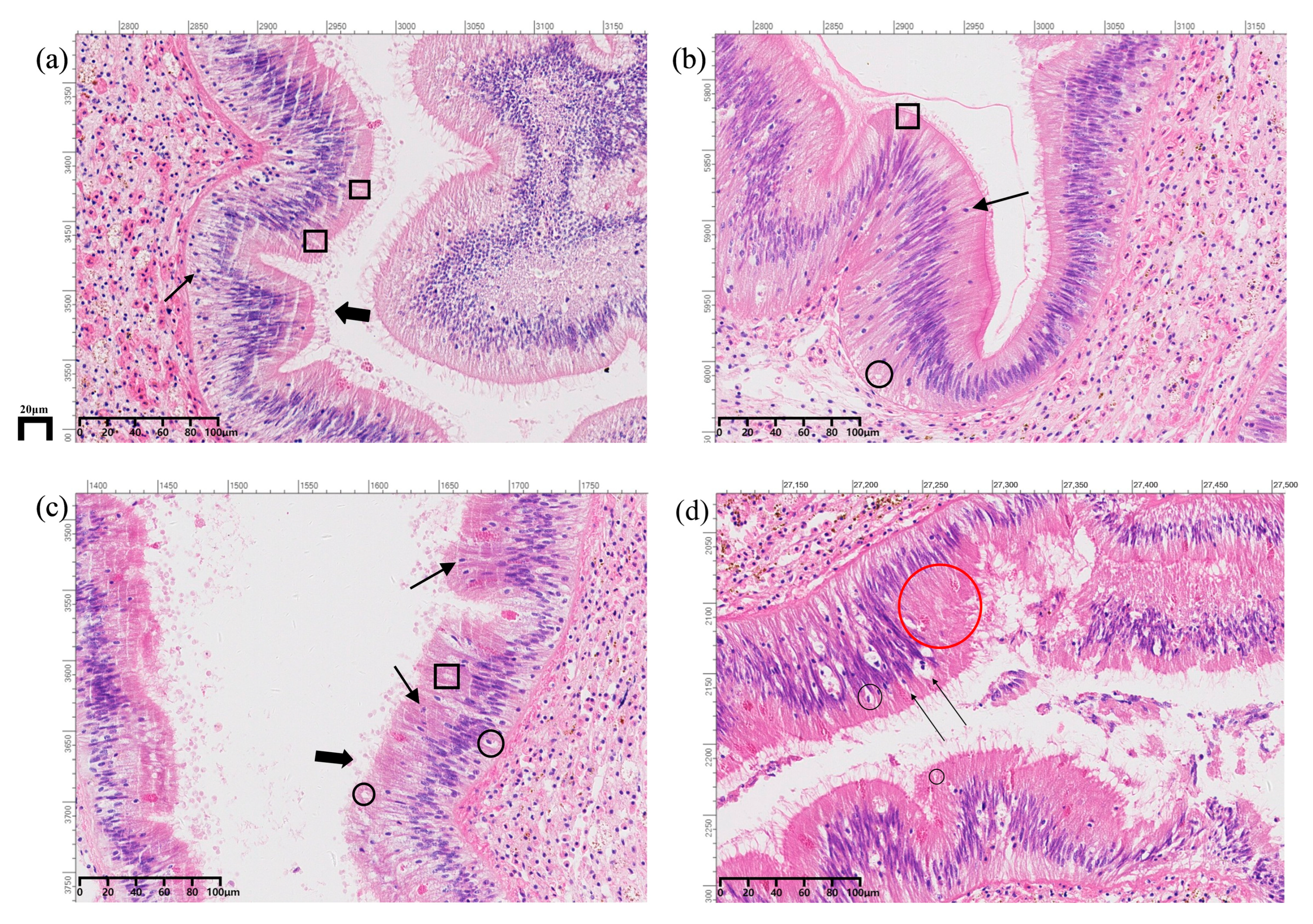
As shown in Figure 1, significant differences were observed in the histological morphology of the intestine of A. granosa across the N0, N14, MH14, and SH14 groups. In comparison to the normoxic control group, after 14 days under normoxia conditions, no substantial changes were detected, with only a minor presence of few inflammatory cells in the intestinal epithelium. However, following 14 days of exposure to 2.0 mg/L dissolved oxygen stress, there was an increase in vacuolar degeneration and inflammatory cell presence, alongside disordered intestinal villi. This indicates that although A. granosa has a strong tolerance to hypoxia, prolonged exposure ultimately leads to severe intestinal damage. Under conditions of 0.5 mg/L dissolved oxygen stress for 14 days, there was an approximate 50% reduction in goblet cells compared to the control group, along with infiltration of inflammatory cells, enterophilic phenomenon, vacuolar degeneration of epithelial cells, necrosis of the lamina propria, and shedding of intestinal villi. These pathological changes are consistent with the effects of oxidative stress and immune response induced by hypoxia conditions, and are associated with damage to intestinal cells and impairment of their function.
Figure 1.
Histological sections of A. granosa intestines under different dissolved oxygen conditions. (a): Control group; (b): Normoxia for 14 days; (c): 2.0 mg/L dissolved oxygen stress for 14 days; (d): 0.5 mg/L dissolved oxygen stress for 14 days. Fine arrows: inflammatory cells; thick arrows: villi; box: goblet cells; black circle: vacuolar degeneration; red circle: necrosis.
3.2. Effects of Hypoxia on Intestinal Immune-Related Gene Expression
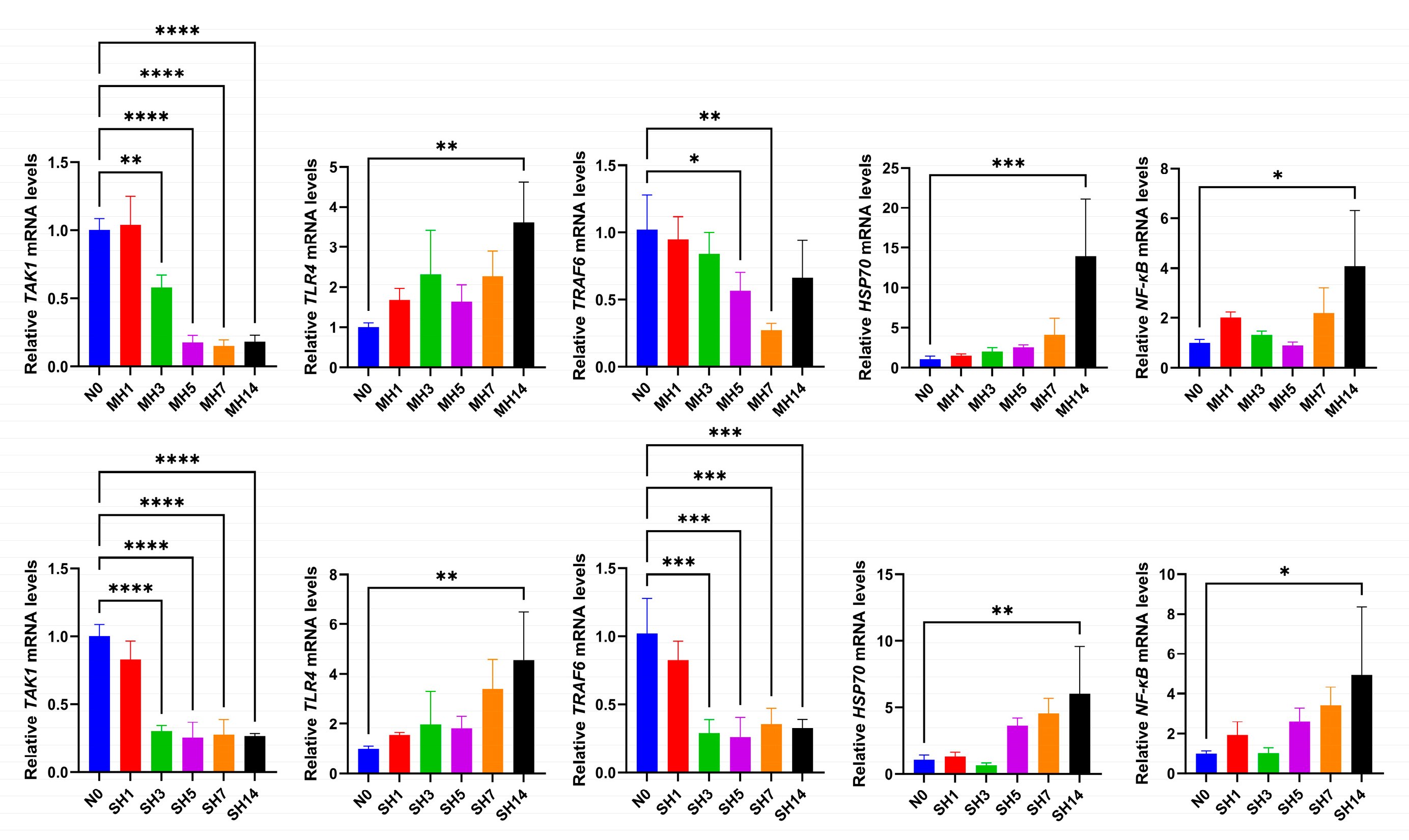
Figure 2 illustrates the expression levels of the immune-related genes TAK1, TLR4, TRAF6, HSP70 and NF-κB in the intestine of A. granosa under varying hypoxia stress conditions, in comparison to a normoxic control. Quantitative analysis revealed significant temporal changes in gene expression. Protective stress responses were markedly upregulated, with HSP70 expression peaking at day 14 in both MH and SH groups, showing approximately 13-fold (p < 0.001) and 5-fold (p < 0.01) increases relative to controls, respectively. Concurrently, pro-inflammatory signaling components TLR4 and NF-κB also exhibited significant upregulation at day 14, with TLR4 increasing by approximately 3.5-fold (MH14, p < 0.01) and 4.5-fold (SH14, p < 0.01), and NF-κB by 4-fold (MH14, p < 0.05) and 5-fold (SH14, p < 0.05). In contrast, genes associated with inflammatory signaling cascades showed early and sustained downregulation. TAK1 expression was significantly suppressed from day 3 in both MH (p < 0.01) and SH (p < 0.0001) groups, with this suppression maintained through days 5, 7, and 14 (p < 0.0001). Similarly, TRAF6 expression decreased significantly from day 5 in the MH group (p < 0.05) and from day 3 in the SH group (p < 0.001). These coordinated expression patterns, particularly the peak upregulation of protective genes coinciding with the period of most severe epithelial damage observed histologically, suggest a complex immune rebalancing mechanism under prolonged hypoxic stress.
Figure 2.
Expression levels of immune-related genes in the intestine of A. granosa under hypoxia stress. * Indicates significant difference (p < 0.05), ** p < 0.01, *** p < 0.001, **** p < 0.0001.
3.3. Response of Intestinal Bacterial Community in A. granosa to Hypoxia Stress
Following the quality control of the raw sequencing data, a total of 4,175,880 high-quality sequences were obtained, these sequences were clustered into 4107 bacterial Operational Taxonomic Units (OTUs) after comparison with the bacterial classification database. The numbers of OTUs identified in the samples N0, N1, MH1, SH1, N3, MH3, SH3, N5, MH5, SH5, N7, MH7, SH7, N14, MH14 and SH14 were 565, 513, 493, 449, 326, 444, 429, 423, 347, 394, 349, 301, 303, 485, 359 and 381, respectively (Figure 3). The Venn diagram analysis revealed that the number of shared bacterial OTUs in the intestines of A. granosa across the groups N0, N1, MH1 and SH1 was 253, with each group containing 96, 100, 74 and 47 unique OTUs, respectively. The number of shared bacterial OTUs in groups N0, N3, MH3 and SH3 was 186, with 169, 35, 94 and 73 unique OTUs, respectively. For groups N0, N5, MH5 and SH5 the number of shared OTUs was 172, with 183, 60, 51 and 71 unique OTUs, respectively. In groups N0, N7, MH7 and SH7, the shared OTUs numbered 144, with 236, 58, 40 and 41 unique OTUs, respectively. Lastly, in groups OTUs in N0, N14, MH14 and SH14, there were 184, with 183, 104, 21 and 64 unique OTUs, respectively.
Figure 3.
Venn diagrams showing the common and unique bacterial OTUs in the intestine of A. granosa under different dissolved oxygen concentrations and stress times.
It is noteworthy that the MH14 group exhibited the lowest number of unique bacterial OTUs, whereas the N0 group demonstrated the highest. These findings suggest that variations in hypoxia treatment durations and dissolved oxygen concentrations significantly influenced the diversity of bacterial OTUs within the intestine of A. granosa.
3.4. Alterations in Ecological Mechanisms Regulating Intestinal Microbiota Assembly in A. granosa During Hypoxia Stress
As illustrated in Figure 4, analysis at the bacterial (sub) phylum level revealed that two bacterial phyla exhibited an average relative abundance exceeding 1% across all groups: Pseudomonadota and Bacillota. The relative abundance of Pseudomonadota was observed to be lowest in the MH3 group with an average relative abundance of 25.69%, and highest in the N3 group, with an average relative abundance of 98.49%. Conversely, the relative abundance of Bacillota was lowest in the N3 group, with an average relative abundance of 1.17%, and highest in the MH3 group, with an average relative abundance of 67.42%.
Figure 4.
Average relative abundance of dominant bacterial phyla (a–c) and genera (d–f) in the intestine of A. granosa under different dissolved oxygen concentrations and stress times.
At the bacterial genus level, Shewanella was consistently observed across all groups, with an average relative abundance exceeding 1%. Notably, compared to the initial control group (N0), the relative abundances of several putative opportunistic pathogens or stress-associated genera, such as Vibrio, Mycoplasma, and Staphylococcus, showed an increasing trend in multiple hypoxic groups (e.g., MH3, SH3, MH7, SH7). In contrast, some core genera like Shewanella and Acinetobacter displayed complex and dynamic variations across different treatments and time points, suggesting potential functional versatility within the community.
3.5. Diversity Differences in Intestinal Bacterial Community in A. granosa
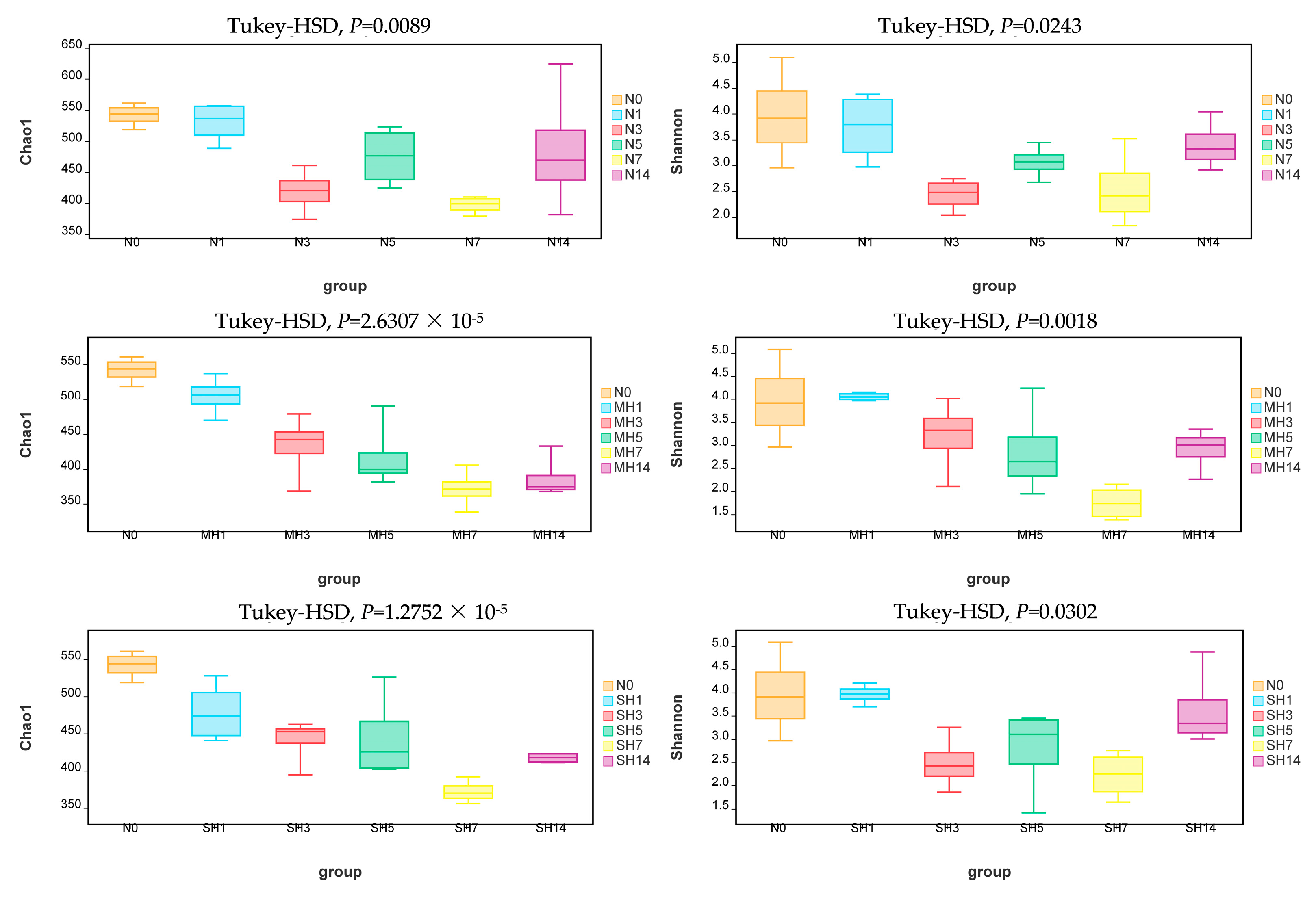
The Chao1 and Shannon indices were employed to assess variations in α-diversity. Statistical differences among groups under different dissolved oxygen levels and stress durations were determined using one-way ANOVA followed by Tukey’s HSD post hoc test. As shown in Figure 5, both the Chao1 and Shannon indices were generally elevated in the N0 group compared to other groups. Under hypoxia stress, both indices demonstrated a declining trend with prolonged stress duration, with significant differences observed (p < 0.05). These findings suggest that hypoxia stress significantly impacts the α-diversity of the intestinal bacterial community in A. granosa.
Figure 5.
Changes in α-diversity indices of the intestinal bacterial community in A. granosa under different dissolved oxygen concentrations and stress times.
3.6. Structural Differences in Intestinal Bacterial Community in A. granosa
Distinct colors denote samples subjected to different conditions, while connecting lines illustrate the similarity between samples. It is evident that with prolonged exposure to hypoxic stress, the spatial distribution of samples under hypoxic conditions progressively diverged from those of the normoxic group, highlighting the impact of hypoxia on the intestinal microbiota structure of A. granosa. Furthermore, samples collected at different time points exhibited a certain degree of dispersion within the figure, suggesting that temporal factors significantly influenced microbial community structure under identical conditions.
As shown in Figure 6, the results of the Non-metric Multidimensional Scaling (NMDS) analysis of the intestinal microbiota in A. granosa under varying dissolved oxygen conditions revealed significant differences in community structure. Over time, alterations in the microbial community composition were observed in both normoxic and hypoxic stress groups. Distinct colors denote samples subjected to different conditions, while connecting lines illustrate the similarity between samples. It is evident that with prolonged exposure to hypoxia stress, the spatial distribution of samples under hypoxic conditions progressively diverged from those of the normoxic group, highlighting the impact of hypoxia on the intestinal microbiota structure of A. granosa. Additionally, samples collected at different time points also showed a certain degree of dispersion in the figure, suggesting that temporal factors significantly influenced microbial community structure under identical conditions. The PERMANOVA analysis based on Bray–Curtis distance indicated that the N, MH, and SH groups caused significant variations of 31.14%, 30.74%, and 39.26% in the gut bacterial communities of A. granosa under different stress durations, respectively (Table 3).
Figure 6.
NMDS showing structural differences in the intestinal bacterial community in A. granosa under different dissolved oxygen concentrations and stress times.
Table 3.
Permanova analysis based on Bray–Curtis distance was used to detect the effects of different dissolved oxygen concentrations and stress duration on the gut microbiota structure of A. granosa.
3.7. Functional Differences in Intestinal Bacterial Community in A. granosa
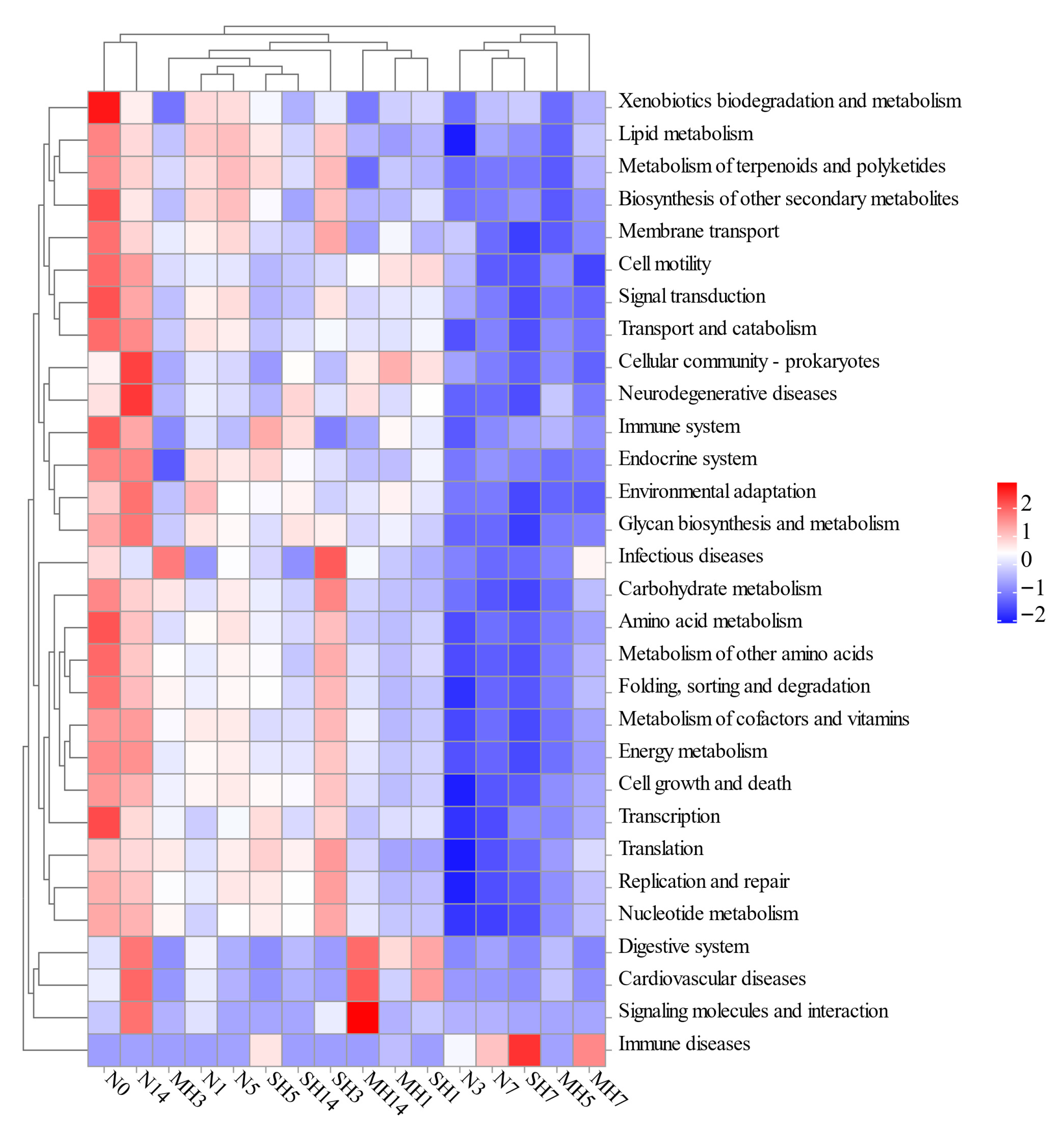
Figure 7 shows the abundance distribution characteristics of potential KEGG functional pathways mediated by the gut microbiota of A. granosa under different dissolved oxygen conditions. The prediction results suggested that the inferred functions of all groups mainly involved four categories of functional pathways: metabolism, genetic information processing, cellular processes, and environmental information processing. The N0 group had small predicted functional differences compared with the N1, N5, and N14 groups, indicating that the overall microbial functions were relatively stable under normal dissolved oxygen conditions. However, notable differences in the predicted functional profiles were observed between the N0 group and the other groups, especially with the MH5, N7, MH7, and SH7 groups, mainly manifested in the decreased abundance of predicted functional pathways such as xenobiotics biodegradation and metabolism, lipid metabolism, metabolism of terpenoids and polyketides, biosynthesis of other secondary metabolites, membrane transport, cell motility, and signal transduction. According to the predictions, pathways related to digestive disorders, circulatory conditions, and signaling pathways molecules and interaction showed notably elevated abundances in both N14 and MH14 groups compared to remaining groups. Regarding immune-related ailments, the N7, MH7, and SH7 clusters demonstrated substantially greater occurrence frequencies than alternative groups. These predictive results suggest that the functional pathways mediated by gut bacteria vary under different dissolved oxygen conditions.
Figure 7.
Heatmap showing the abundance distribution changes in potential functional pathways in the intestinal bacterial community of A. granosa under different dissolved oxygen concentrations and stress times.
4. Discussion
Hypoxia represents a critical and significant environmental stressor that interacts intricately with other environmental factors, exerting a profound impact on the health and performance of marine organisms [,,]. While the detrimental effects of hypoxia on bivalve physiology are well-documented [,,,], previous research in A. granosa has primarily focused on isolated physiological responses, such as oxidative stress, metabolism, and HIF-1 gene expression characteristics in tissues like gills and hemocytes [,]. Our study provides the first integrative analysis of the “microbiota–intestine–immune” axis in this species by combining histology, immune gene expression, and microbiome profiling, revealing how hypoxic stress disrupts this critical homeostatic network.
The current study elucidates that hypoxia stress markedly undermines the structural integrity of intestinal tissue in A. granosa. After 14 days of exposure to hypoxic conditions, pronounced histopathological changes were evident, including vacuolar degeneration, infiltration of inflammatory cells, disorganization of intestinal villi, and villus shedding. These morphological disruptions are likely attributable to heightened oxidative stress and dysregulated immune responses under hypoxic conditions, culminating in cellular damage and functional impairment of the intestine [,,].
Exposure to environmental stressors, such as hypoxia, has been documented to compromise immune barrier functions in bivalves [,,]. Our investigation of immune-related gene expression in A. granosa intestine under hypoxia revealed a coordinated regulatory pattern. To comprehensively assess the hypoxic stress response, we selected key genes representing distinct functional aspects: HSP70 as a core cytoprotective protein critical for hypoxia adaptation [], and NF-κB as the central inflammatory pathway regulator, which together with TLR4 and TAK1/TRAF6 forms an integrated immune regulatory network. The sustained downregulation of TAK1 and TRAF6—key upstream regulators of the NF-κB pathway—that was observed specifically on days 7 and 14 of hypoxia exposure likely represents a crucial negative feedback mechanism to prevent excessive inflammatory responses. Notably, the concurrent upregulation of HSP70 may further fine-tune this response by elevating the activation threshold of NF-κB, thereby preventing cytokine storm while maintaining essential defense functions. This sophisticated balance between pathogen recognition and inflammation control is further reflected in the coordinated expression of TLR4. Additionally, the chaperone function of HSP70 could help rectify membrane protein damage resulting from microbial lipid metabolism dysbiosis, establishing a collaborative “host protection–microecological stability” mechanism to combat hypoxic stress. Collectively, these precisely timed expression changes demonstrate a compensatory strategy to maintain intestinal immune homeostasis under prolonged hypoxic stress []. These mechanisms may represent a compensatory strategy to maintain intestinal immune homeostasis under stressful conditions.
Research has shown that the physicochemical conditions within aquaculture environments can significantly impact the abundance and composition of gut microbial communities in aquatic species []. Correspondingly, our study demonstrates that varying levels of hypoxic stress markedly influence the diversity and structure of the gut bacterial community in A. granosa. High-throughput sequencing revealed a significant decline in both α-diversity and β-diversity under hypoxic conditions. This finding aligns with observations in Macrobrachium nipponense, where a four-week exposure to hypoxia resulted in a substantial reduction in microbial diversity [].
At the phylum level, Pseudomonadota remained the predominant group across all experimental conditions, though its abundance showed dynamic fluctuations under hypoxia. The observed decline in Pseudomonadota, which typically relies on aerobic metabolic pathways [], likely reflects suppression of oxygen-dependent processes under hypoxic conditions. Concurrently, the significant shift toward Bacillota in both MH and SH groups represents a notable microbial adaptation. This transition likely reflects Bacillota’s superior tolerance to low-oxygen conditions, with its increased abundance potentially aiding in maintaining gut homeostasis through mechanisms such as short-chain fatty acid production to support energy metabolism. Importantly, the metabolic products of Bacillota may help reduce intestinal pH, thereby inhibiting the proliferation of opportunistic pathogens like Vibrio, which complements the host’s upregulated TLR4-mediated pathogen recognition to establish a multi-level defense system. However, the precise functional contributions require further validation. The resilience of Pseudomonadota, known for its role in organic matter degradation and nutrient cycling [], along with the expansion of Bacillota, illustrates the complex microbial restructuring in response to hypoxic stress. At the bacterial genus level, the notable enrichment of genera such as Vibrio and Mycoplasma under hypoxia is of particular concern. Vibrio species are well-documented opportunistic pathogens in aquatic animals, whose proliferation is often associated with host immunosuppression and environmental stress []. Similarly, Mycoplasma has been linked to diseases in various hosts, and its increased abundance may signal a state of ecological imbalance (dysbiosis) within the gut ecosystem []. The rise in such taxa likely compromises the gut’s colonization resistance, thereby elevating the host’s susceptibility to disease.
Alterations in the gut bacterial community are recognized to significantly influence microbial-mediated functions in aquatic organisms [,,]. In alignment with these findings, modifications in the composition and diversity of the gut microbiota in A. granosa were hypothesized to be associated with corresponding changes in predicted metabolic potential. In this study, functional prediction analysis using PICRUSt2 revealed that hypoxic stress may have affected the predicted abundance of KEGG functional pathways in the gut microbiota of A. granosa, showing a distinct stress intensity and time-specific response pattern. Compared with the initial control group N0, the predicted functional structure of the normal oxygen groups (N1, N5, N14) was relatively conservative, while notable differences in the predicted functions were observed in the other groups, especially the MH5, N7, MH7, and SH7 groups. This was primarily inferred as a decrease in the predicted abundance of pathways such as xenobiotics biodegradation and metabolism, lipid metabolism, metabolism of terpenoids and polyketides, biosynthesis of other secondary metabolites, membrane transport, cell motility, and signal transduction. PICRUSt2 predictions revealed a significant 29% decrease in the abundance of microbial lipid metabolism pathways in the SH14 group compared to the N0 control. This predicted reduction provides a quantitative link to the observed vacuolar degeneration in intestinal epithelial cells, showing a positive correlation with the approximately 50% incidence of epithelial vacuolization. Specifically, the downregulation of these pathways likely reduces the production of microbial-derived metabolites, particularly short-chain fatty acids, which serve as crucial energy substrates for enterocytes []. This energy deficit, coupled with impaired synthesis of essential membrane components, may disrupt cellular homeostasis and structural integrity. These findings establish a quantifiable relationship between microbial metabolic dysfunction and host cellular damage, demonstrating how predicted metabolic deficiencies directly contribute to the characteristic vacuolization observed in hypoxic intestinal tissues.
Hypoxic stress may broadly suppress the functional activity of the microbial community in aspects such as material metabolism, environmental perception, and transmembrane transport [,]. Notably, on day 7, all treatment groups showed significant enrichment of pathways related to immune diseases, which may be a key stage for the immune stress response of the gut microbiota in A. granosa. On day 14, the N14 and MH14 groups exhibited an up-regulation in the predicted abundance of pathways related to the digestive system, cardiovascular diseases, and signaling molecule interactions, which may reflect the synergistic adaptation mechanisms between the microbial community and the host in physiological regulation under long-term stress. Overall, these predictive results indicate that the functional structure of the gut microbiota in A. granosa appears to be sensitive to changes in dissolved oxygen, with distinct time dynamics and pathway specificity. It is plausible that the microbiota plays a role in immune regulation and host physiological functions, but these functional hypotheses require validation through direct metagenomic or metatranscriptomic approaches.
It is important to acknowledge several limitations in this study. First, although the use of pooled intestinal samples from five individuals provided a community-level perspective on microbiota composition and host gene expression, it prevented the evaluation of inter-individual variation. Future studies involving single-individual analyses would help clarify the full extent of host physiological heterogeneity in response to hypoxia. Second, despite the use of three parallel tanks—each with two pooled samples—to reduce tank effects, subtle microenvironmental differences (e.g., fluctuations in dissolved oxygen, algal distribution) may still have introduced bias. Increasing the number of tank replicates in future experiments could help further minimize such variations. The initial fixation with 4% paraformaldehyde, while preserving basic tissue architecture, resulted in villus edge contraction and blurred measurement benchmarks, precluding precise villus height quantification. Moreover, future studies incorporating standardized morphometric analyses—including villus height measurement and goblet cell counting—should employ optimized fixation protocols such as Bouin’s solution to better preserve tissue morphology, thereby enabling more objective and accurate quantification of intestinal damage under hypoxic stress. Further investigation should also prioritize direct metagenomic and meta-transcriptomic approaches to functionally test the proposed hypotheses. Finally, subsequent research will focus on isolating dominant Bacillota taxa to experimentally validate their effects on the host intestinal epithelium, complemented by targeted metabolomics to establish mechanistic connections between microbial shifts and host physiological responses. Addressing these aspects will be essential for unraveling the detailed mechanisms of the “microbiota–intestine–immune” axis and for developing targeted interventions to alleviate hypoxia stress in aquaculture species.
5. Conclusions
In conclusion, this study demonstrates that hypoxic stress significantly impairs the intestinal health of A. granosa, as indicated by histopathological damage, altered immune gene expression, and reduced diversity and functional capacity of the gut microbiota. These findings highlight the necessity of maintaining adequate dissolved oxygen levels to preserve intestinal integrity and overall health in bivalves. Future studies should focus on elucidating the underlying molecular mechanisms and developing strategies to mitigate hypoxia-induced damage.
Author Contributions
Y.L. and G.C.: writing—original draft, methodology. J.J., Y.J. and X.Z.: methodology, investigation. Y.B. and Z.P.: writing—review and editing, project administration, supervision, funding acquisition. All authors have read and agreed to the published version of the manuscript.
Funding
This work was supported by Zhejiang Provincial Top Discipline of Biological Engineering Level A (NO: CX2025015, KF2025007); Innovation Training Program for College Students of Zhejiang Province (NO: S202510876057); Zhejiang Province Public Welfare Technology Application Research Project (NO: LGN21C190012); Zhejiang Major Program of Science and Technology (2021C02069-7).
Institutional Review Board Statement
Our study involved Anadara granosa, an invertebrate species. According to widely accepted ethical guidelines and common institutional policies, ethical approval is generally not required for research involving invertebrates.
Data Availability Statement
The raw 16S rRNA sequencing reads generated in this study have been deposited in the NCBI Sequence Read Archive (SRA) and are publicly available under the BioProject accession number PRJNA1354393.
Conflicts of Interest
The authors declare no conflicts of interest.
References
- Abdel-Tawwab, M.; Monier, M.N.; Hoseinifar, S.H.; Faggio, C. Fish response to hypoxia stress: Growth, physiological, and immunological biomarkers. Fish Physiol. Biochem. 2019, 45, 997–1013. [Google Scholar] [CrossRef]
- Diaz, R.J.; Rosenberg, R. Spreading Dead Zones and Consequences for Marine Ecosystems. Science 2008, 321, 926–929. [Google Scholar] [CrossRef]
- Guo, Y.; Rong, Z.; Li, B.; Xu, Z.; Li, P.; Li, X. Physical processes causing the formation of hypoxia off the Changjiang estuary after Typhoon Chan-hom, 2015. J. Oceanol. Limnol. 2019, 37, 1–17. [Google Scholar] [CrossRef]
- Lam, P.; Kuypers, M.M.M. Microbial Nitrogen Cycling Processes in Oxygen Minimum Zones. Annu. Rev. Mar. Sci. 2011, 3, 317–345. [Google Scholar] [CrossRef]
- Galic, N.; Hawkins, T.; Forbes, V.E. Adverse impacts of hypoxia on aquatic invertebrates: A meta-analysis. Sci. Total Environ. 2019, 652, 736–743. [Google Scholar] [CrossRef]
- Song, J.; Farhadi, A.; Tan, K.; Lim, L.; Tan, K. Impact of anthropogenic global hypoxia on the physiological response of bivalves. Sci. Total Environ. 2024, 926, 172056. [Google Scholar] [CrossRef]
- Wang, Y.; Jiang, P.; Xia, F.; Bai, Q.; Zhang, X. Transcriptional and physiological profiles reveal the respiratory, antioxidant and metabolic adaption to intermittent hypoxia in the clam Tegillarca granosa. Comp. Biochem. Physiol. Part D Genom. Proteom. 2024, 50, 101215. [Google Scholar] [CrossRef]
- Zhan, Y.; Zha, S.; Peng, Z.; Lin, Z.; Bao, Y. Hypoxia-mediated immunotoxicity in the blood clam Tegillarca granosa. Mar. Environ. Res. 2022, 177, 105632. [Google Scholar] [CrossRef]
- Wen, J.; Wu, Y.; Zhang, F.; Wang, Y.; Yang, A.; Lu, W.; Zhao, X.; Tao, H. Neonatal hypoxia leads to impaired intestinal function and changes in the composition and metabolism of its microbiota. Sci. Rep. 2025, 15, 15285. [Google Scholar] [CrossRef]
- Song, Z.; Ye, W.; Tao, Y.; Zheng, T.; Qiang, J.; Li, Y.; Liu, W.; Xu, P. Transcriptome and 16S rRNA Analyses Reveal That Hypoxic Stress Affects the Antioxidant Capacity of Largemouth Bass (Micropterus salmoides), Resulting in Intestinal Tissue Damage and Structural Changes in Microflora. Antioxidants 2022, 12, 1. [Google Scholar] [CrossRef]
- Niklasson, L.; Sundh, H.; Fridell, F.; Taranger, G.L.; Sundell, K. Disturbance of the intestinal mucosal immune system of farmed Atlantic salmon (Salmo salar), in response to long-term hypoxic conditions. Fish Shellfish Immunol. 2011, 31, 1072–1080. [Google Scholar] [CrossRef]
- Jiang, S.; Zhang, W.; Qian, X.; Ji, J.; Ning, X.; Zhu, F.; Yin, S.; Zhang, K. Effects of hypoxia and reoxygenation on apoptosis, oxidative stress, immune response and gut microbiota of Chinese mitten crab, Eriocheir sinensis. Aquat. Toxicol. 2023, 260, 106556. [Google Scholar] [CrossRef]
- Nie, H.; Wang, H.; Jiang, K.; Yan, X. Transcriptome analysis reveals differential immune related genes expression in Ruditapes philippinarum under hypoxia stress: Potential HIF and NF-κB crosstalk in immune responses in clam. BMC Genom. 2020, 21, 318. [Google Scholar] [CrossRef]
- Falfushynska, H.; Piontkivska, H.; Sokolova, I.M. Effects of intermittent hypoxia on the cell survival and inflammatory responses in the intertidal marine bivalves Mytilus edulis and Crassostrea gigas. J. Exp. Biol. 2020, 223, jeb217026. [Google Scholar] [CrossRef]
- Montufar-Romero, M.; Valenzuela-Munoz, V.; Valenzuela-Miranda, D.; Gallardo-Escarate, C. Hypoxia in the Blue Mussel Mytilus chilensis Induces a Transcriptome Shift Associated with Endoplasmic Reticulum Stress, Metabolism, and Immune Response. Genes 2024, 15, 658. [Google Scholar] [CrossRef]
- Xie, Z.; Li, Y.; Xiong, K.; Tu, Z.; Waiho, K.; Yang, C.; Deng, Y.; Li, S.; Fang, J.K.H.; Hu, M.; et al. Combined effect of salinity and hypoxia on digestive enzymes and intestinal microbiota in the oyster Crassostrea hongkongensis. Environ. Pollut. 2023, 331, 121921. [Google Scholar] [CrossRef]
- Evariste, L.; Barret, M.; Mottier, A.; Mouchet, F.; Gauthier, L.; Pinelli, E. Gut microbiota of aquatic organisms: A key endpoint for ecotoxicological studies. Environ. Pollut. 2019, 248, 989–999. [Google Scholar] [CrossRef]
- Clerissi, C.; de Lorgeril, J.; Petton, B.; Lucasson, A.; Escoubas, J.M.; Gueguen, Y.; Degremont, L.; Mitta, G.; Toulza, E. Microbiota Composition and Evenness Predict Survival Rate of Oysters Confronted to Pacific Oyster Mortality Syndrome. Front. Microbiol. 2020, 11, 311. [Google Scholar] [CrossRef]
- Dai, W.; Dong, Y.; Ye, J.; Xue, Q.; Lin, Z. Gut microbiome composition likely affects the growth of razor clam Sinonovacula constricta. Aquaculture 2022, 550, 737847. [Google Scholar] [CrossRef]
- Montufar-Romero, M.; Valenzuela-Miranda, D.; Valenzuela-Munoz, V.; Morales-Rivera, M.F.; Gallardo-Escarate, C. Microbiota Dysbiosis in Mytilus chilensis Is Induced by Hypoxia, Leading to Molecular and Functional Consequences. Microorganisms 2025, 13, 825. [Google Scholar] [CrossRef]
- Bao, Y.; Zeng, Q.; Wang, J.; Zhang, Z.; Zhang, Y.; Wang, S.; Wong, N.K.; Yuan, W.; Huang, Y.; Zhang, W.; et al. Genomic Insights into the Origin and Evolution of Molluscan Red-Bloodedness in the Blood Clam Tegillarca granosa. Mol. Biol. Evol. 2021, 38, 2351–2365. [Google Scholar] [CrossRef]
- Jin, H.; Zhang, W.; Liu, H.; Bao, Y. Genome-wide identification and characteristic analysis of ETS gene family in blood clam Tegillarca granosa. BMC Genom. 2023, 24, 700. [Google Scholar] [CrossRef]
- Liu, H.; Zha, S.; Yang, Z.; Zhang, W.; Lin, Z.; Wang, S.; Bao, Y. Acute sulfide exposure induces hemocyte toxicity and microbiota dysbiosis in blood clam Tegillarca granosa. Aquat. Toxicol. 2022, 249, 106224. [Google Scholar] [CrossRef]
- Peng, Z.; Liu, X.; Jin, M.; Zhan, Y.; Zhang, X.; Bao, Y.; Liu, M. Hypoxia Activates HIF-1α and Affects Gene Expression and Transcriptional Regulation of PHD in Tegillarca granosa. Fishes 2023, 8, 359. [Google Scholar] [CrossRef]
- Zhang, Y.; Liu, H.; Bao, Y.; Peng, Z. The Structure Analysis and mRNA Expression of CaV2 Gene Responding to Hypoxia Stress in Anadara granosa. Fishes 2024, 9, 409. [Google Scholar] [CrossRef]
- Shao, Y.; Chai, X.; Xiao, G.; Zhang, J.; Lin, Z.; Liu, G. Population Genetic Structure of the Blood Clam, Tegillarca granosa, Along the Pacific Coast of Asia: Isolation by Distance in the Sea. Malacologia 2016, 59, 303–312. [Google Scholar] [CrossRef]
- Zha, S.; Rong, J.; Guan, X.; Tang, Y.; Han, Y.; Liu, G. Immunotoxicity of four nanoparticles to a marine bivalve species, Tegillarca granosa. J. Hazard. Mater. 2019, 377, 237–248. [Google Scholar] [CrossRef]
- Jing, H.; Liu, Z.; Wu, B.; Tu, K.; Liu, Z.; Sun, X.; Zhou, L. Physiological and molecular responses to hypoxia stress in Manila clam Ruditapes philippinarum. Aquat. Toxicol. 2023, 257, 106428. [Google Scholar] [CrossRef]
- Cheng, H.; Peng, Z.; Zhao, C.; Jin, H.; Bao, Y.; Liu, M. The transcriptomic and biochemical responses of blood clams (Tegillarca granosa) to prolonged intermittent hypoxia. Comp. Biochem. Physiol. B Biochem. Mol. Biol. 2024, 270, 110923. [Google Scholar] [CrossRef]
- Zha, S.; Zhang, W.; Liu, H.; Huang, S.; Sun, C.; Bao, Y. Two common nanoparticles exert immunostimulatory and protective effects in Tegillarca granosa against Vibrio parahaemolyticus. Fish Shellfish Immunol. 2023, 137, 108774. [Google Scholar] [CrossRef]
- Su, W.; Zha, S.; Wang, Y.; Shi, W.; Xiao, G.; Chai, X.; Wu, H.; Liu, G. Benzo[a]pyrene exposure under future ocean acidification scenarios weakens the immune responses of blood clam, Tegillarca granosa. Fish Shellfish Immunol. 2017, 63, 465–470. [Google Scholar] [CrossRef]
- Chen, S.; Zhou, Y.; Chen, Y.; Gu, J. fastp: An ultra-fast all-in-one FASTQ preprocessor. Bioinformatics 2018, 34, i884–i890. [Google Scholar] [CrossRef]
- Magoc, T.; Salzberg, S.L. FLASH: Fast length adjustment of short reads to improve genome assemblies. Bioinformatics 2011, 27, 2957–2963. [Google Scholar] [CrossRef]
- Bokulich, N.A.; Subramanian, S.; Faith, J.J.; Gevers, D.; Gordon, J.I.; Knight, R.; Mills, D.A.; Caporaso, J.G. Quality-filtering vastly improves diversity estimates from Illumina amplicon sequencing. Nat. Methods 2013, 10, 57–59. [Google Scholar] [CrossRef]
- Edgar, R.C. Search and clustering orders of magnitude faster than BLAST. Bioinformatics 2010, 26, 2460–2461. [Google Scholar] [CrossRef]
- Edgar, R.C.; Haas, B.J.; Clemente, J.C.; Quince, C.; Knight, R. UCHIME improves sensitivity and speed of chimera detection. Bioinformatics 2011, 27, 2194–2200. [Google Scholar] [CrossRef]
- Chen, H.; Boutros, P.C. VennDiagram: A package for the generation of highly-customizable Venn and Euler diagrams in R. BMC Bioinform. 2011, 12, 35. [Google Scholar] [CrossRef]
- Lozupone, C.; Knight, R. UniFrac: A new phylogenetic method for comparing microbial communities. Appl. Environ. Microbiol. 2005, 71, 8228–8235. [Google Scholar] [CrossRef]
- Langille, M.G.; Zaneveld, J.; Caporaso, J.G.; McDonald, D.; Knights, D.; Reyes, J.A.; Clemente, J.C.; Burkepile, D.E.; Vega Thurber, R.L.; Knight, R.; et al. Predictive functional profiling of microbial communities using 16S rRNA marker gene sequences. Nat. Biotechnol. 2013, 31, 814–821. [Google Scholar] [CrossRef]
- Todgham, A.E.; Stillman, J.H. Physiological responses to shifts in multiple environmental stressors: Relevance in a changing world. Integr. Comp. Biol. 2013, 53, 539–544. [Google Scholar] [CrossRef]
- Clavel, T.; Gomes-Neto, J.C.; Lagkouvardos, I.; Ramer-Tait, A.E. Deciphering interactions between the gut microbiota and the immune system via microbial cultivation and minimal microbiomes. Immunol. Rev. 2017, 279, 8–22. [Google Scholar] [CrossRef]
- Wang, X.; Wei, Z.; Ma, R.; Wang, J.; Liu, Z.; Liu, Y.; Xu, X.; Chu, P.; Zhu, L. Dysregulation of gut barrier and microbiota in Asiatic hard clams (Meretrix petechialis) exposed to environmentally relevant bisphenol A. Environ. Res. 2025, 279, 121913. [Google Scholar] [CrossRef]
- Quigley, E.M.; Stanton, C.; Murphy, E.F. The gut microbiota and the liver. Pathophysiological and clinical implications. J. Hepatol. 2013, 58, 1020–1027. [Google Scholar] [CrossRef]
- Wang, W.-z.; Huang, J.-s.; Zhang, J.-d.; Wang, Z.-l.; Li, H.-j.; Amenyogbe, E.; Chen, G. Effects of hypoxia stress on the intestinal microflora of juvenile of cobia (Rachycentron canadum). Aquaculture 2021, 536, 736419. [Google Scholar] [CrossRef]
- Dai, W.; Zhang, Z.; Dong, Y.; He, L.; Xue, Q.; Lin, Z. Acute Salinity Stress Disrupts Gut Microbiota Homeostasis and Reduces Network Connectivity and Cooperation in Razor Clam Sinonovacula constricta. Mar. Biotechnol. 2023, 25, 1147–1157. [Google Scholar] [CrossRef]
- Steffen, J.B.M.; Falfushynska, H.I.; Piontkivska, H.; Sokolova, I.M. Molecular Biomarkers of the Mitochondrial Quality Control Are Differently Affected by Hypoxia-Reoxygenation Stress in Marine Bivalves Crassostrea gigas and Mytilus edulis. Front. Mar. Sci. 2020, 7, 604411. [Google Scholar] [CrossRef]
- Andreyeva, A.Y.; Kukhareva, T.A.; Gostyukhina, O.L.; Vialova, O.Y.; Tkachuk, A.A.; Chelebieva, E.S.; Podolskaya, M.S.; Borovkov, A.B.; Bogacheva, E.A.; Lavrichenko, D.S.; et al. Impacts of ocean acidification and hypoxia on cellular immunity, oxygen consumption and antioxidant status in Mediterranean mussel. Fish Shellfish Immunol. 2024, 154, 109932. [Google Scholar] [CrossRef]
- Liu, T.; Lu, Y.; Sun, M.; Shen, H.; Niu, D. Effects of acute hypoxia and reoxygenation on histological structure, antioxidant response, and apoptosis in razor clam Sinonovacula constricta. Fish Shellfish Immunol. 2024, 145, 109310. [Google Scholar] [CrossRef]
- He, H.; Huang, S.; Geng, N.; Weng, S.; He, J.; Li, C. Acute hypoxia stress mediates HIF-1alpha-Yki-Cactus axis to facilitate the infection of Vibrio parahaemolyticus in Litopenaeus vannamei. Front. Immunol. 2024, 15, 1476309. [Google Scholar] [CrossRef]
- Wang, Y.; Zhou, S.; Liu, T.; Chen, M.; Li, W.; Zhang, X. The transcriptomic responses of the ark shell, Anadara broughtonii, to sulfide and hypoxia exposure. Mol. Biol. Rep. 2019, 46, 4245–4257. [Google Scholar] [CrossRef]
- Rivera-Ingraham, G.A.; Rocchetta, I.; Meyer, S.; Abele, D. Oxygen radical formation in anoxic transgression and anoxia-reoxygenation: Foe or phantom? Experiments with a hypoxia tolerant bivalve. Mar. Environ. Res. 2013, 92, 110–119. [Google Scholar] [CrossRef]
- Klase, G.; Lee, S.; Liang, S.; Kim, J.; Zo, Y.G.; Lee, J. The microbiome and antibiotic resistance in integrated fishfarm water: Implications of environmental public health. Sci. Total Environ. 2019, 649, 1491–1501. [Google Scholar] [CrossRef]
- Sun, S.; Yang, M.; Fu, H.; Ge, X.; Zou, J. Altered intestinal microbiota induced by chronic hypoxia drives the effects on lipid metabolism and the immune response of oriental river prawn Macrobrachium nipponense. Aquaculture 2020, 526, 735431. [Google Scholar] [CrossRef]
- Carrillo-Barragan, P.; Cassola, G.E.; Burkhardt-Holm, P. Microbial colonisation of polyethylene in offshore marine environments: Insights from the Southern and South Atlantic Oceans. Sci. Total Environ. 2025, 996, 180189. [Google Scholar] [CrossRef]
- Chang, Y.; Ko, H.; Wu, P.; Kumar, R.; Wang, H.; Lu, H. Gut microbiota of Pacific white shrimp (Litopenaeus vannamei) exhibits distinct responses to pathogenic and non-pathogenic Vibrio parahaemolyticus. Microbiol. Spectr. 2023, 11, e0118023. [Google Scholar] [CrossRef]
- Yamada, Y.; Saito, Y.; Shimanuki, K.; Izumi, S.; Gojobori, T.; Akiyama, N. Monitoring the gut microbiota of rainbow trout during seawater acclimation. Fish Sci. 2025, 91, 943–959. [Google Scholar] [CrossRef]
- Akter, S.; Wos-Oxley, M.L.; Catalano, S.R.; Hassan, M.M.; Li, X.; Qin, J.G.; Oxley, A.P. Host Species and Environment Shape the Gut Microbiota of Cohabiting Marine Bivalves. Microb. Ecol. 2023, 86, 1755–1772. [Google Scholar] [CrossRef]
- Pierce, M.L.; Ward, J.E. Gut Microbiomes of the Eastern Oyster (Crassostrea virginica) and the Blue Mussel (Mytilus edulis): Temporal Variation and the Influence of Marine Aggregate-Associated Microbial Communities. mSphere 2019, 4, e00730-19. [Google Scholar] [CrossRef]
- Wang, S.; Li, X.; Zhang, M.; Jiang, H.; Wang, R.; Qian, Y.; Li, M. Ammonia stress disrupts intestinal microbial community and amino acid metabolism of juvenile yellow catfish (Pelteobagrus fulvidraco). Ecotoxicol. Environ. Saf. 2021, 227, 112932. [Google Scholar] [CrossRef]
- Koh, A.; De Vadder, F.; Kovatcheva-Datchary, P.; Backhed, F. From Dietary Fiber to Host Physiology: Short-Chain Fatty Acids as Key Bacterial Metabolites. Cell 2016, 165, 1332–1345. [Google Scholar] [CrossRef]
Disclaimer/Publisher’s Note: The statements, opinions and data contained in all publications are solely those of the individual author(s) and contributor(s) and not of MDPI and/or the editor(s). MDPI and/or the editor(s) disclaim responsibility for any injury to people or property resulting from any ideas, methods, instructions or products referred to in the content. |
© 2025 by the authors. Licensee MDPI, Basel, Switzerland. This article is an open access article distributed under the terms and conditions of the Creative Commons Attribution (CC BY) license (https://creativecommons.org/licenses/by/4.0/).