Communication

Cystic Lung Phantom to Validate Clinical CT Protocols

Laboratory of Imaging Physics, Biochemistry and Biophysics Center, National Heart, Lung and Blood Institute, National Institutes of Health, Bethesda, MD 20892, USA
*
Author to whom correspondence should be addressed.
Methods Protoc. 2025, 8(3), 63; https://doi.org/10.3390/mps8030063
Submission received: 1 April 2025 / Revised: 27 May 2025 / Accepted: 9 June 2025 / Published: 13 June 2025
(This article belongs to the Section Public Health Research)

Abstract

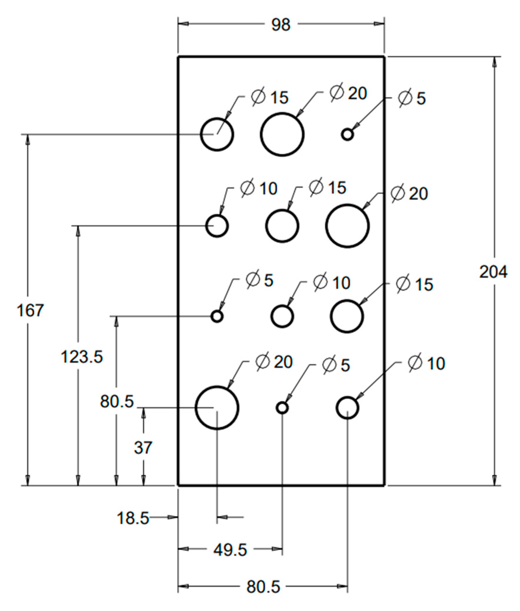
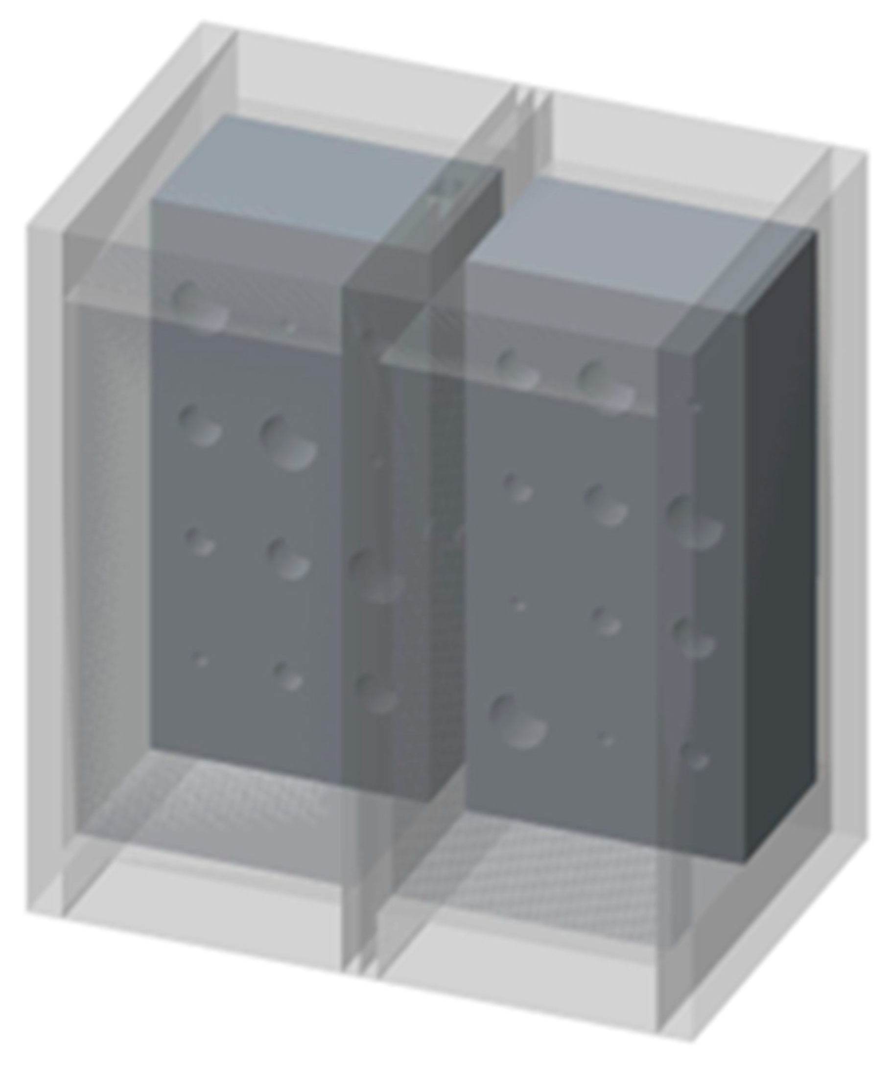
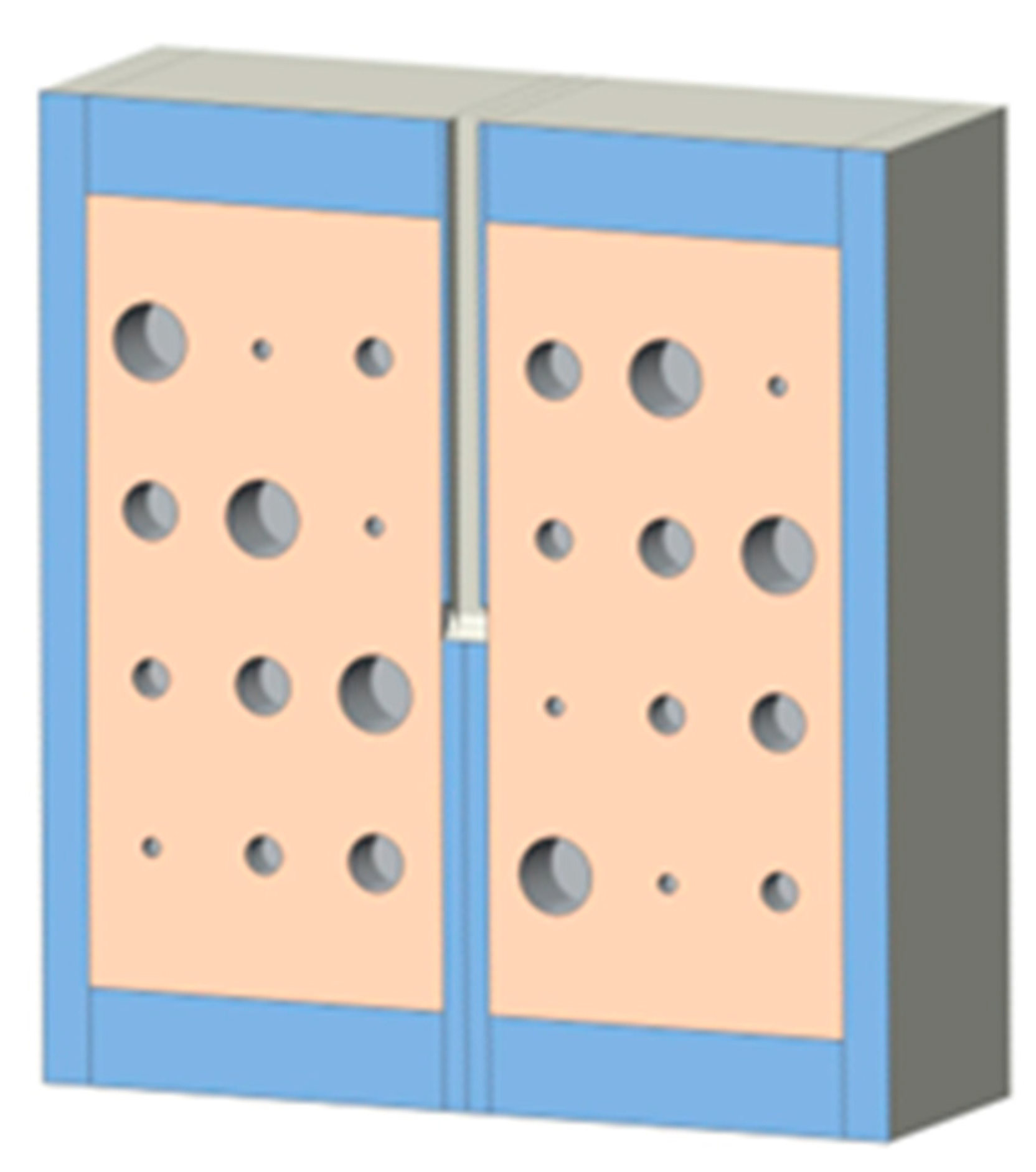
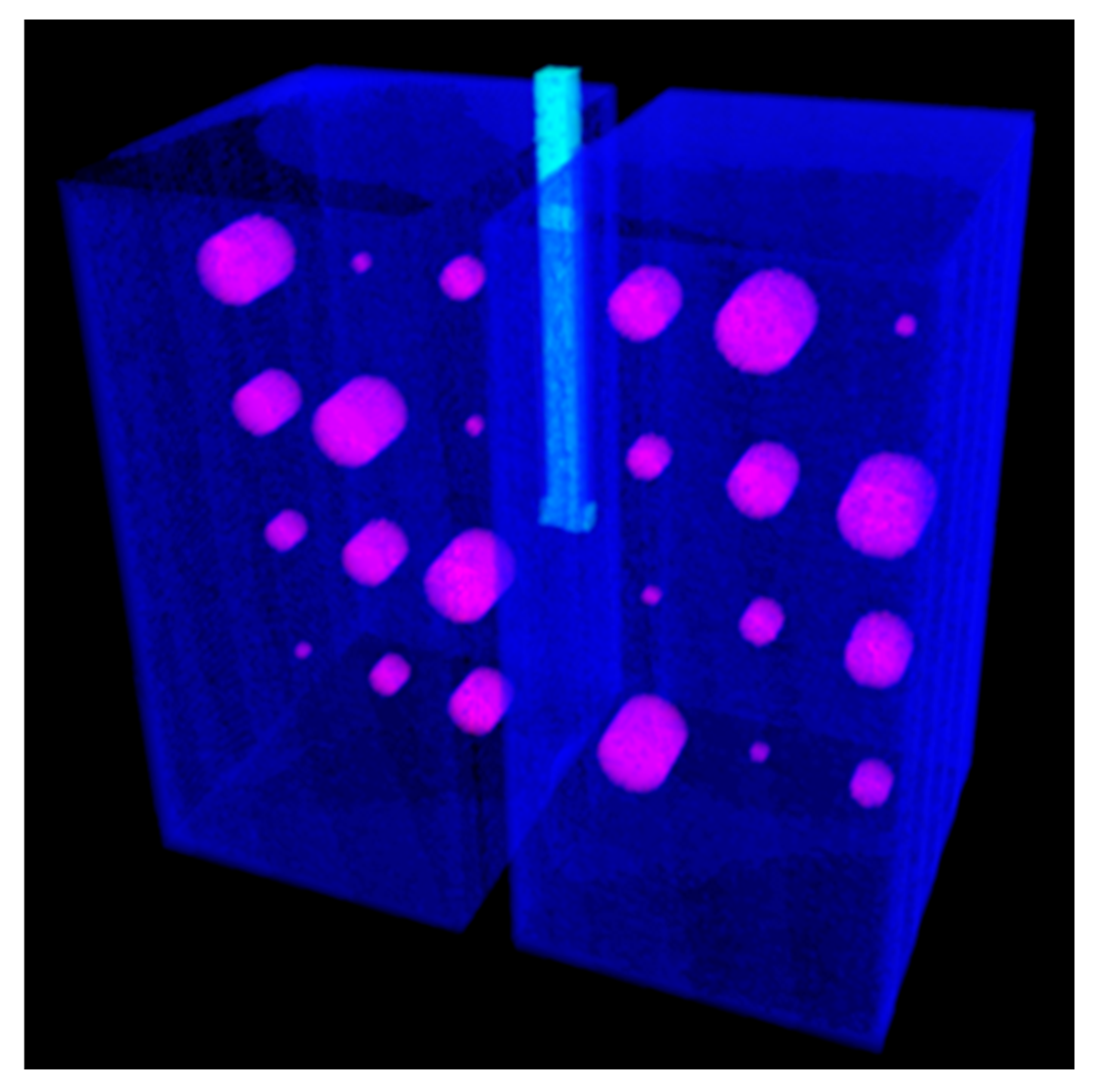
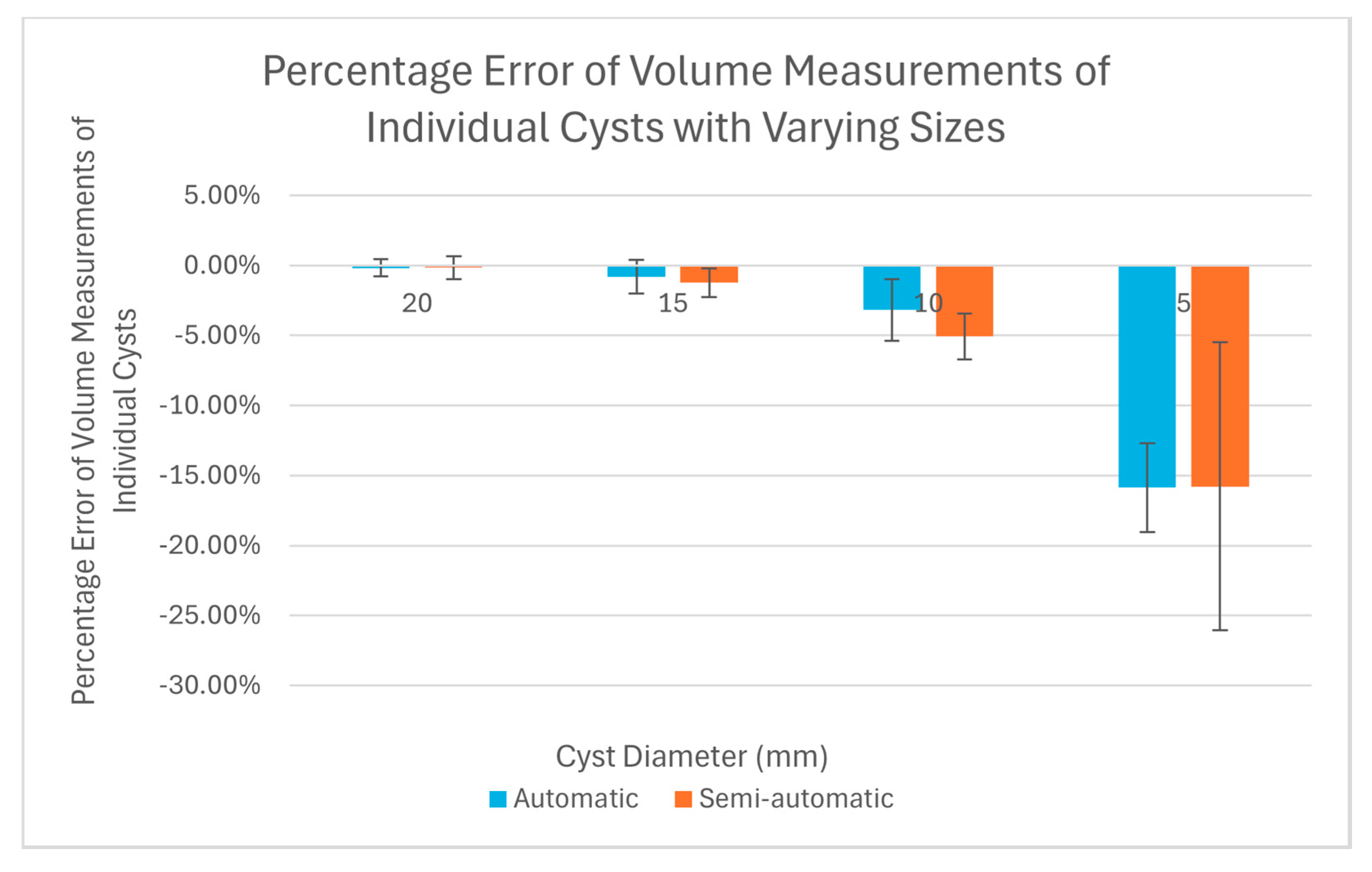
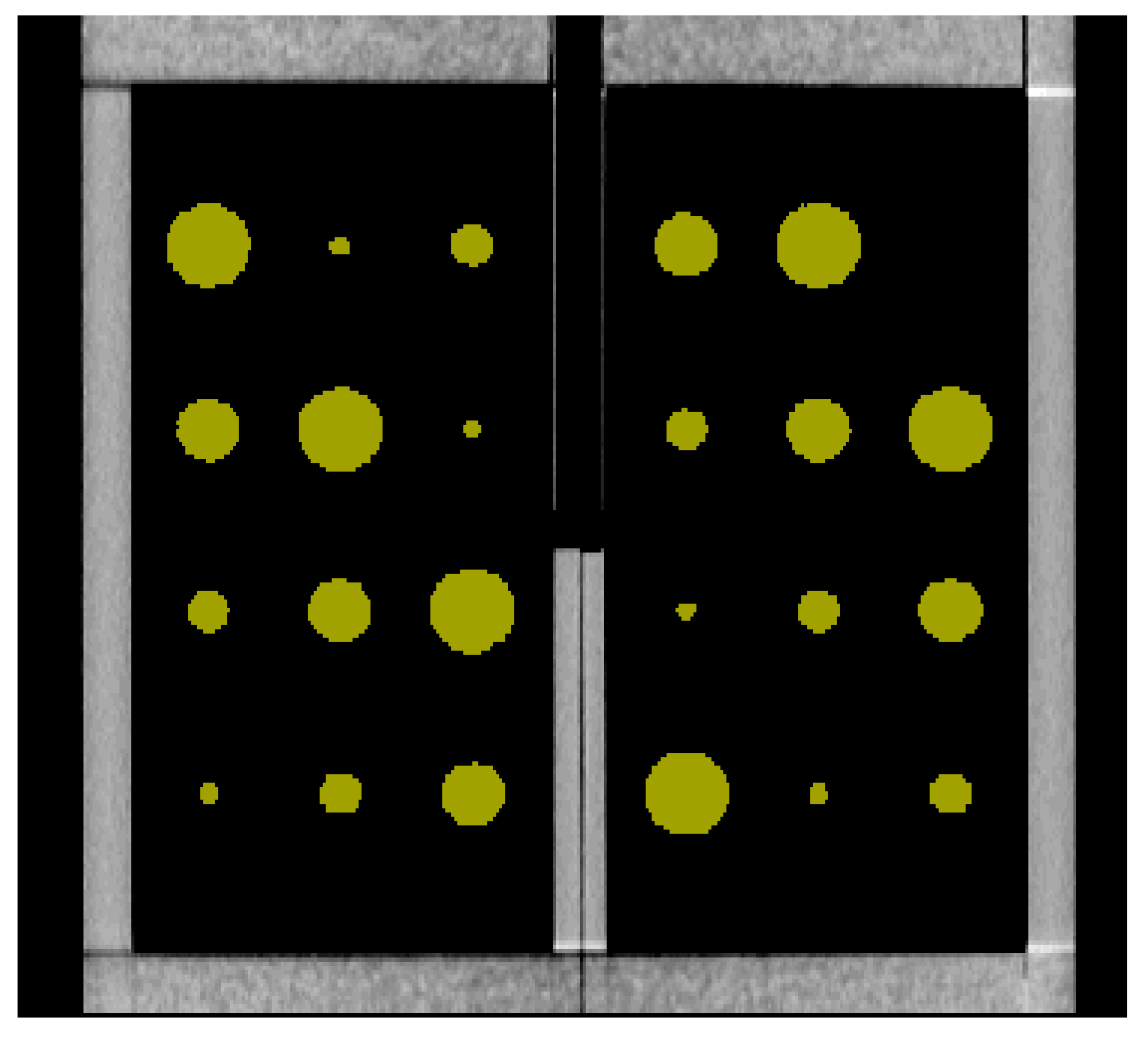
In computed tomography (CT)-based evaluation of the extent of cystic changes in the lungs of patients with cystic lung diseases, such as Lymphangioleiomyomatosis (LAM), there is a lack of a lung phantom containing air-filled cavities that mimic pulmonary cysts to calibrate the measurement of cystic volumes from CT scans. We describe an easy-to-replicate cystic lung phantom consisting of basic lung structures of a trachea and two lung compartments. The lung compartments contain air cavities of varying sizes to mimic cystic lesions. The lung compartments are made of a foam material recommended by NIST to simulate the radiodensity of human lung parenchyma. In tests performed on a clinical scanner, various structures in the lung phantom were correctly recognized by two types of lung analysis software. The resulting cystic volume measurements revealed the relationship between the size of the cysts and the accuracy of the measurement. The significant finding was that the volumes of individual cysts were underestimated for small cysts. The error increased with decreasing cyst sizes. Such underestimation has not been mentioned previously and deserves the attention of clinicians using CT scans to assess the cyst burden in the lungs, particularly in patients presenting with numerous small pulmonary cysts.
Keywords: cystic lung disease; CT scan; CT cystic lung phantom cystic lung disease; CT scan; CT cystic lung phantom