

Large Scale Double-Path Illumination System with Split Field of View for the All-Optical Study of Inter-and Intra-Hemispheric Functional Connectivity on Mice


Abstract
1. Introduction
2. Materials and Methods
2.1. Mice
2.2. Surgery
2.3. Optogenetic Stimulation and Simultaneous Recording of GCaMP6f Activity
2.4. Wide-Filed Fluorescence Microscope
2.5. Calcium Imaging
2.6. Seed Pixel Correlation Map
2.7. Correlation Matrix
3. Results
4. Discussion
Author Contributions
Funding
Acknowledgments
Conflicts of Interest
References
- Emiliani, V.; Cohen, A.E.; Deisseroth, K.; Hausser, M. All-Optical Interrogation of Neural Circuits. J. Neurosci. 2015, 35, 13917–13926. [Google Scholar] [CrossRef] [Scilit] [PubMed]
- Packer, A.M.; Russell, L.E.; Dalgleish, H.W.; Hausser, M. Simultaneous all-optical manipulation and recording of neural circuit activity with cellular resolution in vivo. Nat. Methods 2015, 12, 140–146. [Google Scholar] [CrossRef] [Scilit] [PubMed]
- Ju, N.; Jiang, R.; Macknik, S.L.; Martinez-Conde, S.; Tang, S. Long-term all-optical interrogation of cortical neurons in awake-behaving nonhuman primates. PLoS Biol. 2018, 16, e2005839. [Google Scholar] [CrossRef] [Scilit] [PubMed]
- Zhang, Z.; Russell, L.E.; Packer, A.M.; Gauld, O.M.; Hausser, M. Closed-loop all-optical interrogation of neural circuits in vivo. Nat. Methods 2018. [Google Scholar] [CrossRef] [Scilit] [PubMed]
- Forli, A.; Vecchia, D.; Binini, N.; Succol, F.; Bovetti, S.; Moretti, C.; Nespoli, F.; Mahn, M.; Baker, C.A.; Bolton, M.M.; et al. Two-Photon Bidirectional Control and Imaging of Neuronal Excitability with High Spatial Resolution In Vivo. Cell Rep. 2018, 22, 3087–3098. [Google Scholar] [CrossRef] [Scilit] [PubMed]
- Chen, T.W.; Wardill, T.J.; Sun, Y.; Pulver, S.R.; Renninger, S.L.; Baohan, A.; Schreiter, E.R.; Kerr, R.A.; Orger, M.B.; Jayaraman, V.; et al. Ultrasensitive fluorescent proteins for imaging neuronal activity. Nature 2013, 499, 295–300. [Google Scholar] [CrossRef] [Scilit] [PubMed]
- Palmer, A.E.; Tsien, R.Y. Measuring calcium signaling using genetically targetable fluorescent indicators. Nat. Protoc. 2006, 1, 1057–1065. [Google Scholar] [CrossRef] [Scilit]
- Wright, P.W.; Brier, L.M.; Bauer, A.Q.; Baxter, G.A.; Kraft, A.W.; Reisman, M.D.; Bice, A.R.; Snyder, A.Z.; Lee, J.M.; Culver, J.P. Functional connectivity structure of cortical calcium dynamics in anesthetized and awake mice. PLoS ONE 2017, 12, e0185759. [Google Scholar] [CrossRef] [Scilit]
- Balbi, M.; Vanni, M.P.; Vega, M.J.; Silasi, G.; Sekino, Y.; Boyd, J.D.; LeDue, J.M.; Murphy, T.H. Longitudinal monitoring of mesoscopic cortical activity in a mouse model of microinfarcts reveals dissociations with behavioral and motor function. J. Cereb. Blood Flow Metab. 2018. [Google Scholar] [CrossRef] [Scilit]
- Xiao, D.; Vanni, M.P.; Mitelut, C.C.; Chan, A.W.; LeDue, J.M.; Xie, Y.; Chen, A.C.; Swindale, N.V.; Murphy, T.H. Mapping cortical mesoscopic networks of single spiking cortical or sub-cortical neurons. Elife 2017, 6. [Google Scholar] [CrossRef] [Scilit]
- Tian, L.; Hires, S.A.; Mao, T.; Huber, D.; Chiappe, M.E.; Chalasani, S.H.; Petreanu, L.; Akerboom, J.; McKinney, S.A.; Schreiter, E.R.; et al. Imaging neural activity in worms, flies and mice with improved GCaMP calcium indicators. Nat. Methods 2009, 6, 875–881. [Google Scholar] [CrossRef] [Scilit] [PubMed]
- Mao, T.; O’Connor, D.H.; Scheuss, V.; Nakai, J.; Svoboda, K. Characterization and subcellular targeting of GCaMP-type genetically-encoded calcium indicators. PLoS ONE 2008, 3, e1796. [Google Scholar] [CrossRef] [Scilit] [PubMed]
- Pologruto, T.A.; Yasuda, R.; Svoboda, K. Monitoring neural activity and [Ca2+] with genetically encoded Ca2+ indicators. J. Neurosci. 2004, 24, 9572–9579. [Google Scholar] [CrossRef] [Scilit] [PubMed]
- Reiff, D.F.; Ihring, A.; Guerrero, G.; Isacoff, E.Y.; Joesch, M.; Nakai, J.; Borst, A. In vivo performance of genetically encoded indicators of neural activity in flies. J. Neurosci. 2005, 25, 4766–4778. [Google Scholar] [CrossRef] [Scilit]
- Lorenz-Fonfria, V.A.; Heberle, J. Channelrhodopsin unchained: Structure and mechanism of a light-gated cation channel. Biochim. Biophys. Acta 2014, 1837, 626–642. [Google Scholar] [CrossRef] [Scilit]
- Kim, C.K.; Adhikari, A.; Deisseroth, K. Integration of optogenetics with complementary methodologies in systems neuroscience. Nat. Rev. Neurosci. 2017, 18, 222–235. [Google Scholar] [CrossRef] [Scilit] [PubMed]
- Tennant, K.A.; Taylor, S.L.; White, E.R.; Brown, C.E. Optogenetic rewiring of thalamocortical circuits to restore function in the stroke injured brain. Nat. Commun. 2017, 8, 15879. [Google Scholar] [CrossRef] [Scilit]
- Li, L.; Tang, Y.; Sun, L.; Rahman, K.; Huang, K.; Xu, W.; Yu, J.; Dai, J.; Cao, G. In vivo fiber photometry of neural activity in response to optogenetically manipulated inputs in freely moving mice. J. Innov. Opt. Health Sci. 2017, 10. [Google Scholar] [CrossRef] [Scilit]
- Crocini, C.; Ferrantini, C.; Coppini, R.; Scardigli, M.; Yan, P.; Loew, L.M.; Smith, G.; Cerbai, E.; Poggesi, C.; Pavone, F.S.; et al. Optogenetics design of mechanistically-based stimulation patterns for cardiac defibrillation. Sci. Rep. 2016, 6, 35628. [Google Scholar] [CrossRef] [Scilit]
- Scardigli, M.; Mullenbroich, C.; Margoni, E.; Cannazzaro, S.; Crocini, C.; Ferrantini, C.; Coppini, R.; Yan, P.; Loew, L.M.; Campione, M.; et al. Real-time optical manipulation of cardiac conduction in intact hearts. J. Physiol. 2018, 596, 3841–3858. [Google Scholar] [CrossRef] [Scilit]
- Haupt, D.; Vanni, M.P.; Bolanos, F.; Mitelut, C.; LeDue, J.M.; Murphy, T.H. Mesoscale brain explorer, a flexible python-based image analysis and visualization tool. Neurophotonics 2017, 4, 031210. [Google Scholar] [CrossRef] [Scilit] [PubMed]
- Vanni, M.P.; Murphy, T.H. Mesoscale transcranial spontaneous activity mapping in GCaMP3 transgenic mice reveals extensive reciprocal connections between areas of somatomotor cortex. J. Neurosci. 2014, 34, 15931–15946. [Google Scholar] [CrossRef] [Scilit] [PubMed]
- Mohajerani, M.H.; Chan, A.W.; Mohsenvand, M.; LeDue, J.; Liu, R.; McVea, D.A.; Boyd, J.D.; Wang, Y.T.; Reimers, M.; Murphy, T.H. Spontaneous cortical activity alternates between motifs defined by regional axonal projections. Nat. Neurosci. 2013, 16, 1426–1435. [Google Scholar] [CrossRef] [Scilit] [PubMed]
- White, B.R.; Bauer, A.Q.; Snyder, A.Z.; Schlaggar, B.L.; Lee, J.M.; Culver, J.P. Imaging of functional connectivity in the mouse brain. PLoS ONE 2011, 6, e16322. [Google Scholar] [CrossRef] [Scilit] [PubMed]
- Silasi, G.; Xiao, D.; Vanni, M.P.; Chen, A.C.; Murphy, T.H. Intact skull chronic windows for mesoscopic wide-field imaging in awake mice. J. Neurosci. Methods 2016, 267, 141–149. [Google Scholar] [CrossRef] [Scilit]
- Chan, A.W.; Mohajerani, M.H.; LeDue, J.M.; Wang, Y.T.; Murphy, T.H. Mesoscale infraslow spontaneous membrane potential fluctuations recapitulate high-frequency activity cortical motifs. Nat. Commun. 2015, 6, 7738. [Google Scholar] [CrossRef] [Scilit] [PubMed]
- Szabo, V.; Ventalon, C.; De Sars, V.; Bradley, J.; Emiliani, V. Spatially selective holographic photoactivation and functional fluorescence imaging in freely behaving mice with a fiberscope. Neuron 2014, 84, 1157–1169. [Google Scholar] [CrossRef] [Scilit]
- Obaid, A.L.; Loew, L.M.; Wuskell, J.P.; Salzberg, B.M. Novel naphthylstyryl-pyridium potentiometric dyes offer advantages for neural network analysis. J. Neurosci. Methods 2004, 134, 179–190. [Google Scholar] [CrossRef] [Scilit]
- Gupta, R.K.; Salzberg, B.M.; Grinvald, A.; Cohen, L.B.; Kamino, K.; Lesher, S.; Boyle, M.B.; Waggoner, A.S.; Wang, C.H. Improvements in optical methods for measuring rapid changes in membrane potential. J. Membr. Biol. 1981, 58, 123–137. [Google Scholar] [CrossRef] [Scilit]
- Ross, W.N.; Salzberg, B.M.; Cohen, L.B.; Grinvald, A.; Davila, H.V.; Waggoner, A.S.; Wang, C.H. Changes in absorption, fluorescence, dichroism, and Birefringence in stained giant axons: Optical measurement of membrane potential. J. Membr. Biol. 1977, 33, 141–183. [Google Scholar] [CrossRef] [Scilit]
- Grinvald, A. Real-time optical mapping of neuronal activity: From single growth cones to the intact mammalian brain. Annu. Rev. Neurosci. 1985, 8, 263–305. [Google Scholar] [CrossRef]
- Vanni, M.P.; Chan, A.W.; Balbi, M.; Silasi, G.; Murphy, T.H. Mesoscale Mapping of Mouse Cortex Reveals Frequency-Dependent Cycling between Distinct Macroscale Functional Modules. J. Neurosci. 2017, 37, 7513–7533. [Google Scholar] [CrossRef] [Scilit] [PubMed]
- Lim, D.H.; Mohajerani, M.H.; Ledue, J.; Boyd, J.; Chen, S.B.; Murphy, T.H. In vivo large-scale cortical mapping using channelrhodopsin-2 stimulation in transgenic mice reveals asymmetric and reciprocal relationships between cortical areas. Front. Neural Circuit 2012, 6. [Google Scholar] [CrossRef] [Scilit]
- Mohajerani, M.H.; McVea, D.A.; Fingas, M.; Murphy, T.H. Mirrored Bilateral Slow-Wave Cortical Activity within Local Circuits Revealed by Fast Bihemispheric Voltage-Sensitive Dye Imaging in Anesthetized and Awake Mice. J. Neurosci. 2010, 30, 3745–3751. [Google Scholar] [CrossRef] [Scilit] [PubMed]
- Bauer, A.Q.; Kraft, A.W.; Baxter, G.A.; Wright, P.W.; Reisman, M.D.; Bice, A.R.; Park, J.J.; Bruchas, M.R.; Snyder, A.Z.; Lee, J.M.; et al. Effective Connectivity Measured Using Optogenetically Evoked Hemodynamic Signals Exhibits Topography Distinct from Resting State Functional Connectivity in the Mouse. Cereb. Cortex 2018, 28, 370–386. [Google Scholar] [CrossRef] [Scilit] [PubMed]
- Oh, S.W.; Harris, J.A.; Ng, L.; Winslow, B.; Cain, N.; Mihalas, S.; Wang, Q.; Lau, C.; Kuan, L.; Henry, A.M.; et al. A mesoscale connectome of the mouse brain. Nature 2014, 508, 207–214. [Google Scholar] [CrossRef] [Scilit] [PubMed]
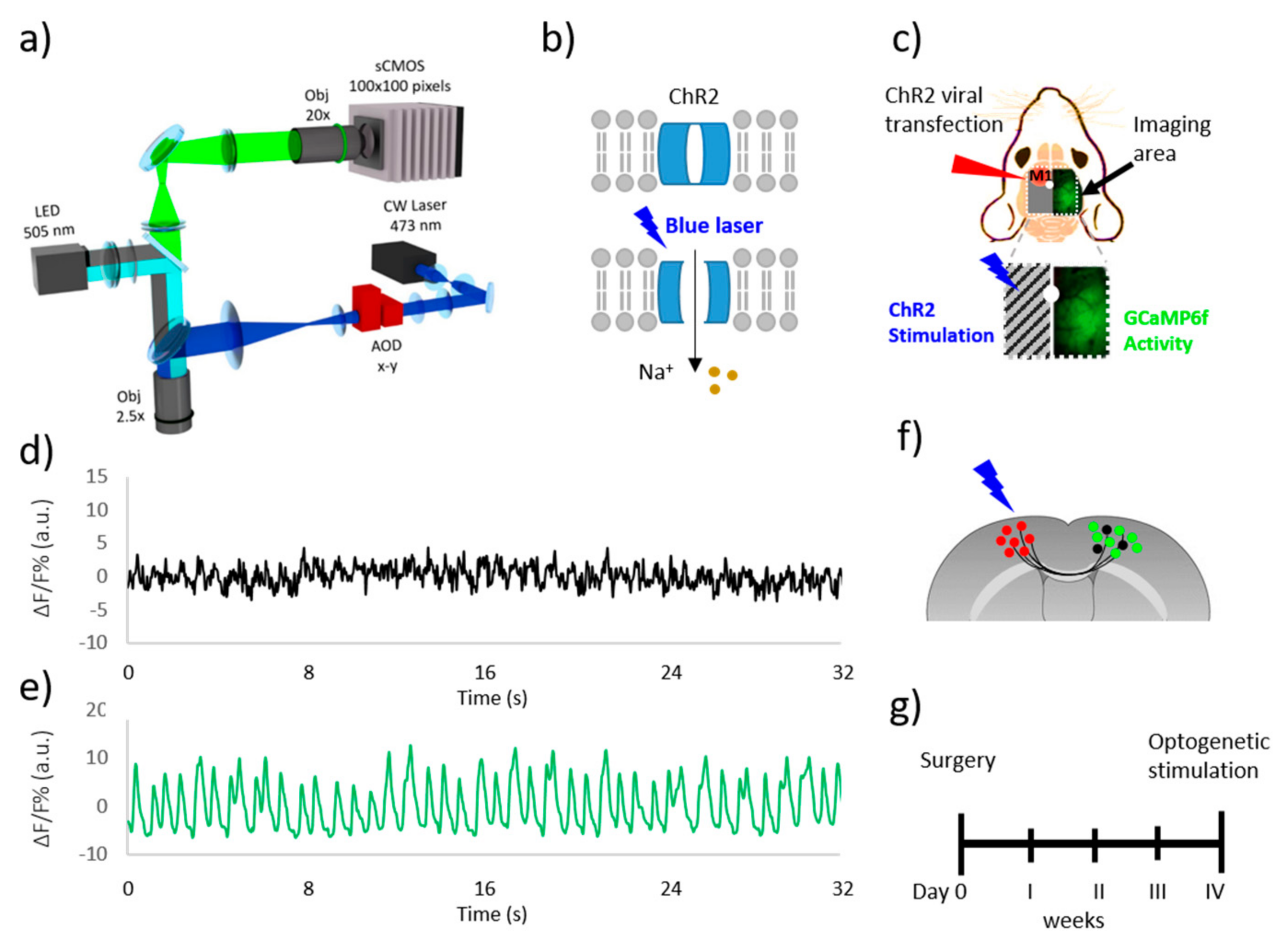
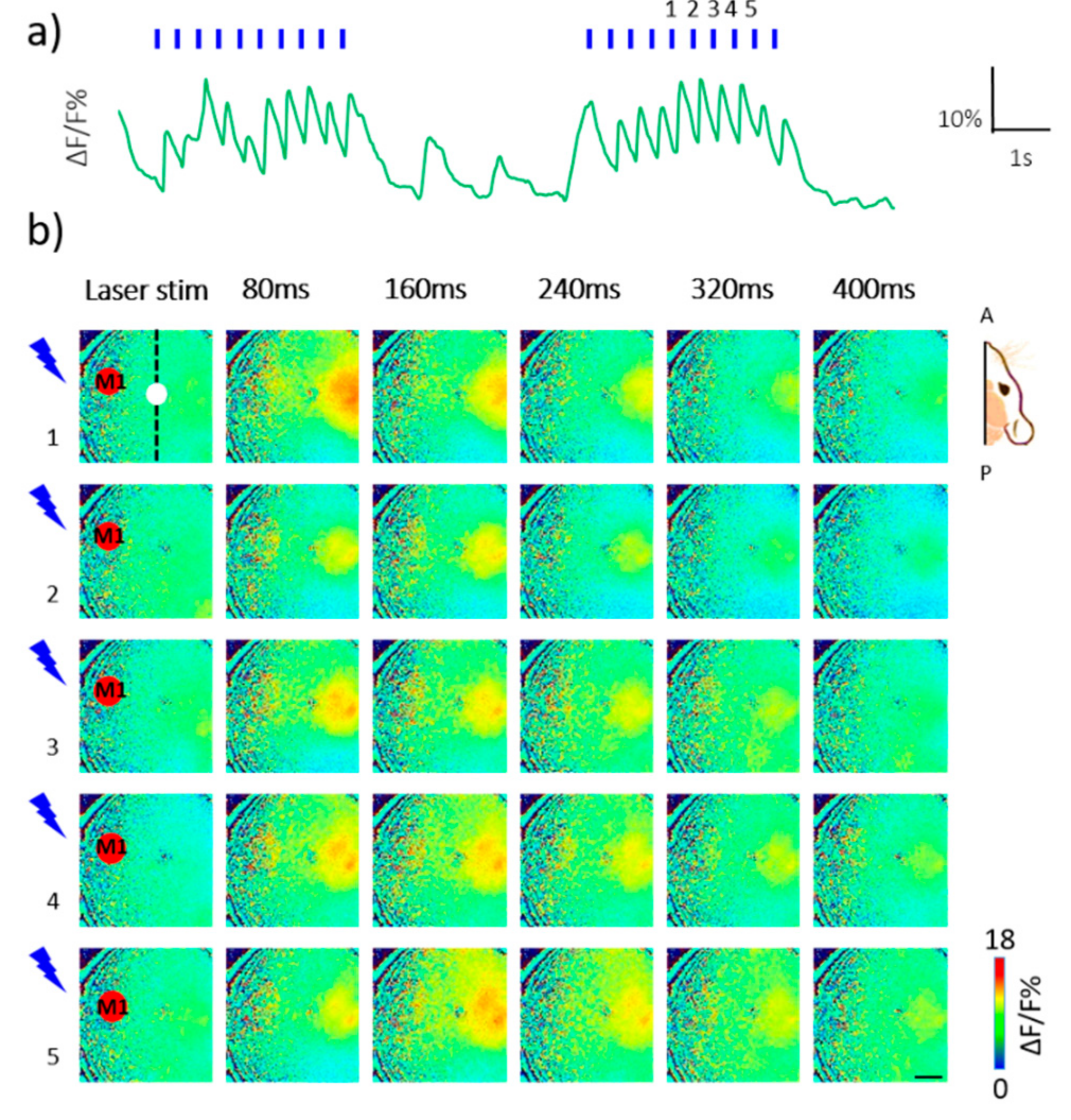
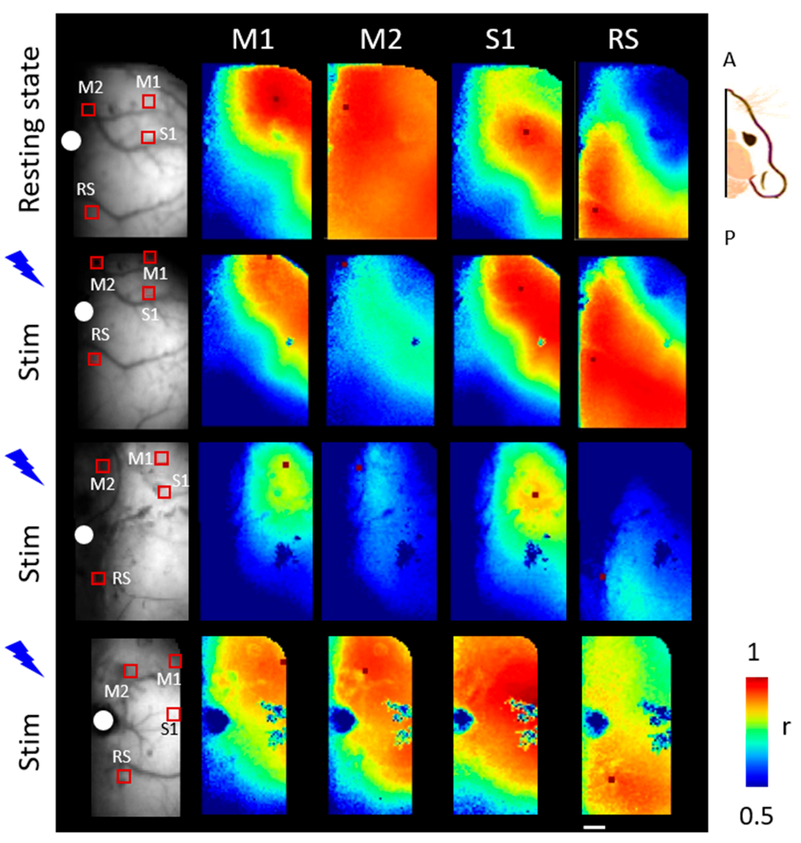
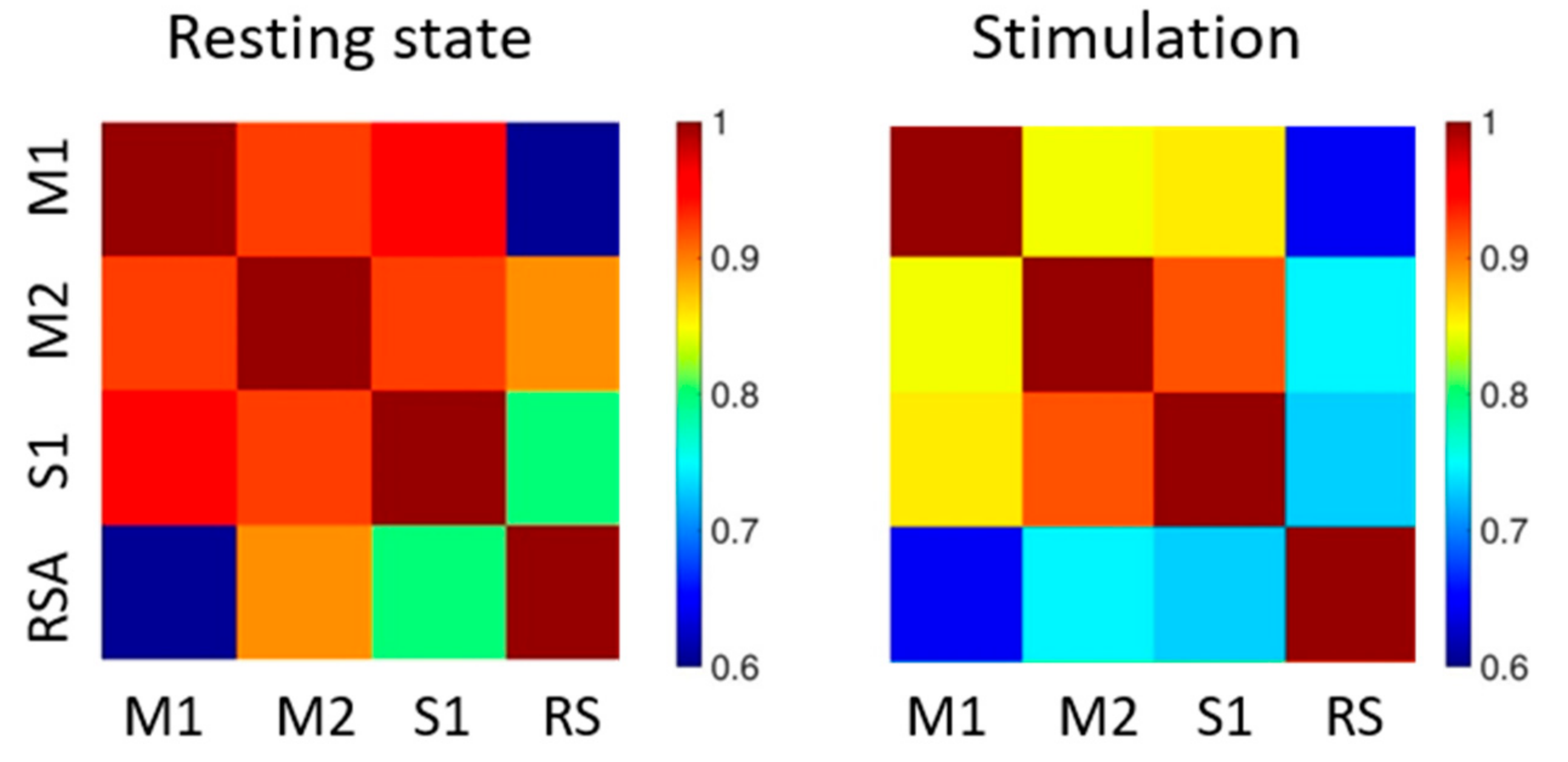

| Functional Areas | Resting State | Stimulation |
|---|---|---|
| M1/M2 | 0.85 ± 0.06 | 0.92 ± 0.005 |
| M1/S1 | 0.85 ± 0.07 | 0.94 ± 0.01 |
| M1/RS | 0.64 ± 0.02 | 0.62 ± 0.1 |
| M2/S1 | 0.92 ± 0.03 | 0.92 ± 0.005 |
| M2/RS | 0.75 ± 0.08 | 0.9 ± 0.03 |
| S1/RS | 0.73 ± 0.05 | 0.81 ± 0.03 |
© 2019 by the authors. Licensee MDPI, Basel, Switzerland. This article is an open access article distributed under the terms and conditions of the Creative Commons Attribution (CC BY) license (http://creativecommons.org/licenses/by/4.0/).
Share and Cite
Conti, E.; Allegra Mascaro, A.L.; Pavone, F.S. Large Scale Double-Path Illumination System with Split Field of View for the All-Optical Study of Inter-and Intra-Hemispheric Functional Connectivity on Mice. Methods Protoc. 2019, 2, 11. https://doi.org/10.3390/mps2010011
Conti E, Allegra Mascaro AL, Pavone FS. Large Scale Double-Path Illumination System with Split Field of View for the All-Optical Study of Inter-and Intra-Hemispheric Functional Connectivity on Mice. Methods and Protocols. 2019; 2(1):11. https://doi.org/10.3390/mps2010011
Chicago/Turabian StyleConti, Emilia, Anna Letizia Allegra Mascaro, and Francesco Saverio Pavone. 2019. "Large Scale Double-Path Illumination System with Split Field of View for the All-Optical Study of Inter-and Intra-Hemispheric Functional Connectivity on Mice" Methods and Protocols 2, no. 1: 11. https://doi.org/10.3390/mps2010011
APA StyleConti, E., Allegra Mascaro, A. L., & Pavone, F. S. (2019). Large Scale Double-Path Illumination System with Split Field of View for the All-Optical Study of Inter-and Intra-Hemispheric Functional Connectivity on Mice. Methods and Protocols, 2(1), 11. https://doi.org/10.3390/mps2010011

