Next Article in Journal
A Phenomenological Take on Mirror Self-Face Perception in Schizophrenia
Previous Article in Journal
Nasal Surgery Versus Pharyngeal Surgery in the Treatment of Obstructive Sleep Apnea
 
 
Journal of Mind and Medical Sciences is published by MDPI from Volume 12 Issue 1 (2025). Previous articles were published by another publisher in Open Access under a CC-BY (or CC-BY-NC-ND) licence, and they are hosted by MDPI on mdpi.com as a courtesy and upon agreement with Valparaiso University (ValpoScholar).
Case Report

Giant Gastric Polyp Mimicking a Duodenal Tumor

by
Ioana Duca
1,
Florin Mihaileanu
2,*,
Alexandra Chira
1,
Romeo Chira
3,
Teodora Surdea Blaga
1,
Diana Popovici
4,
Adriana Albu
1 and
Dan Lucian Dumitrascu
1
1
2nd Department of Internal Medicine, Clinical Emergency County Hospital, Iuliu Hatieganu University for Medicine and Pharmacy, Cluj-Napoca, Romania
2
2nd Department of Surgery, Clinical Emergency County Hospital, Iuliu Hatieganu University for Medicine and Pharmacy, 2nd-4th Clinicilor Street, Cluj-Napoca 400006, Romania
3
Department of Gastroenterology, Clinical Emergency County Hospital, Iuliu Hatieganu University for Medicine and Pharmacy, Cluj-Napoca, Romania
4
2nd Department of Internal Medicine, Clinical Emergency County Hospital, Cluj-Napoca, Romania
*
Author to whom correspondence should be addressed.
J. Mind Med. Sci. 2022, 9(2), 324-329; https://doi.org/10.22543/2392-7674.1316
Submission received: 20 February 2022 / Revised: 1 March 2022 / Accepted: 16 April 2022 / Published: 15 October 2022

Abstract

:
Inflammatory fibroid polyps are very rare gastrointestinal tumors. We present the case of a 66-year-old woman with severe anemia and a giant gastric polyp which had intermittent duodenal intussusception. Ultrasonography showed increased gastric wall thickness and suspected an ampulloma, as revealed also by endoscopy and computed tomography. Ultrasonography reassessment showed later the intragastric mass, which was confirmed by endosonography: giant pediculated hypervascular polyp suggesting malignancy. Challenging phenomena at different investigation methods were due to intermittent protrusion into the first duodenal segment mimicking an ampulloma, but without gastric outlet syndrome or a malignant component, despite the severe anemia. Abnormal US aspect of the stomach in clinical context of anemic syndrome, requires EUS with biopsies in order to confirm underlying lesions. The particularities of this case are the: discordance between imaging aspects and the protrusion into the first duodenal segments with consecutive cholestasis mimicking an ampulloma and the lack of gastric outlet syndrome.

Introduction

Gastric polyps are epithelial or subepithelial protruding lesions including both benign and malignant underlying masses; they have variable presentation and prognosis depending on their histology. Gastric polyps are considered benign on the first impression unless proven otherwise [1]. Frequent types of gastric polyps are: fundic gland polyps, hyperplastic polyps, and adenomas. Rare types include: neuroendocrine tumors (NET, carcinoids), infiltrates (xanthomas, lymphoproliferative neoplasms), mesenchymal proliferations (leiomyomas, gastrointestinal stromal tumors-GIST, inflammatory fibroid polyps-IFP), and hamartomas [2,3].
IFPs are rare benign mesenchymal gastrointestinal tumors, representing 0.1% of all gastric polyps [4]; this type of tumors usually appears in the fifth decades of life, but a wide age range is affected (4-84 years), especially in women, and have been first described by Vanek in 1949 as a submucosal gastric granuloma with eosinophilia [5]. IFPs are found predominant in the antrum, as Garmpis et al. showed in his review [6], being followed by the small bowel [7].
From a histological point of view, IFP is characterized by proliferation of highly vascular fibrous tissue and inflammatory infiltration; 4 types of IFPs have been described: classical fibrovascular, nodular, sclerotic, and edematous [8]. Some authors considered the last form as a result of edema of the small bowel due to intussusception [9]. IFPs are characterized from the immunohistological point of view by spindle cells which are positive for CD34 and vimentin, and negative for CD117 [10].
The etiology of IFP is still unknown, an allergic reaction due to the presence of eosinophils has been suggested as possible cause. Also, other factors seem to be implied in the development of IPFs, such as genetic alterations (mutations in the platelet-derived growth factor receptor alpha-gene), autoimmune reaction, neural hyperplasia, irritants, trauma, and bacterial, physical, or chemical stimulants [11]. Helicobacter pylori infection, parasitic infestation, hypersensitivity reactions, and metabolic factors seem also to be involved [12]. There is no evidence of malignant potential of IFP, but some authors suggest it, as malignant processes have been found adjacent to some IFPs [13].
Ultrasonography (US) and especially hydrosonography is the first imaging, inexpensive and low-invasive technique used to detect large polyps; a more precise characterization and confirmation is possible by endoscopy, endoscopic ultrasonography (EUS) and histological examination.
In this case report we describe a patient with a giant gastric polyp mimicking a duodenal tumor.

Case presentation

A 66-year-old woman, with personal history of essential arterial hypertension and recent deep vein thrombosis-DVT (under treatment with apixaban), ex-smoker, was admitted for asthenia, important weight loss (6 kg in the last 2 months), but with no history of gastric outlet syndrome, nor melena or hematemesis. General physical examination revealed pallor, no pain at the abdominal palpation and negative rectal examination for melena or rectal bleeding.
Laboratory data evidenced: severe microcytic hypochromic anemia (hemoglobin of 3.1 g/dl), low value of ferritin (9 ng/ml) and transferrin saturation (2%), lower levels of B12 vitamin (155 pg/ml), but normal folate, anisocytosis, variable cholestasis (alkaline phosphatase 167-224 U/l, GGT 70-189 U/l), hepatocytolysis (GOT 100 U/l, GPT 105 U/l), inflammatory syndrome in the context of catheter-thrombophlebitis, negative urine and blood cultures, negative hemoccult tests (possibly due to a single sample and intermittent bleeding) and negative stool antigen for H. pylori, normal tumoral markers (CEA 1.68 ng/ml, CA 125 7.7 U/ml, CA 19-9< 0.8U/ml).
The first grey-scale ultrasonography (US) described thick walls of the stomach (11 mm), but with maintenance of the stratification (Figure 1a). In addition, in the duodenum there was a hypoechoic mass of 6 cm, quite well delimited, with vascular signal (color-Doppler US) inside. Ductus choledochus was dilated at 11 mm, with normal intrahepatic biliary ducts and no ascites. The US reassessment done before endosonography (EUS) revealed a large, hypoechoic intragastric mass (Figure 1b).
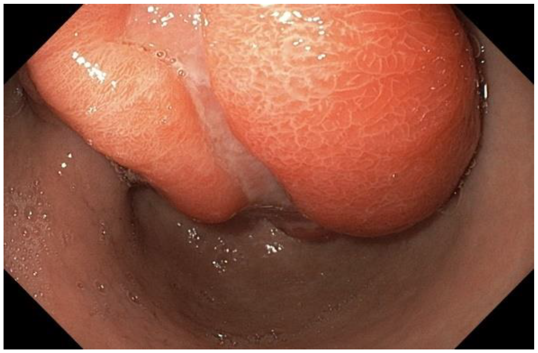
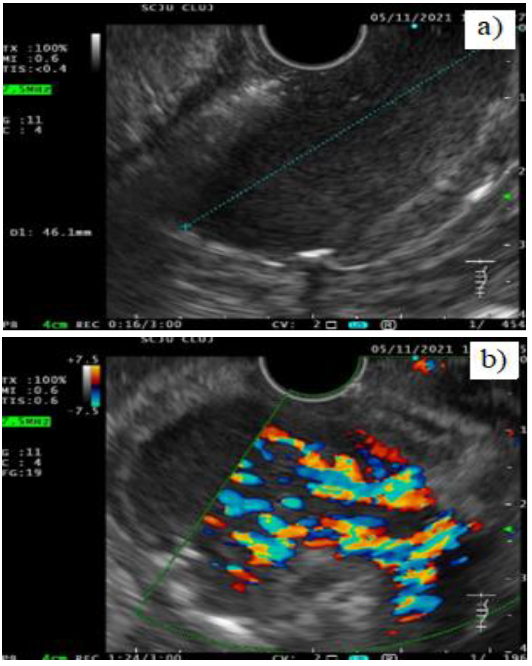
The patient was referred to upper gastrointestinal endoscopy (UGIE), which revealed a deformed antrum in the pyloric region with possible tumoral infiltration; inside the bulbus duodeni and in the descending part (D2), there was a vegetant bleeding tumor of 4 cm which led to relative stenosis of the ampulla of Vater, suggesting duodenal tumor/ampulloma. An intragastric mass was confirmed by the US reassessment and UGIE performed during EUS; EUS revealed a giant (4.6 cm) antral (Figure 2) pediculated hypervascular (color Doppler) polyp with a thick vascularized pedicle (Figure 3), having areas of ulceration on its surface, and extended hypoechoic areas, suggesting malignancy (T1NxMx). No perigastric lymphadenopathies were detected, nor suspect retroperitoneal or intrahepatic lesions. Biopsies taken during UGIE and EUS did not show any tumoral cells, but revealed granular tissue, fibrinoid deposits and inflammatory infiltrate.
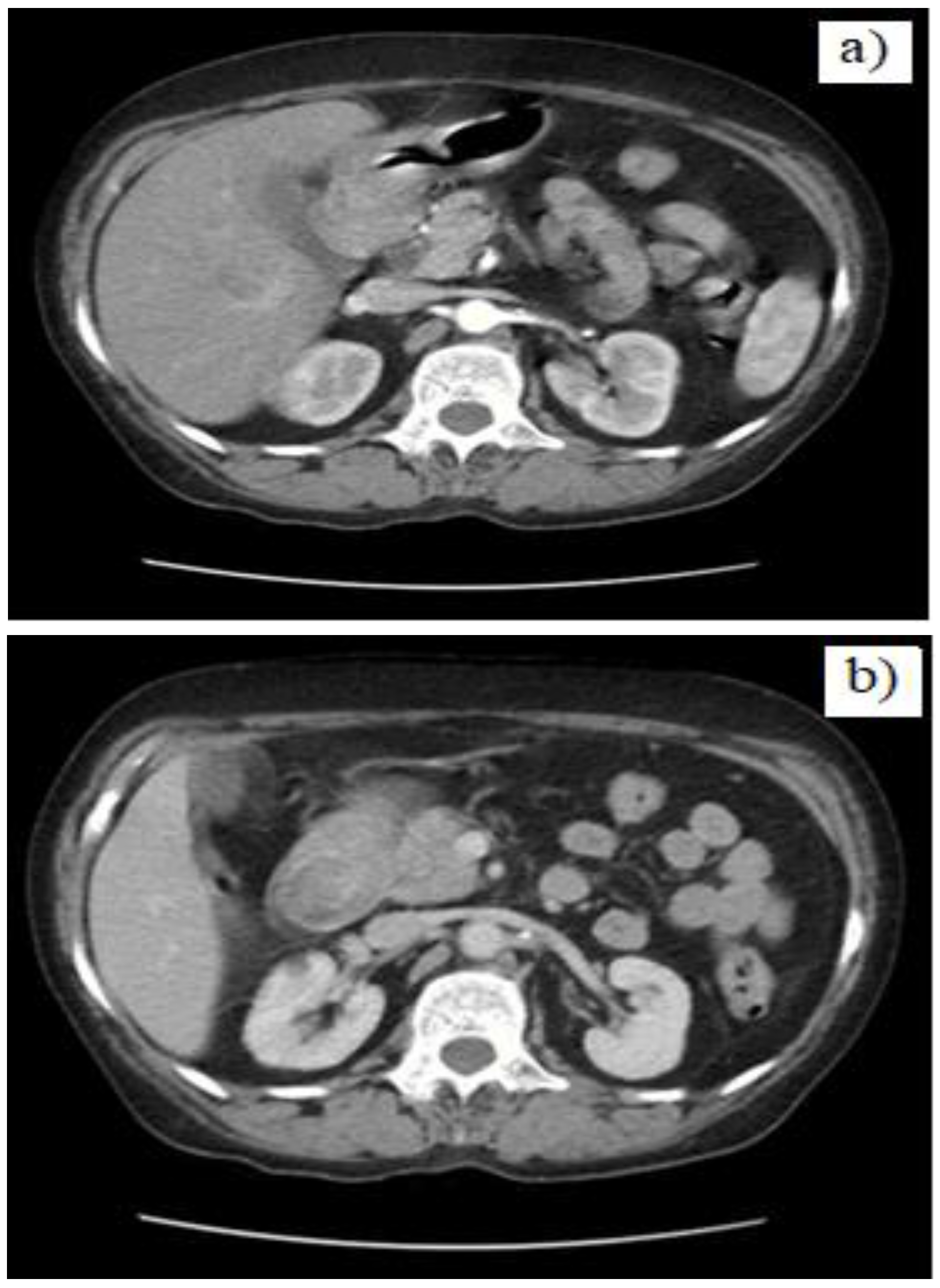
Distance staging has been done by means of contrast enhanced abdominal and pelvic computed tomography (CT). It revealed circumferential inhomogeneous thickening (14 mm) of the D1 and D2 segments of the duodenum, which extended up to the antrum, suggesting its tumoral infiltration (Figure 4). The apparently intraduodenal tumor showed inhomogeneity (after contrast medium), with loss of wall stratification and it was located in close contact with the gallbladder, pancreatic head and uncinate process, without a clear delimitation from them. No intraabdominal metastases were detected.
The case was interpreted as a giant antral pediculated polypoid cancer, mimicking a duodenal tumor (including ampulloma), and complicated with hemorrhage and severe anemia, establishing the necessity of surgery and possible paraneoplastic DVT.
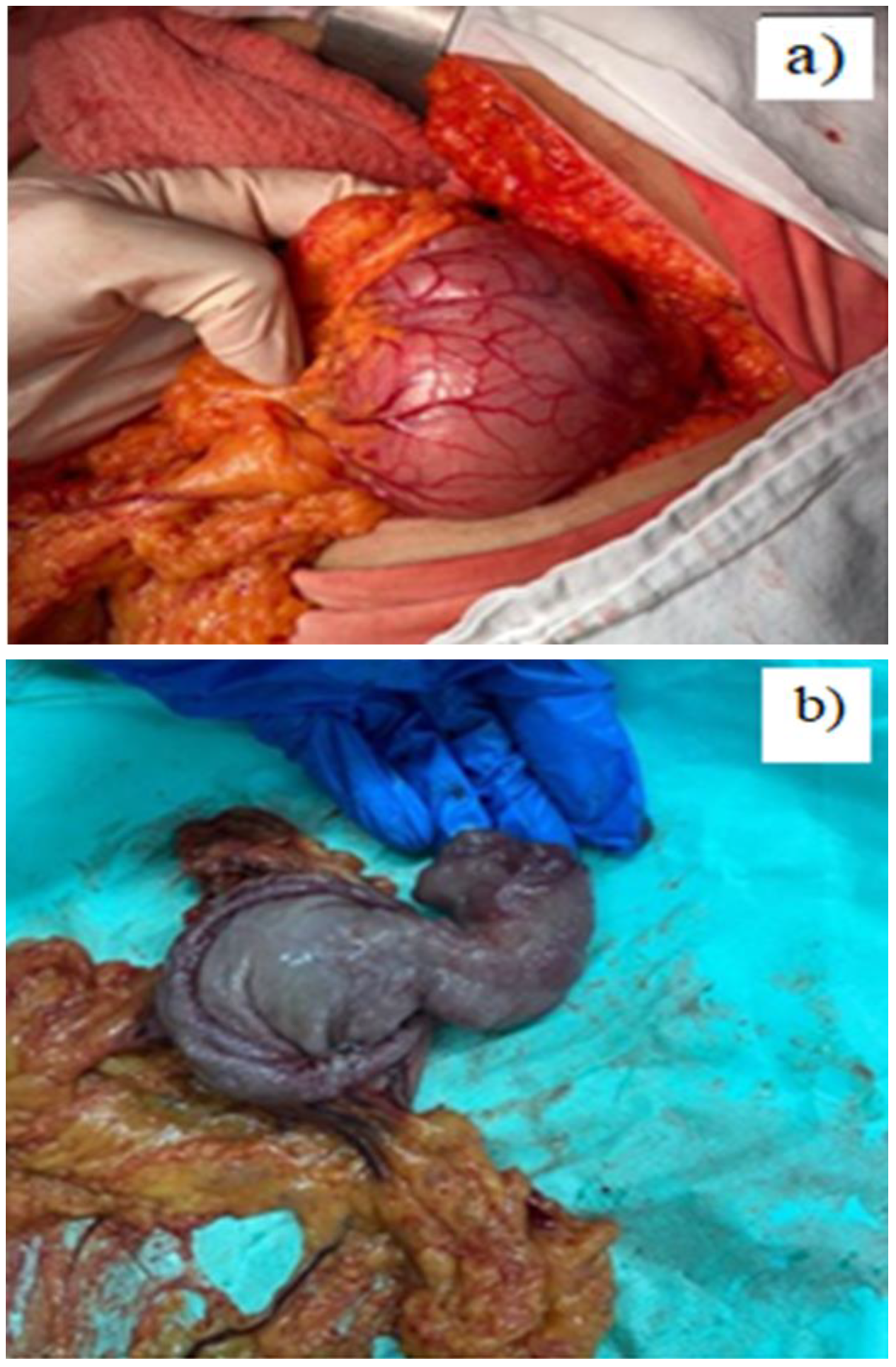
After the correction of the anemic syndrome with several blood transfusions, the patient underwent surgery– 2/3 gastrectomy with gastro-jejunal anastomosis and retrograde cholecystectomy. No hepatic or peritoneal secondary metastasis or intra-abdominal lymphadenopathy have been identified. A tumor of 7 cm in diameter was detected by palpation, located at the antropyloric level, floating intragastrically (Figure 5). The postoperative evolution of the patient was favorable, being discharged on the 10th day. Histopathological examination after surgery (Figure 6) revealed IFP without any malignant cells. Follow-up at 3 months showed no changes from clinical and laboratory point of view, with nFo anemia.

Discussion

Symptoms of IFPs are heterogeneous and depend mainly on the location and size of the tumoral mass. Regarding the clinical picture of intussuscepted gastroduodenal (GD) tumors, most patients have nonspecific symptoms, mimicking other diseases: epigastric pain, nausea, vomiting, upper gastrointestinal hemorrhage or pyloric obstruction [14], weight loss (lipoma, adenocarcinoma [15]), intermittent symptoms of regurgitation and heartburn, anemia [16]. Gastric outlet obstruction mimicking a GIST or carcinoma were also described in a giant antral IFP [17]. Intermittent gastric outlet obstruction ("ball valve syndrome") mimicking a gastrointestinal stromal tumor (GIST) or a gastric carcinoma/lymphoma, with an intestinal obstruction of high origin were also described in literature in a giant (7 cm) antral IFP [17]. Our patient presented atypically, with wasting syndrome suggesting neoplasia.
Gastric polyps can be diagnosed preoperatively starting from the US pattern and through corroborating several noninvasive tests. Indeed, IPF is often described as a duodenal mass that may suggest an invagination; in intussuscepted tumors US may reveal a space-occupying lesion around the liver, a mass with an alternating concentric echogenic lesion (GIST) [18] or hypoechoic gastric tumor [19]. In our case, the preliminary abdominal US examination revealed a thick, multi-layer appearance of the gastric wall, and raised the suspicion of Vater-ampulloma, prompted by the association of anemic and cholestatic syndrome together with the vascularized mass located in the duodenum. CT and UGIE established the duodenum as primary location of the tumor in our case, misdiagnosing its real origin, similar to data in literature [15,20].
Literature mentioned that CT often diagnosed the duodenal masses and lipomatous nature of the tumor [20,21], and also showed diffuse fatty thickening of the gastric wall in addition to multiple lipomas in stomach, duodenum and in the jejunum with jejunal intussusception in gastric lipomatosis [14]. Additionally, diffusion-weighted magnetic resonance imaging can evidence the hyper-vascularity in the IFPs as a peripheral enhancement [17].
As most of the IFPs are asymptomatic, they are diagnosed incidentally at endoscopy [5]. Endoscopic biopsies are often unhelpful, like in our case at UGIE, and diagnosis can be reached only with resection, histological and immunohistochemical analysis; only about 10% of the gastric lesions are diagnosed correctly prior to resection [17]. Biopsies are of limited use for the diagnosis of IFP and diagnosis of foci of high-grade dysplasia/early gastric cancer may not be possible until resection [19]. Ortiz de Solórzapo Aurusa et al. described the non-diagnostic assessment of upper digestive endoscopy in a 5.5 cm large gastric lipoma [20]. The deep location inside the mucosa, or submucosa in gastric IFPs proven at EUS, could explain the bioptic false negative results [22].
Preoperative diagnosis remains a challenge and unclear etiology required sometimes diagnostic laparoscopy, which confirmed the gastroduodenal intussusception by a gastric lipoma [23] and adenocarcinoma [15] in literature.
In our case, repeated US and EUS revealed the real background of the antral origin of the giant polyp with the large pedicle, explaining the intermittent prolapse into the duodenum and hereby also the intermittent cholestasis found in laboratory data and choledochal dilatation in US. We consider that location-variability was due to the intermittent intussusception.
GIST is the most common cause for GD intussusception, caused by transpyloric prolapse of a gastric lesion into the duodenum. Rare cases of gastroduodenal intussusception were associated with: large/giant benign gastric tumors like lipomas [20,21,23,24], diffuse gastroduodenal lipomatosis [14], submucosal lesions like IFP (normally ≤ 2.5 cm) [19] and even more uncommon, malignant gastric neoplasia [15,25]. Gastric tumors arising from collision of a well-differentiated adenocarcinoma with a poorly differentiated neuroendocrine carcinoma prolapsed into the duodenum First case in literature [15] or collision of adenocarcinoma and NET within a single gastric hyperplastic polyp [26] are extremely rare.
Differential diagnosis including gastroduodenal intussusception with gastric cancer invasion to the duodenum, or pancreatic cancer with adherence to the gastric antrum and duodenum has to be taken into account. Large gastric polyps should be carefully examined because there is the possibility of an underlying NET and malignant transformation of surface epithelium [26]. Differentiation from GIST is possible through negative stain for CD117 in IFP. Other differential diagnosis with immuno-histochemistry is: leiomyoma, solitary fibrous tumors, spindle cell carcinoid, hemangioendothelioma, hemangiopericytoma, T-cell lymphoma, schwannoma, fusiform cell leiomyoma, inflammatory myofibroblastic tumor or pseudotumor, inflammatory fibrosarcoma and spindle-cell carcinoid [17,27]. Inflammatory polyps are thought to be part of a diffuse inflammation, with regenerative and/or metaplastic changes of the overlying mucosa.
Surgical treatment with complete resection with safe margins is considered curative, as there exists the possibility of recurrence and because there is no clear imaging (EUS) or histopathologic pattern, that may predict a malignant transformation [28]. Endoscopic polypectomy is appropriate for small-diameter polyps, larger polyps that have risk of intussusception/obstruction require surgical resection [29]. Some of the cases of gastroduodenal intussusception caused by gastric GIST were treated by endoscopic submucosal dissection (ESD), which is also an alternative treatment for elderly patients who are not candidates for surgery [18]. Regression of gastric IFPs after H. pylori eradication therapy was observed in 2% patients by Zinkiewicz et al. [28].
Some authors [30] raised the importance of vigilance for clinicians for occult upper gastrointestinal bleeding from gastric IFPs, and thus the early diagnosis and management of this tumor, in order to combat chronic unexplained iron deficiency anemia.
The particularity of this case is the discordance between the US, endoscopic, EUS and CT aspects and the intermittent protrusion into the first and second duodenal segments with consecutive cholestasis mimicking an ampulloma, but without gastric outlet syndrome or a malignant component, despite the severe anemia, the tumoral bleeding, wasting syndrome and big size of the tumor.

Conclusions

In conclusion, gastric polyps may present as intussuscepted tumors in the duodenum, being diagnosed by US and confirmed by EUS and UGIE. Therefore, multilayer US aspect of the stomach or intragastric masses in the clinical context of anemic and wasting syndrome requires EUS with biopsies in order to confirm underlying lesions.

Conflict of Interest disclosure

There are no known conflicts of interest in the publication of this article. The manuscript was read and approved by all authors.

Compliance with ethical standards

Any aspect of the work covered in this manuscript has been conducted with the ethical approval of all relevant bodies and that such approvals are acknowledged within the manuscript.

References

  1. Carneiro, F. Familial and hereditary gastric cancer, an overview. Best Pract Res Clin Gastroenterol. 2022, 58, 101800. [Google Scholar] [CrossRef] [PubMed]
  2. Islam, R.S.; Patel, N.C.; Lam-Himlin, D.; Nguyen, C.C. Gastric polyps: a review of clinical, endoscopic, and histopathologic features and management decisions. Gastroenterol Hepatol (N Y). 2013, 9, 640–651. [Google Scholar]
  3. Borch, K.; Skarsgård, J.; Franzén, L.; Mårdh, S.; Rehfeld, J.F. Benign gastric polyps: morphological and functional origin. Dig Dis Sci. 2003, 48, 1292–1297. [Google Scholar] [CrossRef] [PubMed]
  4. Carmack, S.W.; Genta, R.M.; Schuler, C.M.; Saboorian, M.H. The current spectrum of gastric polyps: a 1-year national study of over 120,000 patients. Am J Gastroenterol. 2009, 104, 1524–1532. [Google Scholar] [CrossRef] [PubMed]
  5. Joyce, K.M.; Waters, P.S.; Waldron, R.M.; Khan, I.; Orosz, Z.S.; Németh, T.; Barry, K. Recurrent adult jejuno-jejunal intussusception due to inflammatory fibroid polyp - Vanek's tumour: a case report. Diagn Pathol. 2014, 9, 127. [Google Scholar] [CrossRef]
  6. Garmpis, N.; Damaskos, C.; Garmpi, A.; Georgakopoulou, V.E.; Sakellariou, S.; Liakea, A.; Schizas, D.; Diamantis, E.; Farmaki, P.; Voutyritsa, E.; Syllaios, A.; Patsouras, A.; Sypsa, G.; Agorogianni, A.; Stelianidi, A.; Antoniou, E.A.; Kontzoglou, K.; Trakas, N.; Dimitroulis, D. Inflammatory Fibroid Polyp of the Gastrointestinal Tract: A Systematic Review for a Benign Tumor. In Vivo. 2021, 35, 81–93. [Google Scholar] [CrossRef]
  7. Kimura, N.; Hight, M.; Liang, J.; Willy, R.; Liang, K.; Camp, J. Adult Intussusception Secondary to Inflammatory Fibroid Polyp. West J Emerg Med. 2015, 16, 581–582. [Google Scholar] [CrossRef]
  8. Wysocki, A.P.; Taylor, G.; Windsor, J.A. Inflammatory fibroid polyps of the duodenum: a review of the literature. Dig Surg. 2007, 24, 162–168. [Google Scholar] [CrossRef]
  9. Kim, Y.I.; Kim, W.H. Inflammatory fibroid polyps of gastrointestinal tract. Evolution of histologic patterns. Am J Clin Pathol. 1988, 89, 721–727. [Google Scholar] [CrossRef] [PubMed]
  10. Kim, M.K.; Higgins, J.; Cho, E.Y.; Ko, Y.H.; Oh, Y.L. Expression of CD34, bcl-2, and kit in inflammatory fibroid polyps of the gastrointestinal tract. Appl Immunohistochem Mol Morphol. 2000, 8, 147–153. [Google Scholar] [CrossRef]
  11. Bae, J.S.; Song, J.S.; Hong, S.M.; Moon, W.S. An unusual presentation of an inflammatory fibroid polyp of the ileum: A case report. Oncol Lett. 2015, 9, 327–329. [Google Scholar] [CrossRef] [PubMed]
  12. Rockey, D.C. Occult and obscure gastrointestinal bleeding: causes and clinical management. Nat Rev Gastroenterol Hepatol. 2010, 7, 265–279. [Google Scholar] [CrossRef]
  13. Mitsui, Y.; Kagemoto, K.; Itagaki, T.; Inoue, S.; Naruse, K.; Muguruma, N.; Takayama, T. Gastric inflammatory fibroid polyp morphologically changed by Helicobacter pylori eradication. Clin J Gastroenterol. 2015, 8, 77–81. [Google Scholar] [CrossRef]
  14. Jayasundara, J.; Sellahewa, C.S.; Hall, A.D.; Patel, R.T. A case of gastroduodenal lipomatosis. Ann R Coll Surg Engl. 2016, 98, e203–e205. [Google Scholar] [CrossRef] [PubMed]
  15. Kadowaki, Y.; Nishimura, T.; Komoto, S.; Yuasa, T.; Tamura, R.; Okamoto, T.; Ishido, N. Gastroduodenal intussusception caused by a gastric collision tumor consisting of adenocarcinoma and neuroendocrine carcinoma. Case Rep Gastroenterol. 2014, 8, 89–94. [Google Scholar] [CrossRef]
  16. Sulu, B.; Günerhan, Y.; Kösemehmetoğlu, K. A rare ileal tumor causing anemia and intussusception: inflammatory fibroid polyp. Turk J Gastroenterol. 2014, 25, 116–117. [Google Scholar] [CrossRef] [PubMed]
  17. Fleres, F.; Mazzeo, C.; Ieni, A.; Rossitto, M.; Cucinotta, E. Gastric inflammatory fibroid polyp tumor with acute intestinal obstruction-Vanek's tumor can mimick a giant gastrointestinal stromal tumor or a gastric lymphoma. J Vis Surg. 2018, 4, 54. [Google Scholar] [CrossRef]
  18. Hsieh, Y.L.; Hsu, W.H.; Lee, C.C.; Wu, C.C.; Wu, D.C.; Wu, J.Y. Gastroduodenal intussusception caused by gastric gastrointestinal stromal tumor: A case report and review of the literature. World J Clin Cases. 2021, 9, 838–846. [Google Scholar] [CrossRef]
  19. Silva, M.; Albuquerque, A.; Cardoso, H.; Costa, J.; Macedo, G. Gastric inflammatory fibroid polyp mimicking a gastrointestinal stromal tumour. Rev Esp Enferm Dig. 2016, 108, 497–498. [Google Scholar]
  20. Ortiz de Solórzapo Aurusa, F.J.; Yarritu Viilanueva, C.; Ruiz Adrados, E.; Obelar Bernal, L.; Acebo García, M.; Alvarez Rabanal, R.; Viguri Díaz, A. Invaginación gastroduodenal y hemorragia digestiva alta secundaria a un lipoma gástrico [Gastroduodenal invagination and upper gastrointestinal hemorrhage secondary to gastric lipoma]. Gastroenterol Hepatol. 1997, 20, 303–305. [Google Scholar]
  21. Sankaranunni, B.; Ooi, D.S.; Sircar, T.; Smith, R.C.; Barry, J. Gastric lipoma causing gastroduodenal intussusception. Int J Clin Pract. 2001, 55, 731–732. [Google Scholar] [CrossRef] [PubMed]
  22. Matsushita, M.; Hajiro, K.; Okazaki, K.; Takakuwa, H. Gastric inflammatory fibroid polyps: endoscopic ultrasonographic analysis in comparison with the histology. Gastrointest Endosc. 1997, 46, 53–57. [Google Scholar] [CrossRef] [PubMed]
  23. Uysal, E.; Dokur, M.; Maralcan, G. Abdominal surgery in autoimmune and autoimmune related diseases; A review. J Clin Investig Surg. 2021, 6, 76–87. [Google Scholar] [CrossRef]
  24. Lin, F.; Setya, V.; Signor, W. Gastroduodenal intussusception secondary to a gastric lipoma: a case report and review of the literature. Am Surg. 1992, 58, 772–774. [Google Scholar]
  25. Euanorasetr, C.; Suwanthanma, W. Transpyloric prolapse of a pedunculated polypoid gastric carcinoma: a case report and review of the literature. J Med Assoc Thai. 2011, 94, 1008–1012. [Google Scholar]
  26. Park, Y.M.; Na, K.; Sung, J.Y.; Jang, J.Y.; Kim, Y.W. Adenocarcinoma and Neuroendocrine Collision Tumor in a Giant Gastric Hyperplastic Polyp. J Nippon Med Sch. 2020, 87, 157–161. [Google Scholar] [CrossRef]
  27. Dumitriu, B.; Valcea, S.; Andrei, G.; Beuran, M. The impact of patient-dependent risk factors on morbidity and mortality following gastric surgery for malignancies. J Mind Med Sci. 2021, 8, 267–272. [Google Scholar] [CrossRef]
  28. Zinkiewicz, K.; Zgodzinski, W.; Dabrowski, A.; Szumilo, J.; Cwik, G.; Wallner, G. Recurrent inflammatory fibroid polyp of cardia: a case report. World J Gastroenterol. 2004, 10, 767–768. [Google Scholar] [CrossRef]
  29. Spencer, D. Recurrent familial inflammatory fibroid polyps of the small intestine. J Clin Pathol. 1969, 22, 743. [Google Scholar] [CrossRef]
  30. Inayat, F.; Ur Rahman, A.; Wahab, A.; Riaz, A.; Zahid, E.; Bejarano, P.; Pimentel, R. Gastric Inflammatory Fibroid Polyp: A Rare Cause of Occult Upper Gastrointestinal Bleeding. J Investig Med High Impact Case Rep. 2020, 8, 2324709620936840. [Google Scholar] [CrossRef]
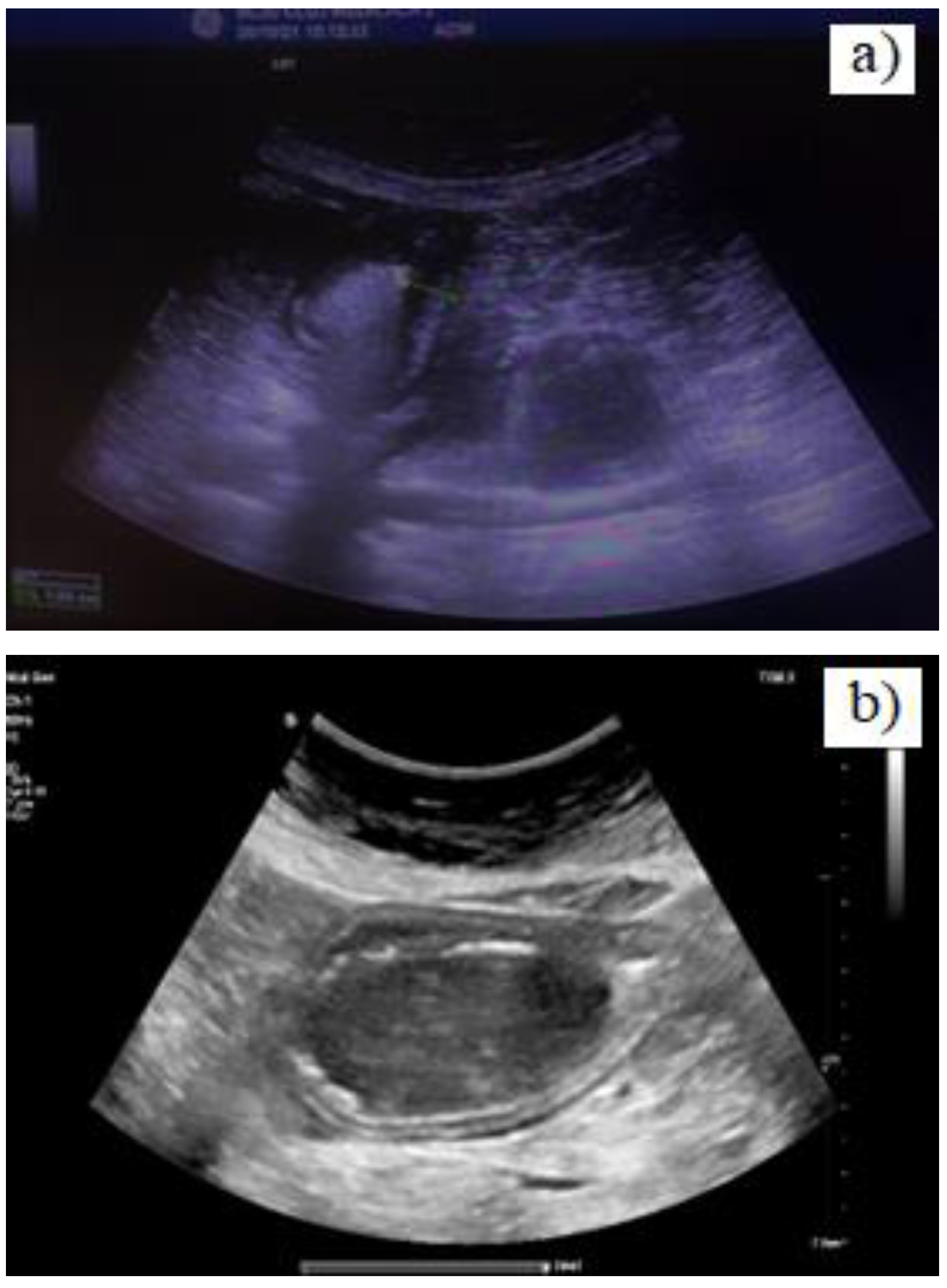
Figure 1. Grey-scale US; a). increased gastric wall thickness and inhomogeneous content; b). large hypoechoic intragastric mass, relatively homogenous, surrounded by gas (hyperechoic inner interface of the gastric wall).
Jmms 09 00037 g001
Figure 2. UGIE during EUS – giant gastric polyp with a deep central ulcer suggesting malignant transformation.
Jmms 09 00037 g002
Figure 3. EUS – large hypoechoic antral mass (a) with intense vascularization visible in Color Doppler mode (b).
Jmms 09 00037 g003
Figure 4. Contrast enhanced abdominal CT-inhomogeneous hypodense enlargement of the duodenal and antral walls.
Jmms 09 00037 g004
Figure 5. Intraoperatory and resection specimen aspects - giant antropyloric polypoid tumor.
Jmms 09 00037 g005
Figure 6. Histology: IFP-mesenchymal cells with chronic inflammatory infiltrate (lymphocytes, eosinophils) and blood vessels.
Jmms 09 00037 g006