Next Article in Journal
A Pedunculated Esophageal Chondromatous Hamartoma in a Child
Previous Article in Journal
Acute Chylous Peritonitis Due to Idiopathic Pancreatitis Mimicking Acute Appendicitis
 
 
Journal of Mind and Medical Sciences is published by MDPI from Volume 12 Issue 1 (2025). Previous articles were published by another publisher in Open Access under a CC-BY (or CC-BY-NC-ND) licence, and they are hosted by MDPI on mdpi.com as a courtesy and upon agreement with Valparaiso University (ValpoScholar).
Case Report

Postoperative Complicated Appendectomy Revealing Crohn’s Disease in a Pediatric Patient

by
Augustina Enculescu
1,*,
Mircea Denis Lupusoru
2,
Catalin Cirstoveanu
3,
Andra-Iulia Suceveanu
4,
Liliana Florina Andronache
3,
Adrian-Paul Suceveanu
4,
Felix Voinea
4 and
Maria Puscasu
5
1
Department of Pathology, Emergency Clinic Hospital for Children Marie Sklodowska Curie, Bucharest, Romania
2
Department of Physiology, Carol Davila University of Medicine and Pharmacy, Bucharest, Romania
3
Faculty of Medicine, Carol Davila University of Medicine and Pharmacy, Bucharest, Romania
4
Department of Gastroenterology, Faculty of Medicine, Ovidius University, Constanta, Romania
5
Department of Pediatric Surgery, Emergency Clinic Hospital for Children Marie Sklodowska Curie, Bucharest, Romania
*
Author to whom correspondence should be addressed.
J. Mind Med. Sci. 2021, 8(1), 154-160; https://doi.org/10.22543/7674.81.P154160
Submission received: 14 December 2020 / Revised: 10 February 2021 / Accepted: 10 February 2021 / Published: 8 April 2021

Abstract

:
Granulomatous appendicitis is a very rare entity in children. It is related to Crohn’s disease in only 5–10% of the cases. The diagnosis of Crohn’s disease is a real challenge in the pediatric population, especially when its initial manifestation is acute appendicitis. Herein, we present a 13-year-old boy admitted for acute appendicitis. The histopathological examination was conclusive for acute granulomatous appendicitis. The development of an entero-cutaneous fistula has complicated the postoperative evolution. The failure of the conservative management required ileocecal resection. The pathological examination revealed Crohn’s disease. The one-year follow-up on the patient din not reveal any complications. We strongly support the importance of the histopathological examination of the entirely removed appendix. Careful assessment of granulomatous appendicitis is needed in order to establish its possible etiology and to initiate the adequate treatment.

Introduction

The retrospective histopathological research shows that granulomatous appendicitis (GA) is a rare condition, responsible for a percentage that ranges from 0.1-0.3% to 2.3% of all cases of appendicitis, regardless the age of the patients [1]. For many years, this type of appendicitis has been considered an initial manifestation of Crohn’s disease [2], but recent studies have shown that only 5 to 10% of the patients with granulomatous appendicitis develop an inflammatory bowel disease [3]. A recent review of 200 published cases of granulomatous appendicitis in the English literature reveals that only 17 (8.5%) of them were children [4]. A large study on 632 appendectomy specimens showing acute appendicitis found isolated granulomatous appendicitis in only 9 cases, with age ranging from 15 to 38 years. The postoperative complications of these patients were minor, no further surgical interventions being necessary for other complications [5]. The post-appendectomy enterocutaneous (EC) fecal fistula is known as a very rare major complication. A large study on open appendectomy conducted by Genier F et al. between 1970 to 1993 shows an occurrence of EC fecal fistulas of 0.133%, which also illustrates a link between a severe form of appendicitis (suppurative, gangrenous, or perforated) involving the adjoining cecum wall and these sequelae [6]. This complication is also known as a challenge for surgeons operating on intestinal malignancies or inflammatory bowel disease, and the doctors should evaluate closely if the fistula closes alone, or surgical intervention is needed [7].
We present the case of a pediatric patient, with no previous medical history, who underwent surgery for acute appendicitis in our clinic. The postoperative course was complicated by the entero-cutaneous fistula, a rare event following open appendectomy. We bring into discussion the surgical and the diagnostic (clinical and histopathological) aspects during the evolution of this patient who was diagnosed with Crohn’s disease upon this series of events.

Case report

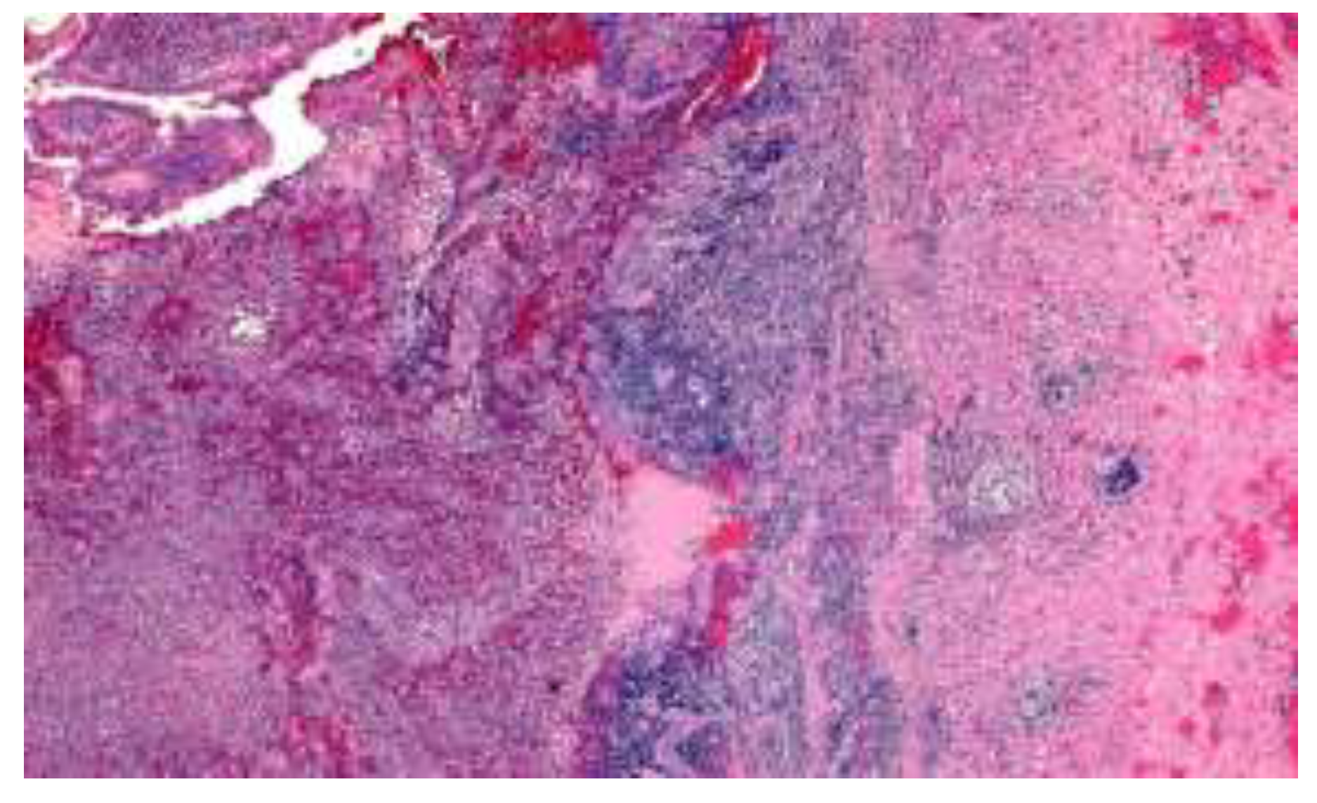
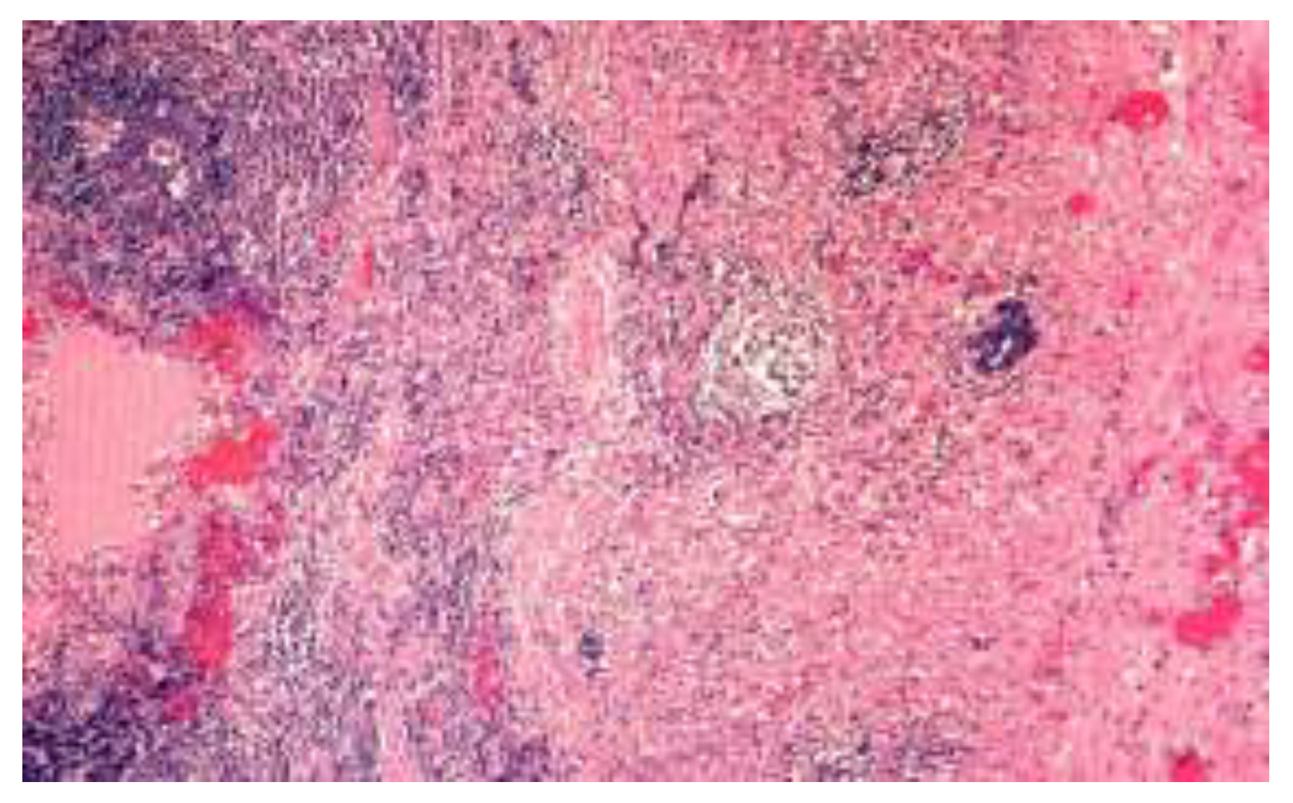
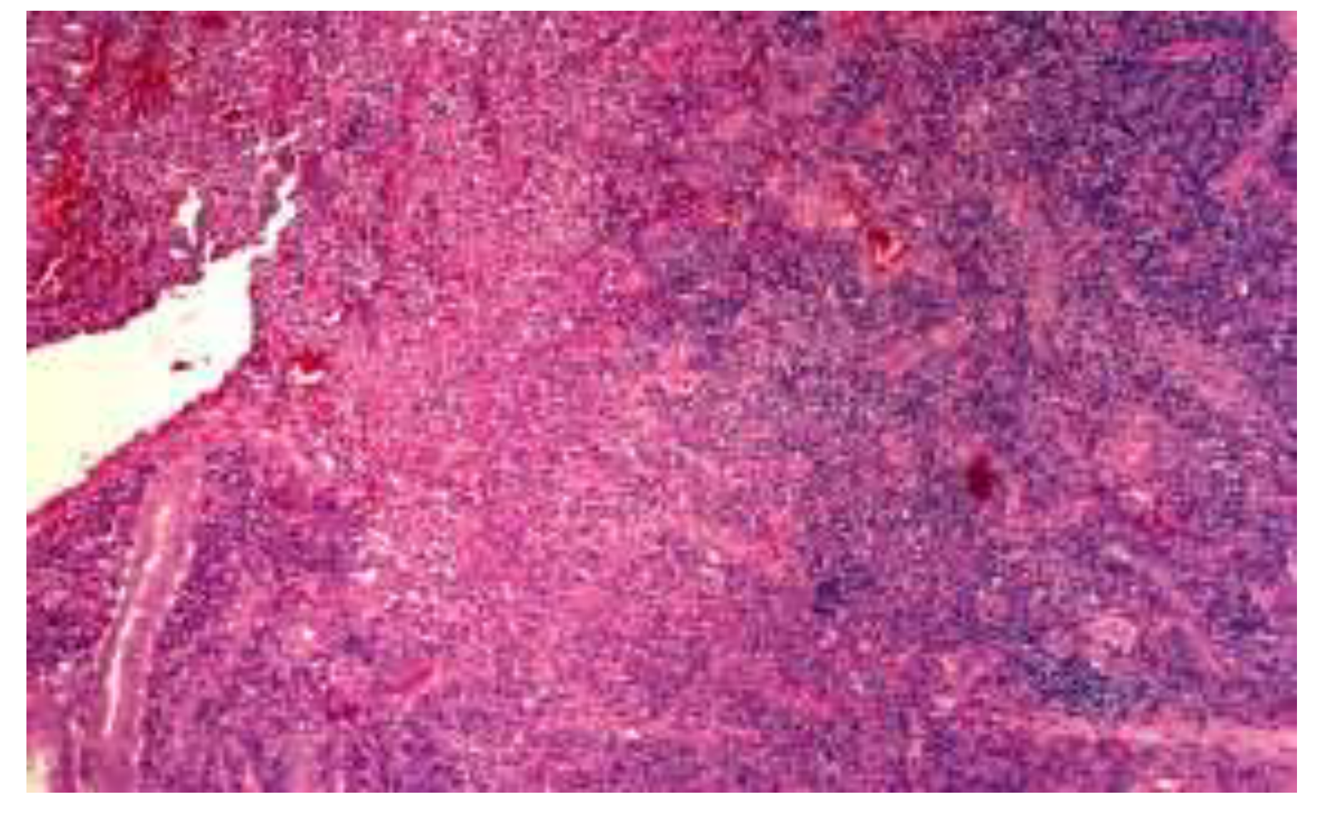
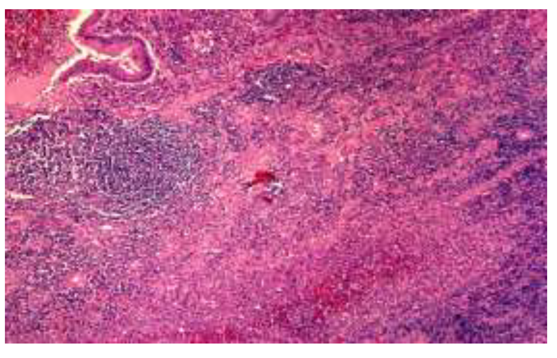
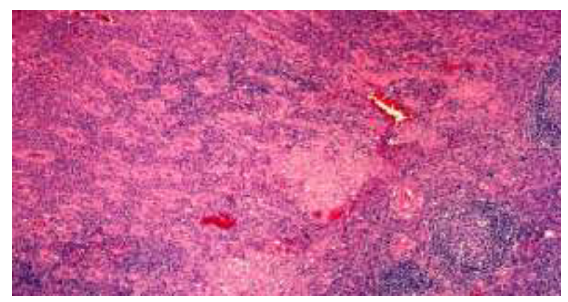
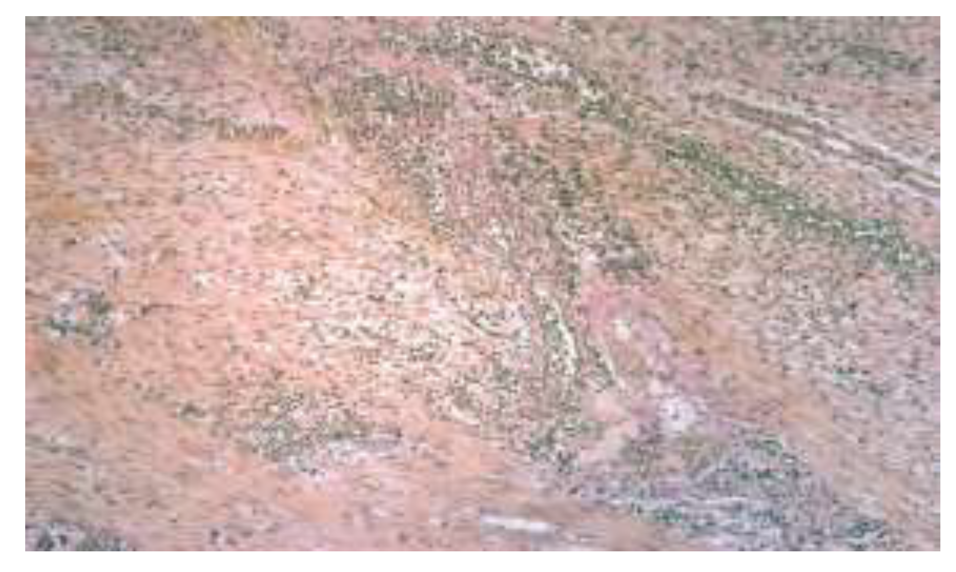
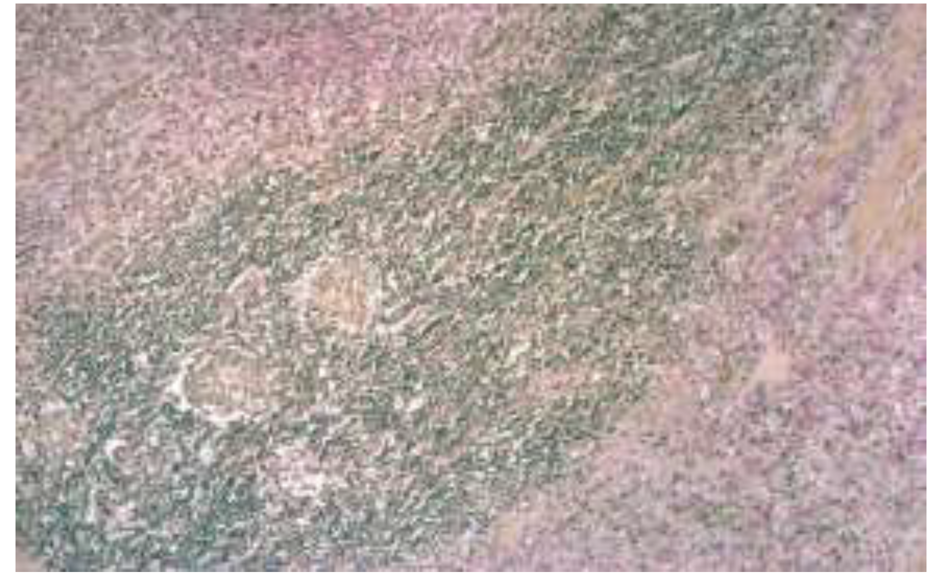
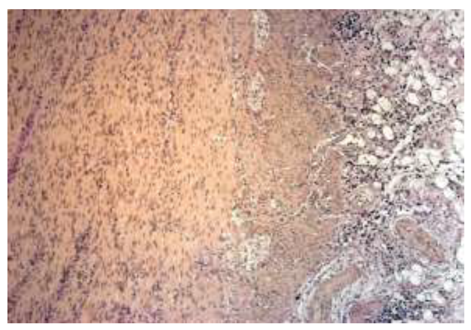
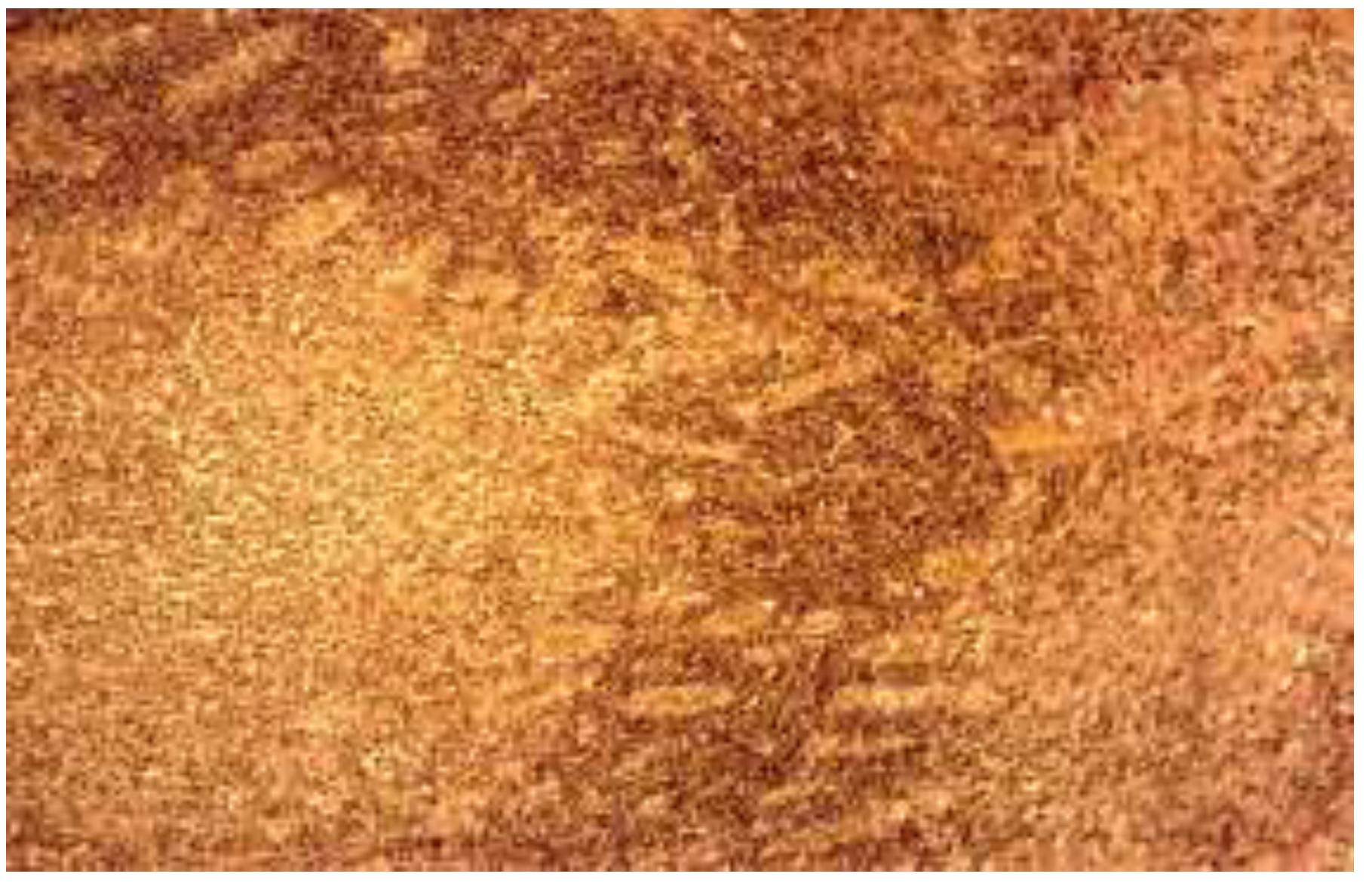
A 13-year-old boy, without any surgical history, has been referred to us for right lower quadrant abdominal pain, nausea and vomiting lasting for three days. He saw a pediatrician for recurrent epigastric pain 7 months before and underwent an abdominal ultrasonography that revealed an enlarged right hepatic lobe and mild hepatic steatosis. He had no remarkable family history. The clinical examination revealed a fixed palpable mass in the RLQ, local tenderness and rebound pain. The blood tests showed leukocytosis (14.850/microL) and no shift to the left of the neutrophils. Liver enzymes, pancreatic enzymes and bilirubin measurements were within normal range. The abdominal ultrasonography performed on the admission day showed a thickened wall of the terminal ileum and an inflamed appendix. The preoperative diagnosis – based on the previous listed findings – was acute appendicitis. After the informed consent was signed by the patient’s parents [8], we found an apparently gangrenous appendix intraoperatively, with a highly thickened and friable wall and multiple adhesions with distal ileal loops, cecum and the greater omentum. Local hyperemia and the thickening of the cecum were also noted and a relatively low quantity of serosanguinous fluid was present locally. Open appendectomy was performed, followed by a postoperative triple antibiotic therapy (ceftriaxone, gentamicin, metronidazole). The patient was discharged 5 days postoperatively. The histopathological examination of the appendix revealed mucosal ulcerations, transmural mixed inflammatory infiltrate with lympho-plasmocytes, neutrophils and eosinophils, smooth muscle disarray and the involvement of the appendicular serosa; epithelioid granulomas were noticed in the appendicular wall. The diagnosis was acute granulomatous appendicitis with periappendicitis (Figure 1, Figure 2, Figure 3, Figure 4, Figure 5, Figure 6 and Figure 7, the archive of the Pediatric Surgery Department, The Emergency Clinical Hospital for Children Marie S. Curie).
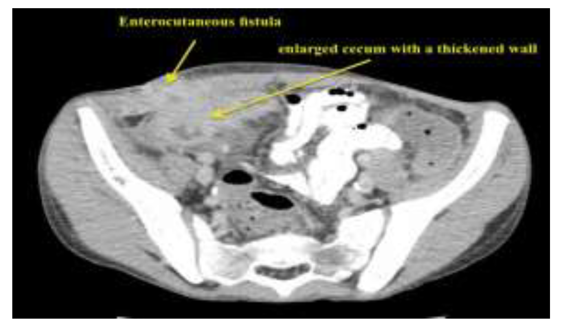
The results of the stool cultures were negative. Two weeks later, the patient developed an entero-cutaneous fistula (Figure 8).
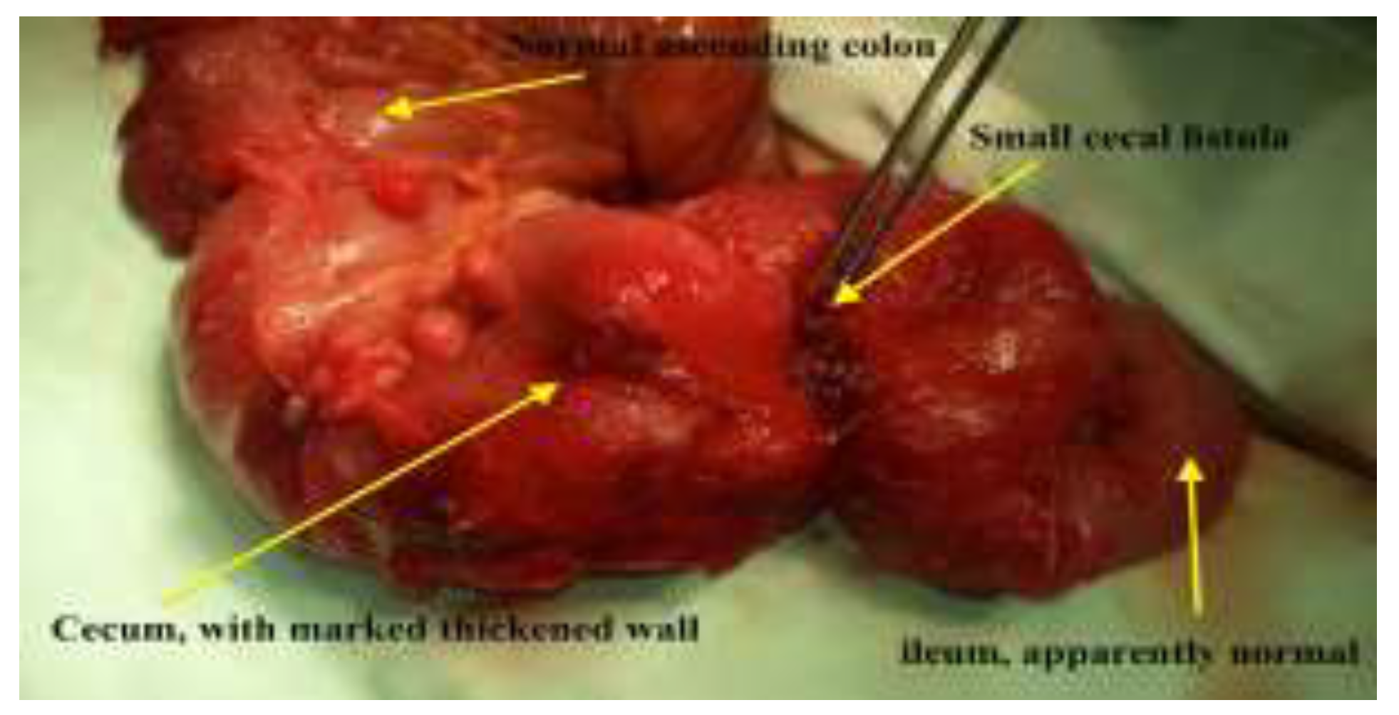
The conservatory management of the fistula was initiated, but the surgical intervention was reconsidered after one month. The intraoperative findings were a highly thickened cecal wall (a tumor-like aspect) with a small cecal orifice at the base level of the appendix (Figure 9).
The evaluation of the peritoneal cavity did not reveal any other abnormalities. The resection of the cecum and the terminal ileum were performed, followed by the anastomosis of the apparently healthy ileum and ascending colon. The macroscopic evaluation of the resected and sectioned terminal ileum and cecum revealed the marked thickened wall of the cecum and the presence of the fistulous orifice at the appendicular base level (Figure 10).
Microscopically, both the ileal and cecal walls revealed mucosal ulcerations, prominent fissures, hyperemia and edema, lamina propria and submucosal lymphoid hyperplasia, transmural mixed inflammatory infiltrate; non-necrotic epithelioid granulomas with multinucleated giant cells were scattered in submucosal and serous layers. The mesenteric lymph nodes had features of lymphoid hyperplasia and a few epithelioid granulomas. The pathological report concluded in favor of Crohn’s disease (Figure 11, Figure 12, Figure 13, Figure 14 and Figure 15).
The postoperative evolution was uneventful. He was referred to the Gastroenterology Department for further management; the fecal calprotectin level was repeatedly within the normal range. The patient is symptom-free after one-year follow-up with the appropriate medical management of CD and he did not have any complications.

Discussions

Crohn’s disease is a multifactorial inflammatory bowel disease, which generally affects people aged between 15-30 years, but occasionally it may be encountered in children or older ages. The transmural inflammation of the digestive wall can affect all the segments from mouth to the rectum and the anus, but most cases are localized at the level of the terminal ileum and colon. Pathophysiology is still a subject of research, but there is evidence involving immune system disorders and autoimmune responses. Several studies showed that vitamin D deficiency plays an important role in immune response disorders and it is common in patients with Crohn’s disease [9,10,11,12]. Gut dysbiosis promoting a chronic low level of inflammation and metabolic syndrome are also encountered more frequently in patients with inflammatory bowel diseases, signaling a possible pathophysiological correlation [13,14,15].
Besides the digestive manifestations, an extensive array of extra-digestive features may be associated, involving a multidisciplinary follow-up: uveitis, sacroiliitis, arthritis. Women with IBD are at an increased risk of severe pre-eclampsia, preterm delivery, infants with a low Apgar score and major congenital malformations, as well as a high incidence of post-partum depression [16,17,18,19,20,21,22].
Appendicitis occurs most often in patients aged between 11-40 years, affecting about 7% of the world population. Time has passed since Claudius Amyand performed the first appendectomy in 1735, thus confirming the efficiency of surgery in treating this potentially deadly condition [23]. Recently laparoscopic approach gains increased popularity due to its quick recovery, less pain and decreased hospital stay, even now in the difficult COVID-19 pandemic era [24,25]. The multimodal approach of the patient in the perioperative period is also important in quickly identifying and treating the possible complications [26]. Meyerding and Bertram described granulomatous appendicitis for the first time in 1953 [2]. The granulomatous aspect has been reported in only 0.1-2% of all appendectomy specimens [27,28]. The defining elements of this condition – granulomas – are chronic inflammatory lesions composed of accumulations of epithelioid histiocytes occasionally accompanied by multinucleated giant cells and variably encompassed by a cuff of lymphocytes and plasma cells. Eosinophils may also be present. Granulomas may be localized at any level of the appendicular wall in GA [3].
Initially, it was thought to be a manifestation of Crohn’s disease. It has been demonstrated in time that this condition is related to Crohn’s disease in only 5-10% of the cases [3]. Using polymerase chain reaction techniques, Lamps et al. showed that the infection with Yersinia enterocolitica and/or Yersinia pseudotuberculosis occurs in 25% of the cases of granulomatous appendicitis [27]. Guo and Greenson [28] demonstrated that over 50% of the patients suffering from delayed (interval) appendectomy, consequently to a prior conservatory treatment of subacute/recurrent appendicitis, develop a granulomatous aspect of the appendix. This is perceived as a secondary inflammatory response. Additional causes of granulomatous appendicitis include different other infectious agents (Enterobius vermicularis, Mycobacterium tuberculosis, Candida, Treponema pallidum, Actinomyces, Bartonella henselae, the measles virus, etc.), foreign body reactions, and sarcoidosis [3].
Some histologic details may suggest an infectious cause of GA: large numbers of granulomas, very large granulomas, necrosis and the central suppuration or the micro-abscess formation within granulomas. However, many investigators state that it is impossible to distinguish between different appendicular granulomas determined by various causes [3,29]. Nevertheless, there are several lesions accepted to be typical of the Crohn’s disease of the appendix:
the focal or the discontinuous inflammation of the mucosa with or without the lengthening and distortion of the crypts, with predomination of histiocytes and lymphocytes in the inflammatory infiltrate.
epithelioid granulomas.
the presence of lymphoid aggregates at the border of the muscularis propria and subserosa, associated with the infiltration of the muscular layer by mononuclear leukocytes in most cases [30].
In a retrospective study of 25 cases of appendicitis as the initial manifestation of Crohn’s disease (1.8% of all the patients with appendicitis undergoing surgery in a 9-year period of the retrospective study), Agha et al. found that Crohn’s disease determined appendicitis with concurrent inflammation of the cecum or the terminal ileum in 5 cases. Through a thorough review of the literature, they observed that Crohn’s appendicitis had been followed by the subsequent involvement of other intestinal segments in 7% of the cases [31].
In a retrospective analysis, Bass and co. state that pediatric CD presenting as appendicitis has statistically significant lower hemoglobin and mean corpuscular volume (MVC) values, and higher platelet values at the initial presentation. They studied a series of 2,718 consecutive appendectomies and found 8 patients who had subsequently developed CD. The period of time between appendectomy and the diagnosis of CD ranged from 1 to 3 months. Four of these 8 patients (50% of the cases) developed postoperative abscess and/or fistula [32].
Kambouri et al. presented the case of a child who suffered from local peritonitis as the first manifestation of CD. After the appendectomy, the patient developed two entero-cutaneous fistulae and two strictures in the ileum and underwent a right limited hemicolectomy [33], for which anastomosis could be performed either manually or using mechanical staplers [34].
Anastomotic leakage is a serious complication in colorectal surgery, being associated with a high rate of postoperative morbidity [35]. It was found that there was a 50% chance that a CD patient would never require further related surgical interventions following the resection of the ileocecal segment [36].
In our case, no systemic manifestations of Crohn’s disease, such as arthralgia, dermatologic signs, or uveitis, were previously noted, acute appendicitis being its first manifestation. We did not encounter preoperative microcytic anemia or thrombocytosis as Bass J. et al. had suggested in their study [37]. The histopathological evaluation of our appendectomy specimen was relevant for granulomatous appendicitis. The postoperative persistent EC fistula required the appropriate surgical attitude, the diagnosis of CD being made through the pathological examination of the specimen.
CD is a rare condition in children and acute appendicitis as its first manifestation is exceptional, but it should be taken into account, due to its possible serious implications [38]. The chance of treating extremely rare conditions in children is obviously higher in tertiary centers [39,40,41]. In our review of the 2,915 consecutive appendectomies routinely evaluated from a histopathological point of view in our institution over the last 4 years, this is the first case of granulomatous appendicitis. Despite the debate of routinely sending all appendicular specimens to be histopathologically evaluated [42], although the appendectomies are one of the most frequent pediatric surgical interventions [43], we strongly support the importance of the microscopic examination of all removed appendices.

Highlights

The entero-cutaneous fistula is a severe postoperative complication of appendectomy. In rare cases, its underlying cause is granulomatous appendectomy – an initial manifestation of Crohn’s disease.
The patient we present did not have any relevant history of Crohn’s disease. The histopathological examination of the appendix played an important role in the final diagnosis, accounting for the aggravating evolution of the patient who had undergone appendectomy.

Conclusions

Acute appendicitis may be the first clinical sign of the onset of Crohn’s disease in children. The careful assessment of granulomatous appendicitis is necessary in order to establish its possible etiology and thus, to initiate the appropriate treatment as soon as possible.

Abbreviations

CD = Crohn’s Disease, EC = entero-cutaneous, RLQ = right lower quadrant, GA = granulomatous appendicitis, MVC = mean corpuscular volume

Conflict of interest disclosure

There are no known conflicts of interest in the publication of this article. The manuscript was read and approved by all authors.

Compliance with ethical standards

Any aspect of the work covered in this manuscript has been conducted with the ethical approval of all relevant bodies and that such approvals are acknowledged within the manuscript.

References

  1. Blair, N.P.; Bugis, S.P.; Turner, L.J.; MacLeod, M.M. Review of the pathologic diagnoses of 2,216 appendectomy specimens. Am J Surg. 1993, 165, 618–620. [Google Scholar] [CrossRef]
  2. Meyerding, E.V.; Bertram, H.F. Nonspecific granulomatous inflammation (Crohn's disease) of the appendix; a case report. Surgery. 1953, 34, 891–894. [Google Scholar] [PubMed]
  3. Bronner, M.P. Granulomatous appendicitis and the appendix in idiopathic inflammatory bowel disease. Semin Diagn Pathol. 2004, 21, 98–107. [Google Scholar] [CrossRef]
  4. Yayla, D.; Alpman, B.N.; Dolek, Y. Granulomatous appendicitis in a 12-year-old boy. J Pediatr Surg. 2010, 45, e27-9. [Google Scholar] [CrossRef] [PubMed]
  5. Richards, M.L.; Aberger, F.J.; Landercasper, J. Granulomatous appendicitis: Crohn's disease, atypical Crohn's or not Crohn's at all? J Am Coll Surg. 1997, 185, 13–17. [Google Scholar] [CrossRef] [PubMed]
  6. Genier, F.; Plattner, V.; Letessier, E.; Armstrong, O.; Heloury, Y.; Le Neel, J.C. Fistules caecales post-appendicectomies. A propos de vingt-deux cas [Post-appendectomy fistulas of the cecum. Apropos of 22 cases]. J Chir (Paris). 1995, 132, 393–398. [Google Scholar]
  7. Orangio, G.R. Enterocutaneous fistula: medical and surgical management including patients with Crohn's disease. Clin Colon Rectal Surg. 2010, 23, 169–175. [Google Scholar] [CrossRef]
  8. Serban, D.; Spataru, R.I.; Vancea, G.; Balasescu, S.A.; Socea, B.; Tudor, C.; Dascalu, A.M. Informed consent in all surgical specialties: from legal obligation to patient satisfaction. Rom J Leg Med. 2020, 28, 317–321. [Google Scholar] [CrossRef]
  9. Ham, M.; Longhi, M.S.; Lahiff, C.; Cheifetz, A.; Robson, S.; Moss, A.C. Vitamin D levels in adults with Crohn's disease are responsive to disease activity and treatment. Inflamm Bowel Dis. 2014, 20, 856–860. [Google Scholar] [CrossRef]
  10. Fletcher, J.; Cooper, S.C.; Ghosh, S.; Hewison, M. The Role of Vitamin D in Inflammatory Bowel Disease: Mechanism to Management. Nutrients. 2019, 11, 1019. [Google Scholar] [CrossRef]
  11. Stanescu, A.M.A.; Grajdeanu, I.V.; Iancu, M.A.; Stoian, A.P.; Bratu, O.G.; Socea, B.; Socea, L.I.; Diaconu, C.C. Correlation of Oral Vitamin D Administration with the Severity of Psoriasis and the Presence of Metabolic Syndrome. Rev Chim. 2018, 69, 1668–1672. [Google Scholar] [CrossRef]
  12. Stoian, A.P.; Elian, V.; Nitipir, C.; Negoita, O.; Iancu, D.M.; Ditu, G. Association between vitamin D insufficiency and metabolic syndrome. INTERDIAB 2017: DIABETES MELLITUS IN INTERNAL MEDICINE Book Series: International Conference on Interdisciplinary Management of Diabetes Mellitus and its Complications. 2017; 232–242. [Google Scholar]
  13. Dragasevic, S.; Stankovic, B.; Kotur, N.; Sokic-Milutinovic, A.; Milovanovic, T.; Lukic, S.; Milosavljevic, T.; Srzentic Drazilov, S.; Klaassen, K.; Pavlovic, S.; Popovic, D. Metabolic Syndrome in Inflammatory Bowel Disease: Association with Genetic Markers of Obesity and Inflammation. Metab Syndr Relat Disord. 2020, 18, 31–38. [Google Scholar] [CrossRef]
  14. Suceveanu, A.I.; Stoian, A.P.; Parepa, I.; Voinea, C.; Hainarosie, R.; Manuc, D.; Nitipir, C.; Mazilu, L.; Suceveanu, A.P. Gut Microbiota Patterns in Obese and Type 2 Diabetes (T2D) Patients from Romanian Black Sea Coast Region. Rev Chim. 2018, 69, 2260–2267. [Google Scholar] [CrossRef]
  15. Yorulmaz, E.; Adali, G.; Yorulmaz, H.; Ulasoglu, C.; Tasan, G.; Tuncer, I. Metabolic syndrome frequency in inflammatory bowel diseases. Saudi J Gastroenterol. 2011, 17, 376–382. [Google Scholar] [CrossRef] [PubMed]
  16. Boyd, H.A.; Basit, S.; Harpsøe, M.C.; Wohlfahrt, J.; Jess, T. Inflammatory Bowel Disease and Risk of Adverse Pregnancy Outcomes. PLoS One. 2015, 10, e0129567. [Google Scholar] [CrossRef] [PubMed]
  17. Dimitriu, M.; Stoian, A.P.; Rosu, G.; Dan, A.; Caimacan, A.; Zygouropoulos, N.; Jacotă – Alexe, F. Postpartum Depression - a serious, dangerous, disabling, extremely frequent condition that is almost completely ignored in Romania. Mediterranean Journal of Clinical Psychology. 2020, 8, 3. [Google Scholar] [CrossRef]
  18. Merlo, E.M.; Stoian, A.P.; Motofei, I.G.; Settineri, S. Clinical Psychological Figures in Healthcare Professionals: Resilience and Maladjustment as the "Cost of Care". Front Psychol. 2020, 11, 607783. [Google Scholar] [CrossRef]
  19. Turcan, N.; Bohiltea, R.E.; Ionita-Radu, F.; Furtunescu, F.; Navolan, D.; Berceanu, C.; Nemescu, D.; Cirstoiu, M.M. Unfavorable influence of prematurity on the neonatal prognostic of small for gestational age fetuses. Exp Ther Med. 2020, 20, 2415–2422. [Google Scholar] [CrossRef]
  20. Grigoriu, C.; Furtunescu, F.; Gaman, L.E.; Balaceanu, A.L.; Buzatu, R.; Grigoriu, M. A Biochemical Marker of Preeclampsia - Hypertriglyceridemia - and the Impact of Secondary Prevention Therapy. Rev Chim. 2018, 69, 2740–2743. [Google Scholar] [CrossRef]
  21. Bandoli, G.; Chambers, C.D. Autoimmune conditions and comorbid depression in pregnancy: examining the risk of preterm birth and preeclampsia. J Perinatol. 2017, 37, 1082–1087. [Google Scholar] [CrossRef]
  22. Yu, A.; Friedman, S.; Ananthakrishnan, A.N. Incidence and Predictors of Flares in the Postpartum Year Among Women With Inflammatory Bowel Disease. Inflamm Bowel Dis. 2020, 26, 1926–1932. [Google Scholar] [CrossRef]
  23. Brănescu, C.; Serban, D.; Dascălu, A.M.; Oprescu, S.M.; Savlovschi, C. Interleukin 6 and lipopolysaccharide binding protein - markers of inflammation in acute appendicitis. Chirurgia (Bucur). 2013, 108, 206–214. [Google Scholar] [PubMed]
  24. Serban, D.; Smarandache, C.G.; Tudor, C.; Duta, L.N.; Dascalu, A.M.; Aliuș, C. Laparoscopic Surgery in COVID-19 Era-Safety and Ethical Issues. Diagnostics (Basel). 2020, 10, 673. [Google Scholar] [CrossRef]
  25. Suceveanu, A.I.; Mazilu, L.; Suceveanu, A.P.; et al. Assertion for Montelukast in the COVID-19 pandemics? Farmacia. 2020, 68, 579–585. [Google Scholar] [CrossRef]
  26. Tănăsescu, C.; Faur, M.; Sabău, D. Day-case Surgery in the Context of Inguinal Hernia Repair by the Modified Lichtenstein Technique - A Single Centre Experience. Chirurgia (Bucur). 2019, 114, 115–120. [Google Scholar] [CrossRef] [PubMed]
  27. Lamps, L.W.; Madhusudhan, K.T.; Greenson, J.K.; Pierce, R.H.; Massoll, N.A.; Chiles, M.C.; Dean, P.J.; Scott, M.A. The role of Yersinia enterocolitica and Yersinia pseudotuberculosis in granulomatous appendicitis: a histologic and molecular study. Am J Surg Pathol. 2001, 25, 508–515. [Google Scholar] [CrossRef]
  28. Guo, G.; Greenson, J.K. Histopathology of interval (delayed) appendectomy specimens: strong association with granulomatous and xanthogranulomatous appendicitis. Am J Surg Pathol. 2003, 27, 1147–1151. [Google Scholar] [CrossRef]
  29. Huang, J.C.; Appelman, H.D. Another look at chronic appendicitis resembling Crohn's disease. Mod Pathol. 1996, 9, 975–981. [Google Scholar]
  30. Stangl, P.C.; Herbst, F.; Birner, P.; Oberhuber, G. Crohn's disease of the appendix. Virchows Arch. 2002, 440, 397–403. [Google Scholar] [CrossRef]
  31. Agha, F.P.; Ghahremani, G.G.; Panella, J.S.; Kaufman, M.W. Appendicitis as the initial manifestation of Crohn's disease: radiologic features and prognosis. AJR Am J Roentgenol. 1987, 149, 515–518. [Google Scholar] [CrossRef]
  32. Bass, J.A.; Goldman, J.; Jackson, M.A.; Gasior, A.C.; Sharp, S.W.; Drews, A.A.; Saunders, C.J.; St Peter, S.D. Pediatric Crohn disease presenting as appendicitis: differentiating features from typical appendicitis. Eur J Pediatr Surg. 2012, 22, 274–278. [Google Scholar] [CrossRef]
  33. Kambouri, K.; Gardikis, S.; Agelidou, M.; Vaos, G. Local peritonitis as the first manifestation of Crohn's disease in a child. J Indian Assoc Pediatr Surg. 2014, 19, 100–102. [Google Scholar] [CrossRef] [PubMed]
  34. Spataru, R. The use of mechanical suture in the treatment of Hirschsprung's disease: experience of 17 cases. Chirurgia (Bucur). 2014, 109, 208–212. [Google Scholar]
  35. Alius, C.; Tudor, C.; Badiu, C.D.; Dascalu, A.M.; Smarandache, C.G.; Sabau, A.D.; Tanasescu, C.; Balasescu, S.A.; Serban, D. Indocyanine Green-Enhanced Colorectal Surgery-between Being Superfluous and Being a Game-Changer. Diagnostics (Basel). 2020, 10, 742. [Google Scholar] [CrossRef]
  36. Kim, N.K.; Senagore, A.J.; Luchtefeld, M.A.; MacKeigan, J.M.; Mazier, W.P.; Belknap, K.; Chen, S.H. Long-term outcome after ileocecal resection for Crohn's disease. Am Surg. 1997, 63, 627–633. [Google Scholar]
  37. Spataru, R.I.; Martius, E.; Ivan, L.E.; Sirbu, D.; Hostiuc, S. Pseudomembranous colitis complicating the natural course of Crohn's disease in a pediatric patient. Rom J Legal Med. 2014, 22, 161–166. [Google Scholar] [CrossRef]
  38. Spataru, R.I.; Sirbu, A.; Sirbu, D. Forensic ramifications in diagnosing and treating high forms of the Hirschsprung's disease. Rom J Leg Med. 2013, 21, 105–110. [Google Scholar] [CrossRef]
  39. Suciu, N.; Serban, A.; Toader, O.; Oprescu, D.; Spataru, R.I. Case report of fetal lingual tumor - perinatal care and neonatal surgical intervention. J Matern Fetal Neonatal Med. 2014, 27, 314–319. [Google Scholar] [CrossRef] [PubMed]
  40. Motofei, I.G.; Rowland, D.L.; Georgescu, S.R.; Tampa, M.; Paunica, S.; Constantin, V.D.; Balalau, C.; Manea, M.; Baleanu, B.C.; Sinescu, I. Post-Finasteride Adverse Effects in Male Androgenic Alopecia: A Case Report of Vitiligo. Skin Pharmacol Physiol. 2017, 30, 42–45. [Google Scholar] [CrossRef]
  41. Spataru, R.I.; Popoiu, M.C.; Ivanov, M. Foregut duplication cyst associated with esophageal atresia-one-stage neonatal surgical repair. Indian J Surg. 2015, 77 (Suppl 1), 52–55. [Google Scholar] [CrossRef]
  42. Jones, A.E.; Phillips, A.W.; Jarvis, J.R.; Sargen, K. The value of routine histopathological examination of appendicectomy specimens. BMC Surg. 2007, 7, 17. [Google Scholar] [CrossRef] [PubMed]
  43. Bischoff, A.; Gupta, A.; D'Mello, S.; Mezoff, A.; Podberesky, D.; Barnett, S.; Keswani, S.; Frischer, J.S. Crohn's disease limited to the appendix: a case report in a pediatric patient. Pediatr Surg Int. 2010, 26, 1125–1128. [Google Scholar] [CrossRef] [PubMed]
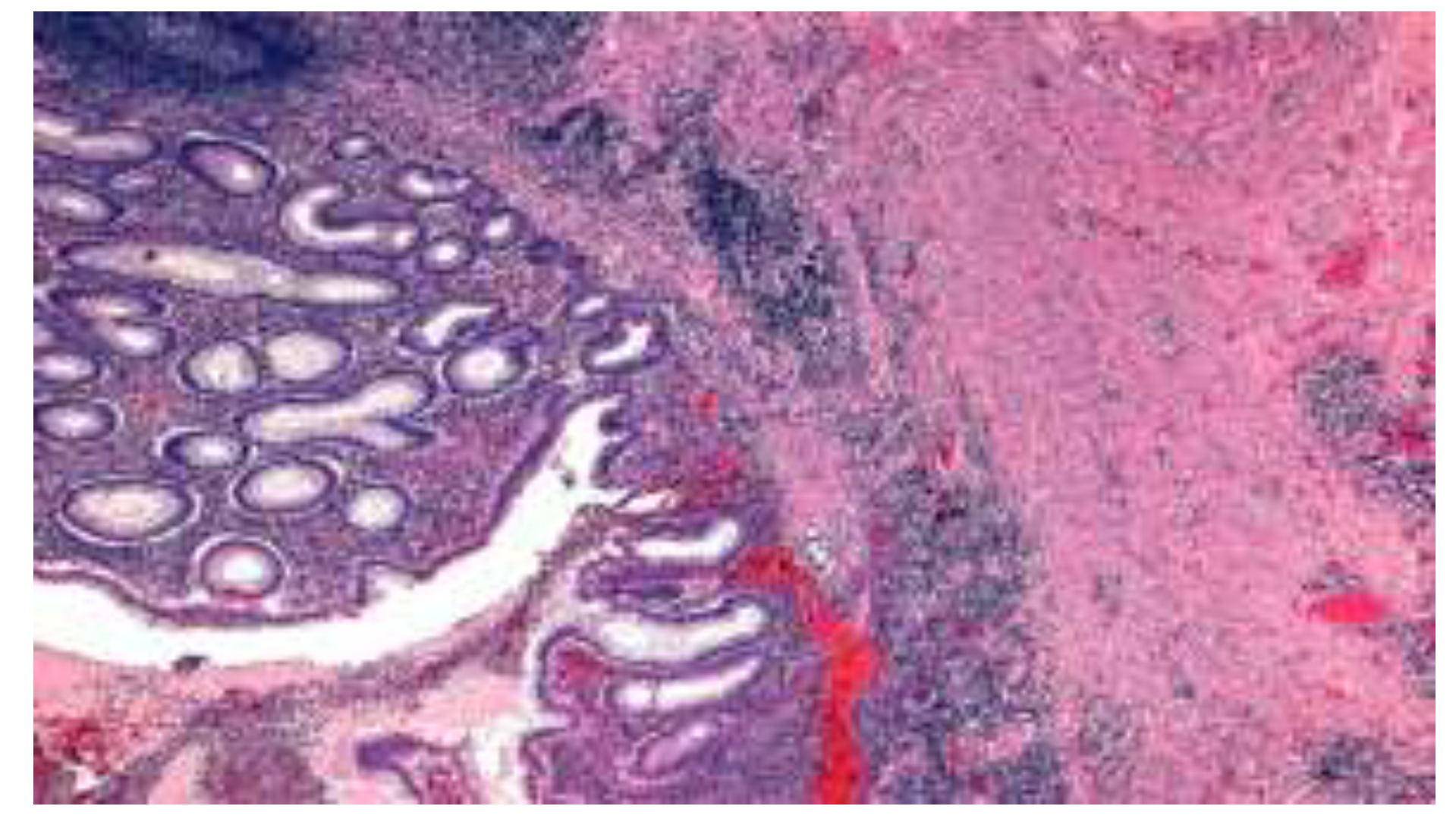
Figure 1. Phlegmonous acute appendicitis with mixed inflammatory infiltrate and lymphoid aggregates; Hematoxylin and eosin staining; Ob. 10X (the archive of the Pediatric Surgery Department, The Emergency Clinical Hospital for Children Marie S. Curie).
Jmms 08 00020 g001
Figure 2. Appendix with mucosal ulceration and epithelioid granuloma in the submucosa; Hematoxylin and eosin staining; Ob. 10X (the archive of the Pediatric Surgery Department, The Emergency Clinical Hospital for Children Marie S. Curie).
Jmms 08 00020 g002
Figure 3. Detail: epithelioid granuloma; Hematoxylin and eosin staining; Ob. 20X.
Jmms 08 00020 g003
Figure 4. Detail: Ulcers in the appendicular wall and granulation tissue; Hematoxylin and eosin staining; Ob. 20X.
Jmms 08 00020 g004
Figure 5. Appendix with mucosal ulcers, mixed inflammatory infiltrate, and abscess; Hematoxylin and eosin staining; Ob. 10X.
Jmms 08 00020 g005
Figure 6. Appendix: polymorph inflammatory infiltrate in muscular layer, with abundant neutrophils and eosinophils; Hematoxylin and eosin staining; Ob. 20X.
Jmms 08 00020 g006
Figure 7. Appendix: epithelioid granulomas in the wall; Hematoxylin and eosin staining; Ob. 20X.
Jmms 08 00020 g007
Figure 8. The CT-scan revealing an enlarged cecum with a thickened wall and an entero-cutaneous fistula.
Jmms 08 00020 g008
Figure 9. The intraoperative aspect illustrating a marked thickening of the cecal wall, a small orifice located at the appendicular base, and apparently normal ascending colon and terminal ileum.
Jmms 08 00020 g009
Figure 10. The resected terminal ileum and the sectioned cecum, revealing a pseudo-tumoral aspect of the cecum and the presence of the fistulous orifice at the appendicular base level.
Jmms 08 00020 g010
Figure 11. Colon with mucosal ulcers and granulation tissue; Van Gieson staining; Ob.10X.
Jmms 08 00020 g011
Figure 12. Colon with chronic transmural inflammatory infiltrate; Van Gieson staining; Ob.10X.
Jmms 08 00020 g012
Figure 13. Colon: epithelioid granulomas in the submucosal and muscular layer; Van Gieson staining; Ob.20X.
Jmms 08 00020 g013
Figure 14. Colon: muscular and serosa layers with chronic inflammatory infiltrate; Van Gieson staining; Ob.10X.
Jmms 08 00020 g014
Figure 15. Colonic wall with fissure and ulceration; Van Gieson staining; Ob.20X.
Jmms 08 00020 g015