Next Article in Journal
Postoperative Complicated Appendectomy Revealing Crohn’s Disease in a Pediatric Patient
Previous Article in Journal
The Crosstalk Between Insulin Resistance, Systemic Inflammation, Redox Imbalance and the Thyroid in Subjects with Obesity
 
 
Journal of Mind and Medical Sciences is published by MDPI from Volume 12 Issue 1 (2025). Previous articles were published by another publisher in Open Access under a CC-BY (or CC-BY-NC-ND) licence, and they are hosted by MDPI on mdpi.com as a courtesy and upon agreement with Valparaiso University (ValpoScholar).
Case Report

Acute Chylous Peritonitis Due to Idiopathic Pancreatitis Mimicking Acute Appendicitis

by
Dan Bratu
1,
Alin Mihetiu
1,*,
Dragoș Virgil Davițoiu
2,
Vlad Dumitru Băleanu
3,
Liliana Florina Andronache
2 and
Oana Popescu
1
1
Lucian Blaga University of Sibiu, Emergency County Hospital of Sibiu, 550245 Sibiu, Romania
2
Faculty of General Medicine, Carol Davila University of Medicine and Pharmacy, Bucharest, Romania
3
Department of General Surgery, St. Pantelimon Hospital, Bucharest, Romania
*
Author to whom correspondence should be addressed.
J. Mind Med. Sci. 2021, 8(1), 149-153; https://doi.org/10.22543/7674.81.P149153
Submission received: 12 November 2020 / Revised: 25 February 2021 / Accepted: 25 February 2021 / Published: 8 April 2021

Abstract

:
The acute development and the accumulation of chyle into the peritoneal space is a rare condition and one needs to be able to distinguish it from chylous ascites, which is most commonly chronic and associated with related diseases. It is frequently idiopathic and additionally, clinical and imaging findings are nonspecific. In almost all cases, the diagnosis is made intraoperatively during laparoscopy or laparotomy, most diagnostic procedures being indicated by acute complaints pertaining to the abdomen. A 23-year-old man was admitted to the Emergency Department with abdominal pain, with right iliac fossa tenderness and peritonism. Laparoscopy was performed due to acute abdomen findings. Further exploration revealed chylous effusion with milky-like fluid. The peritoneal lavage and the insertion of drains were subsequent to the careful inspection of the cavity. The biochemical analysis of the peritoneal fluid was an important aid which provided the diagnosis of chylous peritonitis due to acute pancreatitis.

Introduction

Acute chylous peritonitis has rarely been reported in the literature (fewer than 100 described cases). The accumulation of milky or creamy peritoneal fluid with elevated levels of triglycerides, occurs as a result of the disruption of the lymphatic system. Commonly seen etiologies of this condition are congenital abnormalities, traumatic and inflammatory causes, cirrhosis or intraabdominal malignancy. A sudden outpour of chyle may reflect in the acuteness of the symptoms of the abdominal process. Chylous peritonitis, as a rare event, is usually diagnosed during the surgical procedures which can rarely relieve the location of the disrupted lymphatic channel [1,2,3].

Case Report

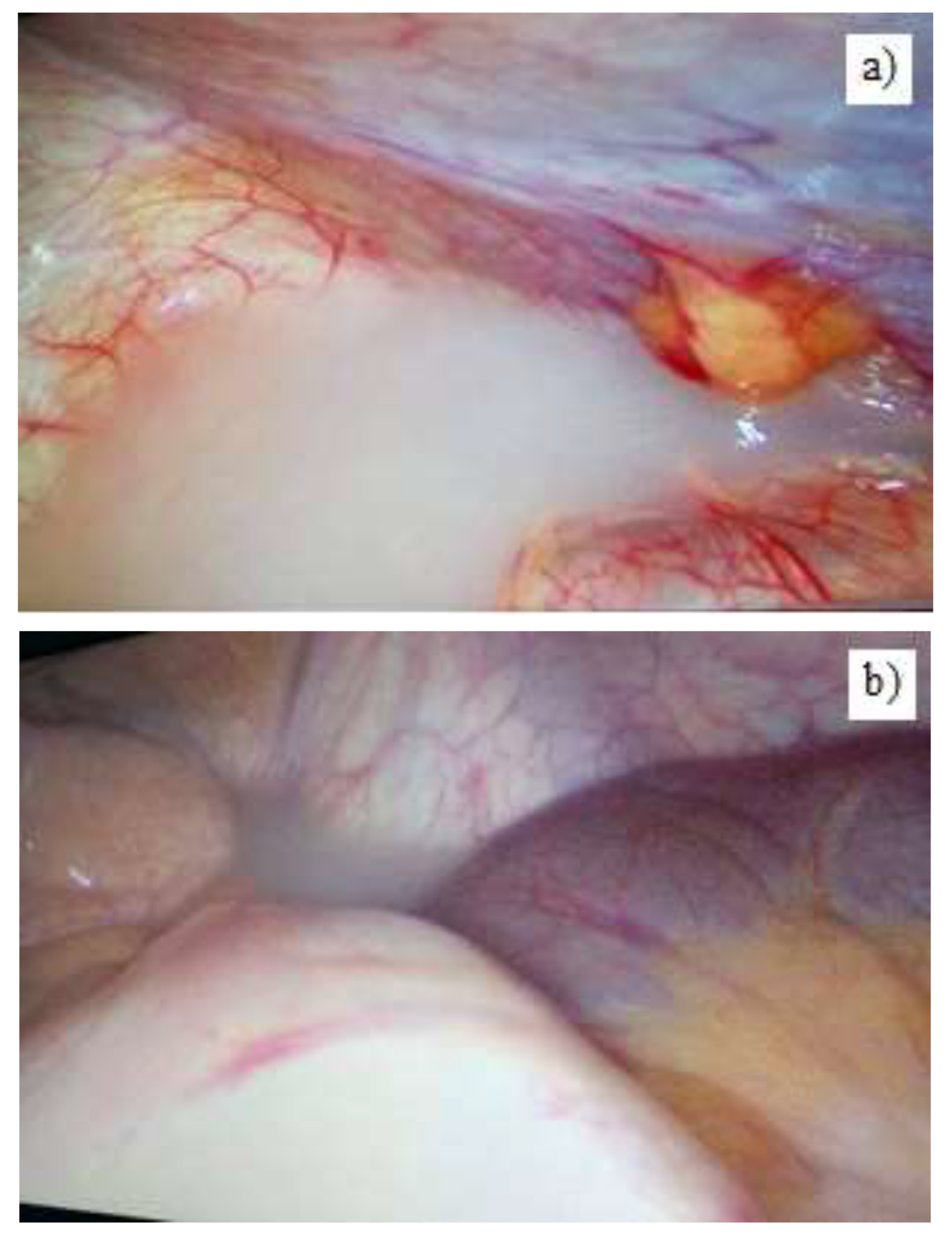
A 23-year-old patient was seen with complaints of approximately 4 hours’ duration, chief of which was the epigastric abdominal pain that migrated in the right iliac fossa, with no other abdominal problem at the clinical evaluation. The past history was not relevant. Hematology and biochemistry investigation were as follows: leukocytosis, 11.75 x 10⁶ /L; neutrophilia, 76.3%. Amylase was elevated at 110 U/L (the normal range being between 28-100 U/L). He denied alcohol consumption and did not have any history of previous abdominal trauma. The CT scans revealed a minimal to moderate amount of fluid collection with no further parenchymal changes. The informed consent was obtained after presenting the presumable preoperative diagnosis and therapeutic alternatives to the patient [2]. Suspecting peritonitis secondary to acute appendicitis, we performed an exploratory laparoscopy on an emergency basis that revealed milky fluid, having a thick, creamy appearance in the subhepatic and perisplenic space, interhepatophrenic area, and the pouch of Douglas (Figure 1).
The abdominal cavity was carefully inspected, but there was no specific sign related to the source of chylous extravasation, acute inflammatory changes or acute appendicitis. At the level of gastrocolic ligament, an area of infiltration and impregnation of the appearance of the peritoneal fluid is highlighted. Abundant lavage and adequate drainage were performed. At the cytological examination of smears from the harvested chylous liquid, low cellularity was highlighted consisting of small lymphocytes, granulocytes, neutrophils and rare mesothelial cells. The biochemical analysis of peritoneal fluid yielded elevated values of triglycerides, 1,348mg/dL (normal range: 50-160 mg/dL); and amylase, 444 mg/dL (normal range below 100 mg/dL). Laboratory tests were helpful in ruling out tuberculous peritonitis.
A tentative diagnosis of chylous peritonitis was made, associated with a recognized but rare cause of chylous effusion: idiopathic pancreatitis. He received somatostatin analogue therapy with subcutaneous administration, 100 mcg/ml, 3 times a day. The patient tolerated parenteral nutrition and gradually turned to per os fat-free diet. Initially, the postoperative drainage amount was 150 cc/24h and decreased thereafter to 75 cc, 25 cc per day, respectively 0 cc on the 5th postoperative day. A low-fat diet was recommended and he was discharged on the 8th postoperative day. There were no problems or recurrences at the 6 months and 1-year postoperative follow-up.

Discussions

Chylous ascites represents the extravasation of lymphatic fluid within the peritoneal cavity, its incidence having been reported at 1/20,000 of the admitted cases. If the accumulation occurs rapidly, the patient may have representative symptoms and signs for acute abdomen leading to the so-called “chylous peritonitis” [1].
The first case of chylous peritonitis was described by Murphy in 1887. In 1999, Fazili reported the existence of 3 cases of chylous peritonitis associated with the clinical picture of acute appendicitis. Two of the patients underwent laparoscopic appendectomy and midline laparotomy was performed for the third patient. Moreover, the etiological factors remained unknown. Vettoretto et al. discussed the existence of 71 cases in the literature up to 2008 [1,2,3,4].
Acute pancreatitis is uncommonly known to cause chylous ascites and the association with peritonism was rarely found. In 1984, Goldfarb reported the first case of acute chylous peritonitis secondary to acute pancreatitis. Since then, only few cases have been recorded [5,6].
Between 2008 and 2020, a number of 12 cases of chylous ascites had been reported on PubMed and the diagnosis was misleading in 5 of them due to a clinical resemblance to appendicitis: three of them had acute pancreatitis as an underlying pathology and one of them was presented as chylous peritonitis due to pancreatitis mimicking acute appendicitis, this pathological entity being a rare, but a significant cause of medical errors in emergency surgery [7,8,9,10].
In adults, chylous ascites is most frequently associated with surgical procedures such as lymph node dissection or aortic surgery [11,12,13]. In cases of refractory lymphatic leakage, indocyanine green may be injected at the level of the previous lymphadenectomy and near infrared (NIR) assisted surgery may be useful to better visualize the lymphatic flow and identify the leakage site [13,14]. Other causes include malignant diseases (retroperitoneal lymphomas, small bowel lymphomas and angiosarcomas), hepatic cirrhosis, abdominal traumatic injuries [15,16].
Chylomicrons are absorbed in the small intestine and afterwards flow from the lymphatic capillaries into the lymphatic vessels, and then into the lymph trunks that are tributary to the receptaculum chyli (cisterna chyli) described by Pequet [17]. Cisterna chyli is joined by descending thoracic, right and left lumbar and liver lymphatic trunks contributing to the formation of the major thoracic duct, which goes through the right posterior mediastinum and eventually enters the venous system at the left subclavian vein level [12,15,18].
It was experimentally proved that the ligation of the thoracic duct is not always linked to the development of chylous ascites, which is considered to appear only in the situation of a poorly developed system of collaterals. The mechanisms by which chylous fluid occurs in acute pancreatitis are debatable: it is considered to arise from the lymphatic fluid extravasation due to increased pressure that can be caused by the pancreatic enzyme erosion or a pancreatic inflammatory process [9].
Clinical aspects in chylous ascites are usually nonspecific and can manifest during weeks or months (abdominal distension, nausea, vomiting, weight loss, loss of appetite), mimicking digestive malignancies, such as pancreatic or colorectal neoplasia. Moreover, malignancies are one of the leading causes of chylous ascites, along with cirrhosis and postsurgical lymphatic disruption. The pathological mechanism involved are lymphatic flow blockage, either by migrant malignant cells, or increased pressure in the portal circulation [19,20,21,22]. On the contrary, in its acute forms, severe abdominal pain may occur. Signs of peritoneal irritation are often maximal in the right iliac fossa, as a result of impregnation with chylous fluid of the right paracolic gutter. Pain may result from the elevated pressure in the retroperitoneum according to fluid accumulation or from irritated mesenteric serosa. Acute appendicitis can be mimicked and testing for specific biomarkers of inflammation, such as IL6, CRP and LBP, may be helpful in making the differential diagnosis [23]. Thus, the symptoms can suggest acute cholecystitis, acute mesenteric ischemia, the perforation of hollow organs or other causes of peritonitis [24,25].
Attempts to make a diagnosis are successful because of the intraoperatively fluid withdrawal and investigation. The milky, creamy, odorless fluid withdrawn from the abdomen and the biochemical evidence with triglyceride levels 2 to 8x fold greater than the plasma level (normal range below 200 mg/dl) are valuable tools for facilitating the early diagnosis. There are noticeable differences between chyliform and pseudochylous effusions considering their source and aspect determined by cellular degradation components in the context of infection or malignancy. In these cases, the triglyceride concentration is low [26,27,28,29,30,31,32].
Radiological investigations are nonspecific, but they can provide guidance by detecting lymphadenopathy or compressive tissue masses. On the CT scan, chyle’s density is similar to that of water, which is why it can be differentiated from the hemoperitoneum, but not from the ascites fluid [18,30,31,32,33,34].
Lymphoscintigraphy has the highest yield of diagnostic information and it is advantageous because of the fewer side effects. It can be repeated and can detect lymphatic leaks or obstructions. The disadvantage is that it is difficult to perform from a technical point of view. Lymphangiography is considered the gold standard in the diagnosis of lymphatic obstruction. Complications such as allergic reactions, necrotic tissue and embolism have been reported [35,36].
Dietary management involves a high protein and low-lipid diet. In some cases, when the therapeutic response is absent, total parenteral nutrition is recommended. The pharmacological treatment describes the use of Orlistat – an inhibitor of pancreatic and gastric lipase together with Somatostatin and sometimes with total parenteral nutrition [35,36,37].
Peritoneovenuous or transjugular shunting are indicated in chronic, non-submissive forms or in cases with surgical contraindication [28].
Lymphangiography with adjunctive embolization is a solution in the case of conservative management failure [37,38].
Surgery can be effective in case of disruption or obstruction of the lymphatic system, the suture ligation of the retroperitoneal fistulas being the most common surgical approach. Additionally, the use of fibrin glue leads to better results than any other surgical technique that is limited to the simple closure, significantly reducing the mortality and morbidity rates. The laparoscopic approach and the accuracy of the surgical procedure ensure a quick recovery, reducing the hospital stay and associated medical expenses, the decision to choose between the classic and the laparoscopic approach remains, according to up to date regulations, at the discretion of the surgical team [39]. For this rare pathological entity, the laparoscopic approach was the best choice, for both the diagnosis and the treatment [40].

Highlights

Chylous peritonitis, as a rare event, is usually diagnosed during the surgical procedures which can rarely relieve the location of the disrupted lymph channel.
Minimally invasive surgery is effective for both the diagnosis and the treatment and carry a low rate of complications when compared to open surgery.

Conclusions

We described a rare case of acute chylous peritonitis with underlying causes of acute pancreatic pathology that presented symptoms and signs of peritonism which clinically mimicked acute appendicitis. A minimally invasive surgical treatment was performed, proving the effectiveness of diagnostic laparoscopy that carries a low rate of complications when compared to open surgery. This condition is a diagnostic and therapeutic challenge due to the rarity encountered in the current medical and surgical practice.

Conflict of interest disclosure

There are no known conflicts of interest in the publication of this article. The manuscript was read and approved by all authors.

Compliance with ethical standards

Any aspect of the work covered in this manuscript has been conducted with the ethical approval of all relevant bodies and that such approvals are acknowledged within the manuscript.

References

  1. Vettoretto, N.; Odeh, M.; Romessis, M.; Pettinato, G.; Taglietti, L.; Giovanetti, M. Acute abdomen from chylous peritonitis: a surgical diagnosis. Case report and literature review. Eur Surg Res. 2008, 41, 54–57. [Google Scholar] [CrossRef] [PubMed]
  2. Serban, D.; Spataru, R.I.; Vancea, G.; Balasescu, S.A.; Socea, B.; Tudor, C.; Dascalu, A.M. Informed consent in all surgical specialties: from legal obligation to patient satisfaction. Rom J Leg Med. 2020, 28, 317–321. [Google Scholar] [CrossRef]
  3. Ozgüç, H.; Narmanlı, M.; Keskin, M.K. Acute chylous peritonitis: Report of a case. Int J Surg Case Rep. 2013, 4, 419–421. [Google Scholar] [CrossRef]
  4. Fazili, F.M.; Khawaja, F.I. Acute chylous peritonitis simulating acute appendicitis: A case report and review of the literature. Ann Saudi Med. 1999, 19, 236–238. [Google Scholar] [CrossRef] [PubMed]
  5. Ben-Ami, H.; Nagachandran, P.; Assalia, A.; Edoute, Y. Acute transient chylous ascites associated with acute biliary pancreatitis. Am J Med Sci. 1999, 318, 122–123. [Google Scholar] [CrossRef]
  6. Chuang, S.C.; Lee, K.T.; Wang, S.N.; Kuo, K.K.; Chen, J.S. Hypertriglyceridemia-associated acute pancreatitis with chylous ascites in pregnancy. J Formos Med Assoc. 2006, 105, 583–587. [Google Scholar] [CrossRef]
  7. Georgiou, G.K.; Harissis, H.; Mitsis, M.; Batsis, H.; Fatouros, M. Acute chylous peritonitis due to acute pancreatitis. World J Gastroenterol. 2012, 18, 1987–1990. [Google Scholar] [CrossRef]
  8. D'Amata, G.; Rega, M.; Viola, V.; Bove, V.; Simeone, P.; Baiano, G. Chyloperitoneum associated with idiopathic pancreatitis: case report and review of the literature. G Chir. 2016, 37, 167–170. [Google Scholar] [CrossRef]
  9. Smith, E.K.; Ek, E.; Croagh, D.; Spain, L.A.; Farrell, S. Acute chylous ascites mimicking acute appendicitis in a patient with pancreatitis. World J Gastroenterol. 2009, 15, 4849–4852. [Google Scholar] [CrossRef]
  10. Serban, D.; Smarandache, A.M.; Cristian, D.; Tudor, C.; Duta, L.; Dascalu, A.M. Medical errors and patient safety culture – shifting the healthcare paradigm in Romanian hospitals. Rom J Leg Med. 2020, 28, 195–201. [Google Scholar] [CrossRef]
  11. Spataru, R.I.; Sirbu, A.; Sirbu, D. Forensic ramifications in diagnosing and treating high forms of the Hirschsprung's disease. Rom J Leg Med. 2013, 21, 105–110. [Google Scholar] [CrossRef]
  12. Spataru, R. The use of mechanical suture in the treatment of Hirschsprung's disease: experience of 17 cases. Chirurgia (Bucur). 2014, 109, 208–212. [Google Scholar]
  13. Shimajiri, H.; Egi, H.; Yamamoto, M.; Kochi, M.; Mukai, S.; Ohdan, H. Laparoscopic management of refractory chylous ascites using fluorescence navigation with indocyanine green: A case report. Int J Surg Case Rep, 1: 49. [CrossRef]
  14. Alius, C.; Tudor, C.; Badiu, C.D.; Dascalu, A.M.; Smarandache, C.G.; Sabau, A.D.; Tanasescu, C.; Balasescu, S.A.; Serban, D. Indocyanine Green-Enhanced Colorectal Surgery-between Being Superfluous and Being a Game-Changer. Diagnostics (Basel). 2018, 49, 149–152. [Google Scholar] [CrossRef]
  15. Rogdakis, A.; Bouras, P.; Giannakakis, P.; et al. Spontaneous chylous peritonitis presenting as acute surgical abdomen. Hellenic J Surg 2011, 83, 166–169. [Google Scholar] [CrossRef]
  16. Kypson, A.P.; Onaitis, M.W.; Feldman, J.M.; Tyler, D.S. Carcinoid and chylous ascites: an unusual association. J Gastrointest Surg. 2002, 6, 781–783. [Google Scholar] [CrossRef]
  17. Bhardwaj, R.; Vaziri, H.; Gautam, A.; Ballesteros, E.; Karimeddini, D.; Wu, G.Y. Chylous Ascites: A Review of Pathogenesis, Diagnosis and Treatment. J Clin Transl Hepatol. 2018, 6, 105–113. [Google Scholar] [CrossRef]
  18. Al-Busafi, S.A.; Ghali, P.; Deschênes, M.; Wong, P. Chylous Ascites: Evaluation and Management. ISRN Hepatol. 2014, 2014, 240473. [Google Scholar] [CrossRef]
  19. de la Torre-Saldaña, V.A.; Cruz-Fabián, S.; Ayala-Haro, N.; et al. Chylous Ascites as Initial Manifestation of Colon Cancer. Med Int Mex. 2014, 30, 99–107. [Google Scholar]
  20. Ciocirlan, M.; Draghia, L.; Manuc, D.; et al. Nutritional status of patients with digestive cancers. In Proceedings of the Conference: 3rd International Conference on Interdisciplinary Management of Diabetes Mellitus and its Complications (INTERDIAB), Bucharest, Romania, 2–4 March 2017; Sponsor(s): Assoc Renal Metab & Nutrit Studies; AstraZeneca Diabetes; MSD Diabetes; novo nordisk; SANOFI, INTERDIAB 2017: DIABETES MELLITUS IN INTERNAL MEDICINE Book SeriesInternational Conference on Interdisciplinary Management of Diabetes Mellitus and its Complications. 2017; pp. 132–138. [Google Scholar]
  21. Draghici, T.; Negreanu, L.; Bratu, O.; et al. Paraneoplastic syndromes in digestive tumors: a review. Rom Biotechnol Lett. 2019, 24, 813–819. [Google Scholar] [CrossRef]
  22. Apikotoa, S.; Wijesuriya, R. Idiopathic acute chylous peritonitis during pregnancy, mimicking perforated acute appendicitis: A case report. Int J Surg Case Rep. 2021, 81, 105790. [Google Scholar] [CrossRef]
  23. Brănescu, C.; Serban, D.; Dascălu, A.M.; Oprescu, S.M.; Savlovschi, C. Interleukin 6 and lipopolysaccharide binding protein - markers of inflammation in acute appendicitis. Chirurgia (Bucur). 2013, 108, 206–214. [Google Scholar]
  24. Tănăsescu, C.; Faur, M.; Sabău, D. Day-case Surgery in the Context of Inguinal Hernia Repair by the Modified Lichtenstein Technique - A Single Centre Experience. Chirurgia (Bucur). 2019, 114, 115–120. [Google Scholar] [CrossRef] [PubMed]
  25. Popoiag, R.E.; Pantea-Stoian, A.; Suceveanu, A.P.; et al. The relationship between gut microbiota and spontaneous bacterial peritonitis in patients with liver cirrhosis - a literature review. J Mind Med Sci. 2019, 6, 26–30. [Google Scholar] [CrossRef]
  26. Tanasescu, C.; Patrusel, D.; Perisanu, S.; Chis, F.; Antonescu, M. Difficult case of thoraco-abdominal injuries due to a motor vehicle accident. Chirurgia (Bucur). 2011, 106, 265–268. [Google Scholar] [PubMed]
  27. Fang, F.C.; Hsu, S.D.; Chen, C.W.; Chen, T.W. Spontaneous chylous peritonitis mimicking acute appendicitis: a case report and review of literature. World J Gastroenterol. 2006, 12, 154–156. [Google Scholar] [CrossRef] [PubMed]
  28. Kiss, L.; Kiss, R.; Porr, P.J.; Nica, C.; Nica, C.; Bardac, O.; Tănăsescu, C.; Bărbulescu, B.; Bundache, M.; Ilie, S.; Maniu, D.; Zaharie, S.I.; Hulpuş, R. Pathological evidence in support of total mesorectal excision in the management of rectal cancer. Chirurgia (Bucur). 2011, 106, 347–52. [Google Scholar]
  29. Grigoriu, M.E.; Costea, R.V.; Grigoriu, C.I.; Furtunescu. Endoscopic management of choledocolithiasis related to periampullary duodenal divericula. Medical-Surgical Journal-Revista Medico-Chirurgicala. 2018, 122, 102–108. [Google Scholar]
  30. Huang, L.L.; Xia, H.H.; Zhu, S.L. Ascitic Fluid Analysis in the Differential Diagnosis of Ascites: Focus on Cirrhotic Ascites. J Clin Transl Hepatol. 2014, 2, 58–64. [Google Scholar] [CrossRef]
  31. Cárdenas, A.; Chopra, S. Chylous ascites. Am J Gastroenterol. 2002, 97, 1896–1900. [Google Scholar] [CrossRef]
  32. Hibbeln, J.F.; Wehmueller, M.D.; Wilbur, A.C. Chylous ascites: CT and ultrasound appearance. Abdom Imaging. 1995, 20, 138–140. [Google Scholar] [CrossRef]
  33. Tofolean, D.E.; Mazilu, L.; Stăniceanu, F.; Mocanu, L.; Suceveanu, A.I.; Baz, R.O.; Parepa, R.I.; Suceveanu, A.P.; Bondari, S.; Bondari, D.; Voinea, F. Clinical presentation of a patient with cutis laxa with systemic involvement: a case report. Rom J Morphol Embryol. 2015, 56, 1205–1210. [Google Scholar] [PubMed]
  34. Spataru, R.I.; Martius, E.; Ivan, L.E.; Sirbu, D.; Hostiuc, S. Pseudomembranous colitis complicating the natural course of Crohn's disease in a pediatric patient. Rom J Leg Med. 2014, 22, 161–166. [Google Scholar] [CrossRef]
  35. Widjaja, A.; Gratz, K.F.; Ockenga, J.; Wagner, S.; Manns, M.P. Octreotide for therapy of chylous ascites in yellow nail syndrome. Gastroenterology. 1999, 116, 1017–1018. [Google Scholar] [CrossRef]
  36. Motofei, I.G.; Rowland, D.L.; Georgescu, S.R.; Tampa, M.; Paunica, S.; Constantin, V.D.; Balalau, C.; Manea, M.; Baleanu, B.C.; Sinescu, I. Post-Finasteride Adverse Effects in Male Androgenic Alopecia: A Case Report of Vitiligo. Skin Pharmacol Physiol. 2017, 30, 42–45. [Google Scholar] [CrossRef]
  37. Mittleider, D.; Dykes, T.A.; Cicuto, K.P.; Amberson, S.M.; Leusner, C.R. Retrograde cannulation of the thoracic duct and embolization of the cisterna chyli in the treatment of chylous ascites. J Vasc Interv Radiol. 2008, 19 Pt 1, 285–290. [Google Scholar] [CrossRef]
  38. Benedix, F.; Lippert, H.; Meyer, F. Post-surgical lymphocutaneous fistula, chylous ascites and chylothorax--infrequent but serious complications: etiology, diagnosis and therapeutic options. Zentralbl Chir. 2007, 132, 529–538. [Google Scholar] [CrossRef] [PubMed]
  39. Serban, D.; Smarandache, C.G.; Tudor, C.; Duta, L.N.; Dascalu, A.M.; Aliuș, C. Laparoscopic Surgery in COVID-19 Era-Safety and Ethical Issues. Diagnostics (Basel). 2020, 10, 673. [Google Scholar] [CrossRef]
  40. Ozgüç, H.; Narmanlı, M.; Keskin, M.K. Acute chylous peritonitis: Report of a case. Int J Surg Case Rep. 2013, 4, 419–421. [Google Scholar] [CrossRef]
Figure 1. (a, b). Intraoperative aspect of chylous peritonitis.
Jmms 08 00019 g001