Next Article in Journal
The Molecular Mechanisms Linking Metabolic Syndrome to Endometrial and Breast Cancers
Previous Article in Journal
Postoperative Complicated Appendectomy Revealing Crohn’s Disease in a Pediatric Patient
 
 
Journal of Mind and Medical Sciences is published by MDPI from Volume 12 Issue 1 (2025). Previous articles were published by another publisher in Open Access under a CC-BY (or CC-BY-NC-ND) licence, and they are hosted by MDPI on mdpi.com as a courtesy and upon agreement with Valparaiso University (ValpoScholar).
Case Report

A Pedunculated Esophageal Chondromatous Hamartoma in a Child

by
Dan-Alexandru Iozsa
1,*,#,
Maria Puscasu
2,#,
Catalin Gabriel Cirstoveanu
3,#,
Vlad Denis Constantin
3,#,
Anca Silvia Dumitriu
4,#,
Stana Paunica
4,# and
Nicolae Sebastian Ionescu
1,#
1
Carol Davila University of Medicine and Pharmacy, Department of Pediatric Surgery, "Marie S. Curie" Emergency Clinic Hospital, Bucharest, Romania
2
Department of Pediatric Surgery, "Marie S. Curie" Emergency Clinic Hospital for Children, Bucharest, Romania
3
Faculty of General Medicine, Carol Davila University of Medicine and Pharmacy, Bucharest, Romania
4
Carol Davila University of Medicine and Pharmacy, Dan Theodorescu Hospital, Bucharest, Romania
*
Author to whom correspondence should be addressed.
#
All authors contributed equally to this work and thus share first authorship.
J. Mind Med. Sci. 2021, 8(1), 161-166; https://doi.org/10.22543/7674.81.P161166
Submission received: 11 September 2020 / Revised: 16 December 2020 / Accepted: 16 December 2020 / Published: 8 April 2021

Abstract

:
Esophageal tumors are uncommon in pediatric population and most of them are benign. Esophageal hamartomas have been reported extremely rare in children. These can present as intramural tumors of the esophageal wall or as polyps. Dysphagia is the main symptom described in their case, but other specific symptoms are also reported. Such symptoms encountered in clinical practice are represented by obstructive apnea episodes and bradycardia, poor weight gain, epigastric or retrosternal pain, hematemesis or melena, dysphonia or tracheal sounds. Diagnosis can be delayed due to the insidious onset and non-specific symptoms, therefore patient’s compliance to follow-up and broad, careful evaluation are mandatory. Preoperative imagistic assessment is extremely important for a precise definition of the tumor’s anatomical relations, especially when facing large tumors located in the posterior mediastinum. Herein we report a case of a large chondromatous polypoid hamartoma of the esophagus in a 9 years old boy, emphasizing over the diagnosis and surgical challenges we have met, along with comments on illustrative similar cases reported in the literature.

Introduction

The posterior mediastinal masses in the pediatric population have a neurogenic origin in 90% of the cases [1]. Less than 5% of all tumors detected during childhood or adolescence originate in the gastrointestinal tract, from which a small part arises from the esophagus, 70% of them being benign [2].
Regarding their configuration in relation with the esophageal lumen, esophageal polypoid lesions are very unusual endoscopic findings in children, being found by Septer et al. in only 0.14% of the 9,438 pediatric patients who underwent routine upper endoscopies [3].
Hamartomas are non-neoplastic developmental malformations. They are made of a variety of cytologically normal, mature cells and tissues representative for the anatomical region they arise from. The architectural pattern is chaotic, displaying a dominant component. Hamartomas can be found arising in any part of the body, but most of them are identified in the lungs, liver, pancreas and spleen, and extremely rarely in the esophagus, in both pediatric and adult population [4].
Because of their rarity, being reported in isolated cases or small series, literature is not consistent enough regarding the management of the suspected cases of esophageal tumors in children.
We report the case of a large pedunculated esophageal chondromatous hamartoma in a young boy. Further on, we bring into discussion the history, the clinical presentation, the work-up, the surgical approach and the histopathologic diagnosis of this extremely rare entity.

Case Report

A 9-year-old boy was admitted to our clinic for stridor and mild dysphagia. The informed consent was signed by the patient’s parents, after having discussed the reasonable disclosure and the therapeutic possibilities [5]. The onset of the patient’s symptoms was 2 years prior to the present admission, when he had begun to exhibit recurrent upper airway infections and chronic coughing, being treated by the local family physician without any additional work-up. Progressive dysphagia and stridor subsequently came into the clinical picture, so he presented to the Pediatrics Department, where asthma was suspected, and a pulmonary X-ray was done, revealing a large mass presumably in the posterior mediastinum. Therefore, the evaluation was continued through an esophagogram (Figure 1), which identified a large cervical-thoracic esophageal mass. In order to better define the tumor, including its relationship with the esophageal wall, MRI scans (Figure 2) and CT scans (Figure 3) were done.
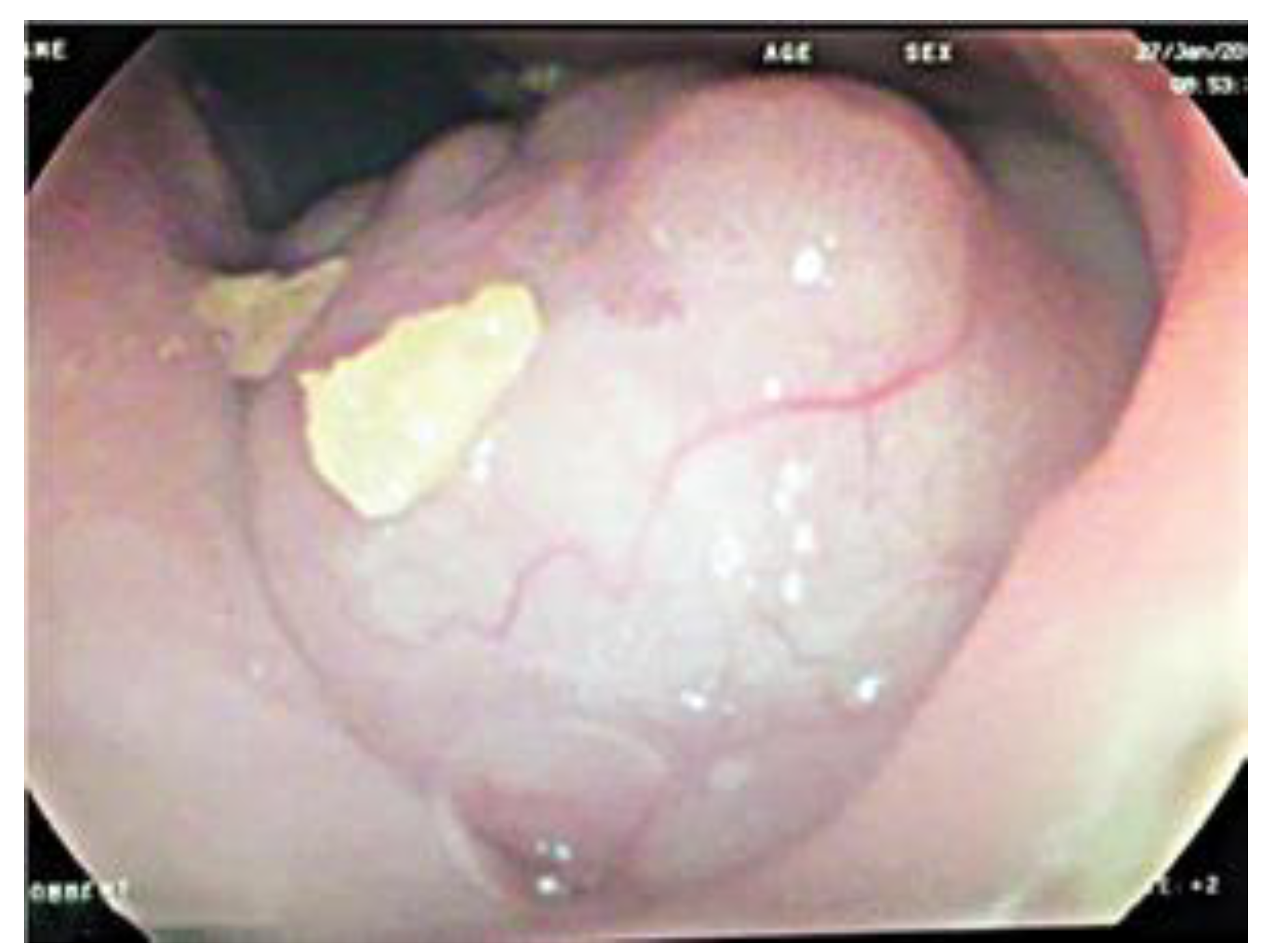
A preoperative esophagoscopy was performed, showing the intraluminal protrusion of the esophageal tumor, retained food remnants, but no additional information was obtained in order to describe the mass, since its size was making the evaluation very difficult (Figure 4).
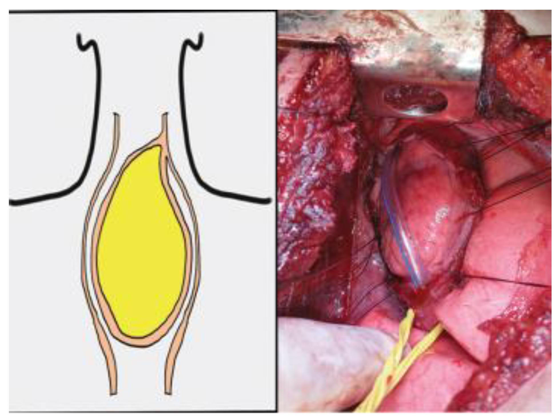
The patient underwent surgery via right thoracotomy. After the mediastinal inspection, oblique esophagotomy was done. A massive cervical-thoracic pedunculated intra-luminal tumor (102 x 52 x 48 mm) was noted in the cervical-thoracic esophagus lumen, which was significantly dilated from the T4 level upward to the lower cervical region (Figure 5). The mass was found to have arisen from the esophageal wall at the distal cervical segment.
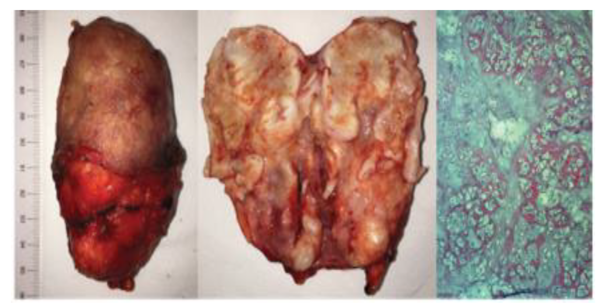
The complete excision and the esophageal preservation were possible without the cervical approach. Dissection revealed that the tumor has developed in the esophageal wall – no invasion was noted in the stretched mucosa and muscularis of the esophagus. The histopathological diagnosis was chondromatous polypoid hamartoma (Figure 6).
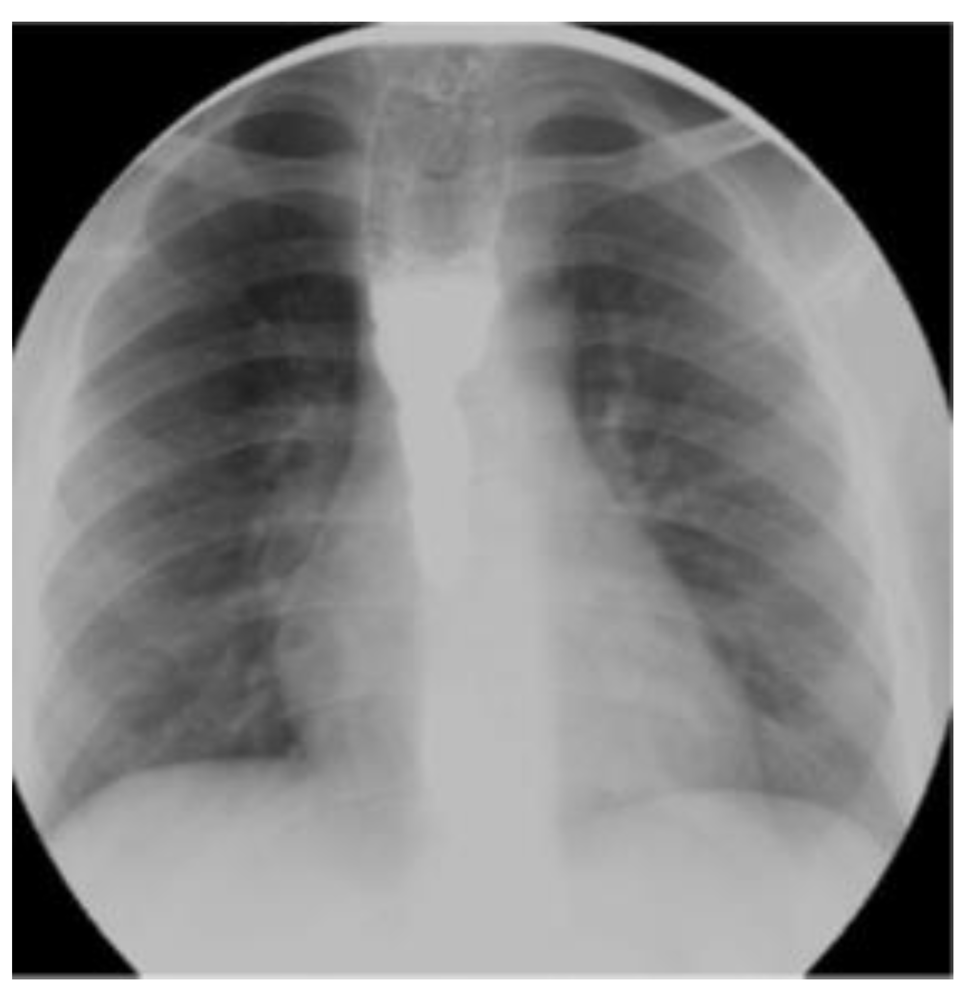
The one-year follow-up did not reveal any complications, the follow-up esophagogram showed the persistence of a mild esophageal dilatation (Figure 7), but no swallowing disorders were noted. A significant 6 kg weight gain was observed – from 39 Kg (percentile 86.7) to 45 (percentile 92.4).

Discussions

The division process of the primitive foregut underlies many ways of embryo pathogenesis. The tracheal-bronchial bud arises from the ventral part and grows caudally separating from the future esophagus. A disturbed tracheal-esophageal separation process sets up the origin of the congenital tracheal-esophageal fistulas or foregut duplication cysts – which, in exceptional cases, may associate esophageal atresia [6,7]. In the same course, respiratory tissue sequestration in the esophageal wall during the tracheal-esophageal separation process may explain the origins of congenital esophageal stenoses due to tracheal-bronchial remnants or chondromatous hamartomas (either polypoid or intramural) if the heterotopic tissue is organized in tumor-like lesions [8-10].
Dysphagia is the main manifestation of the esophageal tumor overall. However, clinical presentations may vary depending on their benign or malignant nature. A careful pre- and intraoperative assessment is necessary to prevent medical errors, and the therapeutic decision should be taken following the discussion of the alternatives with the child’s legal representative and signing the informed consent [11,12]. In children, benign tumors, having a slowly growing pattern, often lead to airway compression and micro-aspirations, therefore a chronic respiratory distress picture (recurrent airway infections, wheezing, shortness of breath) is common. By contrast, some benign tumors generate the total obstruction of the digestive tract since birth [11]. Malignant esophageal tumors of childhood present signs of nutritional deficiency such as weight loss, anemia or dehydration, more often secondary to dysphagia [2]. Decreased serum melatonin may be a relevant biomarker for malignancy [12]. Our patient’s history is similar to the case presented by Samad, et al., where the diagnosis of a pedunculated lipoma of the esophagus has been delayed for 2 years, the tumor’s clinical presentation dissimulating asthma [13]. On the other hand, Coury, et al. report a neonate whose initial symptoms (dyspnea and cyanosis) were disguised by the presumption of severe gastro-esophageal reflux and peripheral pulmonary stenosis and the tumor’s diagnosis was delayed until the 40th day of life, when due to recurrent obstructive apnea episodes and bradycardia (despite of nasal-jejunal feeding), a laryngoscopy was done revealing a polypoid cervical esophageal hamartoma prolapsing intermittently in the upper airway [14,15]. Other clinical signs reported in the esophageal tumors of childhood are poor weight gain, epigastric or retrosternal pain, hematemesis or melena, dysphonia or tracheal sounds [2]. The malabsorption of micronutrients related to dysphagia, with low serum Zn/Cu ratio may lead to attention deficit spectrum disorders [16]. In our opinion, considering the broad spectrum of subtle signs and the frequency of the gastroesophageal reflux disease symptoms in pediatric population, endoscopic studies of the esophagus and airways should be more seriously taken into consideration as an early diagnostic tool, in case of an inadequate response therapy, in order to exclude unlikely, but threatening conditions.
Dysphagia in children should be carefully assessed, mainly because of the aspiration risk. The primary evaluation tools are video fluoroscopy and fiber optic endoscopy. All swallowing aspects should be checked, from the oro-pharyngeal phase to the esophageal passage of the barium solution [17]. On the other hand, from our experience, we consider that all children (especially toddlers), presenting with respiratory symptoms, should undergo systematically plain X-ray evaluations. These can identify any mediastinal masses much earlier or exclude life-threatening situations such as unsuspected esophageal foreign bodies (for example, button batteries). However, if esophageal masses remain undetected at the initial evaluation, parents’ compliance and return for follow-up plays an essential role in the early detection in children and the health professional should never hesitate to consider barium swallowing examination and both endoscopic and digestive endoscopic tools in case of doubtful results or persistent symptoms. Additional means, such as CT and MRI scans, may be required for a better mass description as a differential diagnosis, anatomical relations or preoperative planning [18]. In selected cases, endoscopic ultrasounds and fine needle aspiration biopsies might be considered in the preoperative evaluation and treatment planning, which has the ad
Esophageal hamartomas are extremely rare and there is no commonly accepted guidance for their treatment. The vast underlying molecular network connecting these disorders opened a vast area of research [19]. The surgical attitude may vary widely depending on their aspect – intramural or polypoid, their volume and depth of invasion into the esophageal layers. Throughout the years, minimally invasive methods have gained ground when considering the surgical field [20]. Endoscopic resection using snare coagulation or simple ligation is possible in carefully selected cases [8], with all the advantages of the minimally invasive approach: quick recovery, minimal scaring, reduced pain and postoperative complications [21,22]. If the nature of the tumor is not suitable for a minimally invasive approach, esophagotomy and enucleation may be attempted [23,24], but this may not be possible all the time, therefore esophagectomy and end-to-end anastomosis have to be done [25]. In these cases, an ICG tissue angiography is a feasible and reliable technical support in the evaluation of the anastomotic perfusion and for the prevention of any anastomotic leak [24-26].
Similar to other surgical situations, in selected cases, one can use a linear stapler in order to resect a pedunculated esophageal mass [25]. In our case, enucleation was possible due to a relatively simple dissection between the tumor and the esophageal muscularis. The mucosa excess was also removed along with the tumor and esophageal suturing was done in safe conditions. Moreover, in terms of preoperative planning, in large esophageal masses – the accurate evaluation of the position related to the thoracic aperture is an important matter, in order to decide between thoracotomy, cervical approach or both. The accuracy of the surgical procedure ensures a quick recovery, reducing the hospital stay and the associated medical expenses [26]. The differential diagnosis of the esophageal polypoid hamartomas in childhood may include squamous papilloma, giant fibrovascular polyps, lipomas, inflammatory fibroid polyps, glomangiomas, pyogenic granulomas [3] or teratomas [27-29]. Other common causes of benign esophageal masses in children are leiomyomas, papillomas, hemangiomas, lymphangiomas or neurofibromas. More rarely, malignant causes are also reported, the most common being squamous cell carcinomas and adenocarcinomas [2]. However, the most frequent site of chondromatous hamartoma is the lung [9,30], accounting for 3% of all lung tumors in the adult population, its possibility of malignant degeneration toward carcinoma or sarcoma being well-known [31].

Highlights

Esophageal tumors in the pediatric population are extremely rare entities with a low incidence of occurrence in the clinical practice of a pediatrician.
The diagnosis is delayed by nonspecific symptoms and radiologic findings.
The wide range of pathological entities similar to it require extensive planning and interdisciplinary collaboration between the radiologist, oncologist, pediatric surgeon and endoscopist. The surgical treatment should preserve the native esophagus and prevent long term complications.

Conclusions

Esophageal tumors are uncommon in the pediatric population. The diagnosis can be delayed due to their insidious onset of symptoms and their unspecific clinical signs, therefore the patient’s compliance to follow-up and broad, careful evaluation are mandatory. The respiratory symptoms and the history or clinical signs of nutritional difficulties should lead to a more careful evaluation of the esophagus.
The preoperative evaluation may be difficult because of the size of the tumors. Because of their rarity along with the virtual space defined by the esophageal lumen, when facing large tumors of the posterior mediastinum in the pediatric population, the surgeon should never hesitate to use preoperative imaging tools for a precise definition of the tumor’s anatomical relations.

Conflict of interest disclosure

There are no known conflicts of interest in the publication of this article. The manuscript was read and approved by all authors.

Compliance with ethical standards

Any aspect of the work covered in this manuscript has been conducted with the ethical approval of all relevant bodies and that such approvals are acknowledged within the manuscript.

References

  1. McCarville, M.B. Malignant pulmonary and mediastinal tumors in children: differential diagnoses. Cancer Imaging. 2010, 10, S35–S41. [Google Scholar] [CrossRef] [PubMed]
  2. Theilen, T.M.; La Quaglia, M. Esophageal Tumors in Childhood and Adolescence: Benign and Malignant. In Esophageal and Gastric Disorders in Infancy and Childhood; Till, H., Thomson, M., Foker, J., Holcomb, G., III, Khan, K., Eds.; Springer: Berlin, Heidelberg, 2017. [Google Scholar] [CrossRef]
  3. Septer, S.; Cuffari, C.; Attard, T.M. Esophageal polyps in pediatric patients undergoing routine diagnostic upper gastrointestinal endoscopy: a multicenter study. Dis Esophagus. 2014, 27, 24–29. [Google Scholar] [CrossRef]
  4. Hou, S.; Wei, J.; Lu, W.; Wang, L.; Liu, H.; Zhang, Q. Intramural chondroid hamartoma in the distal esophagus in an adult: A case report and review of the literature. Mol Clin Oncol. 2017, 6, 19–22. [Google Scholar] [CrossRef] [PubMed]
  5. Serban, D.; Spataru, R.I.; Vancea, G.; Balasescu, S.A.; Socea, B.; Tudor, C.; Dascalu, A.M. Informed consent in all surgical specialties: from legal obligation to patient satisfaction. Rom J Leg Med. 2020, 28, 317–321. [Google Scholar] [CrossRef]
  6. Ioannides, A.S.; Copp, A.J. Embryology of oesophageal atresia. Semin Pediatr Surg. 2009, 18, 2–11. [Google Scholar] [CrossRef] [PubMed]
  7. Spataru, R.I.; Popoiu, M.C.; Ivanov, M. Foregut duplication cyst associated with esophageal atresia-one-stage neonatal surgical repair. Indian J Surg. 2015, 77 (Suppl 1), 52–55. [Google Scholar] [CrossRef]
  8. Everett, C.; Volberg, F.; Ulshen, M.; Murray, G. Intramural esophageal hamartoma: a report of two cases and review of the literature. Gastrointest Radiol. 1980, 5, 317–319. [Google Scholar] [CrossRef]
  9. Zhang, C.; Xu, J.; Wang, Z.; He, Z.; Yang, H.; Hu, J. Chondromatous hamartoma of cervical esophagus: a case report and literature review. J Thorac Dis. 2017, 9, E236–E244. [Google Scholar] [CrossRef]
  10. Rebelo, P.G.; Ormonde, J.V.; Ormonde Filho, J.B. Congenital esophageal stenosis owing to tracheobronchial remnants. Rev Paul Pediatr. 2013, 31, 406–410. [Google Scholar] [CrossRef]
  11. Serban, D.; Smarandache, A.M.; Cristian, D.; Tudor, C.; Duta, L.; Dascalu, A.M. Medical errors and patient safety culture - shifting the healthcare paradigm in Romanian hospitals. Rom J Leg Med. 2020, 28, 195–201. [Google Scholar] [CrossRef]
  12. Stanciu, A.E.; Zamfir-Chiru-Anton, A.; Stanciu, M.M.; Stoian, A.P.; Jinga, V.; Nitipir, C.; Bucur, A.; Pituru, T.S.; Arsene, A.L.; Dragoi, C.M.; Hainarosie, R.; Nicolae, A.C.; Gherghe, M.; Gheorghe, D.C.; Spandidos, D.A.; Tsatsakis, A.; Papasavva, M.; Drakoulis, N. Clinical significance of serum melatonin in predicting the severity of oral squamous cell carcinoma. Oncol Lett. 2020, 19, 1537–1543. [Google Scholar] [CrossRef] [PubMed]
  13. Suciu, N.; Serban, A.; Toader, O.; Oprescu, D.; Spataru, R.I. Case report of fetal lingual tumor - perinatal care and neonatal surgical intervention. J Matern Fetal Neonatal Med. 2014, 27, 314–319. [Google Scholar] [CrossRef]
  14. Samad, L.; Ali, M.; Ramzi, H.; Akbani, Y. Respiratory distress in a child caused by lipoma of the esophagus. J Pediatr Surg. 1999, 34, 1537–1538. [Google Scholar] [CrossRef]
  15. Motofei, I.G. Malignant Melanoma: Autoimmunity and Supracellular Messaging as New Therapeutic Approaches. Curr Treat Options Oncol. 2019, 20, 45. [Google Scholar] [CrossRef] [PubMed]
  16. Tudosie, M.S.; Truta, E.; Davitoiu, A.M.; Stanciulescu, L.; Jinescu, G.; Mitu, A.M.; Forje, M.; Horhota, L.; Bojescu, A.A.; Mares, A.M.; Ionica, M. The Impact of Copper in Children with Attention Deficit Hyperactivity Disorder. Rev. Chim. 2017, 68, 279–283. [Google Scholar] [CrossRef]
  17. Coury, J.; Steinfeld, J.; Zwillenberg, D.; Zwillenberg, S. Esophageal hamartoma as an unusual cause of neonatal apnea and bradycardia. Ear Nose Throat J. 2010, 89, E7–E11. [Google Scholar]
  18. Dodrill, P.; Gosa, M.M. Pediatric Dysphagia: Physiology, Assessment, and Management. Ann Nutr Metab. 2015, 66 (Suppl 5), 24–31. [Google Scholar] [CrossRef] [PubMed]
  19. Tofolean, D.E.; Mazilu, L.; Stăniceanu, F.; Mocanu, L.; Suceveanu, A.I.; Baz, R.O.; Parepa, R.I.; Suceveanu, A.P.; Bondari, S.; Bondari, D.; Voinea, F. Clinical presentation of a patient with cutis laxa with systemic involvement: a case report. Rom J Morphol Embryol. 2015, 56, 1205–1210. [Google Scholar]
  20. Rusescu, A.; Pietrosanu, C.; Ionita, I.; Stefanescu, C.D.; Stoian, A.M.P.; Camen, A.; Zainea, V.; Hainarosie, R. Chemical Ablation of the Submucosal Tissue in Volumetric Reduction of Inferior Turbinate. Rev. Chim. 2018, 69, 642–644. [Google Scholar] [CrossRef]
  21. Serban, D.; Smarandache, C.G.; Tudor, C.; Duta, L.N.; Dascalu, A.M.; Aliuș, C. Laparoscopic Surgery in COVID-19 Era- Safety and Ethical Issues. Diagnostics (Basel). 2020, 10, 673. [Google Scholar] [CrossRef]
  22. Hryhorczuk, A.L.; Lee, E.Y.; Eisenberg, R.L. Esophageal abnormalities in pediatric patients. AJR Am J Roentgenol. 2013, 201, W519-32. [Google Scholar] [CrossRef] [PubMed]
  23. Räsänen, J.; Ilonen, I.; Ristimäki, A.; Salo, J.A.; Mäkitie, A.A. A hamartoma presenting as an intramural upper oesophageal tumour. J Thorac Dis. 2017, 9, E698–E701. [Google Scholar] [CrossRef] [PubMed]
  24. Alius, C.; Tudor, C.; Badiu, C.D.; Dascalu, A.M.; Smarandache, C.G.; Sabau, A.D.; Tanasescu, C.; Balasescu, S.A.; Serban, D. Indocyanine Green-Enhanced Colorectal Surgery-between Being Superfluous and Being a Game-Changer. Diagnostics (Basel). 2020, 10, 742. [Google Scholar] [CrossRef] [PubMed]
  25. Smith, C.W.; Murray, G.F.; Wilcox, B.R. Intramural esophageal hamartoma. An unusual cause of progressive stricture in a child. J Thorac Cardiovasc Surg. 1976, 72, 315–318. [Google Scholar] [CrossRef]
  26. Savlovschi, C.; Serban, D.; Andreescu, C.; Dascalu, A.; Pantu, H. Economic analysis of medical management applied for left colostomy. Chirurgia (Bucur). 2013, 108, 666–669. [Google Scholar]
  27. Motofei, I.G. Biology of Cancer; From Cellular Cancerogenesis to Supracellular Evolution of Malignant Phenotype. Cancer Invest. 2018, 36, 309–317. [Google Scholar] [CrossRef] [PubMed]
  28. Spataru, R. The use of mechanical suture in the treatment of Hirschsprung's disease: experience of 17 cases. Chirurgia (Bucur). 2014, 109, 208–212. [Google Scholar]
  29. Ryan, M.L.; Zhuge, Y.; Smothers, C.D.; Zhang, J.; Huang, E.Y. Intraluminal esophageal teratoma in a neonate. J Ped Surg Case Reports. 2015, 3, 451–454. [Google Scholar] [CrossRef]
  30. Kiss, L.; Kiss, R.; Porr, P.J.; Nica, C.; Nica, C.; Bardac, O.; Tănăsescu, C.; Bărbulescu, B.; Bundache, M.; Ilie, S.; Maniu, D.; Zaharie, S.I.; Hulpuş, R. Pathological evidence in support of total mesorectal excision in the management of rectal cancer. Chirurgia (Bucur). 2011, 106, 347–352. [Google Scholar]
  31. Ekinci, G.H.; Hacıömeroğlu, O.; Ersev, A.; Alpay, L.; Özgen, H.; Yılmaz, A. The frequency of lung cancer in patients with pulmonary hamartomas: An evaluation of clinical, radiological, and pathological features and follow-up data of 96 patients with pulmonary hamartomas. Rev Port Pneumol (2006). 2017, 23, 280–286. [Google Scholar] [CrossRef]
Figure 1. The esophagogram revealing a large cervical-thoracic esophageal mass.
Jmms 08 00021 g001
Figure 2. The MRI aspect of the esophageal tumor.
Jmms 08 00021 g002
Figure 3. CT scan showing the esophageal tumor.
Jmms 08 00021 g003
Figure 4. The endoscopic aspect of the lesion.
Jmms 08 00021 g004
Figure 5. The diagram and the intraoperative aspect of the large, intra-luminal tumor.
Jmms 08 00021 g005
Figure 6. The pathological exam – gross and microscopic aspect of the esophageal chondromatous hamartoma.
Jmms 08 00021 g006
Figure 7. The follow-up esophagogram showing the persistence of a mild esophageal dilatation.
Jmms 08 00021 g007