Next Article in Journal
Acute Chylous Peritonitis Due to Idiopathic Pancreatitis Mimicking Acute Appendicitis
Previous Article in Journal
D-Dimer Levels and Acute Pulmonary Embolism Development in COVID-19 Patients
 
 
Journal of Mind and Medical Sciences is published by MDPI from Volume 12 Issue 1 (2025). Previous articles were published by another publisher in Open Access under a CC-BY (or CC-BY-NC-ND) licence, and they are hosted by MDPI on mdpi.com as a courtesy and upon agreement with Valparaiso University (ValpoScholar).
Article

The Crosstalk Between Insulin Resistance, Systemic Inflammation, Redox Imbalance and the Thyroid in Subjects with Obesity

by
Nicoleta Răcătăianu
1,
Nicoleta Valentina Leach
2,*,
Sorana D. Bolboacă
3,
Maria Loredana Soran
4,
Mirela Flonta
5,
Ana Valea
1,
Andrada-Luciana Lazăr
6 and
Cristina Ghervan
1
1
Department of Endocrinology, Iuliu HaţIeganu University of Medicine and Pharmacy, Cluj-Napoca, Romania
2
The 5th Department of Internal Medicine, Iuliu HaţIeganu University of Medicine and Pharmacy, Cluj-Napoca, Romania
3
Department of Medical Informatics and Biostatistics, Iuliu HaţIeganu University of Medicine and Pharmacy, Cluj-Napoca, Romania
4
National Institute for Research and Development of Isotopic and Molecular Technologies, Cluj-Napoca, Romania
5
Laboratory Department, Infectious Diseases Clinical Hospital, Cluj-Napoca, Romania
6
Department of Dermatology, Iuliu HaţIeganu University of Medicine and Pharmacy, Cluj-Napoca, Romania
*
Author to whom correspondence should be addressed.
J. Mind Med. Sci. 2021, 8(1), 139-148; https://doi.org/10.22543/7674.81.P139148
Submission received: 13 November 2020 / Revised: 26 January 2021 / Accepted: 26 January 2021 / Published: 8 April 2021

Abstract

:
We aimed at assessing the interaction between visceral adipose tissue (VAT), insulin resistance (IR), circulating levels of monocyte chemoattractant protein-1 (MCP-1) and malondialdehyde (MDA) and the thyroid parameters in obese subjects. Methods. Obese subjects without thyroid pathologies or diseases associated with systemic inflammation and OS were recruited. Insulinemia, visceral fat thickness, metabolic and thyroid parameters were assayed. Circulating levels of MCP-1 and MDA were used to quantify inflammation and OS. Results. A number of 160 obese subjects were included. The MCP-1 level increased with the degree of obesity and HOMA-IR. MCP 1 was positively associated with antithyroperoxidase antibody (TPOab) levels and the frequency of Hashimoto’s thyroiditis (HT). The MDA level was positively correlated with the degree of obesity, aspartate aminotransferase and MCP-1. MDA was an independent predictor for the occurrence of hypothyroidism. IR patients showed higher fT3 levels and a positive association between insulin and TPOab levels. Conclusions. Systemic inflammation increased with VAT, IR and OS and was correlated with the frequency and the severity of HT, suggesting that, in obesity, MCP-1 could be part of the etiopathogenesis of autoimmune thyroiditis. MDA was an independent risk factor for hypothyroidism; therefore, redox imbalance associated with obesity can produce cell damage and thyroid dysfunction. FT3 is increased in IR patients, thus being a marker for the severity of metabolic impairment.

Introduction

Obesity has become a public health problem with a highly increased prevalence among adults and children. Furthermore, the risk of developing a metabolic syndrome is higher in patients with obesity; metabolic syndrome is the result of the interplay between an unhealthy lifestyle and genetic predisposition [1,2].
There is an interconnection between obesity and altered thyroid function [3]. Thus, patients suffering from obesity seem to have lower levels of free T4 (fT4) together with a higher volume of the thyroid gland compared to those with a body mass index (BMI) within normal limits. Moreover, the relationship between thyroid nodules and insulin resistance (IR) has been established [4]. Several studies [5,6] have shown that patients with obesity have a high prevalence of thyroid disorders, although the mechanisms linking obesity to thyroid pathology are not completely understood. IR, low-grade systemic chronic inflammation, and increased oxidative stress (OS) associated with obesity can directly cause the impairment of thyroid cells and may trigger factors for autoimmune thyroiditis [7,8].
Additionally, an increased resistance to thyroid hormones was established in patients suffering from obesity, a condition which significantly improved after bariatric surgery together with the improvement of the thyrotroph thyroxine resistance index and the thyroid-stimulating hormone index [8]. Baseline thyroid stimulating hormone (TSH) levels might be related to weight-loss after laparoscopic gastric banding. Thus, the amount of short-term weight loss after the aforementioned procedure is greater in patients with low normal TSH than those with normal and high-normal values [9].
Visceral adipose tissue (VAT) is a significant determinant of IR vi increased production of proinflammatory cytokines (leptin, TNFα, IL-6), and chemokines such as monocyte chemoattractant protein-1 (MCP-1). MCP-1 determines monocytes' recruitment and their differentiation into macrophages, increasing the inflammatory cascade and interfering with local and systemic insulin signaling [10]. Although several studies have assessed serum MCP-1 levels in patients with fatty liver [11] or thyroid diseases [12], not enough data have been reported regarding the effect of "low grade" systemic inflammation associated with obesity on thyroid morpho-functioning. The interplay between high sensitive-C reactive protein (hs-CRP) levels and subclinical hypothyroidism (SH) has also been analyzed in a cross-sectional study; however, no association between hs-CRP and SH was found [13].
It is well-known that both hypothyroidism and hyperthyroidism determine increased OS, but the mechanism by which obesity-associated OS may alter the thyroid remains unclear [14]. IR causes mitochondrial dysfunction and the activation of microsomal and peroxisomal oxidation pathways, generating increased reactive oxygen species (ROS) and lipid peroxidation products, such as malondialdehyde (MDA) [15]. Mitochondrial dysfunction leads to low cellular energy reserves and may be involved in the decreased transmembrane transport of thyroid hormones with impaired intracellular triiodothyronine (T3) and thyroxine (T4) levels, which explains the peripheral resistance to thyroid hormones observed in obesity [16]. Moreover, MDA may directly cause thyroid damage, having a systemic proinflammatory and profibrogenic effect [17,18]. Furthermore, the metabolic dysfunction related to obesity and IR may alter the deiodinase-1 activity with decreased intracellular T4-to-T3 conversion and intracellular T3 levels, and increased deiodinase-3 expression, which stimulates T4 conversion to the inactive rT3 form, responsible for the occurrence of the “low T3” syndrome [19,20,21].
Based on the hypothesis that IR, inflammation and OS associated with obesity could be involved in the etiopathogenesis of thyroid disorders, this study assesses the interaction between metabolic markers, serum MCP-1&MDA levels and thyroid parameters in subjects with obesity. Our research's second objective was to divide the subjects with obesity at a cut-off HOMA-IR ≥ 2.5 and compare IR to non-IR groups in terms of inflammation, redox imbalance, and thyroid parameters, considering IR as a marker of severe metabolic impairment.
Our study's novelty consists of the complex, integrative analyses of the associations between metabolic markers, systemic inflammation, OS and thyroid parameters, and their evaluation as possible risk factors for the changes in thyroid morpho-functioning, commonly observed in subjects with obesity.

Materials and Methods

A cross-sectional, observational study was conducted with the recruitment of eligible subjects, using a non-probabilistic sampling method, i.e. convenient sampling. Patients with obesity, presenting from November 2015 to December 2017 to the Endocrinology outpatient clinic for obesity investigation, were consecutively enrolled. Subjects aged >18 years and with a BMI ≥ 30 kg/m2 were included in the sample. Patients with previously known thyroid disorders or diseases associated with increased systemic inflammation and OS, including diabetes mellitus, depression, epilepsy, schizophrenia, cancer, rheumatoid arthritis, decompensated liver disease, congestive heart failure, or chronic renal failure were excluded.
Each patient was informed before recruitment regarding the protocol and was asked for written consent in order to participate in the study. The study protocol was approved by the Ethics Committee of “Iuliu Haţieganu” University of Medicine and Pharmacy and aligned with the ethical principles in the Declaration of Helsinki.
All participants underwent clinical, laboratory and ultrasound assessments.

Clinical evaluation

Demographic (gender, age) data and the anthropometric data, weight, height, waist circumference (WC) of each participant, were measured and recorded. BMI was calculated as the ratio between weight/height2 (kg/m2). Obesity was considered at BMI values ≥30 kg/m2 and was classified as degree I (30-34.9 kg/m2), II (35-39.9 kg/m2), or III (BMI ≥40 kg/m2) [22]. The diagnosis of abdominal obesity was established at WC ≥80 cm in women and ≥94 cm in men [23]. The same examiner performed all the measurements.

Laboratory investigations

Blood samples were collected in the morning after overnight fasting. The biochemical determinations (glycemia, aspartate aminotransferase [AST], alanine aminotransferase [ALT], gamma-glutamyl transpeptidase [GGT], triglycerides [TG], total cholesterol [TC], HDL-cholesterol, and LDL-cholesterol) were performed on an automated analyzer (Beckman Coulter Unicell DXC600, USA), using specific commercially available kits. Basal insulinemia was determined on an automatic analyzer (Beckman Coulter Unicell DXI600, USA) by chemiluminescence method (CMIA), using a specific kit with the serum detection range from 1.9 μIU/mL to 23 μIU/mL. IR was assessed with the homeostatic model assessment IR (HOMA-IR) index, using the following formula: HOMA-IR = basal glycemia (mg/dL) × basal insulinemia (μIU/mL)/405. A HOMA-IR index ≥2.5 was considered as a criterion for IR [24,25]. At this threshold value, the study group was divided into two subgroups: obese-IR (HOMA-IR≥2.5) and obese non-IR (HOMA-IR<2.5) and were compared in terms of metabolic impairment, inflammation, redox imbalance, and thyroid parameters.
The TSH, fT4, free T3 [fT3], anti-thyroperoxidase antibody [TPOab], and anti-thyroglobulin antibody [TGab] were evaluated by ELISA on an automated analyzer (Beckman Coulter Unicel DXI 600, USA) and specific kits. The fT3/fT4 ratio was subsequently calculated and not directly determined. The serum MCP-1 as a marker of inflammation was measured on a semi-automated analyzer (Tecan Trading AG, Switzerland), using the quantitative sandwich ELISA method and protocol described in the kit with a serum detection range from 31.25 pg/ml to 2,000 pg/ml. The plasma MDA was measured as a marker of OS, using an isocratic high-performance liquid chromatography (HPLC) method based on fluorescence detection (Shimadzu LC 2010 Chromatographic System) and a specific kit (Chromsystems Instruments & Chemicals GmbH, Germany). The limit of detection (7.752×10⁻⁴ μmol/L) and quantification (15.48×10⁻⁴ μmol/L) was determined using the SMAC program.

Ultrasound evaluation

The measurement of the abdominal subcutaneous fat thickness (SFT, cm) and visceral fat thickness (VFT, cm) was performed in all patients using Mindray DC-N3 Doppler ultrasound with a 5-MHz convex probe. VFT is considered a reliable indicator of VAT and was measured with the probe located 1 cm above the navel, on the xiphoid-umbilical line, both longitudinally and transversally, and defined as the distance between the white line and the anterior aortic wall, while SFT was measured as the distance between the white line and the skin [26]. The ultrasound evaluation was performed by the same radiologist, who was blinded to the study.

Statistical analysis

Statistical analysis was done with Statistics (StatSoft v.8, USA). Qualitative data are reported as absolute and relative frequencies (%) with 95% confidence interval bounds using an exact method [27] provided in squared brackets (CI). The Shapiro-Wilk test was applied to assess the normality of the measurements and the data were expressed as mean (SD=standard deviation) for normally distributed data, and median and interquartile range (quartile 1 to quartile 3) otherwise. The association between the qualitative data was tested with the Chi-square test. The comparison between two independent groups was performed using Student’s t-test for normally distributed data, and Mann-Whitney test otherwise. The comparisons between more than two groups were performed with the Kruskal-Wallis test. The association between variables was evaluated using the Spearman correlation coefficient. P-values <0.05 were considered significant for the comparison between two groups and 0.017 for the comparison between three groups. Logistic regression analysis was used to test univariate and multivariate associations, and the OR (odds ratio) with associated 95% confidence intervals were reported.

Results

One hundred and sixty patients with a mean age of 45±12.44 years (range 18–68 years) were included in the study. The majority of patients were women (91.25% [85.63 to 95.00]). Most frequently, patients were obesity degree I (65% [56.88 to 72.50]), while obesity degree II was observed in 21.25% [15.00 to 28.12] and degree III in 13.75% [8.75 to 20.00]. The frequency of IR (HOMA-IR≥2.5) was 53.75% [45.63 to 61.87]. The prevalence of thyroid dysfunction in the studied group was 18.12%, as follows: 0.625% had hyperthyroidism and 17.5% had hypothyroidism. Autoimmune and non-autoimmune hypothyroidism affected 12% and 5.5% of the patients, respectively. Hashimoto's thyroiditis (HT) was present in 24.5% of the patients.
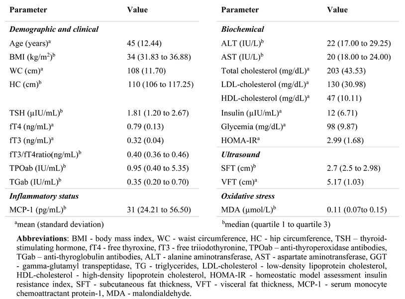
The summary of the investigated clinical, thyroid, metabolic, inflammatory, and OS parameters is presented in Table 1.

Metabolic and thyroid parameters

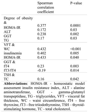
A low correlation was identified between the degree of obesity (BMI) and the degree of hepatic impairment expressed by the GGT level and TSH on the one hand, and some blood markers, on the other hand (Table 2). Moderate correlations of VFT with some blood markers were also identified (Table 2).
Although the frequency of thyroid disorders increased with the degree of obesity (degree I: 27.88%, degree II: 35.29%, degree III: 36.36%), no significant differences between the degrees of obesity and the presence of thyroid dysfunction or HT were identified (p > 0.5).
As expected, the values of VFT (Kruskal-Wallis test: 19.67, p=0.0001) were significantly different between patients with different degrees of obesity (Figure 1).

Systemic inflammation and OS

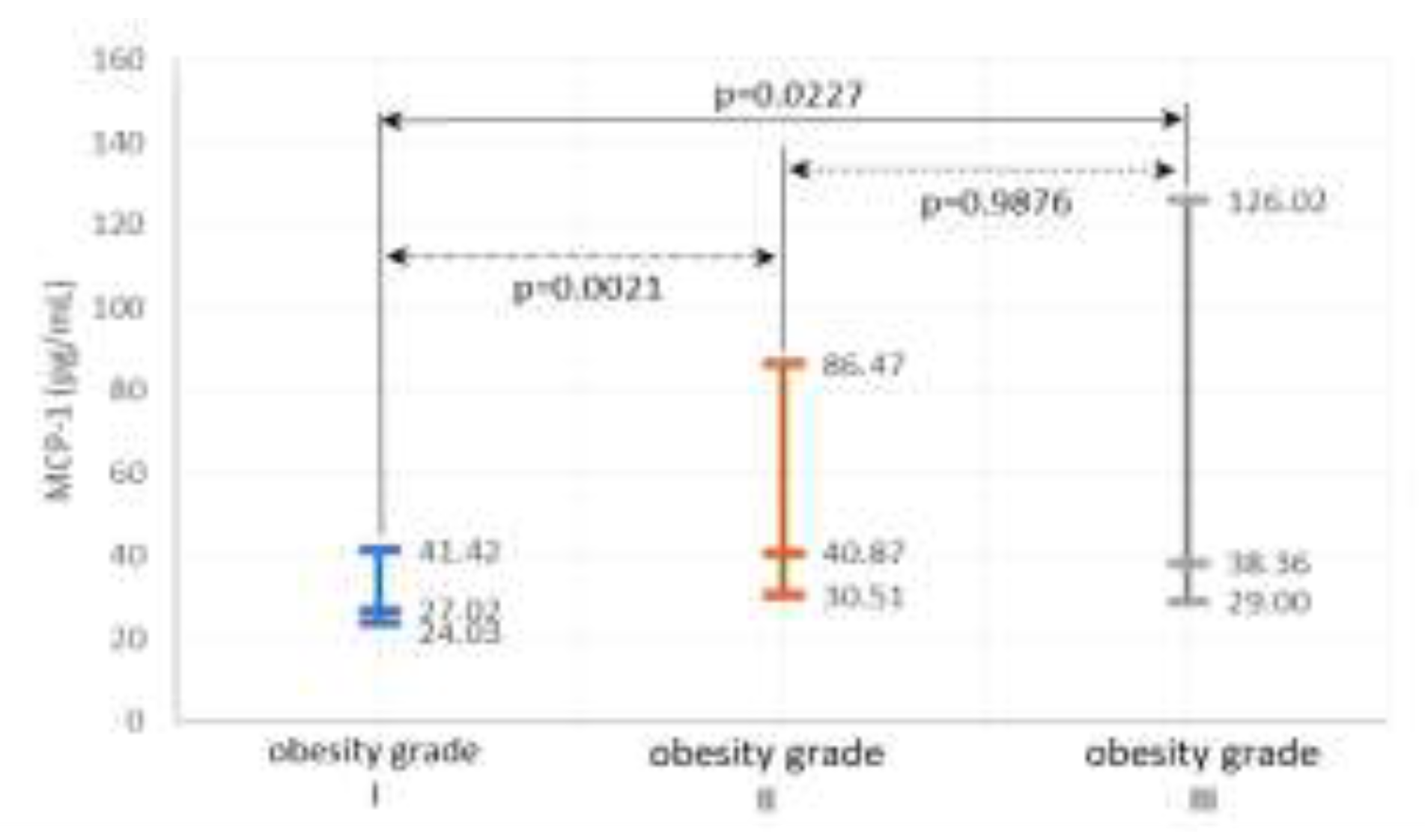
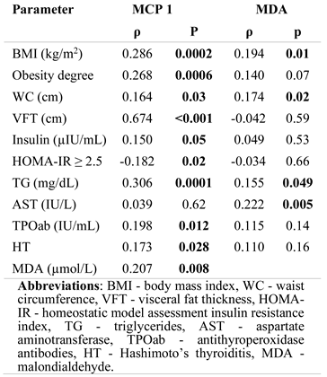
The serum MCP-1 level correlated positively with BMI (Table 3) and increased with the degrees of obesity (Kruskal-Wallis test: 12.24, p=0.002) (Figure 2).
Moreover, serum MCP-1 level increased with abdominal obesity (WC), TG level (Table 3) and at a cut-off HOMA-IR ≥2.5, serum MCP-1 levels were higher in IR versus non-IR patients (Table 3).
Regarding the association between thyroid parameters, serum MCP-1 level correlated significantly with the level of TPOab and the frequency of HT (Table 3).
A tendency for an association between MCP-1 and Hashimoto's thyroiditis was evident in the logistic regression analysis (CI95% [1.000-1005]; p = 0.071).
The OS's assessment in the study group revealed that serum MDA level increased with BMI and WC, TG, and AST, and positively correlated with the systemic inflammation expressed by MCP-1 (Table 3).
Moreover, multiple regression analysis showed that MDA is an independent risk factor associated with the presence of hypothyroidism (CI95% [1.082-781.39]; p = 0.045).
Comparison between the IR and non-IR group at cut-off HOMA-IR ≥ 2.5
The group with obesity was divided according to HOMA-IR ≥ 2.5 in order to analyze the differences between the groups regarding the metabolic impairment, the systemic inflammation, OS and the changes in thyroid parameters at this threshold value of IR. Table 4 shows the differences between IR and non-IR patients.
Factors significantly associated with IR in the univariate analysis included obesity (both in terms of degree [BMI] and distribution [WC, VFT]), ALT, GGT, and hypertriglyceridemia (Table 4), but the multiple linear regression analysis established that only ALT (CI95% [1.008-1.074], p=0.01) and VFT (CI95% [1.167-2.331], p=0.005) were independent factors associated with IR. Additionally, IR patients showed significantly higher serum MCP-1 levels than non-IR ones, but no significant difference was noticed regarding the MDA level (Table 4). Hypothyroidism had a higher frequency of 19.76% (n=17) in IR patients versus non-IR patients 14.86% (n=11), but the difference did not reach the statistical significance threshold (p>0.05). One of the non-IR patients had hyperthyroidism, while 23% of the IR and 26% of the non-IR patients had HT (p=0.72).
As shown in Table 4, IR patients showed significantly higher fT3 levels than non-IR patients. Moreover, insulinemia was positively associated with TPOab level (ρ=0.21, p=0.048) and serum fT4 levels correlated negatively with TG levels (ρ=-0.2483, p=0.0212) in IR-patients.
In the regression analysis, IR proved not to be a risk factor for thyroid dysfunction.

Discussions

One of the main findings of our study was that systemic inflammation (MCP-1) increased significantly with the obesity degree (BMI), abdominal distribution (VFT, WC), and IR. It was also significantly positively correlated with the severity (TPOab) and frequency of Hashimoto’s thyroiditis in subjects with obesity. In addition, the OS expressed by MDA increased significantly with systemic inflammation and it was a predictive factor associated with the presence of hypothyroidism in the study group of patients with obesity.
These results are consistent with the literature data reported and support the idea that increased serum MCP-1 in patients with obesity may be a direct consequence of abdominal obesity and IR [28,29]. The relationship between obesity, fT4, MCP-1 and nerve growth factor-β (NGF-β) was investigated in a recent study conducted by Molnár exclusively on women [30] which demonstrated the correlation between high MCP-1, NGF-β levels and low serum fT4 levels in women with obesity [30]. Likewise, Sartipy et al. revealed increased microRNA MCP-1 expression at VAT and an increase of MCP-1 level after the induction of hyperinsulinemic status both in vitro and in vivo, showing that visceral obesity and IR are the causes of increased MCP-1 serum levels [29]. Moreover, systemic inflammation triggers altered insulin response, making IR both the cause and the effect of systemic inflammation in obesity [11,31].
Regarding the low-grade inflammation associated with obesity and the possible thyroid involvement, our study revealed a significant correlation between MCP-1 and both severity (TPOab level) and HT's frequency in patients with obesity, but without any significant association with the thyroid function. Although there were no significant differences between IR versus non-IR patients regarding the frequency of HT, a significant correlation between insulinemia and TPOab levels was seen in IR versus non-IR patients, indicating the possible pathogenetic contribution of hyperinsulinism in the production of autoantibodies. Several studies support the role of inflammatory cytokines in HT etiopathogenesis; MCP-1 and other cytokines (TNFα, IL-1, INFδ) may induce and aggravate autoimmune thyroid diseases through leukocyte chemotaxis and the perpetuation of a chronic inflammatory response, leading to thyroid cell injury and impaired functioning [7,32]. Several researchers [33,34,35] have shown that hyperleptinemia associated with VAT accumulation may be a pathogenic link between obesity, immune system changes, and HT by activating the inflammatory induction signals in susceptible patients. Our study's result is consistent with that of Kokkotou et al. [12], which revealed a positive correlation between MCP-1 and antithyroid antibodies in HT patients without reporting a significant association with the thyroid function.
Inflammation and OS are closely related processes in obesity, and both interfere with insulin signaling, modifying the synthesis, activity, and metabolism of thyroid hormones [17,18,19,21]. Our study showed a significant association between MDA and the obesity degree & distribution (BMI, WC), along with the hepatic impairment (AST) and its significant increase with systemic inflammation (MCP-1). Instead, no significant difference was found in MDA levels related to IR, probably because most patients had degree I obesity, which is not associated with a significant increase in OS or IR. In regression analyses, MDA was an independent factor associated with hypothyroidism in the studied obesity group.
The possible mechanism by which OS may alter the thyroid function is complex. The balance between oxidants and antioxidants is critical for the proper functioning of the thyroid gland, and the direct effect of increased ROS in thyroid diseases has been investigated by several studies [36]. Moreover, reduced cellular energy production secondary to mitochondrial dysfunction is associated with the decreased transportation of thyroid hormones to the cells, leading to the intracellular deficiency of thyroid hormones. Redox imbalance can also alter the expression and the activity of deiodinases with decreased intracellular activation of T4 in T3 and increased production of reversed-T3, resulting in “low-T3” syndrome [16].
Along with the adipose tissue, the liver is the main source of ROS in obesity. The increased influx of FFA and inflammatory cytokines into the portal vein from VAT lipolysis may cause mitochondrial liver dysfunction, increased ROS release, promoting necroinflammation and aggravating IR, resulting in fatty liver disease (HS/NASH) [37].
The synthesis of thyroid hormone-binding proteins and the activation of T4 into T3 (by deiodinase-1) occur in the liver, and thus liver dysfunction associated with the severity of obesity and IR might influence hepatic T3 output by altering the transportation of the thyroid hormones to the cellular level and their feedback at the hypothalamic-pituitary level [38,39]. In this respect, our study revealed a significant positive correlation between GGT and serum fT4 levels and a negative correlation between GGT levels and fT3/fT4 ratio. Moreover, GGT levels increased with IR and obesity degree and represented cellular response expression against OS [40,41]. Along with the positive association between OS (MDA) and AST, this supports the effect of liver dysfunction on changes in the thyroid parameters observed in obesity.
Regarding the frequency of thyroid dysfunction (18.12%) or HT (24.5%), our results are consistent with the literature data for patients with obesity [35,42,43], but unlike other studies [5,6,34,38] we found no significant differences between the degrees of obesity according to the IR presence, possibly due to the predominance of degree I obesity, which is not accompanied by marked metabolic and endocrine changes.
Consistent with other studies suggesting that fT3 and/or fT3/fT4 ratio increase are markers for the severity of metabolic damage in patients with obesity, our study results show that IR patients exhibited significantly higher fT3 levels than non-IR patients. Furthermore, serum TG level was significantly higher in IR patients and associated with increased VFT and systemic inflammation (MCP-1) (which is explained by VAT lipolysis due to IR) and negatively correlated with fT4 levels as compared to non-IR ones. These results are most likely secondary to increased deiodinase-2 activity at the VAT level, with higher conversion of T4 to T3. The increase in local T3 production is probably a defense mechanism to limit further adipose gain by increasing thermogenesis and stimulating the metabolic activity [20].
Concerning the association between the thyroid and lipid parameters, our study highlighted a significant positive correlation between serum TSH and cholesterol levels, as hypothyroidism decreases the rate of lipoprotein degradation and LDL-cholesterol receptor synthesis. Other studies reported similar results regarding the association between thyroid hormones and metabolic parameters, predictive for the severity of the metabolic impairment [33,37,42,43,44].

Study limitations

Our study's main limitation is the applied non-probabilistic sampling method that led to the unequal distribution of genders in the investigated sample, since it is known that women seek medical attention more frequently than men. Notwithstanding, the existing data in the literature demonstrated the predominance of thyroid disorders among the female population [45]. Thus, there are a considerable number of studies that have been carried out on female population alone [30,46]. A more accurate view could be obtained by investigating subjects with obesity from the general population rather than those who presented for medical consultations.

Conclusions

Systemic inflammation increased with visceral adiposity, IR, and was significantly correlated with the frequency and the severity (TPOab levels) of HT, suggesting that, in obesity, MCP-1 could be part of the etiopathogenesis of autoimmune thyroiditis. OS (MDA) increased significantly with systemic inflammation (MCP-1) and it was an independent risk factor for the occurrence of hypothyroidism, sustaining that redox imbalance associated with obesity can produce cell damage and contribute to the pathophysiology of thyroid dysfunction. A significantly increased level of fT3 in insulin-resistant vs. non-insulin-resistant patients supports the value of fT3 as a marker for the severity of metabolic impairments in patients with obesity.

Conflict of interest disclosure

There are no known conflicts of interest in the publication of this article. The manuscript was read and approved by all authors.

Compliance with ethical standards

Any aspect of the work covered in this manuscript has been conducted with the ethical approval of all relevant bodies and that such approvals are acknowledged within the manuscript.

Acknowledgments

We would like to express our gratitude to the Management Unit of the Infectious Diseases Clinical Hospital, Cluj-Napoca, for its assistance and support.

References

  1. Di Domenico, M.; Pinto, F.; Quagliuolo, L.; Contaldo, M.; Settembre, G.; Romano, A.; Coppola, M.; Ferati, K.; Bexheti-Ferati, A.; Sciarra, A.; Nicoletti, G.F.; Ferraro, G.A.; Boccellino, M. The Role of Oxidative Stress and Hormones in Controlling Obesity. Front Endocrinol (Lausanne). 2019, 10, 540. [Google Scholar] [CrossRef] [PubMed]
  2. Cozma, A.; Sitar-Taut, A.; Urian, L.; Fodor, A.; Suharoschi, R.; Muresan, C.; Negrean, V.; Sampelean, D.; Zdrenghea, D.; Pop, D.; Leucuta, D.; Orasan, O.H. Unhealthy lifestyle and the risk of metabolic syndrome - the Romanian experience. J Mind Med Sci. 2018, 5, 218–229. [Google Scholar] [CrossRef]
  3. Cozma, A.; Sitar-Taut, A.; Orasan, O.; et al. The Relationship Between eNOS (G894T) Gene Polymorphism and Arterial Stiffness in Patients with Metabolic Syndrome. REV. CHIM. (Bucharest). 2018, 69, 2351–2356. [Google Scholar] [CrossRef]
  4. Layegh, P.; Asadi, A.; Jangjoo, A.; Layegh, P.; Nematy, M.; Salehi, M.; Shamsian, A.; Ranjbar, G. "Comparison of thyroid volume, TSH, free t4 and the prevalence of thyroid nodules in obese and non-obese subjects and correlation of these parameters with insulin resistance status". Caspian J Intern Med. 2020, 11, 278–282. [Google Scholar] [CrossRef]
  5. Anil, C.; Akkurt, A.; Ayturk, S.; Kut, A.; Gursoy, A. Impaired glucose metabolism is a risk factor for increased thyroid volume and nodule prevalence in a mild-to-moderate iodine deficient area. Metabolism. 2013, 62, 970–975. [Google Scholar] [CrossRef]
  6. Rezzonico, J.; Rezzonico, M.; Pusiol, E.; Pitoia, F.; Niepomniszcze, H. Introducing the thyroid gland as another victim of the insulin resistance syndrome. Thyroid. 2008, 18, 461–464. [Google Scholar] [CrossRef] [PubMed]
  7. Rasmussen, A.K. Cytokine actions on the thyroid gland. Dan Med Bull. 2000, 47, 94–114. [Google Scholar] [PubMed]
  8. Malaguarnera, R.; Vella, V.; Nicolosi, M.L.; Belfiore, A. Insulin Resistance: Any Role in the Changing Epidemiology of Thyroid Cancer? Front Endocrinol (Lausanne). 2017, 8, 314. [Google Scholar] [CrossRef]
  9. Juiz-Valiña, P.; Cordido, M.; Outeiriño-Blanco, E.; Pértega, S.; Varela-Rodríguez, B.M.; García-Brao, M.J.; Mena, E.; Pena-Bello, L.; Sangiao-Alvarellos, S.; Cordido, F. Central Resistance to Thyroid Hormones in Morbidly Obese Subjects Is Reversed after Bariatric Surgery-Induced Weight Loss. J Clin Med. 2020, 9, 359. [Google Scholar] [CrossRef]
  10. Muraca, E.; Oltolini, A.; Pizzi, M.; Villa, M.; Manzoni, G.; Perra, S.; Zerbini, F.; Bianconi, E.; Cannistraci, R.; Ciardullo, S.; Pizzi, P.; Lattuada, G.; Perseghin, G. Baseline TSH levels and short-term weight loss after different procedures of bariatric surgery. Int J Obes (Lond). 2021, 45, 326–330. [Google Scholar] [CrossRef]
  11. Reaven, G.M. Pathophysiology of insulin resistance in human disease. Physiol Rev. 1995, 75, 473–486. [Google Scholar] [CrossRef] [PubMed]
  12. Kanda, H.; Tateya, S.; Tamori, Y.; Kotani, K.; Hiasa, K.; Kitazawa, R.; Kitazawa, S.; Miyachi, H.; Maeda, S.; Egashira, K.; Kasuga, M. MCP-1 contributes to macrophage infiltration into adipose tissue, insulin resistance, and hepatic steatosis in obesity. J Clin Invest. 2006, 116, 1494–1505. [Google Scholar] [CrossRef] [PubMed]
  13. Kokkotou, E.; Marafelia, P.; Mantzos, E.I.; Tritos, N.A. Serum monocyte chemoattractant protein-1 is increased in chronic autoimmune thyroiditis. Metabolism. 2002, 51, 1489–1493. [Google Scholar] [CrossRef]
  14. Peixoto de Miranda, É.J.; Bittencourt, M.S.; Santos, I.S.; Lotufo, P.A.; Benseñor, I.M. Thyroid Function and High-Sensitivity C-Reactive Protein in Cross-Sectional Results from the Brazilian Longitudinal Study of Adult Health (ELSA-Brasil): Effect of Adiposity and Insulin Resistance. Eur Thyroid J. 2016, 5, 240–246. [Google Scholar] [CrossRef]
  15. Al-Rubae'i, S.H.N.; Al-Musawi, A.K. An evaluation of antioxidants and oxidative stress in Iraqi patients with thyroid gland dysfunction. African J Biochem Res. 2011, 5, 188–196. [Google Scholar]
  16. Leclercq, I.A. Antioxidant defence mechanisms: new players in the pathogenesis of non-alcoholic steatohepatitis? Clin Sci (Lond). 2004, 106, 235–237. [Google Scholar] [CrossRef]
  17. Kent Holtorf. Thyroid Hormone Transport into Cellular Tissue. J Restor Med. 2014, 3, 53–68. [Google Scholar] [CrossRef]
  18. Evans, J.L.; Goldfine, I.D.; Maddux, B.A.; Grodsky, G.M. Oxidative stress and stress-activated signaling pathways: a unifying hypothesis of type 2 diabetes. Endocr Rev. 2002, 23, 599–622. [Google Scholar] [CrossRef]
  19. Codoñer-Franch, P.; Navarro-Ruiz, A.; Fernández-Ferri, M.; Arilla-Codoñer, A.; Ballester-Asensio, E.; Valls-Bellés, V. A matter of fat: insulin resistance and oxidative stress. Pediatr Diabetes. 2012, 13, 392–399. [Google Scholar] [CrossRef]
  20. Yesilova, Z.; Yaman, H.; Oktenli, C.; Ozcan, A.; Uygun, A.; Cakir, E.; Sanisoglu, S.Y.; Erdil, A.; Ates, Y.; Aslan, M.; Musabak, U.; Erbil, M.K.; Karaeren, N.; Dagalp, K. Systemic markers of lipid peroxidation and antioxidants in patients with nonalcoholic Fatty liver disease. Am J Gastroenterol. 2005, 100, 850–855. [Google Scholar] [CrossRef]
  21. Bianco, A.C.; Kim, B.W. Deiodinases: implications of the local control of thyroid hormone action. J Clin Invest. 2006, 116, 2571–2579. [Google Scholar] [CrossRef] [PubMed]
  22. Huang, S.A.; Mulcahey, M.A.; Crescenzi, A.; Chung, M.; Kim, B.W.; Barnes, C.; Kuijt, W.; Turano, H.; Harney, J.; Larsen, P.R. Transforming growth factor-beta promotes inactivation of extracellular thyroid hormones via transcriptional stimulation of type 3 iodothyronine deiodinase. Mol Endocrinol. 2005, 19, 3126–3136. [Google Scholar] [CrossRef]
  23. Alberti, K.G.; Eckel, R.H.; Grundy, S.M.; Zimmet, P.Z.; Cleeman, J.I.; Donato, K.A.; Fruchart, J.C.; James, W.P.; Loria, C.M.; Smith, S.C., Jr. International Diabetes Federation Task Force on Epidemiology and Prevention; Hational Heart, Lung, and Blood Institute; American Heart Association; World Heart Federation; International Atherosclerosis Society; International Association for the Study of Obesity. Harmonizing the metabolic syndrome: a joint interim statement of the International Diabetes Federation Task Force on Epidemiology and Prevention; National Heart, Lung, and Blood Institute; American Heart Association; World Heart Federation; International Atherosclerosis Society; and International Association for the Study of Obesity. Circulation. 2009, 120, 1640–1645. [Google Scholar] [CrossRef] [PubMed]
  24. Obesity: preventing and managing the global epidemic. Report of a WHO consultation. World Health Organ Tech Rep Ser. 2000, 894, i.
  25. Matthews, D.R.; Hosker, J.P.; Rudenski, A.S.; Naylor, B.A.; Treacher, D.F.; Turner, R.C. Homeostasis model assessment: insulin resistance and beta-cell function from fasting plasma glucose and insulin concentrations in man. Diabetologia. 1985, 28, 412–419. [Google Scholar] [CrossRef]
  26. Arkadievich, O.D. Metabolic markers of myocardium insulin resistance in dogs with heart failure. Open Vet J. 2021, 10, 363–370. [Google Scholar] [CrossRef]
  27. Kim, S.K.; Kim, H.J.; Hur, K.Y.; Choi, S.H.; Ahn, C.W.; Lim, S.K.; Kim, K.R.; Lee, H.C.; Huh, K.B.; Cha, B.S. Visceral fat thickness measured by ultrasonography can estimate not only visceral obesity but also risks of cardiovascular and metabolic diseases. Am J Clin Nutr. 2004, 79, 593–599. [Google Scholar] [CrossRef] [PubMed]
  28. Jäntschi, L.; Bolboacã, S.D. Exact probabilities and confidence limits for binomial samples: applied to the difference between two proportions. ScientificWorldJournal. 2010, 10, 865–878. [Google Scholar] [CrossRef]
  29. Xu, H.; Barnes, G.T.; Yang, Q.; Tan, G.; Yang, D.; Chou, C.J.; Sole, J.; Nichols, A.; Ross, J.S.; Tartaglia, L.A.; Chen, H. Chronic inflammation in fat plays a crucial role in the development of obesity-related insulin resistance. J Clin Invest. 2003, 112, 1821–1830. [Google Scholar] [CrossRef]
  30. Sartipy, P.; Loskutoff, D.J. Monocyte chemoattractant protein 1 in obesity and insulin resistance. Proc Natl Acad Sci U S A. 2003, 100, 7265–7270. [Google Scholar] [CrossRef]
  31. Molnár, I. Interactions among thyroid hormone (FT4), chemokine (MCP-1) and neurotrophin (NGF-β) levels studied in Hungarian postmenopausal and obese women. Cytokine. 2020, 127, 154948. [Google Scholar] [CrossRef] [PubMed]
  32. Torres-Leal, F.L.; Fonseca-Alaniz, M.H.; De Oliveira, A.C.; Alonso-Vale, M.I.C. Adipose Tissue Inflammation and Insulin Resistance. Intech. 2012, 6, 137–156. [Google Scholar]
  33. García-López, M.A.; Sancho, D.; Sánchez-Madrid, F.; Marazuela, M. Thyrocytes from autoimmune thyroid disorders produce the chemokines IP-10 and Mig and attract CXCR3+ lymphocytes. J Clin Endocrinol Metab. 2001, 86, 5008–5016. [Google Scholar] [CrossRef] [PubMed]
  34. Marina, C.N.; Danciu, R.; Raducu, L.; Scaunasu, R.V.; Jecan, C.R.; Florescu, P.I. The surgical treatment of diabetic foot ulcers. J Clin Invest Surg. 2019, 4, 96–100. [Google Scholar] [CrossRef]
  35. Duntas, L.H.; Biondi, B. The interconnections between obesity, thyroid function, and autoimmunity: the multifold role of leptin. Thyroid. 2013, 23, 646–653. [Google Scholar] [CrossRef] [PubMed]
  36. Marzullo, P.; Minocci, A.; Tagliaferri, M.A.; Guzzaloni, G.; Di Blasio, A.; De Medici, C.; Aimaretti, G.; Liuzzi, A. Investigations of thyroid hormones and antibodies in obesity: leptin levels are associated with thyroid autoimmunity independent of bioanthropometric, hormonal, and weight-related determinants. J Clin Endocrinol Metab. 2010, 95, 3965–3972. [Google Scholar] [CrossRef]
  37. Ohye, H.; Sugawara, M. Dual oxidase, hydrogen peroxide and thyroid diseases. Exp Biol Med (Maywood). 2010, 235, 424–433. [Google Scholar] [CrossRef]
  38. Girard, J.; Lafontan, M. Impact of visceral adipose tissue on liver metabolism and insulin resistance. Part II: Visceral adipose tissue production and liver metabolism. Diabetes Metab. 2008, 34, 439–445. [Google Scholar] [CrossRef]
  39. Pacifico, L.; Bonci, E.; Ferraro, F.; Andreoli, G.; Bascetta, S.; Chiesa, C. Hepatic steatosis and thyroid function tests in overweight and obese children. Int J Endocrinol. 2013, 2013, 381014. [Google Scholar] [CrossRef]
  40. Farasat, T.; Cheema, A.M.; Khan, M.N. Hyperinsulinemia and insulin resistance is associated with low T₃/T₄ ratio in pre diabetic euthyroid Pakistani subjects. J Diabetes Complications. 2012, 26, 522–525. [Google Scholar] [CrossRef]
  41. Lee, D.H.; Gross, M.D.; Jacobs, D.R., Jr. Cardiovascular Risk Development in Young Adults Study. Association of serum carotenoids and tocopherols with gamma-glutamyltransferase: the Cardiovascular Risk Development in Young Adults (CARDIA) Study. Clin Chem. 2004, 50, 582–588. [Google Scholar] [CrossRef] [PubMed]
  42. Albu, A.; Moldovan, A.; Petra, C.; Para, I. Serum Gamma-Glutamyl Transferase is associated with epicardial fat thickness in middle aged woman. Revista de Chimie. 2020, 71, 430–435. [Google Scholar] [CrossRef]
  43. Rotondi, M.; Leporati, P.; La Manna, A.; Pirali, B.; Mondello, T.; Fonte, R.; Magri, F.; Chiovato, L. Raised serum TSH levels in patients with morbid obesity: is it enough to diagnose subclinical hypothyroidism? Eur J Endocrinol. 2009, 160, 403–408. [Google Scholar] [CrossRef] [PubMed]
  44. Lathia, T. Rising prevalence of thyroid disorders. J Mahatma Gandhi Inst Med Sci. 2015, 20, 125–127. [Google Scholar] [CrossRef]
  45. Chubb, S.A.; Davis, W.A.; Davis, T.M. Interactions among thyroid function, insulin sensitivity, and serum lipid concentrations: the Fremantle diabetes study. J Clin Endocrinol Metab. 2005, 90, 5317–5320. [Google Scholar] [CrossRef]
  46. Farishta, F.; Farishta, S. Insulin resistance and thyroid hypofunction in obese women – A cross sectional study. Integr Obesity Diabetes. 2015, 1. [Google Scholar] [CrossRef]
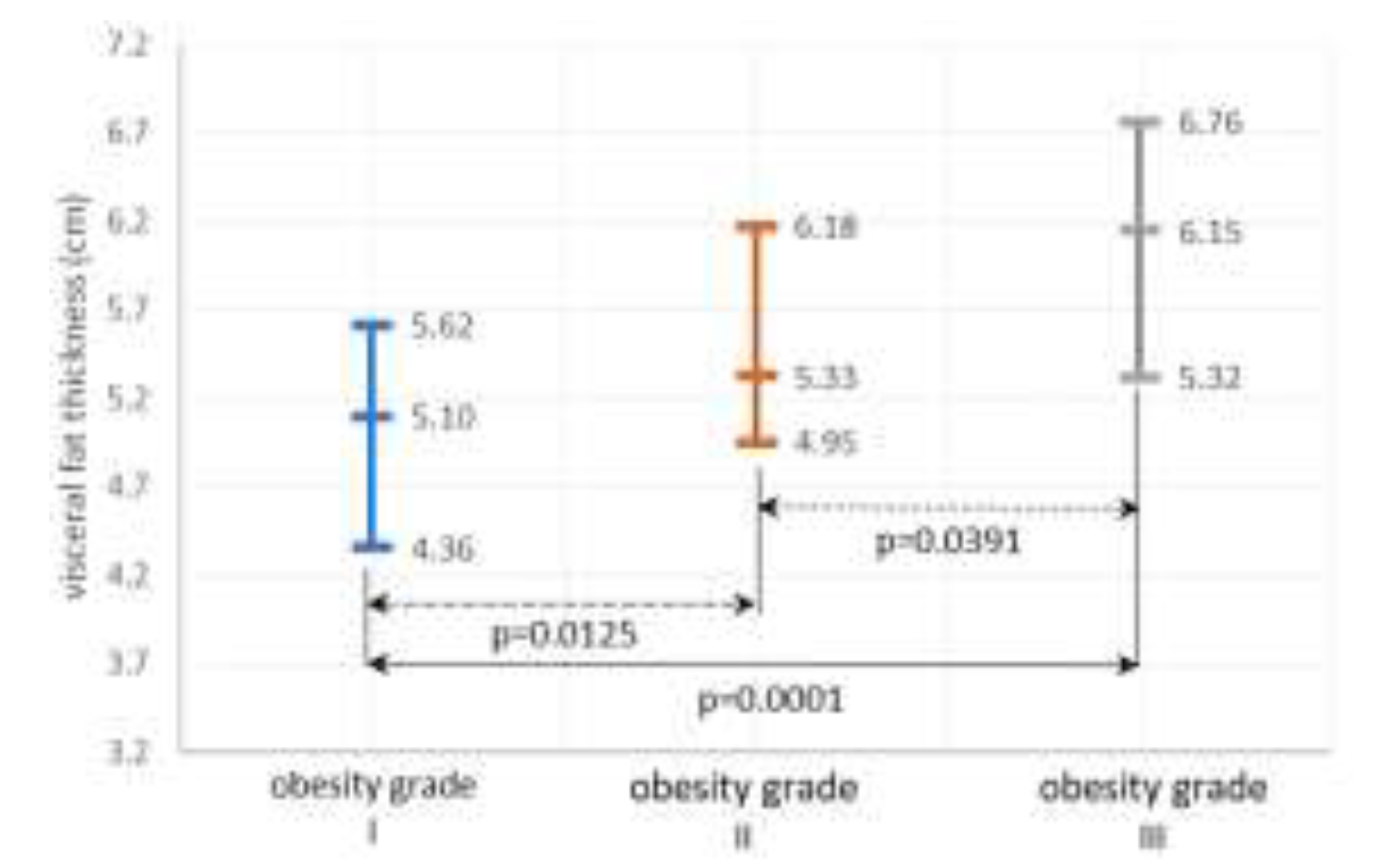
Figure 1. The variability of visceral fat thickness according to the degree of obesity. The middle line represents the median value, the lower line is given by the value of the first quartile, and the upper line represents the value of the third quartile.
Jmms 08 00018 g001
Figure 2. The variability of serum monocyte chemoattractant protein-1 (MCP-1) according to the degrees of obesity. The middle line represents the median value, the lower line is given by the value of the first quartile, and the upper line represents the value of the third quartile.
Jmms 08 00018 g002
Table 1. Clinical, biochemical, thyroid, inflammatory status, oxidative stress, and ultrasound parameters in the study group.
Jmms 08 00018 i001
Table 2. Degree of obesity, VFT, GGT and TSH significant correlations with blood markers.
Jmms 08 00018 i002
Table 3. Significant Spearman correlations between serum MCP-1, MDA and clinical, metabolic, inflammatory status, oxidative stress and thyroid parameters.
Jmms 08 00018 i003
Table 4. Clinical, metabolic, inflammatory status, oxidative stress and thyroid parameters according to the presence of insulin resistance (IR).
Jmms 08 00018 i004
Jmms 08 00018 i005