Article

Clinical Value of Diffusion-Weighted Whole-Body Imaging with Background Body Signal Suppression (DWIBS) for Staging of Patients with Suspected Head and Neck Cancer

1
Department of Radiology, University Medical Center Regensburg, 93053 Regensburg, Germany
2
Department of Othorhinolaryngology, Merciful Brothers Hospital St. Elisabeth, 94315 Straubing, Germany
3
Department of Diagnostic and Interventional Radiology, University Hospital Brandenburg, Brandenburg Medical School Theodor Fontane, 14770 Neuruppin, Germany
4
Department of Diagnostic and Interventional Radiology, Merciful Brothers Hospital St. Elisabeth, 94315 Straubing, Germany
*
Author to whom correspondence should be addressed.
Tomography 2022, 8(5), 2522-2532; https://doi.org/10.3390/tomography8050210
Submission received: 12 September 2022 / Revised: 27 September 2022 / Accepted: 2 October 2022 / Published: 9 October 2022
(This article belongs to the Special Issue New Advances in Medical Imaging and Applied Radiology in Cancers)

Abstract

:
(1) Background: To determine the importance of diffusion-weighted whole-body MRI with background body signal suppression (DWIBS) in the staging process of patients with suspected head and neck carcinomas. (2) Methods: A total of 30 patients (24 male, 6 female) with a median age of 67 years with clinically suspected head and neck carcinoma with pathologic cervical nodal swelling in ultrasound underwent the staging procedure with computed tomography (CT) and whole-body MRI including DWIBS. (3) Results: In a total of 9 patients, abnormalities in the routine work-up of pretherapeutic staging were found. Five cases of either secondary cancer or distant metastases were only visible in DWIBS, while being missed on CT. One diagnosis was only detectable in CT and not in DWIBS, whereas three diagnoses were recognizable in both modalities. (4) Conclusions: DWIBS in addition to a standard neck MRI in cervical lymphadenopathy suspicious for head and neck cancer yielded additional clinically relevant diagnoses in 17% of cases that would have been missed by current staging routine procedures. DWIBS offered a negative predictive value of 98.78% for ruling out distant metastases or secondary malignancies.

1. Introduction

Head and neck cancer has both a high mortality and morbidity due to impeding impairment of speaking, breathing, and swallowing. Worldwide, its incidence is rising with now about 14.500 new cases per year. In many cases, patients with head and neck cancer first present with cervical lymphadenopathy of unknown etiology, implicating a metastatic nodal stage in cases of malignoma detection [1].
In head and neck squamous cell carcinoma (HNSCC) patients, up to 20% present with a clinically identified distant spread of disease, while autopsy incidences have been reported to be up to 57% [2]. With proven distant metastases, palliative treatment remains the only option. Therefore, early detection of distant metastases is crucial to avoid curative treatment and associated side effects in a non-curable situation, and to direct patients with non-curable disease to the most promising palliative treatment [2].
Furthermore a high rate of patients with head and neck cancer is diagnosed with a secondary cancer in the pretherapeutic work-up, leading to a significant change in the therapy regimen [2].
Thus, in the situation of a cervical lymphadenopathy of suspected head and neck carcinomas, early whole body imaging would be of benefit for three reasons: first, to detect or rule out distant metastases in the assumption of a nodal-metastasized head and neck malignoma; second, to identify possible non head and neck primary tumors in the remaining cases, otherwise classified as carcinoma of unknown primary (CUP); third, to rule out secondary malignomas.
The German S3-guideline for laryngeal cancer recommends contrast-enhanced computed tomography (CT) or contrast-enhanced magnetic resonance imaging (MRI) supplemental to the clinical and endoscopic findings for the local staging routine, leaning slightly to MRI due to its better soft tissue depiction. To rule out distant metastases, a thoracoabdominal CT scan is recommended [1,3,4].

Whole-Body Imaging

In addition, for screening for distant metastases or in the diagnostic work-up of patients with a cervical CUP syndrome, 18 F-fluorodeoxyglucose positron emission tomography/computed tomography (18F-FDG-PET/CT) is used in select cases for whole-body imaging. The shortcomings of PET/CT, such as limited accuracy, time, cost, and nuclear invasivity, and the technical advancements in MRI on the other hand now open the possibility to offer whole-body-staging scans by MRI in a reasonable time without further patient preparation or technical prerequisites in a wider oncological spectrum.
Due to the achievement of several technical upgrades, it is now clinically feasible to perform high-resolution whole-body magnetic resonance imaging protocols in a reasonable amount of time. In patients with head and neck carcinoma, whole-body magnetic resonance imaging already showed a promising role for the evaluation of distant metastases in such patients despite a spread in diagnostic accuracy compared to FDG-PET/CT [5,6,7].
In addition to conventional whole-body magnetic resonance imaging, diffusion-weighted imaging has shown potential. In order to deal with motion artifacts, Takahara et al. developed diffusion-weighted whole-body imaging with background body signal suppression (DWIBS). This sequence allows for the acquisition of diffusion-weighted imaging under free-breathing [2,8,9,10,11,12].
We here report on the clinical value of whole-body diffusion-weighted MR-imaging in patients with suspected head and neck cancer due to lymphadenopathy, who otherwise would only undergo local cervical MRI-based-staging and secondary CT- or PET/CT-staging if indicated. The standard clinical work-flow as recommended by the national guidelines compared to the study work-flow is presented in Figure 1.

2. Materials and Methods

2.1. Patients

The study was approved by the institutional ethics committee of the Medical Faculty of the University of Regensburg (No. 19-1492-104) and written informed consent was obtained from all patients.
We enrolled 30 patients between December 2018 and November 2020 in our study.
Patient inclusion criteria were: (1) Clinically suspected head and neck carcinoma in patients with pathologic cervical nodal swelling in ultrasound; (2) Staging with contrast-enhanced CT and MRI including whole-body DWIBS, from which clear images without apparent artifacts were obtained.
Patient exclusion criteria were: (1) history of head/neck chemoradiotherapy or other tumor treatment; (2) Contraindications for either contrast-enhanced staging CT (such as intolerance to iodine contrast agent) or MRI including whole-body DWIBS (such as severe claustrophobia, ferromagnetic foreign material).
After initial diagnostic work-up including histopathology, all patients and cases were discussed in a head and neck specialized tumor conference before initiation of treatment.

2.2. MRI Examination

MRI was performed using a 1.5 Tesla scanner (Ingenia, Philips Medical Systems DMC GmbH, Hamburg, Germany). Studies were performed with a MULTICOIL system (Philips Medical Systems DMC GmbH, Hamburg, Germany). No bowel preparation was performed before examination. The patients were placed in the supine position and positioned head-first on the table platform, ensuring that the body was covered from head to thigh. For the acquisition of the post-contrast-studies, intravenous injection of gadoteric acid (Dotarem, Guerbet Deutschland GmbH, Sulzbach/Taunus, Germany) was performed adapted to patient weight at a ratio of 0.2 mL/kg, varying in a dose of 15–20 mL.
Examinations consisted of (1) transversal T1-weighted images of the neck without fat saturation (turbospin echo; repetition time (TR) 590, echo time (TE) 10 ms, flip angle 90°) with a slice thickness of 4 mm, section gap of 1 mm, field of view (FOV) of 260 × 250 × 190 mm, matrix of 430 × 300 × 45 mm, and a scan time of about 2 min 50 s;
(2) transversal T2-weighted scans of the neck without fat saturation (turbospin echo; TR 9200 ms, TE 100 ms, flip angle 90°) with a slice thickness of 4 mm, section gap of 0 mm, FOV of 260 × 250 × 190 mm, matrix of 320 × 240 × 45 mm, and scan time of about 2 min 55 s;
(3) whole-body DWIBS in the axial plane with the following parameters: TR 10,030 ms, TE 70 ms, flip angle 90°, FOV 430 mm × 333 × 280 mm, matrix 144 × 110 × 70, 4 mm slice thickness, no section gap, b value 0 and 1000 s/mm 2, and a scan time of approximately 12 min;
(4) 3D-dixon-T1-weighted fat-saturated post-contrast study of the neck (TR 6 ms, TE 2 ms, flip angle 15°) with a FOV of 260 × 250 × 220 mm, matrix of 260 × 250 × 220 mm and scan time of about 3 min. with isotropic voxels of 1 mm and reformatted transversal, sagittal and coronal with a slice thickness of 3 mm, section gap of 0 mm.

2.3. CT Examination

Thoracic and abdominal scans were obtained with a 64-row MDCT scanner (Aquilion Prime, Toshiba Medical Systems Deutschland GmbH, Neuss, Germany). Enhanced CT images were obtained in all patients after the intravenous administration of iomeprol (Imeron, Bracco Imaging Deutschland GmbH, Konstanz, Germany) at a dose of 100 mL (350 mg of iodine per milliliter) and a rate of 3 mL/s. Thoracic scans were obtained during the arterial and abdominal scans during the portal venous phase that were determined with bolus tracking and automated triggering technology. Scan delay time in the arterial phase and portal venous phase were 15 and 50 s, respectively
Furthermore, an oral contrast agent was administered in all patients with 30 mL amidotrizioate (Peritrast-oral-GI, Dr. Franz Köhler Chemie GmbH, Bensheim, Germany) at a dose of 300 mg iodine per milliliter diluted in 1 L of drinking water one hour prior to the examination.
CT scans were performed in the supine position with the following parameters:
(1) thoracic scans: 120 kV; 40 mAs; transversal, sagittal and coronal reformations of 5 mm in thickness with a 5 mm gap and a collimation width of 0.5 mm;
(2) abdominal scans: 130 kV; 50 mAs; transversal, sagittal and coronal reformations of 5 mm in thickness with a 5 mm gap and a collimation width of 0.5 mm.
The transverse section data were additionally reconstructed with 1-mm-thick sections and then evaluated in the institutional PACS.

2.4. Image Analysis

All acquired images were analyzed in the PACS (Agfa Healthcare Deutschland GmbH, München, Germany) with dedicated diagnostic monitors (MDNC-2221, Barco Deutschland GmbH, Karlsruhe, Germany) prior to surgery and before the histopathologic results were known. Two radiologic residents with 10 and 20 years of experience, respectively, who were blind to the clinical and histopathological information interpreted each of the MR images independently. Differences in assessment were resolved by means of consensus.

2.5. Statistical Analysis

For data analysis, SPSS (IBM SPSS Statistics 28, IBM, Armonk, New York, USA) was used, into which an Excel file containing all the information collected was imported.
It was used to create all cross-tabulations and frequency tables in order to obtain a clear presentation of absolute and relative frequencies and to allow meaningful comparisons between the individual groups. SPSS was consequently also used to perform further descriptive analysis of the data set and illustrations. The McNemar test was used to test the research hypothesis. The statistical significance level was set as p < 0.05.
The intrinsic test characteristics (sensitivity and specificity) and the performance in the selected population (positive and negative predictive values) were calculated according to standard formulas using a 2 × 2 contingency table as follows: sensitivity (true positive rate, TPR) = TP/P; specificity (true negative rate, TNR) = TN/N; positive prediction value (PPV) = TP/(TP + FP); negative prediction value (NPV) = TN/(TN + FN); positive likelihood ratio (LR+) = TPR/FPR; negative likelihood ratio (LR−) = FNR/TNR; false negative rate (FNR) = 1 − TPR; where TP = true positive, P = positive, TN = true negative, N = negative, FP = false positive, FN = false negative, FPR = false positive rate, and FNR = false negative rate.

3. Results

3.1. Patient Characteristics

Of the 30 patients included, 24 (80%) were male, 6 (20%) were female. Median age was 67 years (male 65 years, women 73 years). The youngest patient was 34 years of age, the oldest was 89 years of age. A prevalence for relevant risk factors for head and neck carcinomas is shown in Table 1.
In all 30 patients included, a final malignant diagnosis was made. Six of the thirty malignancies (20%) were of non-head and -neck origin (leukemia (1), lymphoma (2), metastatic melanoma (1), metastatic bronchial carcinoma (2) after final histopathologic diagnosis), and five of the thirty malignancies were diagnosed as head and neck CUP (carcinoma of unknown primary) after complete diagnostic work-up (Table 2).

3.2. Findings of DWIBS

In a total of 9 patients, unexpected non-head and -neck diagnoses were found in the initial staging with neck MRI, DWIBS, and a thoraco-abdominal contrast-enhanced CT (Table 3) and were verified by histopathology.
One diagnosis of bronchial carcinoma was only detectable in computed tomography and not in DWIBS, whereas three diagnoses (bronchial carcinoma, breast lymphoma and liver metastases) were detectable in both modalities. Remarkably, five cases of either non-head and -neck primary or metastases were only visible in DWIBS (representing 16,7% of 30 patients), while being missed on computed tomography.
Overall, 5 out of 30 patients remained unclassified (CUP; 16,7%). The standard diagnostic work-up led to a correct diagnosis in 20 of 25 patients (80%), the improved diagnostic work-up with DWIBS diagnosed 24 of 25 patients (96%) correctly without CT and 25 of 25 patients (100%) with CT.
The results are statistically not significant (p = 0.219) but DWIBS was clearly superior to computed tomography only in the diagnostic work up of head and neck carcinoma.
The measures of diagnostic accuracy were calculated as follows, assuming the aggregate of both tests (DWIBS and CT) to correctly reflect the presence or absence of diseases assessable by imaging.
CT had a sensitivity of 44.4% (CI95 13.7–78.8%), a negative predictive value of 94.2% (CI95 90.0–96.7%), and a negative diagnostic likelihood ratio of 0.556 (CI95 0.31–0.99) for finding the primary malignancy.
DWIBS had a sensitivity of 88.89% (CI95 51.75–99.72%), a negative predictive value of 98,78% (CI95 92,73–99.81%), and a negative diagnostic likelihood ratio of 0.11 (CI95 0.018–0.705) for finding the primary malignancy.

3.3. Patients’ Further Clinical Course

All therapeutic decisions made for the 30 patients included are based on an institutional multidisciplinary tumor conference consensus and can be found in Table 4.

4. Discussion

In patients presenting with cervical lymphadenopathy suspected of head and neck cancer, diagnostic work-up and staging needs to be reliable and efficient. Distant metastases need to be identified before treatment is initiated. The incidence of synchronous second cancer or distant metastases in carcinomas of the head and neck region varies between 4% and 33%, depending on the size of the primary tumor with stages of T3 and T4 and patients with lymph node involvement being particularly frequently affected [13,14,15]. For example, a significantly higher rate of a secondary bronchial carcinoma was demonstrated in patients with an increase in T stage by Kaanders et al. in 2002 [16].
Depending on the results of initial staging, optimized and customized therapy recommendation can be made. The curative intended therapy is amenable only for patients with locoregionally confined disease. By standard, staging of thorax and abdomen is performed by contrast-enhanced computed tomography, ultrasound, and/or chest X-ray [2].
We here tested the diagnostic performance of a whole-body diffusion-weighted imaging sequence added to the neck MRI as a possible substitute of, or addition to, the CT.
In our study, five diagnoses were made by the DWIBS sequence that would otherwise have been missed by CT. Only in one case, a bronchial carcinoma, DWIBS-staging was inferior to CT. In four cases, both DWIBS and CT showed the pathology. Noji et al. reported a sensitivity and specificity of 93.8% and 73.3% for qualitative scoring of 18F-FDG-PET/CT in patients with head and neck cancer of unknown primary. They found no added clinical value through diffusion-weighted imaging. We here found a negative predictive value of 98.78% for DWIBS in the staging for distant metastases or secondary malignancies. DWIBS has the advantage of being easy to add to any head-and-neck MRI without the need of scheduling a tracer-dependent, time and cost consuming second modality. Highly specialized diagnostic techniques such as PET-CT or PET-MRI should only be used in non-conclusive cases [2,17,18]. Fine-needle aspiration increases the accuracy of diagnosis according to studies but is an invasive measure, as is trans-oral robotic surgery or trans-oral laser microsurgery for tonsillectomy. Further studies should elaborate not only on the clinical value of different diagnostic strategies, but on diagnostic’s morbidity, individual, and socio-economic burden as well, while whole-body MRI is increasingly used as the staging method [18,19].
Based on the here presented results, a possible diagnostic routine for patients with cervical lymphadenopathy suspicious for metastases of an unknown primary should contain a neck MRI with DWIBS and a two plane chest X-ray to compensate for the low sensitivity of MRI in the detection of pulmonary nodules. This approach combines the advantages of being fast, low cost, and low radiation while having a high-sensitivity and -specificity.
The main limitation of the study is the low number of patients enrolled and the single-center design. Both can be addressed in further follow-up studies.
Furthermore, the inclusion criteria of clinically suspected head and neck carcinoma in patients with cervical lymphadenopathy may lead to the bias of advanced tumor disease with a higher rate of findings in staging examinations.

5. Conclusions

Whole-body diffusion-weighted imaging in addition to a neck MRI in cervical lymphadenopathy suspicious for head and neck cancer yields additional clinically relevant diagnoses in 17% of cases, that would have been missed by conventional neck MRI and CT alone. Regarding the rule-out of distant metastases or secondary malignancies, DWIBS has a high negative predictive value of 98.78%. Larger studies should now verify the value of a modified diagnostic work-up including DWIBS for patients presenting with cervical lymphadenopathy.

Author Contributions

Conceptualization, A.S. and S.S.; methodology, W.H.; software, Q.S.; validation, A.S., C.W. and C.S.; formal analysis, S.S.; investigation, W.H.; resources, M.D.; data curation, W.H.; writing—original draft preparation, A.G.S.; writing—review and editing, S.S.; supervision, C.S. and C.W.; project administration, A.S. and S.S. All authors have read and agreed to the published version of the manuscript.

Funding

This research received no external funding.

Institutional Review Board Statement

The study was conducted in accordance with the Declaration of Helsinki and approved by the Institutional Ethics Committee of the University Medical Center Regensburg (No. 19-1492-104 on the 31 July 2019).

Informed Consent Statement

Informed consent was obtained from all subjects involved in the study.

Data Availability Statement

Data is available from the authors on request.

Acknowledgments

We thank Dietlinde Ulsperger for her valuable assistance in preparing the manuscript. Results were obtained in the scope of the doctoral thesis of W. Habicher.

Conflicts of Interest

The authors declare no conflict of interest.

Abbreviations

CE-CTcontrast-enhanced computed tomography
CI9595% Confidence interval
CTcomputed tomography
CUPcancer of unknown primary
DWIBSdiffusion-weighted imaging with background suppression
FNfalse negative
FNRfalse negative rate
FPfalse positive
FPRfalse positive rate
LR+positive likelihood ratio
LR−negative likelihood ratio
Nnegative
NPVnegative prediction value
Ppositive
PPVpositive prediction value
SDstandard deviation
TNtrue negative
TNRtrue negative rate
TPtrue positive
TPRtrue positive rate

References

  1. Leitlinienprogramm Onkologie (Deutsche Krebsgesellschaft, Deutsche Krebshilfe, AWMF): Diagnostik, Therapie und Nachsorge des Larynxkarzinoms, Langversion 1.1, 2019, AWMF-Registernummer: 017/076OL. Available online: https://www.leitlinienprogramm-onkologie.de/leitlinien/larynxkarzinom/ (accessed on 25 August 2022).
  2. Noij, P.; Boerhout, E.; Pieters-van den Bos, I.; Comans, E.F.; Oprea-Lager, D.; Reinhard, R.; Hoekstra, O.S.; de Bree, R.; de Graaf, P.; Castelijns, J.A. Whole-body-MR imaging including DWIBS in the work-up of patients with head and neck squamous cell carcinoma: A feasibility study. Eur. J. Radiol. 2014, 83, 1144–1151. [Google Scholar] [CrossRef] [Green Version]
  3. Cramer, J.D.; Reddy, A.; Ferris, R.L.; Duvvuri, U.; Samant, S. Comparison of the seventh and eighth edition American Joint Committee on Cancer oral cavity staging systems. Laryngoscope 2021, 128, 2351–2360. [Google Scholar] [CrossRef] [PubMed]
  4. Amin, M.B.; Edge, S.; Greene, F.; Byrd, D.R.; Brookland, R.K.; Washington, M.K.; Gershenwald, J.E.; Compton, C.C.; Hess, K.R.; Sullivan, D.C.; et al. American Joint Committee on Cancer (AJCC) Cancer Staging Manual, 8th ed.; AJCC Cancer Staging Form Supplement; Springer: New York, NY, USA, 2017. [Google Scholar]
  5. Fruehwald-Pallamar, J.; Czerny, C.; Mayerhoefer, M.E.; Halpern, B.S.; Eder-Czembirek, C.; Brunner, M.; Schuetz, M.; Weber, M.; Fruehwald, L.; Herneth, A.M. Functional imaging in head and neck squamous cell carcinoma: Correlation of PET/CT and diffusion-weighted imaging at 3 T. Eur. J. Nucl. Med. Mol. Imaging 2011, 38, 1009–1019. [Google Scholar] [CrossRef] [PubMed]
  6. Chan, S.C.; Wang, H.M.; Yen, T.C.; Lin, C.Y.; Chin, S.C.; Liao, C.T.; Wai, Y.Y.; Wang, J.J.; Ng, S.H. 18F-FDGPET/CT and 3.0-T whole-body MRI for the detection of distant metastases and second primary tumours in patients with untreated oropharyngeal/hypopharyngeal carcinoma: A comparative study. Eur. J. Nucl. Med. Mol. Imaging 2011, 38, 1607–1619. [Google Scholar] [CrossRef]
  7. Ng, S.H.; Chan, S.C.; Yen, T.C.; Chang, J.T.; Liao, C.T.; Ko, S.F.; Wang, H.M.; Wai, Y.Y.; Wang, J.J.; Chen, M.C. Pretreatment evaluation of distant-site status in patients with nasopharyngeal carcinoma: Accuracy of whole-body MRI at 3-T and FDG-PET-CT. Eur. Radiol. 2009, 19, 2965–2976. [Google Scholar] [CrossRef]
  8. Takahara, T.; Imai, Y.; Yamashita, T.; Yasuda, S.; Nasu, S.; Van Cauteren, M. Diffusion weighted whole body imaging with background body signal suppression (DWIBS): Technical improvement using free breathing, STIR and high resolution3D display. Radiat. Med. 2004, 22, 275–282. [Google Scholar] [PubMed]
  9. Mürtz, P.; Krautmacher, C.; Träber, F.; Gieseke, J.; Schild, H.H.; Willinek, W.A. Diffusion-weighted whole-body MR imaging with background body signal suppression: A feasibility study at 3.0 T. Eur. Radiol. 2007, 17, 3031–3037. [Google Scholar] [CrossRef]
  10. Tomizawa, M.; Shinozaki, F.; Uchida, Y.; Uchiyama, K.; Tanaka, S.; Sunaoshi, T.; Kano, D.; Sugiyama, E.; Shite, M.; Haga, R.; et al. Comparison of DWIBS/T2 image fusion and PET/CT for the diagnosis of cancer in the abdominal cavity. Exp. Ther. Med. 2017, 14, 3754–3760. [Google Scholar] [CrossRef] [PubMed] [Green Version]
  11. Zhao, C.; Deng, D.; Ye, W.; Long, L.; Lu, Y.; Wei, Y. Diffusion-weighted imaging with background body signal suppression (DWIBS) distinguishes benign lesions from malignant pulmonary solitary lesions. Am. J. Transl. Res. 2021, 13, 88–101. [Google Scholar] [PubMed]
  12. Usuda, K.; Iwai, S.; Yamagata, A.; Iijima, Y.; Motono, N.; Matoba, M.; Doai, M.; Yamada, S.; Ueda, Y.; Hirata, K.; et al. Diffusion-weighted whole-body imaging with background suppression (DWIBS) is effective and economical for detection of metastasis or recurrence of lung cancer. Thorac. Cancer 2021, 12, 676–684. [Google Scholar] [CrossRef]
  13. Haughey, B.H.; Gates, G.A.; Arfken, C.L.; Harvey, J. Meta-analysis of second malignant tumors in head and neck cancer: The case for an endoscopic screening protocol. Ann. Otol. Rhinol. Laryngol. 1992, 101 Pt 1, 105–112. [Google Scholar] [CrossRef]
  14. de Bree, R.; Deurloo, E.E.; Snow, G.B.; Leemans, C.R. Screening for distant metastases in patients with head and neck cancer. Laryngoscope 2000, 110 Pt 1, 397–401. [Google Scholar] [CrossRef]
  15. Warner, G.C.; Cox, G.J. Evaluation of chest radiography versus chest computed tomography in screening for pulmonary malignancy in advanced head and neck cancer. J. Otolaryngol. 2003, 32, 107–109. [Google Scholar] [CrossRef]
  16. Kaanders, J.H.; Hordijk, G.J. Dutch Cooperative Head and Neck Oncology Group. Carcinoma of the larynx: The Dutch national guideline for diagnostics, treatment, supportive care and rehabilitation. Radiother. Oncol. 2002, 63, 299–307. [Google Scholar] [CrossRef]
  17. Rezk, M.; Nasr, I.; Ali, I.; Abdelhamed, H. Comparative Study between 18F FDG-PET/CT and Whole Body MRI DWIBS in Assessment of Recurrent Breast Cancer (Prospective, Comparative, Cross-sectional Study Design). Indian J. Nucl. Med. 2019, 34, 1–9. [Google Scholar]
  18. Civantos, F.J.; Vermorken, J.B.; Shah, J.P.; Rinaldo, A.; Suárez, C.; Kowalski, L.P.; Rodrigo, J.P.; Olsen, K.; Strojan, P.; Mäkitie, A.A.; et al. Metastatic Squamous Cell Carcinoma to the Cervical Lymph Nodes From an Unknown Primary Cancer: Management in the HPV Era. Front. Oncol. 2020, 10, 593164. [Google Scholar] [CrossRef] [PubMed]
  19. Herrmann, J.; Esser, M.; Brecht, I.; Tsiflikas, I.; Schäfer, J.F. Ganzkörper-MRT bei Tumorprädispositionssyndromen [Whole-body MRI in cancer predisposition syndromes]. Radiologie, 2022; Epub ahead of print. (In German). [Google Scholar] [CrossRef]
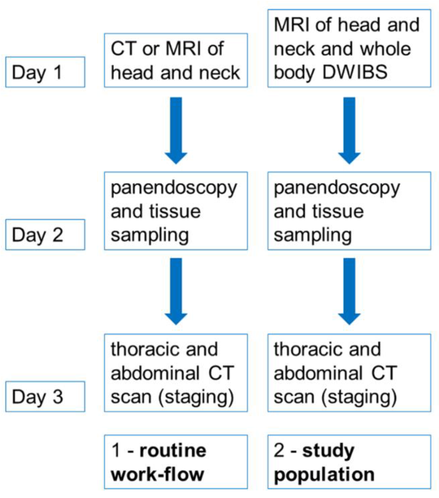
Figure 1. Routine diagnostic work-flow of patients with suspected head and neck cancer due to suspicious cervical lymphadenopathy compared to patients of our study population.
Tomography 08 00210 g001
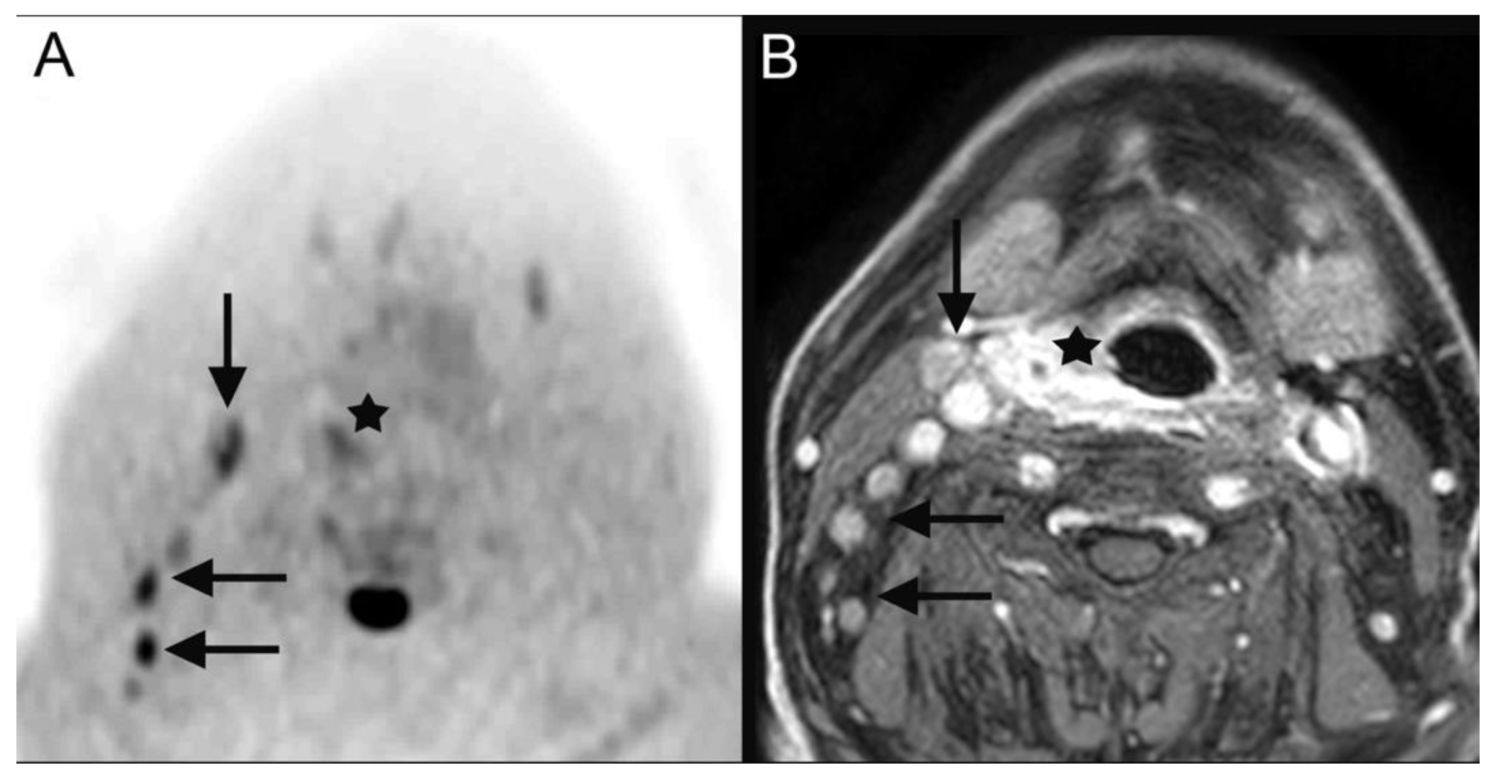
Figure 2. DWIBS (A) as well as contrast-enhanced fat-saturated T1-weighted MR images (B) depict the primary cancer (A,B: black star) lesion as well as the nodal metastasis (A,B: black arrows) in a 70-year-old male patient who was diagnosed with a right-sided hypopharyngeal carcinoma (cT2 cN2b cM0).
Tomography 08 00210 g002
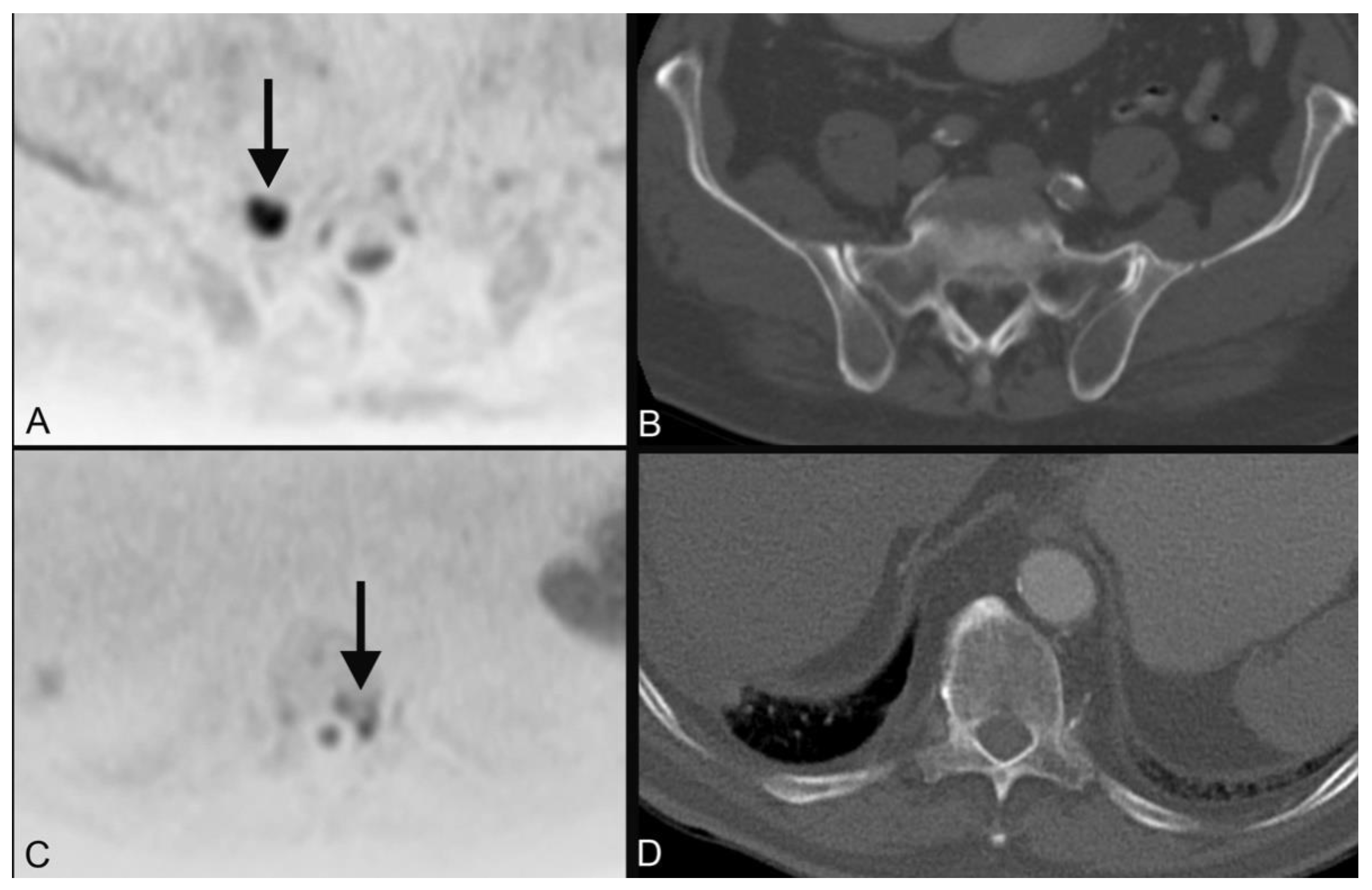
Figure 3. An 85-year-old male patient who was diagnosed with cancer of the right palatine tonsil (cT1 cN2b cM0) showed diffuse spotty diffusion restrictions in DWIBS of the skeletal system with the largest lesions in the right sacral mass (A, black arrow) and the 11th thoracic vertebrae on the left (C, black arrow) without correlate in the CT (B,D) and was later diagnosed with multiple myeloma.
Tomography 08 00210 g003
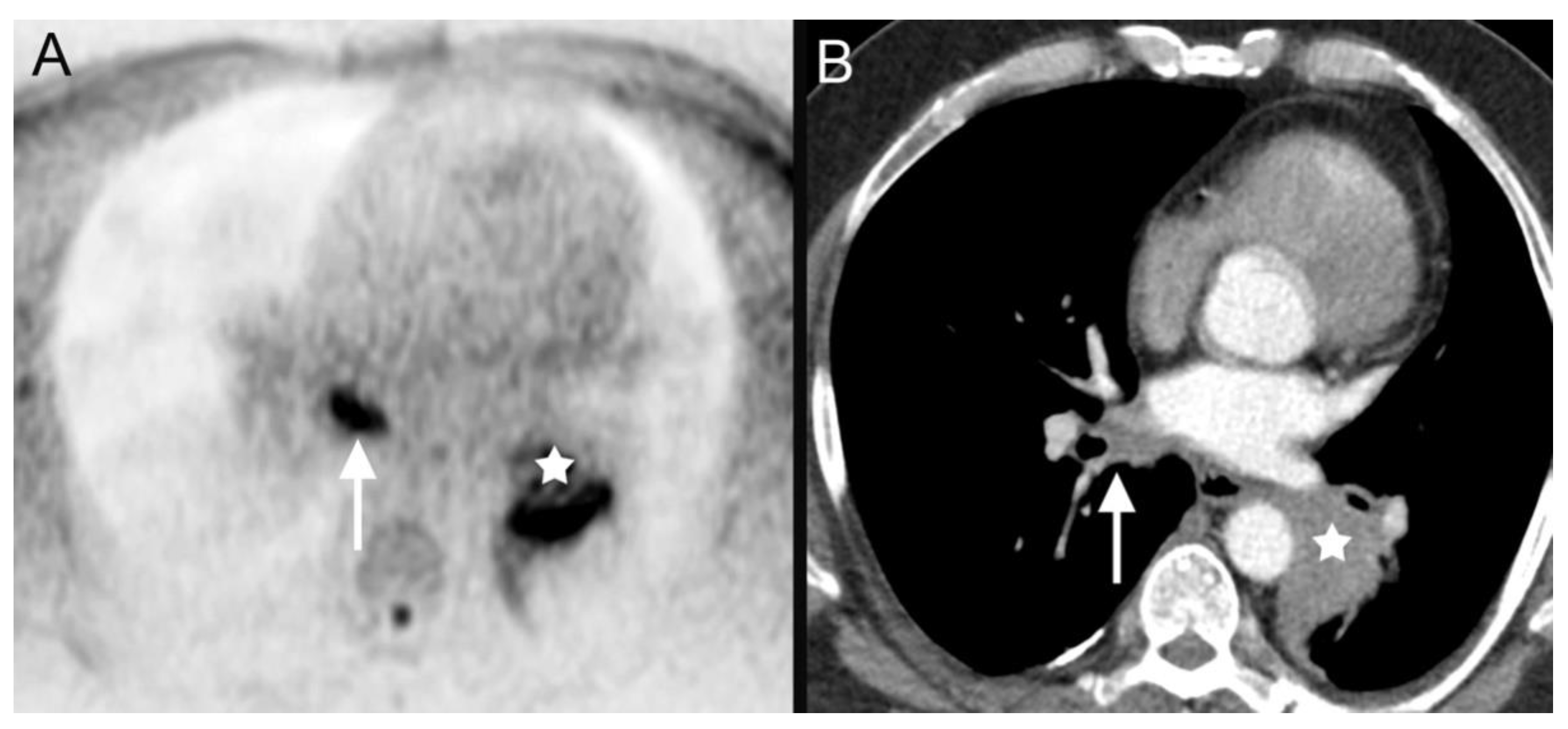
Figure 4. DWIBS (A) as well as contrast-enhanced thoracal CT (B) depicts the primary cancer lesion (A,B: white star) as well as the nodal metastases (A,B: white arrows) in a 60-year-old male patient who was diagnosed with a non-small cell lung cancer (cT2b cN3 cM0) in the diagnostic work-up of clinically suspected head and neck carcinoma.
Tomography 08 00210 g004
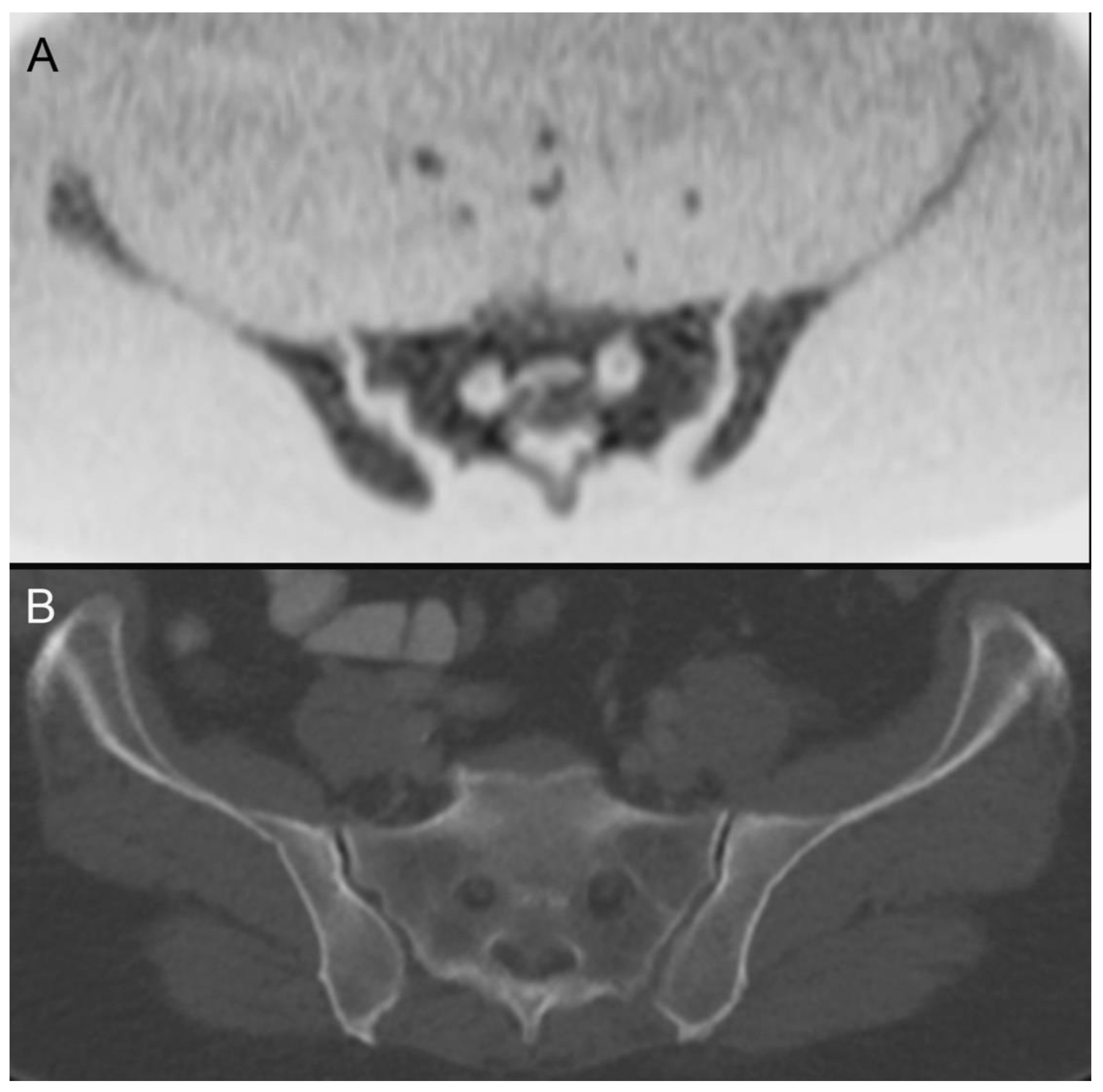
Figure 5. DWIBS (A) contrary to contrast-enhanced abdominal CT (B) reveals diffuse diffusion restriction of the bone marrow with no abnormalities in CT, which lead to the diagnosis of acute myeloid leukemia after further clinical work-up in this 76-year-old female patient.
Tomography 08 00210 g005
Table 1. Risk factors.
Alcohol Consumption (n/%)History of Smoking (n/%)HPV+ (n/%)
13/43%21/70%6/20%
Table 2. Final diagnosis and TNM stage as well as histopathologic subtype of all included patients with clinically suspected head and neck carcinoma and cervical nodal swelling.
Patient NumberDiagnosistnm StageHistopathology
1tonsil carcinomapT1 cN2b cM0squamous cell carcinoma
2laryngeal carcinomacT2 cN1 cM0squamous cell carcinoma
3tonsil carcinomapT1 pN3b cM0squamous cell carcinoma
4 bronchial carcinomanot applicableadenocarcinoma
5CUP syndromepTx pN+ cM0squamous cell carcinoma
6CUP syndromepTx pN+ cM0squamous cell carcinoma
7parotid cancerpT3 pN2b cM0squamous cell carcinoma
8laryngeal carcinomacT4a cN2a cM0squamous cell carcinoma
9tonsil carcinomapT1 pN2b cM0squamous cell carcinoma
10hypopharyngeal carcinomacT4a cN2b cM0squamous cell carcinoma
11tonsil carcinomacT4a cN2c cM0squamous cell carcinoma
12CUP syndromepTx pN+ cM0squamous cell carcinoma
13tongue base carcinomapT1 pN2b cM0squamous cell carcinoma
14CUP syndromepTx pN+ cM0squamous cell carcinoma
15tonsil carcinomacT2 cN2c cM0squamous cell carcinoma
16lymphomanot applicableanaplastic large cell lymphoma
17tonsil carcinomapT1 pN2b cM0squamous cell carcinoma
18leukemianot applicableacute myeloid leukemia
19bronchial carcinomanot applicablesquamous cell carcinoma
20hypopharyngeal carcinomapT2 pN3b cM1squamous cell carcinoma
21tongue base carcinomapT1 cN2b cM0squamous cell carcinoma
22metastatic melanomanot applicablemalignant melanoma
23CUP syndromepTx pN+ cM0squamous cell carcinoma
24hypopharyngeal carcinomacT3 cN2b cM0squamous cell carcinoma
25thyroid carcinomapT4a pN1b cM1follicular thyroid carcinoma
26lymphomanot applicablefollicular b-cell lymphoma
27epiglottic carcinomacT1 cN2b cM0squamous cell carcinoma
28hypopharyngeal carcinoma (Figure 2)pT2 pN2b cM0squamous cell carcinoma
29hypopharyngeal carcinomacT4a cN2c cM0squamous cell carcinoma
30sinunasal carcinomacT2 cN2b cM0neuroendocrine carcinoma
Table 3. Additional diagnosis made with computed tomography (CT) and/or whole-body DWIBS MRI in the routine work-up of pretherapeutic staging.
Patient NumberAdditional Diagnosis CT OnlyAdditional Diagnosis DWIBS OnlyAdditional Diagnosis in Both Modalities
1 plasmocytoma (Figure 3)
4 bronchial carcinoma (Figure 4)
10 esophageal cancer
16 breast lymphoma
18 leukemia (Figure 5)
19bronchial carcinoma
20 single liver metastases
22 several metastases of malignant melanoma
25 several metastases of thyroid cancer
Table 4. Excerpt of the further therapeutic course of the 30 included patients based on the decision of the institutional multidisciplinary tumor conference.
Patient NumberDiagnosisTherapeutic Decision (Multidisciplinary Tumor Conference)
1tonsil carcinomasurgical resection, adjuvant chemoradiotherapy
2laryngeal carcinomaprimary chemoradiotherapy
3tonsil carcinomasurgical resection, adjuvant chemoradiotherapy
4 bronchial carcinomasystemic chemotherapy
5CUP syndromeprimary chemoradiotherapy
6CUP syndromeprimary chemoradiotherapy
7parotid cancersurgical resection only as patient denies chemotherapy as well as radiotherapy
8laryngeal carcinomaprimary chemoradiotherapy as patient denies tracheotomy
9tonsil carcinomasurgical resection, adjuvant chemoradiotherapy
10hypopharyngeal carcinomapalliative radiotherapy
11tonsil carcinomabest supportive care
12CUP syndromeprimary chemoradiotherapy
13tongue base carcinomasurgical resection, adjuvant chemoradiotherapy
14CUP syndromeprimary chemoradiotherapy
15tonsil carcinomaprimary chemoradiotherapy
16lymphomachemoradiotherapy
17tonsil carcinomasurgical resection, adjuvant chemoradiotherapy
18leukemiachemotherapy
19bronchial carcinomasystemic chemotherapy
20hypopharyngeal carcinomapalliative chemotherapy
21tongue base carcinomasurgical resection, adjuvant chemoradiotherapy
22metastatic melanomaimmunotherapy
23CUP syndromeprimary chemoradiotherapy
24hypopharyngeal carcinomaprimary chemoradiotherapy
25thyroid carcinomasurgical resection and radio-iodine treatment
26lymphomachemoradiotherapy
27epiglottic carcinomasurgical resection, adjuvant chemoradiotherapy
28hypopharyngeal carcinoma (Figure 2)primary chemoradiotherapy
29hypopharyngeal carcinomabest supportive care
30sinunasal carcinomaneoadjuvant chemoradiotherapy, surgical resection
Publisher’s Note: MDPI stays neutral with regard to jurisdictional claims in published maps and institutional affiliations.