Article

Novel Deep CNNs Explore Regions, Boundaries, and Residual Learning for COVID-19 Infection Analysis in Lung CT


by
Bader Khalid Alshemaimri
Software Engineering Department, College of Computing and Information Sciences, King Saud University, Riyadh 11671, Saudi Arabia
Tomography 2024, 10(8), 1205-1221; https://doi.org/10.3390/tomography10080091

Submission received: 2 June 2024 / Revised: 6 July 2024 / Accepted: 17 July 2024 / Published: 3 August 2024
(This article belongs to the Section Artificial Intelligence in Medical Imaging)

Abstract

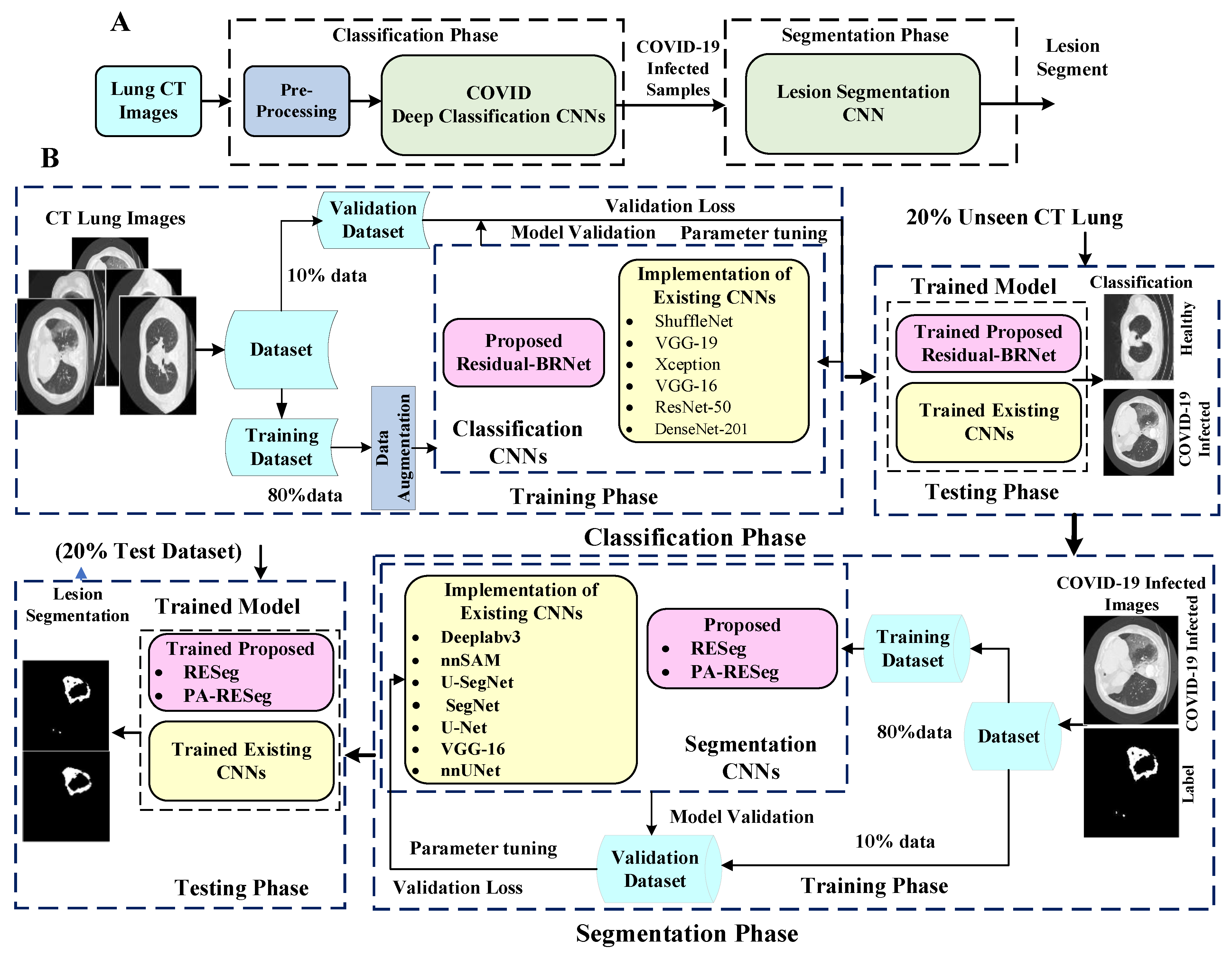
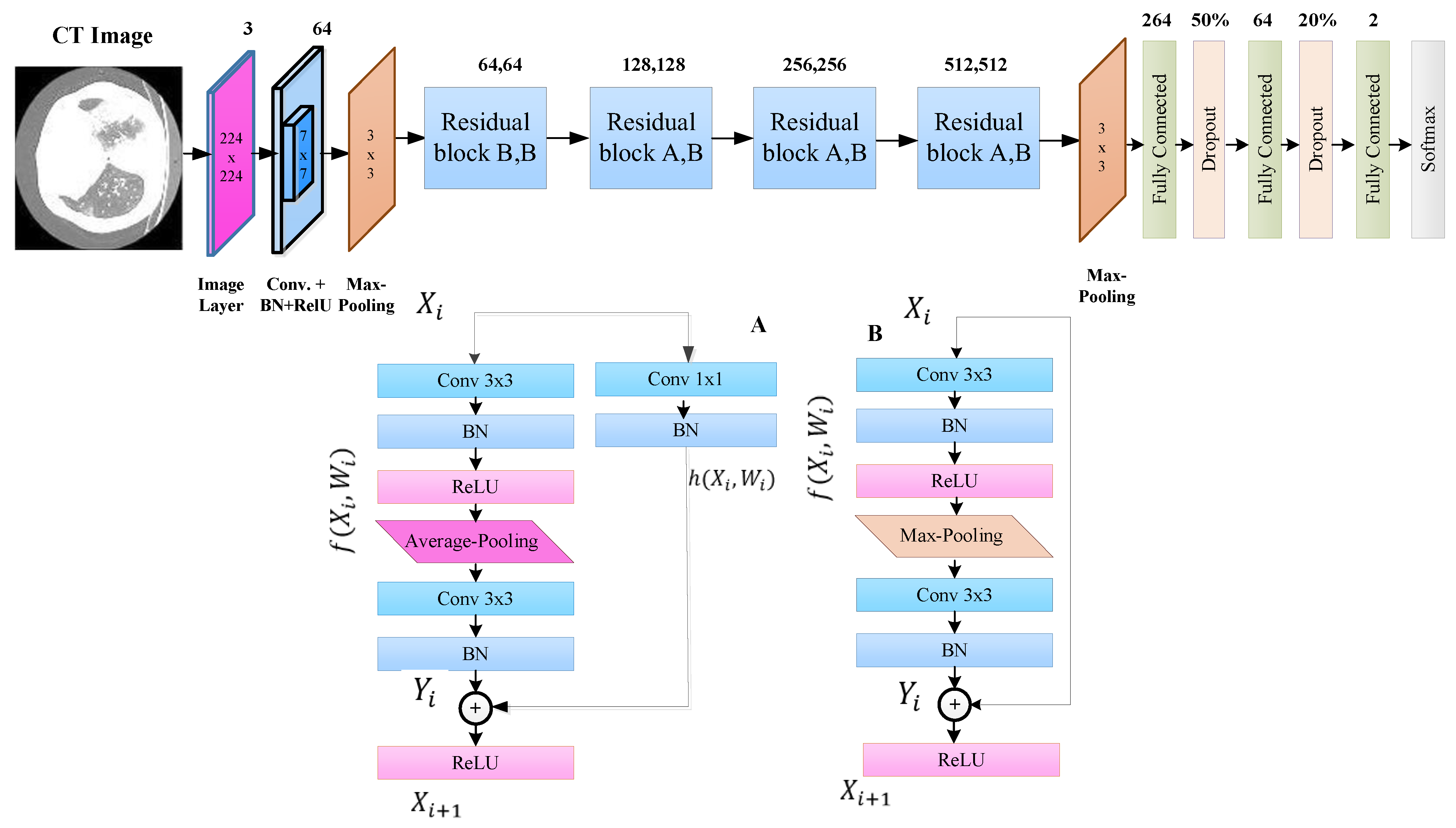
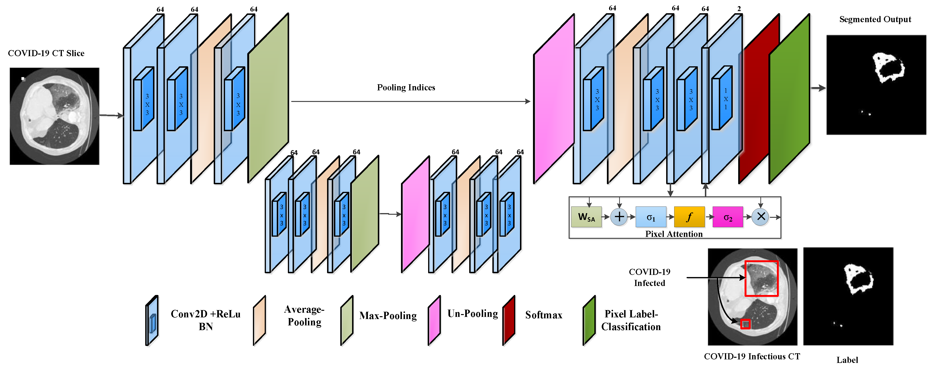
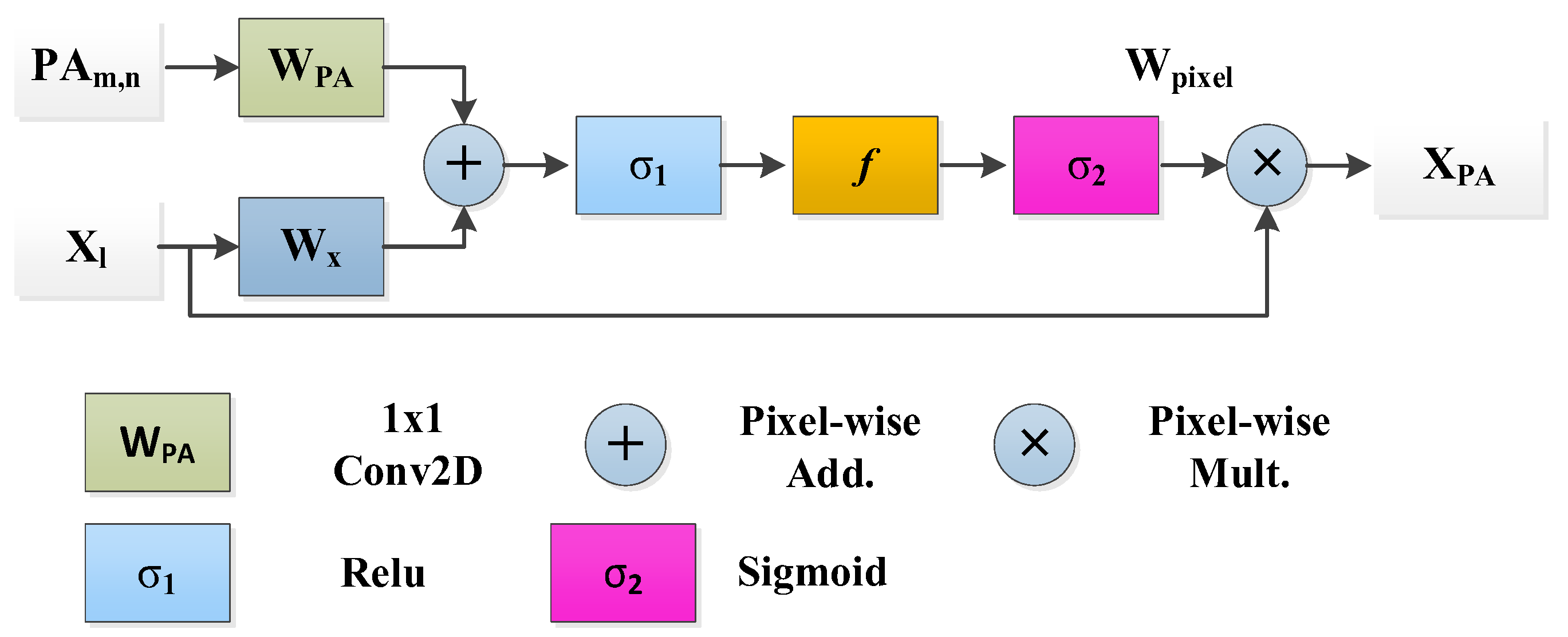
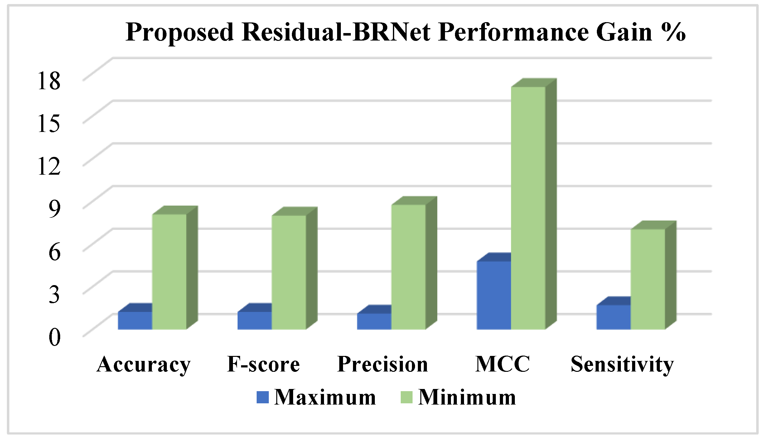
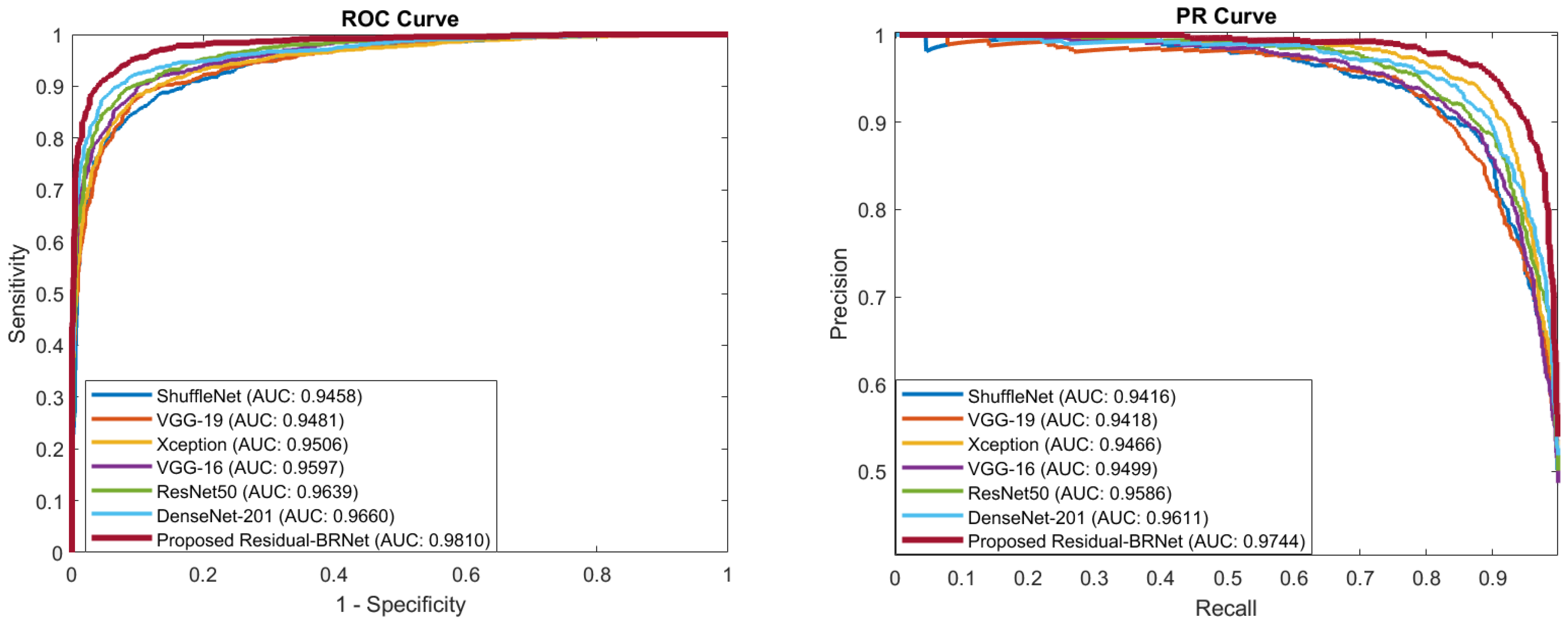
COVID-19 poses a global health crisis, necessitating precise diagnostic methods for timely containment. However, accurately delineating COVID-19-affected regions in lung CT scans is challenging due to contrast variations and significant texture diversity. In this regard, this study introduces a novel two-stage classification and segmentation CNN approach for COVID-19 lung radiological pattern analysis. A novel Residual-BRNet is developed to integrate boundary and regional operations with residual learning, capturing key COVID-19 radiological homogeneous regions, texture variations, and structural contrast patterns in the classification stage. Subsequently, infectious CT images undergo lesion segmentation using the newly proposed RESeg segmentation CNN in the second stage. The RESeg leverages both average and max-pooling implementations to simultaneously learn region homogeneity and boundary-related patterns. Furthermore, novel pixel attention (PA) blocks are integrated into RESeg to effectively address mildly COVID-19-infected regions. The evaluation of the proposed Residual-BRNet CNN in the classification stage demonstrates promising performance metrics, achieving an accuracy of 97.97%, F1-score of 98.01%, sensitivity of 98.42%, and MCC of 96.81%. Meanwhile, PA-RESeg in the segmentation phase achieves an optimal segmentation performance with an IoU score of 98.43% and a dice similarity score of 95.96% of the lesion region. The framework’s effectiveness in detecting and segmenting COVID-19 lesions highlights its potential for clinical applications.
Keywords: COVID-19; CT scan; CNN; region; boundary; residual; transfer learning; classification; segmentation COVID-19; CT scan; CNN; region; boundary; residual; transfer learning; classification; segmentation