

Biomimetic Scaffolds Based on Mn2+-, Mg2+-, and Sr2+-Substituted Calcium Phosphates Derived from Natural Sources and Polycaprolactone


Abstract
1. Introduction
2. Experimental
2.1. Materials
2.2. Characterization Techniques
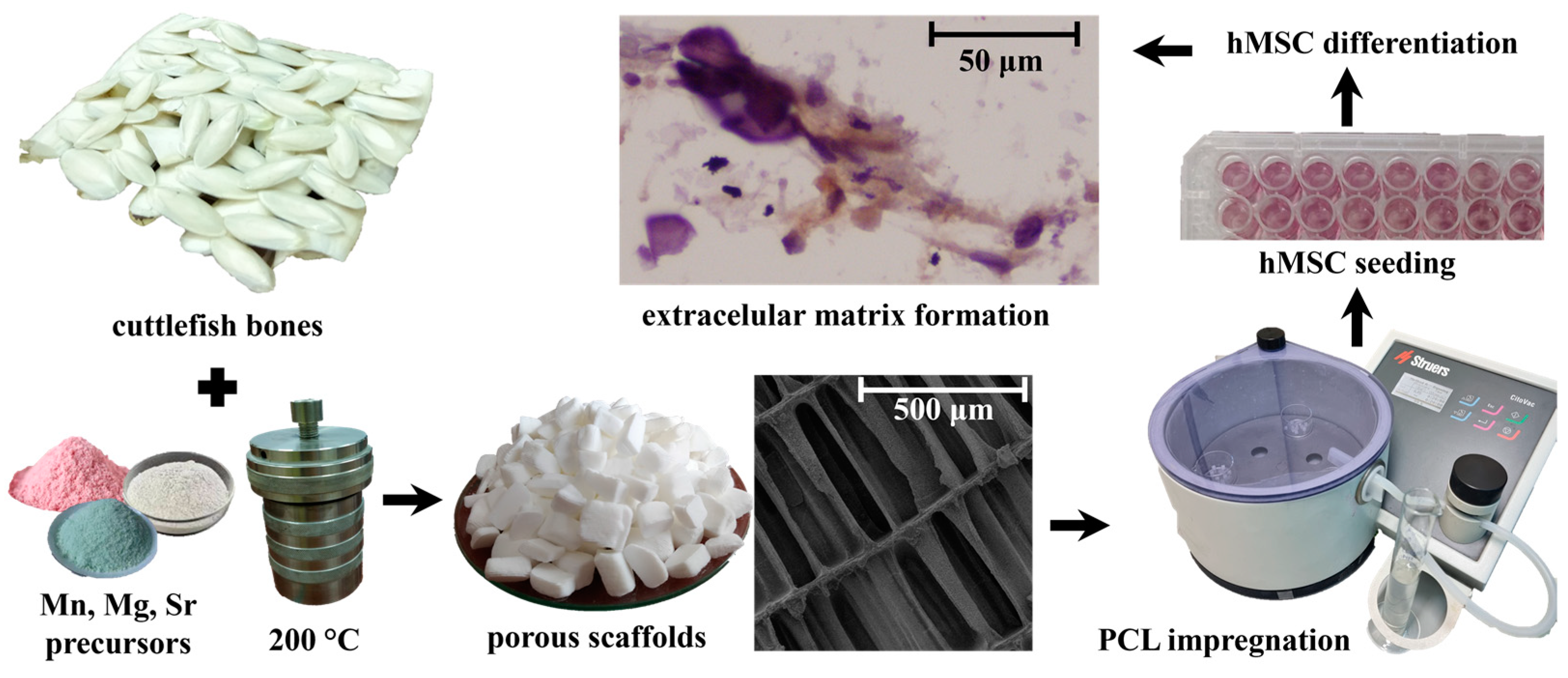
2.3. Preparation of Composite Scaffolds
2.4. Stem Cell Studies
2.5. Statistical Analysis
3. Results
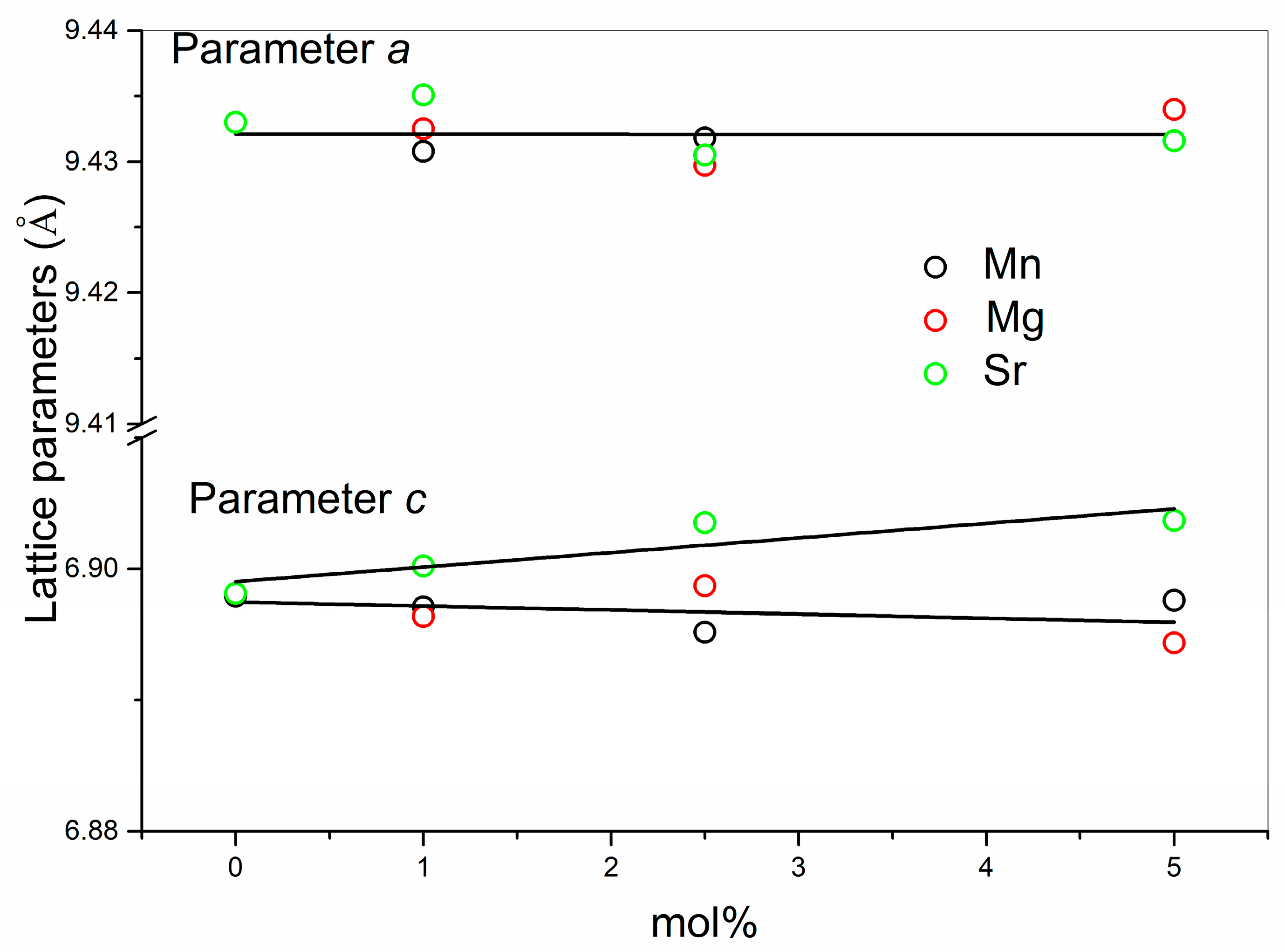
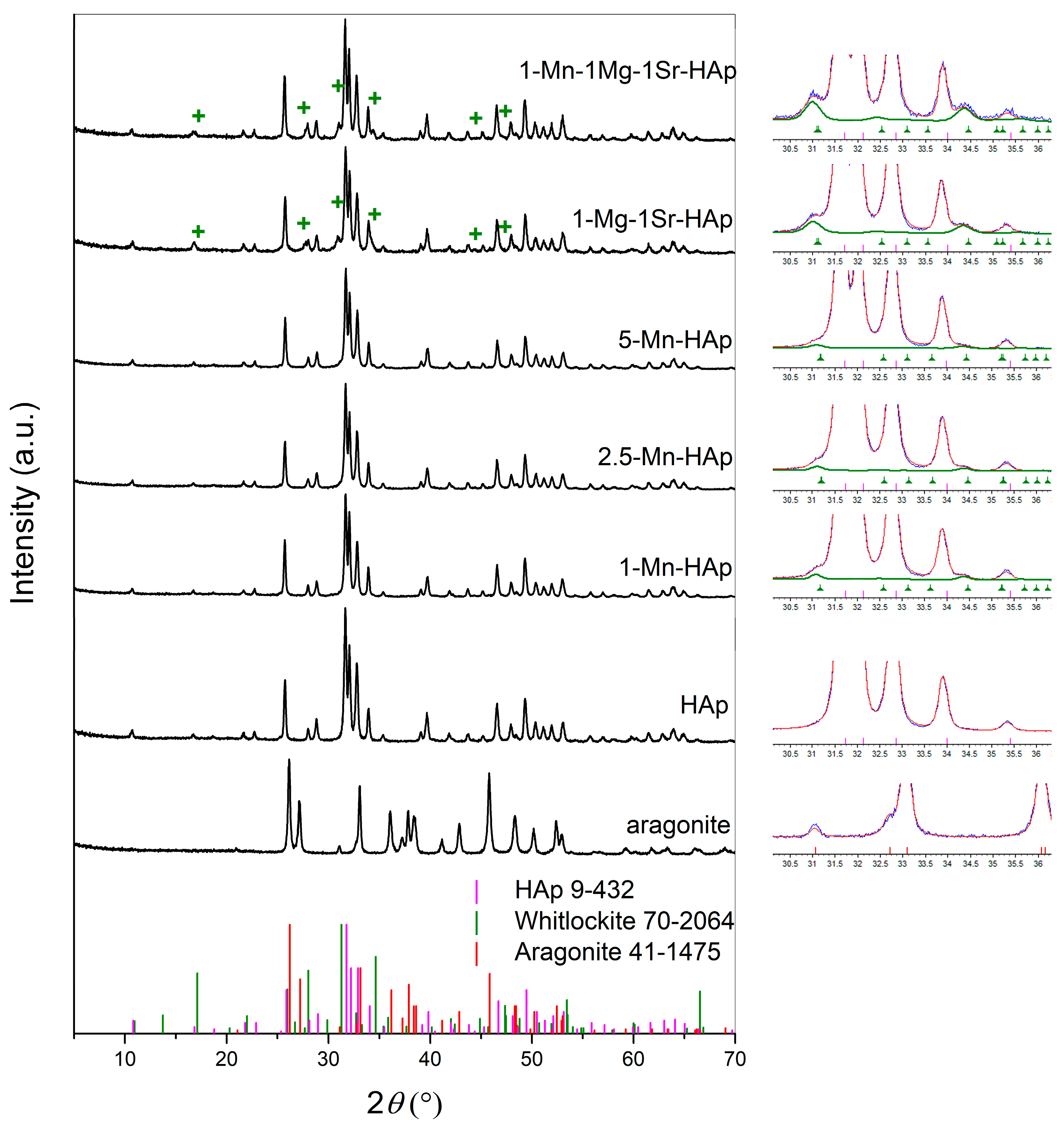
3.1. XRD Analysis
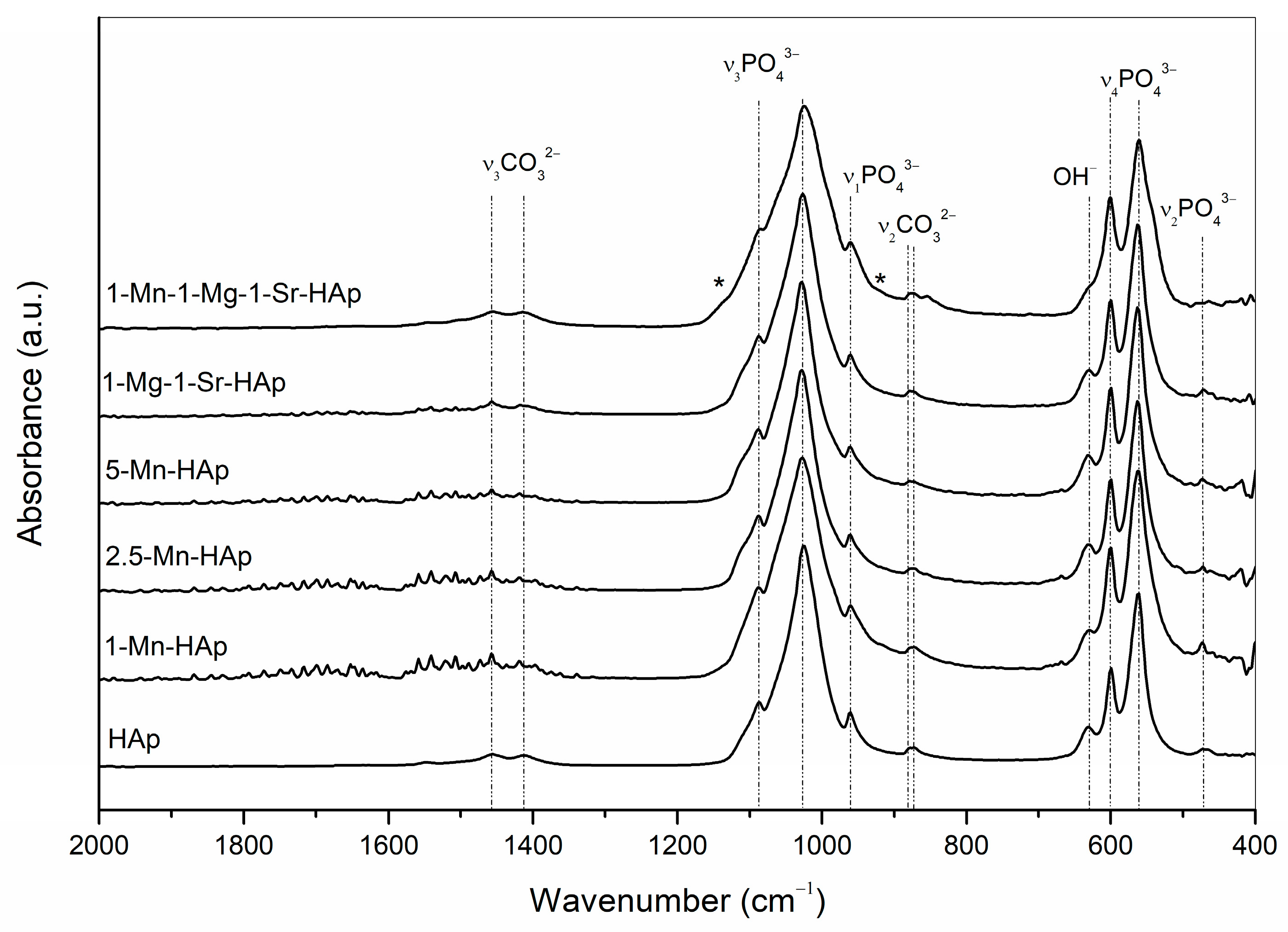
3.2. FTIR Analysis
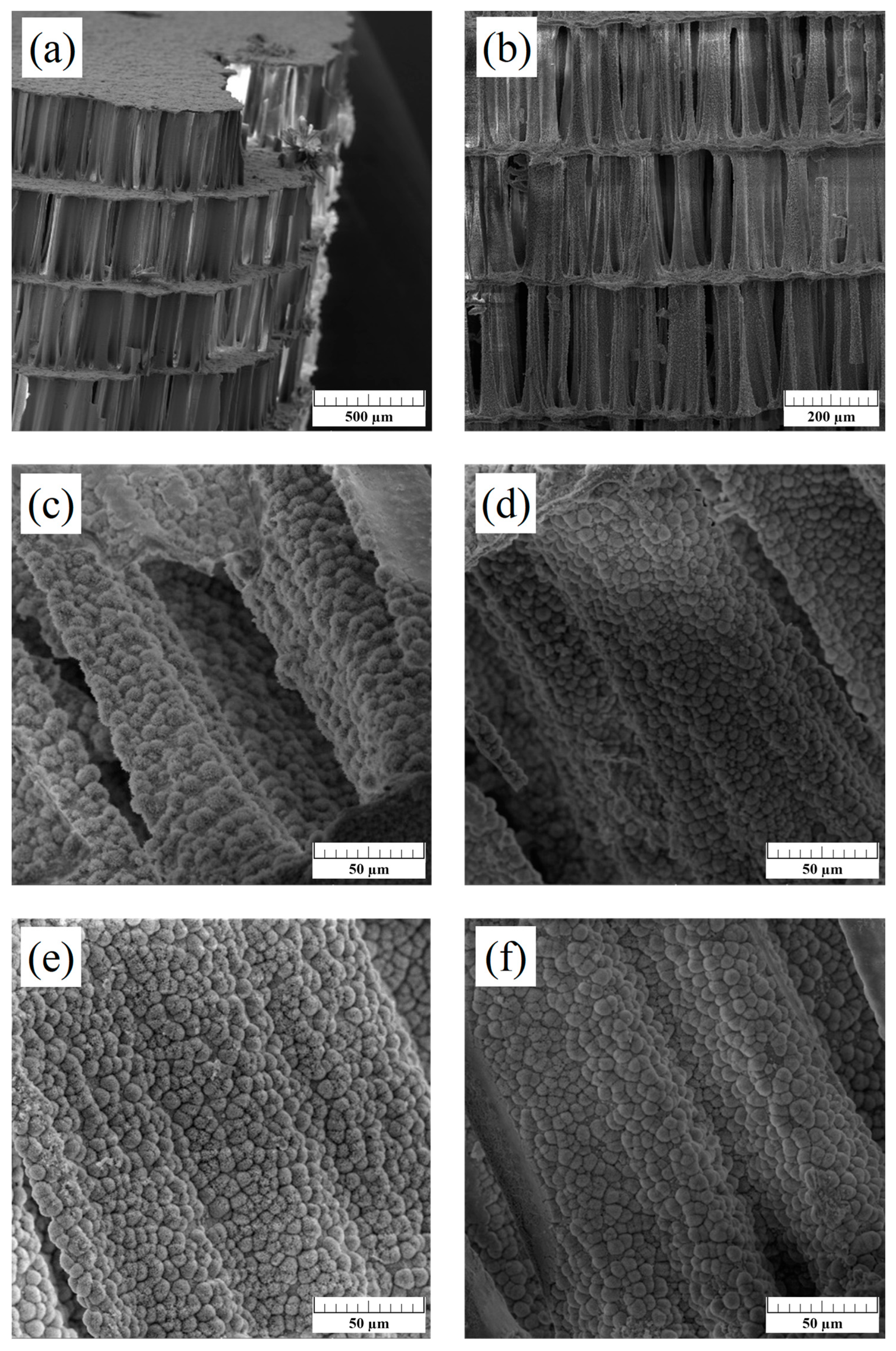
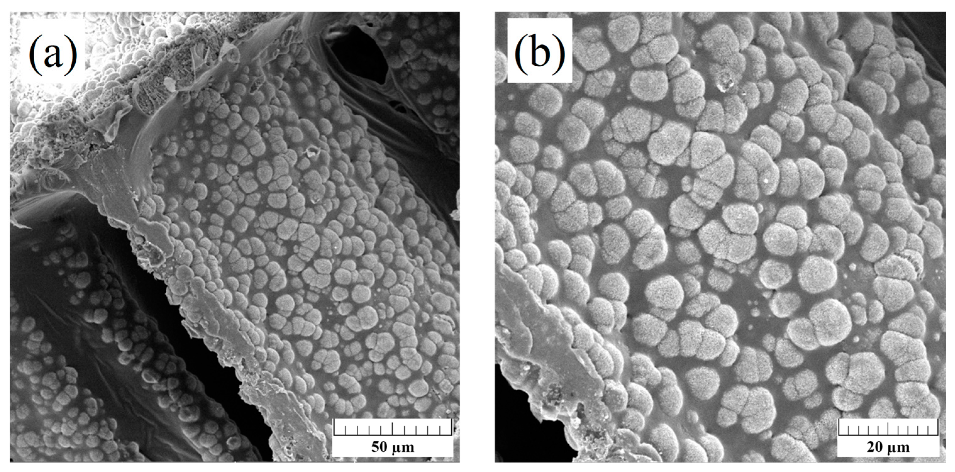
3.3. SEM Analysis
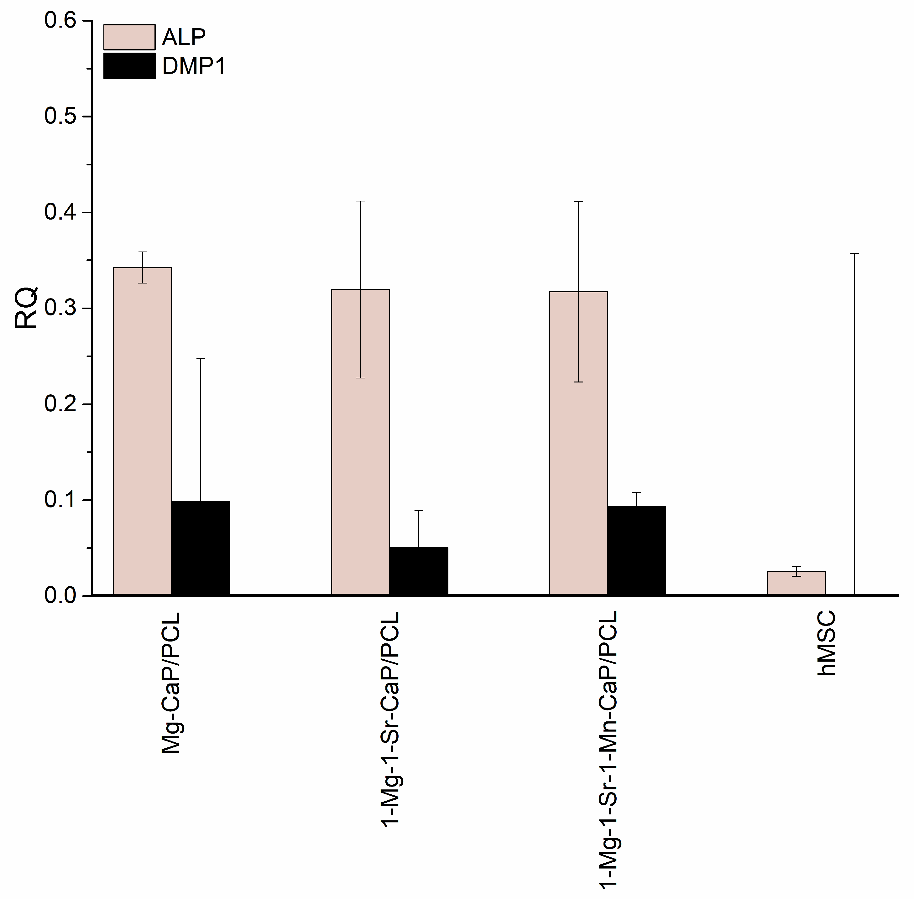
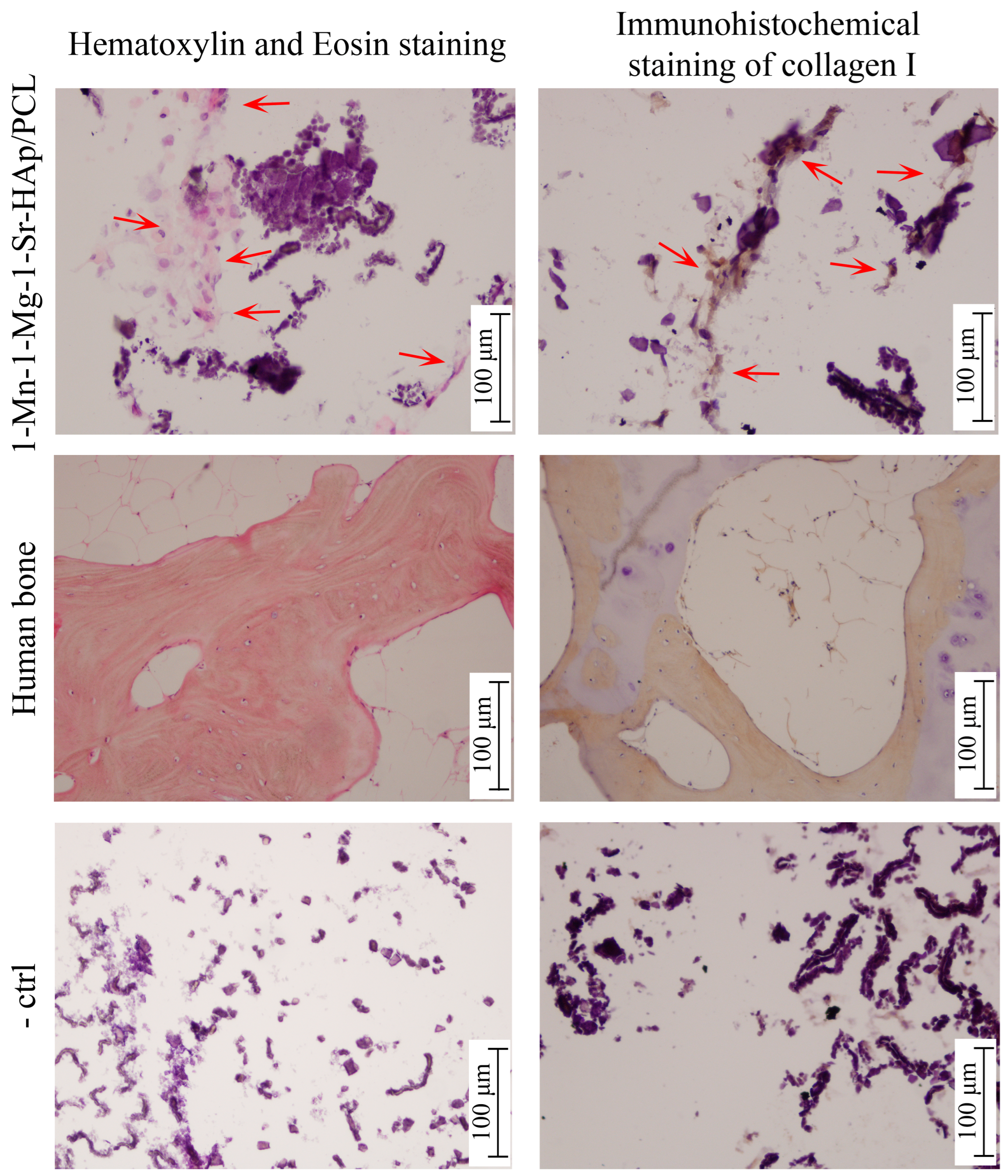
3.4. Stem Cell Studies
4. Discussion
5. Conclusions
Author Contributions
Funding
Institutional Review Board Statement
Informed Consent Statement
Data Availability Statement
Acknowledgments
Conflicts of Interest
References
- Wang, W.; Yeung, K.W.K. Bone grafts and biomaterials substitutes for bone defect repair: A review. Bioact. Mater. 2017, 2, 224–247. [Google Scholar] [CrossRef]
- Hutmacher, D.W. Scaffolds in Tissue Engineering Bone and Cartilage. Biomaterials 2001, 21, 2529–2543. [Google Scholar] [CrossRef]
- Ielo, I.; Calabrese, G.; De Luca, G.; Conoci, S. Recent Advances in Hydroxyapatite-Based Biocomposites for Bone Tissue Regeneration in Orthopedics. Int. J. Mol. Sci. 2022, 23, 9721. [Google Scholar] [CrossRef]
- Boanini, E.; Gazzano, M.; Bigi, A. Ionic substitutions in calcium phosphates synthesized at low temperature. Acta Biomater. 2010, 6, 1882–1894. [Google Scholar] [CrossRef]
- Ratnayake, J.T.B.; Mucalo, M.; Dias, G.J. Substituted Hydroxyapatites for Bone Regeneration: A Review of Current Trends. J. Biomed. Mater. Res. Part B 2017, 105, 1285–1299. [Google Scholar] [CrossRef]
- O’Neill, E.; Awale, G.; Daneshmandi, L.; Umerah, O.; Lo, K.W.-H. The roles of ions on bone regeneration. Drug Discov. Today 2018, 23, 879–890. [Google Scholar] [CrossRef]
- Nabiyouni, M.; Brückner, T.; Zhou, H.; Gbureck, U.; Bhaduri, S.B. Magnesium-based bioceramics in orthopedic applications. Acta Biomater. 2018, 66, 23–43. [Google Scholar] [CrossRef]
- Anwar, A.; Akbar, S. Novel continuous microwave assisted flow synthesis of nanosized manganese substituted hydroxyapatite. Ceram Int. 2018, 44, 10878–10882. [Google Scholar] [CrossRef]
- Frasnelli, M.; Cristofaro, F.; Sglavo, V.M.; Dirè, S.; Callone, E.; Ceccato, R.; Bruni, G.; Cornaglia, A.I.; Visai, L. Synthesis and characterization of strontium-substituted hydroxyapatite nanoparticles for bone regeneration. Mater. Sci. Eng. C 2017, 71, 653–662. [Google Scholar] [CrossRef] [PubMed]
- Legeros, R.Z.; Kijkowska, R.; Bautista, C.; Legeros, J.P. Synergistic effects of magnesium and carbonate on properties of biological and synthetic apatites. Connect. Tissue Res. 1995, 33, 203–209. [Google Scholar] [CrossRef]
- Castiglioni, S.; Cazzaniga, A.; Albisetti, W.; Maier, J.A.M. Magnesium and Osteoporosis: Current State of Knowledge and Future Research Directions. Nutrients 2013, 5, 3022–3033. [Google Scholar] [CrossRef]
- Marie, P.J. Strontium ranelate: A novel mode of action optimizing bone formation and resorption. Osteoporos. Int. 2005, 16, S7–S10. [Google Scholar] [CrossRef]
- Bonnelye, E.; Chabadel, A.; Saltel, F.; Jurdic, P. Dual effect of strontium ranelate: Stimulation of osteoblast differentiation and inhibition of osteoclast formation and resorption in vitro. Bone 2008, 42, 129–138. [Google Scholar] [CrossRef]
- Bigi, A.; Boanini, E.; Capuccini, C.; Gazzano, M. Strontium-substituted hydroxyapatite nanocrystals. Inorganica Chim. Acta 2007, 360, 1009–1016. [Google Scholar] [CrossRef]
- Torres, P.M.C.; Vieira, S.I.; Cerqueir, A.R.; Pina, S.; da Cruz Silva, O.A.; Abrantes, J.C.C.; Ferreira, J.M. Effects of Mn-doping on the structure and biological properties of beta-tricalcium phosphate. J. Inorg. Biochem. 2014, 136, 57–66. [Google Scholar] [CrossRef]
- Huang, Y.; Ding, Q.Q.; Han, S.G.; Yan, Y.J.; Pang, X.F. Characterisation, corrosion resistance and in vitro bioactivity of manganese-doped hydroxyapatite films electrodeposited on titanium. J. Mater. Sci. Mater. Med. 2013, 24, 1853–1864. [Google Scholar] [CrossRef]
- Szurkowska, K.; Drobniewska, A.; Kolmas, J. Dual doping of silicon and manganese in hydroxyapatites: Physicochemical properties and preliminary biological studies. Materials 2019, 12, 2566. [Google Scholar] [CrossRef]
- Armulik, A.; Svineng, G.; Wennerberg, K.; Faessler, R.; Johansson, S. Expression of Integrin Subunit β1B in Integrin β1-Deficient GD25 Cells Does Not Interfere with αVβ3 Functions. Exp. Cell. Res. 2000, 254, 55–63. [Google Scholar] [CrossRef]
- Li, J.M.; Deng, C.J.; Liang, W.Y.; Kang, F.; Bai, Y.; Ma, B.; Wu, C.; Dong, S. Mn-containing bioceramics inhibit osteoclastogenesis and promote osteoporotic bone regeneration via scavenging ROS. Bioact. Mater. 2021, 6, 3839–3850. [Google Scholar] [CrossRef]
- Milovac, D.; Gallego Ferrer, G.; Ivanković, M.; Ivanković, H. PCL-coated hydroxyapatite scaffold derived from cuttlefish bone: Morphology, mechanical properties and bioactivity. Mater. Sci. Eng. C 2014, 34, 437–445. [Google Scholar] [CrossRef]
- Chuenjitkuntaworn, B.; Inrung, W.; Damrongsri, D.; Mekaapiruk, K.; Supaphol, P.; Pavasant, P. Polycaprolactone/hydroxyapatite composite scaffolds: Preparation, characterization, and in vitro and in vivo biological responses of human primary bone cells. J. Biomed. Mater. Res. A 2010, 94, 241–251. [Google Scholar] [CrossRef]
- Yang, X.; Wang, Y.; Zhou, Y.; Chen, J.; Wan, Q. The Application of Polycaprolactone in Three-Dimensional Printing Scaffolds for Bone Tissue Engineering. Polymers 2021, 13, 2754. [Google Scholar] [CrossRef]
- Antoniac, I.V.; Lesci, I.G.; Blajan, A.I.; Vitioanu, G.; Antoniac, A. Bioceramics and Biocomposites from Marine Sources. Key Eng. Mater. 2016, 672, 276–292. [Google Scholar] [CrossRef]
- Palaveniene, A.; Tamburaci, S.; Kimna, C.; Glambaite, K.; Baniukaitiene, O.; Tihminlioğlu, F.; Liesiene, J. Osteoconductive 3D porous composite scaffold from regenerated cellulose and cuttlebone-derived hydroxyapatite. J. Biomater. Appl. 2019, 33, 876–890. [Google Scholar] [CrossRef]
- Borciani, G.; Fischetti, T.; Ciapetti, G.; Montesissa, M.; Baldini, N.; Graziani, G. Marine biological waste as a source of hydroxyapatite for bone tissue engineering applications. Ceram. Int. 2023, 49, 1572–1584. [Google Scholar] [CrossRef]
- Bauer, L.; Antunović, M.; Gallego-Ferrer, G.; Ivanković, M.; Ivanković, H. PCL-Coated Multi-Substituted Calcium Phosphate Bone Scaffolds with Enhanced Properties. Materials 2021, 14, 4403. [Google Scholar] [CrossRef]
- Bauer, L.; Antunović, M.; Rogina, A.; Ivanković, M.; Ivanković, H. Bone-mimetic porous hydroxyapatite/whitlockite scaffolds: Preparation, characterization and interactions with human mesenchymal stem cells. J. Mater. Sci. 2021, 56, 3947–3969. [Google Scholar] [CrossRef]
- Bruker. TOPAS V5: General Profile and Structure Analysis Software for Powder Diffraction Data; Bruker: Karlsruhe, Germany, 2014. [Google Scholar]
- Sudarsanan, K.; Young, R.A. Significant precision in crystal structural details. Holly Springs hydroxyapatite. Acta Crystallogr. Sect. B Struct. Crystallogr. Cryst. Chem. 1969, 25, 1534–1543. [Google Scholar] [CrossRef]
- Gopal, R.; Calvo, C.; Ito, J.; Sabine, W.K. Crystal Structure of Synthetic Mg-Whitlockite, Ca18Mg2H2(PO4)4. Can. J. Chem. 1974, 52, 1155–1164. [Google Scholar] [CrossRef]
- Matić, I.; Antunović, M.; Brkić, S.; Josipović, P.; Caput Mihalić, K.; Karlak, I.; Ivković, A.; Marijanović, I. Expression of OCT-4 and SOX-2 in Bone Marrow-Derived Human Mesenchymal Stem Cells during Osteogenic Differentiation. Open Access Maced. J. Med. Sci. 2016, 4, 9–16. [Google Scholar] [CrossRef]
- Rogina, A.; Antunović, M.; Pribolšan, L.; Caput Mihalić, K.; Vukasović, A.; Ivković, A.; Marijanović, I.; Gallego Ferrer, G.; Ivanković, M.; Ivanković, H. Human Mesenchymal Stem Cells Differentiation Regulated by Hydroxyapatite Content within Chitosan-Based Scaffolds under Perfusion Conditions. Polymers 2017, 9, 387. [Google Scholar] [CrossRef]
- Capitelli, F.; Bosi, F.; Capelli, S.C.; Radica, F.; Ventura, G.D. Neutron and XRD Single- Crystal Diffraction Study and Vibrational Properties of Whitlockite, the Natural Counterpart of Synthetic Tricalcium Phosphate. Crystals 2021, 11, 225. [Google Scholar] [CrossRef]
- Ivanković, H.; Tkalčec, E.; Orlić, S.; Gallego Ferrer, G.; Schauperl, Z. Hydroxyapatite formation from cuttlefish bones: Kinetics. J. Mater. Sci. Mater. Med. 2010, 21, 2711–2722. [Google Scholar] [CrossRef]
- Deligianni, D.D.; Katsala, N.D.; Koutsoukos, P.G.; Missirlis, Y.F. Effect of surface roughness of hydroxyapatite on human bone marrow cell adhesion, proliferation, differentiation and detachment strength. Biomaterials 2001, 22, 87–96. [Google Scholar] [CrossRef]
- Vecstaudža, J.; Gasik, M.; Ločs, J. Amorphous Calcium Phosphate Materials: Formation, Structure and Thermal Behaviour. J. Eur. Ceram. Soc. 2019, 39, 1642–1649. [Google Scholar] [CrossRef]
- Cheng, H.; Chabok, R.; Guan, X.F.; Chawla, A.; Li, Y.X.; Khademhosseini, A.; Jang, H.L. Synergistic interplay between the two major bone minerals, hydroxyapatite and whitlockite nanoparticles, for osteogenic differentiation of mesenchymal stem cells. Acta Biomater. 2018, 69, 342–351. [Google Scholar] [CrossRef]
- Bracci, B.; Torricelli, P.; Panzavolta, S.; Boanini, E.; Giardino, R.; Bigi, A. Effect of Mg(2+), Sr(2+), and Mn(2+) on the chemico-physical and in vitro biological properties of calcium phosphate biomimetic coatings. J. Inorg. Biochem. 2009, 103, 1666–1674. [Google Scholar] [CrossRef]
- Karunakaran, G.; Cho, E.B.; Thirumurugan, K.; Kumar, G.S.; Kolesnikov, E.; Boobalan, S.; Janarthanan, G.; Pillai, M.M.; Rajendran, S. Mesoporous Mn-doped hydroxyapatite nanorods obtained via pyridinium chloride enabled microwave-assisted synthesis by utilizing Donax variabilis seashells for implant applications. Mater. Sci. Eng. C 2021, 126, 112170. [Google Scholar] [CrossRef]
- Da Silva, L.M.; dos Santos Tavares, D.; dos Santos, E.A. Isolating the Effects of Mg2+, Mn2+ and Sr2+ Ions on Osteoblast Behavior from those Caused by Hydroxyapatite Transformation. Mat. Res. 2020, 23, e20200083. [Google Scholar] [CrossRef]
- Ren, F.; Ding, Y.; Leng, Y. Infrared spectroscopic characterization of carbonated apatite: A combined experimental and computational study. J. Biomed. Mater. Res. A 2014, 102, 496–505. [Google Scholar] [CrossRef] [PubMed]
- Bigi, A.; Cojazzi, G.; Panzavolta, S.; Ripamonti, A.; Roveri, N.; Romanello, M.; Suarez, K.N.; Moro, L. Chemical and structural characterization of the mineral phase from cortical and trabecular bone. J. Inorg. Biochem. 1997, 68, 45–51. [Google Scholar] [CrossRef] [PubMed]
- Wopenka, B.; Pasteris, J.D. A mineralogical perspective on the apatite in bone. Mater. Sci. Eng. C 2005, 25, 131–143. [Google Scholar] [CrossRef]
- Ellies, L.G.; Carter, J.M.; Natiella, J.R.; Featherstone, J.D.B.; Nelson, D.G.A. Quantitative analysis of early in vivo tissue response to synthetic apatite implants. J. Biomed. Mater. Res. A 1988, 22, 137–148. [Google Scholar] [CrossRef]
- Ruffini, A.; Sprio, S.; Preti, L.; Tampieri, A. Synthesis of Nanostructured Hydroxyapatite via Controlled Hydrothermal Route. In Biomaterial-Supported Tissue Reconstruction or Regeneration; Barbeck, M., Jung, O., Smeets, R., Koržinskas, T., Eds.; IntechOpen: London, UK, 2019. [Google Scholar] [CrossRef]
- Takeuchi, Y.; Suzawa, M.; Kikuchi, T.; Nishida, E.; Fujita, T.; Matsumoto, T. Differentiation and transforming growth factor-beta receptor down-regulation by collagen-alpha2beta1 integrin interaction is mediated by focal adhesion kinase and its downstream signals in murine osteoblastic cells. J. Biol. Chem. 1997, 272, 29309–29316. [Google Scholar] [CrossRef]
- Kundu, A.K.; Putnam, A.J. Vitronectin and collagen I differentially regulate osteogenesis in mesenchymalstem cells. Biochem. Biophys. Res. Commun. 2006, 347, 347–357. [Google Scholar] [CrossRef]
- Stucki, U.; Schmid, J.; Hämmerle, C.F.; Lang, N.P. Temporal and local appearance of alkaline phosphatase activity in early stages of guided bone regeneration. Clin. Oral Implants Res. 2001, 12, 121–127. [Google Scholar] [CrossRef] [PubMed]
- Rezai Rad, M.; Liu, D.; He, H.; Brooks, H.; Xiao, M.; Wise, G.E.; Yao, S. The role of dentin matrix protein 1 (DMP1) in regulation of osteogenic differentiation of rat dental follicle stem cells (DFSCs). Arch. Oral Biol. 2015, 60, 546–556. [Google Scholar] [CrossRef]


| HAp | 1-Mn- HAp | 2.5-Mn- HAp | 5-Mn- HAp | 1-Mg-1-Sr- HAp | 1-Mn-1-Mg-1-Sr-HAp | |
|---|---|---|---|---|---|---|
| Hydroxyapatite, wt.% | 82.55 | 82.05 | 84.92 | 85.77 | 83.54 | 67.33 |
| Whitlockite, wt.% | 0 | 2.00 | 1.92 | 1.62 | 7.65 | 9.88 |
| Amorphous phase, wt.% | 17.45 | 15.95 | 13.16 | 12.61 | 8.81 | 22.79 |
| Rwp | 6.222 | 6.222 | 6.953 | 6.937 | 6.788 | 7.325 |
| HAp parameters | ||||||
| a, Å | 9.4330 | 9.4308 | 9.4318 | 9.4316 | 9.4316 | 9.4333 |
| c, Å | 6.8979 | 6.8971 | 6.8952 | 6.8976 | 6.9014 | 6.9042 |
| Cell volume (Å3) | 531.55 | 531.24 | 531.21 | 531.37 | 531.67 | 532.07 |
| Crystallite size, nm | 47.4 | 48.4 | 58.7 | 61.7 | 61.4 | 46.5 |
| PO43− | CO32− | OH− | |||||
|---|---|---|---|---|---|---|---|
| Code | ν2 | ν4 | ν1 | ν3 | ν2 | ν3 | |
| HAp | 472 | 561 600 | 960 | 1024 1088 | 873 877 | 1413 1456 1549 | 632 |
| 1-Mn-HAp | 473 | 563 600 | 960 | 1028 1088 | 872 876 | 629 | |
| 2.5-Mn-HAp | 473 | 563 600 | 960 | 1028 1088 | 872 877 | 631 | |
| 5-Mn-HAp | 473 | 563 600 | 960 | 1028 1088 | 873 879 | 631 | |
| 1-Mg-1-Sr-HAp | 472 | 563 600 | 960 | 1026 1087 | 873 878 | 631 | |
| 1-Mn-1-Mg-1-Sr-HAp | 473 | 561 601 | 960 | 1024 1085 | 873 877 | 1413 1456 1546 | |
Disclaimer/Publisher’s Note: The statements, opinions and data contained in all publications are solely those of the individual author(s) and contributor(s) and not of MDPI and/or the editor(s). MDPI and/or the editor(s) disclaim responsibility for any injury to people or property resulting from any ideas, methods, instructions or products referred to in the content. |
© 2024 by the authors. Licensee MDPI, Basel, Switzerland. This article is an open access article distributed under the terms and conditions of the Creative Commons Attribution (CC BY) license (https://creativecommons.org/licenses/by/4.0/).
Share and Cite
Bauer, L.; Antunović, M.; Ivanković, H.; Ivanković, M. Biomimetic Scaffolds Based on Mn2+-, Mg2+-, and Sr2+-Substituted Calcium Phosphates Derived from Natural Sources and Polycaprolactone. Biomimetics 2024, 9, 30. https://doi.org/10.3390/biomimetics9010030
Bauer L, Antunović M, Ivanković H, Ivanković M. Biomimetic Scaffolds Based on Mn2+-, Mg2+-, and Sr2+-Substituted Calcium Phosphates Derived from Natural Sources and Polycaprolactone. Biomimetics. 2024; 9(1):30. https://doi.org/10.3390/biomimetics9010030
Chicago/Turabian StyleBauer, Leonard, Maja Antunović, Hrvoje Ivanković, and Marica Ivanković. 2024. "Biomimetic Scaffolds Based on Mn2+-, Mg2+-, and Sr2+-Substituted Calcium Phosphates Derived from Natural Sources and Polycaprolactone" Biomimetics 9, no. 1: 30. https://doi.org/10.3390/biomimetics9010030
APA StyleBauer, L., Antunović, M., Ivanković, H., & Ivanković, M. (2024). Biomimetic Scaffolds Based on Mn2+-, Mg2+-, and Sr2+-Substituted Calcium Phosphates Derived from Natural Sources and Polycaprolactone. Biomimetics, 9(1), 30. https://doi.org/10.3390/biomimetics9010030