Standardizing TEER Measurements in Blood-Brain Barrier-on-Chip Systems: A Systematic Review of Electrode Designs and Configurations



Abstract
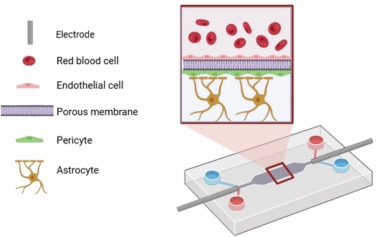
1. Introduction
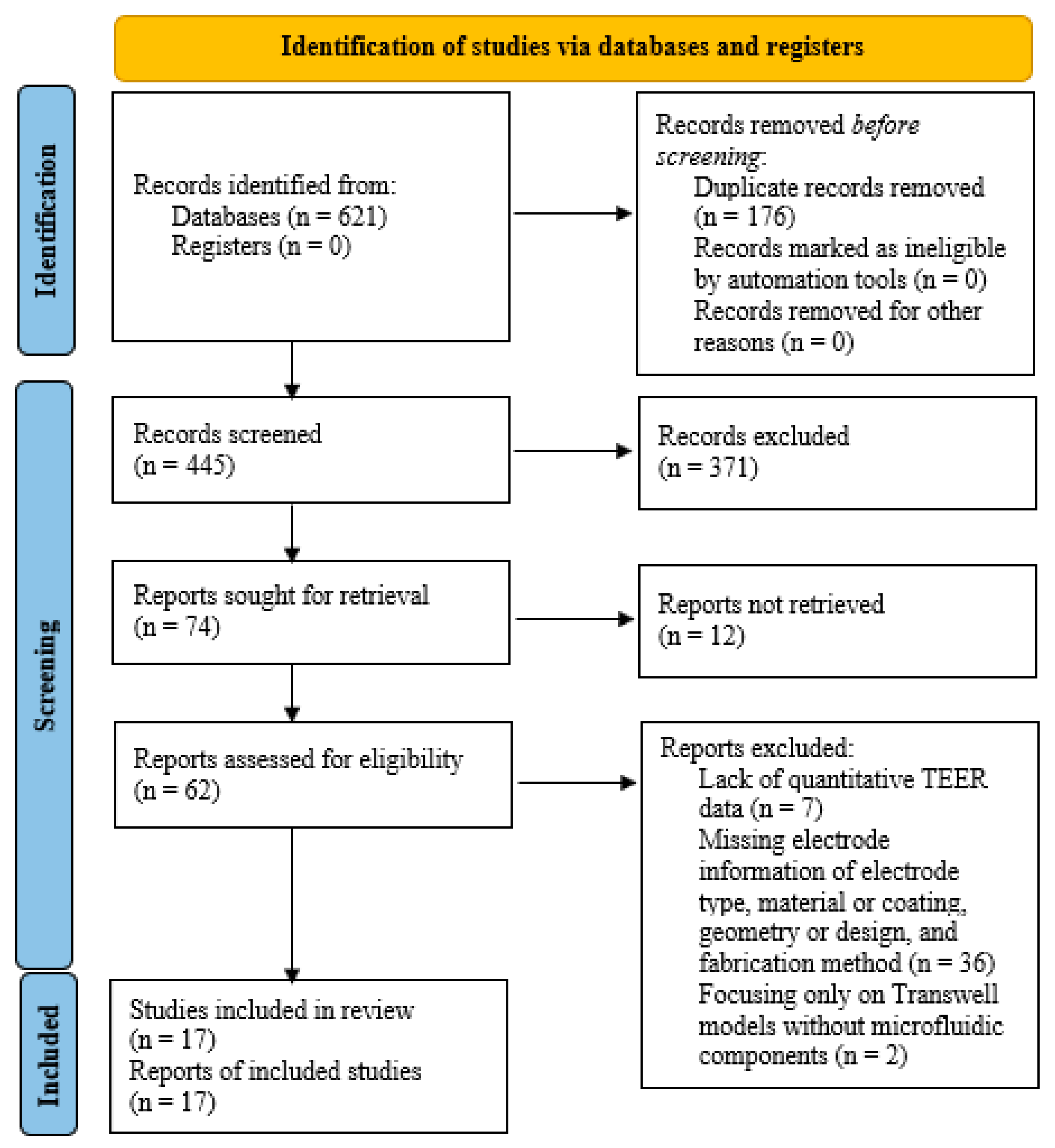
2. Search Strategy and Study Selection
- Title and abstract screening by two independent reviewers;
- Full-text assessment for inclusion or exclusion.
PRISMA Flowchart and Screening Process
3. Overview of BBB Structure and Functions
3.1. BBB Features and Functions
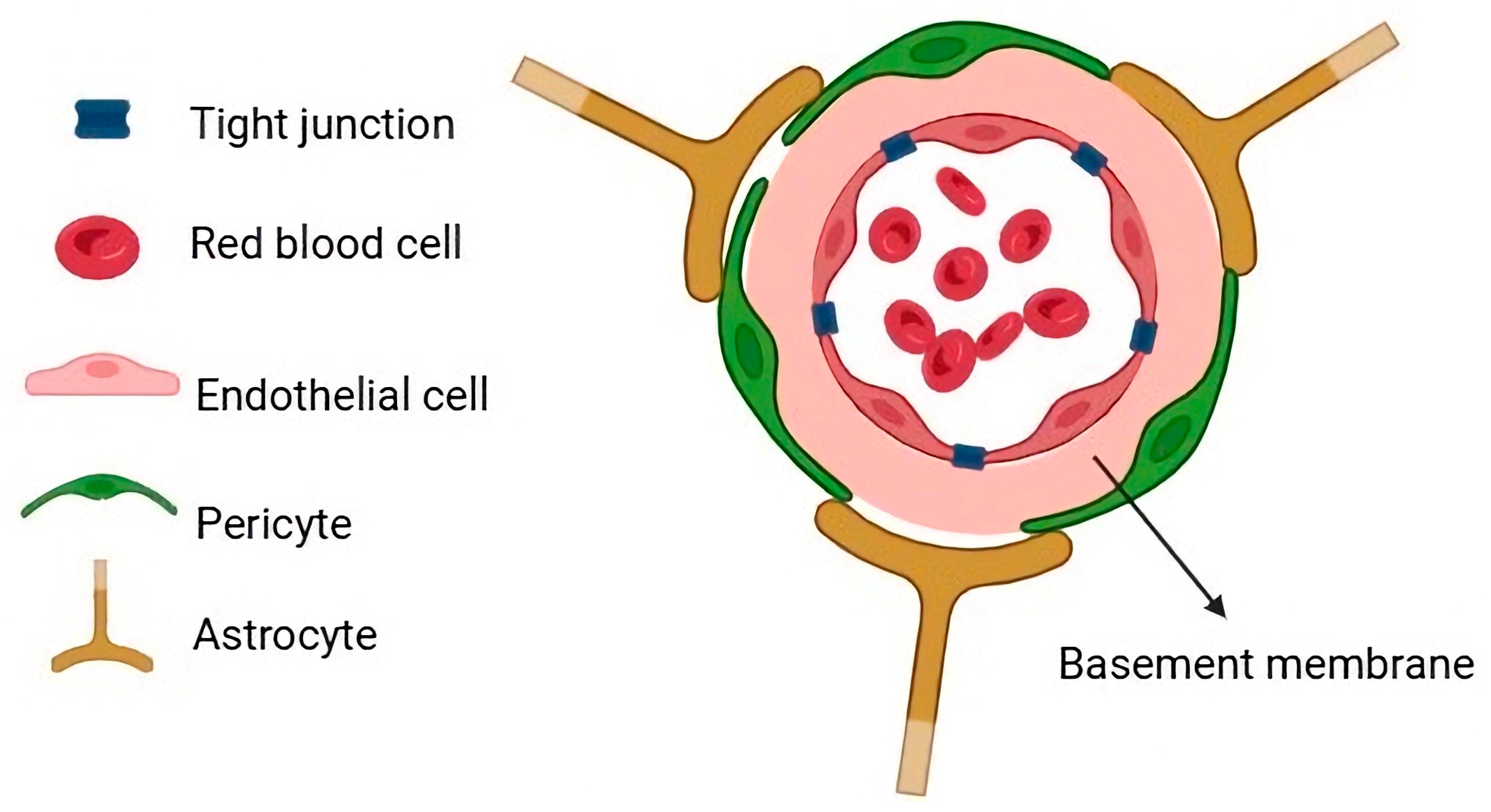
3.2. Cellular Components of the BBB
4. BBB Models
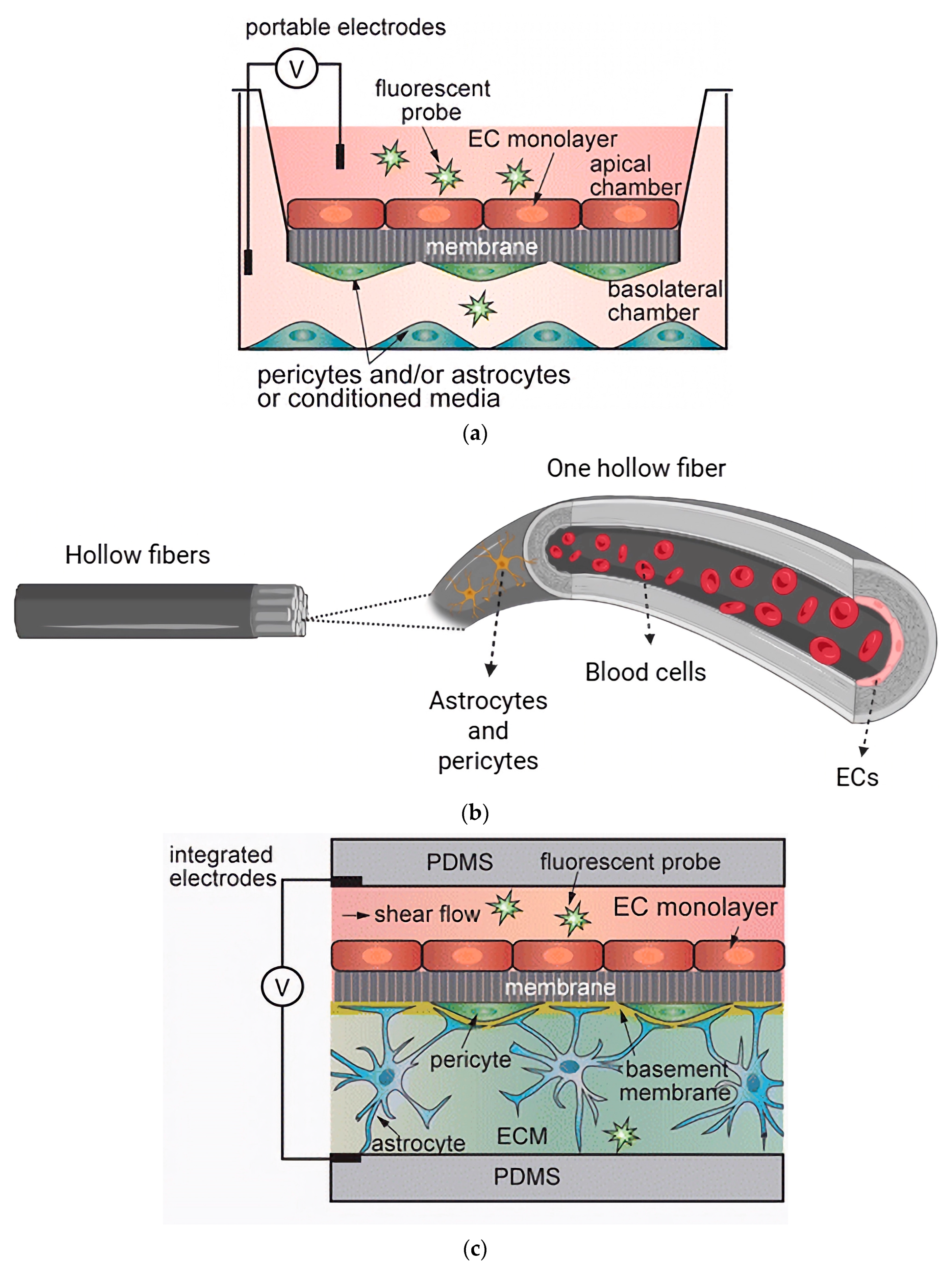
4.1. Static In Vitro BBB Models
4.2. Dynamic In Vitro BBB Models
4.2.1. Hollow Fiber Models
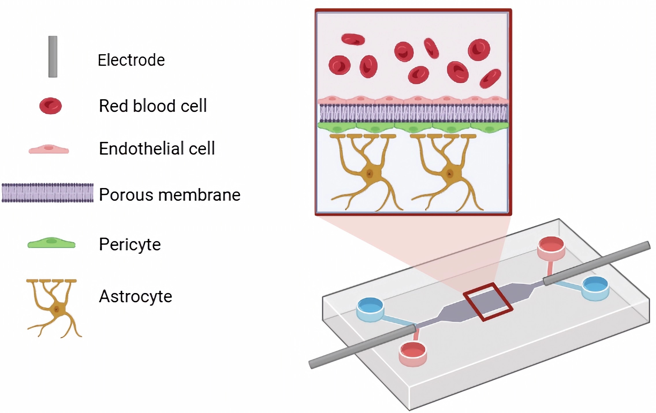
4.2.2. Microfluidic BBB-on-Chip Models

| Device Architecture | Biological Configuration | Membrane Characteristics | |||||
|---|---|---|---|---|---|---|---|
| Device | Chip Materials | Fabrication Method | Surface Coating | Cultured Cells | Membrane Type | Pore Size/Thickness | Ref. |
| Lab-on-chip | PDMS | Spin-coating and lasers-patterning | Fibronectin | ECs, and astrocytes | PC | 0.4 µm/10 µm | [43] |
| Organ-on-chip | PDMS | Molding | Collagen I | Human cardiac microvascular endothelial cells (hCMECs)/D3 | PC | 0.4 µm/10 µm | [44] |
| Organ-on-chip | PDMS | Soft lithography | Fibronectin and collagen IV | Cortical brain microvascular ECs, astrocytes, pericytes | PET | 0.4 µm | [37] |
| Organ-on-chip | Not mentioned | A two and three-lane OrganoPlate | Collagen I | Human brain microvascular endothelial cells (hBMECs), hB astrocytes, hB pericytes | Phase guide | - | [55] |
| Microfluidic model | PDMS | Purchased from SynVivo, Inc. | Collagen I, human fibronectin, and laminin | hCMECs/D3, astrocytes | - | - | [56] |
| Organ-on-chip | PDMS | Soft lithography | Collagen IV and fibronectin | iPS-Brain microvascular endothelial cells (BMVECs), astrocytes, pericytes | PET | 0.4 µm | [31] |
| Organ-on-chip | PDMS | Soft lithography | Fibronectin | hCMECs/D3, astrocytes | PET | 3 µm | [38] |
| Organ-on-chip | PDMS | Soft lithography | Collagen I | ECs, astrocytes, pericytes | - | - | [53] |
| Organ-on-chip | PDMS | A two-step photolithography technique | - | hCMECs/D3, astrocytes, pericytes, human glioblastoma | - | - | [57] |
| Organ-on-chip | Not mentioned | Donated by AIM Biotech | Fibrin hydrogels | Human umbilical vein ECs, human astrocytes | - | - | [58] |
| Organ-on-chip | PDMS | Soft lithography | Collagen I | hCMECs/D3, human astrocytes | PDMS | 5 µm through-hole pores/~2 µm | [46] |
| Organ-on-chip | PDMS | Microelectromechanical processes and soft lithography | Fibronectin | ECs, astrocytes | PET | - | [41] |
| Organ-on-chip | PDMS | Soft lithography | Collagen I | Human brain derived microvascular ECs | - | - | [34] |
| Organ-on-chip | PDMS | Soft lithography | Collagen I | hBMECs | - | - | [59] |
| Dual Channel Microfluidics | Biocompatible photoresist containing bisphenol-A-glycidyl dimethacrylate, ethoxylated bisphenol-A-dimethacrylate, carboxyethyl acrylate, PETA, biotinylated resin, and photo initiator (2,4,6–trimethylbenzoyl)–phenylphosphineoxide | 3D multiphoton lithography | Gelatin and fibrinogen | Immortalized human umbilical vein ECs, pericytes | PET | -/30 µm | [42] |
| Organ-on-chip | PDMS | Not mentioned | Collagen IV, fibronectin, laminin | BMECs, astrocytes, pericytes, microglia | PDMS | Multiple pores of 7 µm/50 µm | [47] |
| Vertical design of a microfluidic chip | PDMS | Not mentioned | Poly-l-lysine, collagen I, hyaluronic acid | NSCs, brain ECs and brain PCs | - | - | [60] |
| Lab-on-chip | Not mentioned | Not mentioned | Matrigel, gelatin | hEC, pericytes | Polyester | - | [61] |
| Organ-on-chip | PDMS | Soft lithography and replica molding methods | - | hBMECs, astrocytes, pericytes | - | - | [62] |
| Organ-on-chip | PDMS | Stereolithography 3D printer and soft lithography | - | BMECs and h pericytes | - | - | [63] |
| Organ-on-chip | PDMS | Any cubic Photon LCD printer and soft lithography | - | hBMECs | Polyester | 0.4 µm | [49] |
| Organ-on-chip | PDMS | Soft lithography | Collagen and fibronectin | iPSC-derived BMECs, primary pericytes, astrocytes | Permeable PDMS | - | [48] |
| Organ-on-chip | PDMS | Soft lithography | Injected collagen | hBMECs, hBV pericytes, human astrocytes | - | - | [64] |
| Organ-on-chip | PDMS | Soft lithography | Fibrin | hiPS-ECs, human brain pericytes, astrocytes | - | - | [65] |
| Microfluidic model | PDMS | Photomasks and molding | Fibrinogen solution | Human iPS-ECs or HBMECs, astrocytes, pericytes | - | - | [66] |
| Organ-on-chip | PDMS | Soft lithography | Fibronectin | Human umbilical vein endothelial cells (hUVECs), human astrocytes | Porous Transwell wall immersed in ethylene-glycol-terminated octadecanethiol | - | [50] |
| Organ-on-chip | PDMS | Lamination photolithography technique and soft lithography | Collagen and fibronectin | hBMVECs, human brain vascular pericytes (hBVPs), astrocytes | PET | - | [40] |
| Organ-on-chip | PDMS | Soft lithography | Collagen I | hCMECs/D3, human astrocytes, microglial cells | PET porous film | - | [36] |
| Organ-on-chip | PDMS | Soft lithography | Collagen | hECs, astrocytes, pericytes | - | - | [67] |
| Organ-on-chip | PDMS | Purchased from Sylgard 184, Dow Corning Corp., Midland, MI | Collagen I | hCMECs/D3, human astrocytes, h pericytes | Semipermeable PET | 0.4 µm/12 µm | [39] |
| Organ-on-chip | PDMS | Soft lithography | Collagen | hCMECs/D3, astrocytes, two breast cancer cell lines | PC | 5 µm | [45] |
| Organ-on-chip | Not mentioned | Purchased an OrganoPlate | Collagen I | hUVECs | - | - | [68] |
5. BBB-on-Chip Assays
5.1. Tight Junction Immunolabeling
5.2. Permeability Assays
5.3. TEER Measurements
- Geometry and configuration of electrodes: Type (chopstick, interdigitated electrode, etc.), material (Ag/AgCl, Pt, Au, ITO, etc.), dimensions, spacing, and distance between electrode and membrane;
- Environmental control: Measurement temperature, medium, and flow conditions (static or dynamic with rate or shear stress);
- Data normalization: Report both raw resistance (Ω) and normalized TEER (Ω.cm2) using Equation (1);
- Replication and statistics: Biological and technical replicates, dispersion (SD or SE), and number of devices (n);
- Calibration and blank correction: Electrode calibration date, blank medium resistance, and stability tests;
- Cell and membrane metadata: Cell origin (primary, immortalized, iPSC-derived), co-culture type, membrane material, and coating.
6. Structures of TEER Electrodes
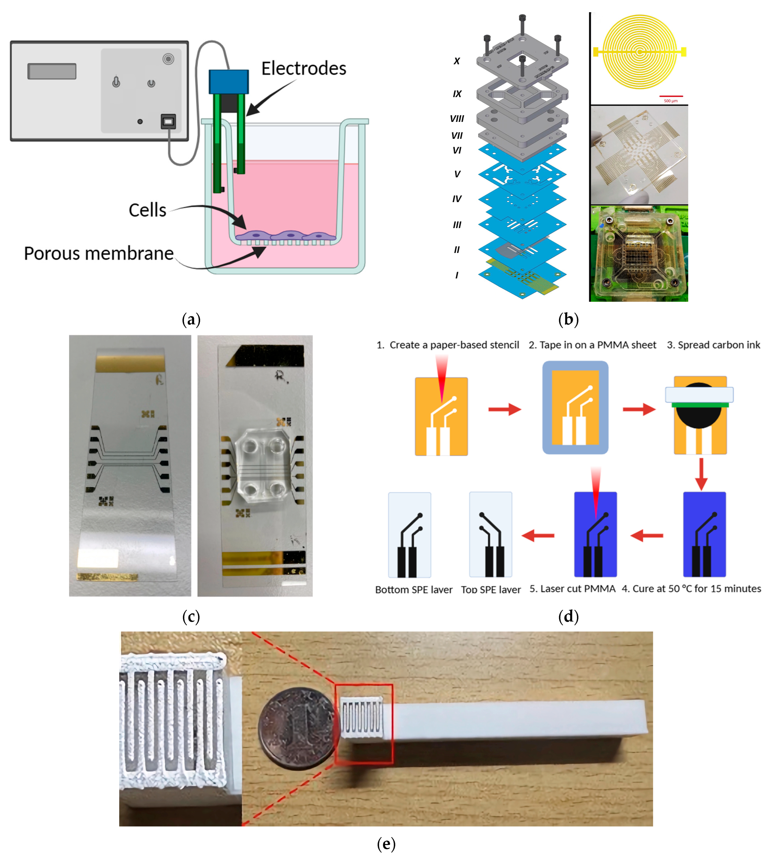
6.1. Chopstick Electrodes
6.2. Integrated Electrodes
6.2.1. Array of Concentric Interdigitated Electrodes
6.2.2. Aluminum Sheet
6.2.3. Deposited Electrodes
6.2.4. Screen-Printed Electrodes
6.2.5. Dynamic External Electrode Configuration (Silver-Finger with ITO Design)

| Electrode Type | Material/Coating | Geometry/Design | Integration Location | Fabrication Method | Distinct Feature/Advantage | Ref. |
|---|---|---|---|---|---|---|
| Chopstick electrode | Pt, Ag/AgCl | Cylindrical wire pair | External inlet/outlet | Manual placement | Simple setup; compatible with standard TEER devices; ideal for Transwell models | [12,44,77,79] |
| Concentric interdigitated array | Au on cyclic-olefin polymer | Circular interdigitated rings | Top and bottom microchannels | Photolithography + sputter deposition | Homogeneous current field; suitable for impedance spectroscopy; high precision | [82,89,100] |
| Aluminum sheet electrode | Aluminum | Planar rectangular sheet | Integrated-side walls | Manual cutting and embedding | Low cost; eliminates the need for clean-room conditions; compatible with cellulose fiber membranes | [75,83] |
| Deposited thin-film electrode | Au/Cr or Au/Ag/Cr multilayer | Planar patterned film | Glass substrate/PDMS interface | Sputter coating + liftoff | High stability and real-time TEER monitoring; uniform field distribution | [10,43,67,84] |
| Screen-printed electrode | Carbon, Au, or Pt-based conductive ink | Flat printed pad | Integrated on polymer substrate | Screen printing/laser processing | Scalable; flexible; low cost; minimal signal drift (~0.02 Ω); suitable for dynamic flow | [85,89,98] |
| Dynamic Ag–ITO hybrid | Ag-coated fork + ITO glass | Movable fork (apical) + planar basal layer | External dynamic configuration | Physical coating + stepper motor positioning | Multifunctional: real-time TEER + reversible electroporation + live-cell imaging | [86] |
7. Comparative Analysis of Electrode Architectures for TEER Measurements in BBB-on-Chip Systems
8. Challenges and Future Perspectives
9. Conclusions
Author Contributions
Funding
Institutional Review Board Statement
Informed Consent Statement
Data Availability Statement
Conflicts of Interest
Abbreviations
| BBB | Blood-brain barrier |
| TEER | Transendothelial electrical resistance |
| CNS | Central nervous system |
| iPSC | Induced pluripotent stem cell |
| ECs | Endothelial cells |
| ZO | Zonula occludens |
| HES | Hydroxyethyl starch |
| PET | Polyethylene terephthalate |
| PC | Polycarbonate |
| PDMS | Polydimethylsiloxane |
| hCMECs | Human cardiac microvascular endothelial cells |
| hBMECs | Human brain microvascular endothelial cells |
| BMVECs | Brain microvascular endothelial cells |
| hUVECs | Human umbilical vein endothelial cells |
| hBVPs | Human brain vascular pericytes |
| FITC | Fluorescein isothiocyanate |
| Pt | Platinum |
| Ag/AgCl | Silver/silver chloride |
| Au | Gold |
| ITO | Indium tin oxide |
| LY | Lucifer yellow |
| EBA | Evans blue-albumin |
| cerebEND | Cerebellar capillary endothelial cells |
| Cr | Chromium |
| Ag | Silver |
| PMMA | Methyl methacrylate |
| RTEER | Ohmic resistance of the cell layer |
| RM | Cell culture medium |
| RI | Porous membrane |
| REMI | Electrode-medium interface |
| CAD | Computer-aided design |
| MEA | Multielectrode array |
References
- Chen, X.; Liu, C.; Muok, L.; Zeng, C.; Li, Y. Dynamic 3D on-chip BBB model design, development, and applications in neurological diseases. Cells 2021, 10, 3183. [Google Scholar] [CrossRef]
- Ghane, N.; Beigi, M.-H.; Labbaf, S.; Nasr-Esfahani, M.-H.; Kiani, A. Design of hydrogel-based scaffolds for the treatment of spinal cord injuries. J. Mater. Chem. B 2020, 8, 10712–10738. [Google Scholar] [CrossRef]
- Ghane, N.; Khalili, S.; Nouri Khorasani, S.; Esmaeely Neisiany, R.; Das, O.; Ramakrishna, S. Regeneration of the peripheral nerve via multifunctional electrospun scaffolds. J. Biomed. Mater. Res. Part A 2021, 109, 437–452. [Google Scholar] [CrossRef]
- Ghane, N.; Khalili, S.; Khorasani, S.N.; Das, O.; Ramakrishna, S.; Neisiany, R.E. Antiepileptic drug-loaded and multifunctional iron oxide@ silica@ gelatin nanoparticles for acid-triggered drug delivery. Sci. Rep. 2024, 14, 11400. [Google Scholar] [CrossRef]
- Pardridge, W.M. A historical review of brain drug delivery. Pharmaceutics 2022, 14, 1283. [Google Scholar] [CrossRef]
- Yau, A.; Jogdand, A.; Chen, Y. Blood-brain-barrier modeling with tissue chips for research applications in space and on Earth. Front. Space Technol. 2023, 4, 1176943. [Google Scholar] [CrossRef] [PubMed]
- Jiang, L.; Li, S.; Zheng, J.; Li, Y.; Huang, H. Recent progress in microfluidic models of the blood-brain barrier. Micromachines 2019, 10, 375. [Google Scholar] [CrossRef] [PubMed]
- Hajal, C.; Le Roi, B.; Kamm, R.D.; Maoz, B.M. Biology and models of the blood–brain barrier. Annu. Rev. Biomed. Eng. 2021, 23, 359–384. [Google Scholar] [CrossRef]
- Van Norman, G.A. Limitations of animal studies for predicting toxicity in clinical trials: Is it time to rethink our current approach? JACC Basic Transl. Sci. 2019, 4, 845–854. [Google Scholar] [CrossRef]
- Kincses, A.; Vigh, J.P.; Petrovszki, D.; Valkai, S.; Kocsis, A.E.; Walter, F.R.; Lin, H.-Y.; Jan, J.-S.; Deli, M.A.; Dér, A. The use of sensors in blood-brain barrier-on-a-chip devices: Current practice and future directions. Biosensors 2023, 13, 357. [Google Scholar] [CrossRef]
- Chin, E.; Goh, E. Blood–brain barrier on a chip. In Methods in Cell Biology; Elsevier: Amsterdam, The Netherlands, 2018; Volume 146, pp. 159–182. [Google Scholar]
- Vigh, J.P.; Kincses, A.; Ozgür, B.; Walter, F.R.; Santa-Maria, A.R.; Valkai, S.; Vastag, M.; Neuhaus, W.; Brodin, B.; Dér, A. Transendothelial electrical resistance measurement across the blood–brain barrier: A critical review of methods. Micromachines 2021, 12, 685. [Google Scholar] [CrossRef]
- Srinivasan, B.; Kolli, A.R.; Esch, M.B.; Abaci, H.E.; Shuler, M.L.; Hickman, J.J. TEER measurement techniques for in vitro barrier model systems. J. Lab. Autom. 2015, 20, 107–126. [Google Scholar] [CrossRef]
- Bellettato, C.M.; Scarpa, M. Possible strategies to cross the blood–brain barrier. Ital. J. Pediatr. 2018, 44, 131. [Google Scholar] [CrossRef]
- Zeiadeh, I.; Najjar, A.; Karaman, R. Strategies for enhancing the permeation of CNS-active drugs through the blood-brain barrier: A review. Molecules 2018, 23, 1289. [Google Scholar] [CrossRef]
- Guarino, V.; Zizzari, A.; Bianco, M.; Gigli, G.; Moroni, L.; Arima, V. Advancements in modelling human blood brain-barrier on a chip. Biofabrication 2023, 15, 022003. [Google Scholar] [CrossRef]
- Musafargani, S.; Mishra, S.; Gulyás, M.; Mahalakshmi, P.; Archunan, G.; Padmanabhan, P.; Gulyás, B. Blood brain barrier: A tissue engineered microfluidic chip. J. Neurosci. Methods 2020, 331, 108525. [Google Scholar] [CrossRef]
- DeStefano, J.G.; Xu, Z.S.; Williams, A.J.; Yimam, N.; Searson, P.C. Effect of shear stress on iPSC-derived human brain microvascular endothelial cells (dhBMECs). Fluids Barriers CNS 2017, 14, 20. [Google Scholar] [CrossRef] [PubMed]
- Lee, H.-G.; Wheeler, M.A.; Quintana, F.J. Function and therapeutic value of astrocytes in neurological diseases. Nat. Rev. Drug Discov. 2022, 21, 339–358. [Google Scholar] [CrossRef] [PubMed]
- Kantawala, B.; Shariff, S.; Ramadan, N.; Fawaz, V.; Hassan, Y.; Mugisha, N.; Yenkoyan, K.; Nazir, A.; Uwishema, O. Revolutionizing neurotherapeutics: Blood-brain barrier-on-a-chip technologies for precise drug delivery. Ann. Med. Surg. 2024, 86, 2794–2804. [Google Scholar] [CrossRef]
- Cai, Y.; Fan, K.; Lin, J.; Ma, L.; Li, F. Advances in BBB on chip and application for studying reversible opening of blood–brain barrier by sonoporation. Micromachines 2022, 14, 112. [Google Scholar] [CrossRef] [PubMed]
- Naik, P.; Cucullo, L. In vitro blood–brain barrier models: Current and perspective technologies. J. Pharm. Sci. 2012, 101, 1337–1354. [Google Scholar] [CrossRef] [PubMed]
- Delsing, L.; Herland, A.; Falk, A.; Hicks, R.; Synnergren, J.; Zetterberg, H. Models of the blood-brain barrier using iPSC-derived cells. Mol. Cell. Neurosci. 2020, 107, 103533. [Google Scholar] [CrossRef]
- Schreiner, T.G.; Creangă-Murariu, I.; Tamba, B.I.; Lucanu, N.; Popescu, B.O. In vitro modeling of the blood–brain barrier for the study of physiological conditions and Alzheimer’s disease. Biomolecules 2022, 12, 1136. [Google Scholar] [CrossRef]
- Kaisar, M.A.; Abhyankar, V.V.; Cucullo, L. In vitro BBB models: Working with static platforms and microfluidic systems. In Blood-Brain Barrier; Springer: New York, NY, USA, 2019; pp. 55–70. [Google Scholar]
- Gaillard, P.J.; Voorwinden, L.H.; Nielsen, J.L.; Ivanov, A.; Atsumi, R.; Engman, H.; Ringbom, C.; de Boer, A.G.; Breimer, D.D. Establishment and functional characterization of an in vitro model of the blood–brain barrier, comprising a co-culture of brain capillary endothelial cells and astrocytes. Eur. J. Pharm. Sci. 2001, 12, 215–222. [Google Scholar] [CrossRef]
- Nakagawa, S.; Deli, M.A.; Kawaguchi, H.; Shimizudani, T.; Shimono, T.; Kittel, A.; Tanaka, K.; Niwa, M. A new blood–brain barrier model using primary rat brain endothelial cells, pericytes and astrocytes. Neurochem. Int. 2009, 54, 253–263. [Google Scholar] [CrossRef] [PubMed]
- Sivandzade, F.; Cucullo, L. In-vitro blood–brain barrier modeling: A review of modern and fast-advancing technologies. J. Cereb. Blood Flow Metab. 2018, 38, 1667–1681. [Google Scholar] [CrossRef] [PubMed]
- Bagchi, S.; Chhibber, T.; Lahooti, B.; Verma, A.; Borse, V.; Jayant, R.D. In-vitro blood-brain barrier models for drug screening and permeation studies: An overview. Drug Des. Dev. Ther. 2019, 13, 3591–3605. [Google Scholar] [CrossRef]
- Cucullo, L.; Hossain, M.; Puvenna, V.; Marchi, N.; Janigro, D. The role of shear stress in Blood-Brain Barrier endothelial physiology. BMC Neurosci. 2011, 12, 40. [Google Scholar] [CrossRef]
- Park, T.-E.; Mustafaoglu, N.; Herland, A.; Hasselkus, R.; Mannix, R.; FitzGerald, E.A.; Prantil-Baun, R.; Watters, A.; Henry, O.; Benz, M. Hypoxia-enhanced Blood-Brain Barrier Chip recapitulates human barrier function and shuttling of drugs and antibodies. Nat. Commun. 2019, 10, 2621. [Google Scholar] [CrossRef]
- Walker, A.M.; Xiao, Y.; Johnston, C.R.; Rival, D.E. The viscous characterization of hydroxyethyl starch (HES) plasma volume expanders in a non-Newtonian blood analog. Biorheology 2013, 50, 177–190. [Google Scholar] [CrossRef]
- Liang, Y.; Yoon, J.-Y. In situ sensors for blood-brain barrier (BBB) on a chip. Sens. Actuators Rep. 2021, 3, 100031. [Google Scholar] [CrossRef]
- Salman, M.; Marsh, G.; Küsters, I.; Delincé, M.; Di Caprio, G.; Upadhyayula, S.; de Nola, G.; Hunt, R.; Ohashi, K.G.; Shimizu, F. An in-vitro BBB-on-a-chip open model of human blood-brain barrier enabling advanced optical imaging. bioRxiv 2020. [Google Scholar] [CrossRef]
- Mantecón-Oria, M.; Rivero, M.J.; Diban, N.; Urtiaga, A. On the quest of reliable 3D dynamic in vitro blood-brain barrier models using polymer hollow fiber membranes: Pitfalls, progress, and future perspectives. Front. Bioeng. Biotechnol. 2022, 10, 1056162. [Google Scholar] [CrossRef]
- Wang, P.; Wu, Y.; Chen, W.; Zhang, M.; Qin, J. Malignant melanoma-derived exosomes induce endothelial damage and glial activation on a human BBB chip model. Biosensors 2022, 12, 89. [Google Scholar] [CrossRef] [PubMed]
- Maoz, B.M.; Herland, A.; FitzGerald, E.A.; Grevesse, T.; Vidoudez, C.; Pacheco, A.R.; Sheehy, S.P.; Park, T.-E.; Dauth, S.; Mannix, R. A linked organ-on-chip model of the human neurovascular unit reveals the metabolic coupling of endothelial and neuronal cells. Nat. Biotechnol. 2018, 36, 865–874. [Google Scholar] [CrossRef] [PubMed]
- Chaing, Y.-Y.; Tu, K.-H. In Vitro Microfluidics-based Blood–brain Barrier Model. In Proceedings of the 2019 IEEE 14th International Conference on Nano/Micro Engineered and Molecular Systems (NEMS), Bangkok, Thailand, 11–14 April 2019; pp. 351–354. [Google Scholar]
- Wei, W.; Cardes, F.; Hierlemann, A.; Modena, M.M. 3D in vitro blood-brain-barrier model for investigating barrier insults. Adv. Sci. 2023, 10, 2205752. [Google Scholar] [CrossRef] [PubMed]
- Boghdeh, N.A.; Risner, K.H.; Barrera, M.D.; Britt, C.M.; Schaffer, D.K.; Alem, F.; Brown, J.A.; Wikswo, J.P.; Narayanan, A. Application of a human blood brain barrier organ-on-a-chip model to evaluate small molecule effectiveness against Venezuelan equine encephalitis virus. Viruses 2022, 14, 2799. [Google Scholar] [CrossRef]
- Tu, K.-H.; Yu, L.-S.; Sie, Z.-H.; Hsu, H.-Y.; Al-Jamal, K.T.; Wang, J.T.-W.; Chiang, Y.-Y. Development of real-time transendothelial electrical resistance monitoring for an in vitro blood-brain barrier system. Micromachines 2020, 12, 37. [Google Scholar] [CrossRef]
- Buchroithner, B.; Mayr, S.; Hauser, F.; Priglinger, E.; Stangl, H.; Santa-Maria, A.R.; Deli, M.A.; Der, A.; Klar, T.A.; Axmann, M. Dual channel microfluidics for mimicking the blood–brain barrier. ACS Nano 2021, 15, 2984–2993. [Google Scholar] [CrossRef]
- Booth, R.; Kim, H. Characterization of a microfluidic in vitro model of the blood-brain barrier (μBBB). Lab Chip 2012, 12, 1784–1792. [Google Scholar] [CrossRef]
- Griep, L.M.; Wolbers, F.; de Wagenaar, B.; ter Braak, P.M.; Weksler, B.B.; Romero, I.A.; Couraud, P.O.; Vermes, I.; van der Meer, A.D.; van den Berg, A. BBB on chip: Microfluidic platform to mechanically and biochemically modulate blood-brain barrier function. Biomed. Microdevices 2013, 15, 145–150. [Google Scholar] [CrossRef]
- Westerhof, T.M.; Yang, B.A.; Merrill, N.M.; Yates, J.A.; Altemus, M.; Russell, L.; Miller, A.J.; Bao, L.; Wu, Z.; Ulintz, P.J. Blood–Brain Barrier Remodeling in an Organ-on-a-Chip Device Showing Dkk1 to be a Regulator of Early Metastasis. Adv. Nanobiomed Res. 2023, 3, 2200036. [Google Scholar] [CrossRef]
- Zakharova, M.; Do Carmo, M.P.; Van Der Helm, M.; De Graaf, M.; Orlova, V.; Van Den Berg, A.; Van Der Meer, A.; Broersen, K.; Segerink, L. Multiplexed blood–brain barrier organ-on-chip. Lab Chip 2020, 20, 3132–3143. [Google Scholar] [CrossRef]
- Pediaditakis, I.; Kodella, K.R.; Manatakis, D.V.; Le, C.Y.; Hinojosa, C.D.; Tien-Street, W.; Manolakos, E.S.; Vekrellis, K.; Hamilton, G.A.; Ewart, L. Modeling alpha-synuclein pathology in a human brain-chip to assess blood-brain barrier disruption. Nat. Commun. 2021, 12, 5907. [Google Scholar] [CrossRef]
- Noorani, B.; Bhalerao, A.; Raut, S.; Nozohouri, E.; Bickel, U.; Cucullo, L. A quasi-physiological microfluidic blood-brain barrier model for brain permeability studies. Pharmaceutics 2021, 13, 1474. [Google Scholar] [CrossRef]
- Kim, M.-H.; Kim, D.; Sung, J.H. A Gut-Brain Axis-on-a-Chip for studying transport across epithelial and endothelial barriers. J. Ind. Eng. Chem. 2021, 101, 126–134. [Google Scholar] [CrossRef]
- Singh, A.V.; Chandrasekar, V.; Laux, P.; Luch, A.; Dakua, S.P.; Zamboni, P.; Shelar, A.; Yang, Y.; Pandit, V.; Tisato, V. Micropatterned neurovascular interface to mimic the blood–brain barrier’s neurophysiology and micromechanical function: A BBB-on-CHIP model. Cells 2022, 11, 2801. [Google Scholar] [CrossRef] [PubMed]
- Hosic, S.; Bindas, A.J.; Puzan, M.L.; Lake, W.; Soucy, J.R.; Zhou, F.; Koppes, R.A.; Breault, D.T.; Murthy, S.K.; Koppes, A.N. Rapid prototyping of multilayer microphysiological systems. ACS Biomater. Sci. Eng. 2020, 7, 2949–2963. [Google Scholar] [CrossRef] [PubMed]
- Dai, C.; Liu, X.; Tang, R.; He, J.; Arai, T. A review on microfluidic platforms applied to nerve regeneration. Appl. Sci. 2022, 12, 3534. [Google Scholar] [CrossRef]
- Yu, F.; Kumar, N.D.O.S.; Foo, L.C.; Ng, S.H.; Hunziker, W.; Choudhury, D. A pump-free tricellular blood–brain barrier on-a-chip model to understand barrier property and evaluate drug response. Biotechnol. Bioeng. 2020, 117, 1127–1136. [Google Scholar] [CrossRef]
- Jamieson, J.J.; Searson, P.C.; Gerecht, S. Engineering the human blood-brain barrier in vitro. J. Biol. Eng. 2017, 11, 37. [Google Scholar] [CrossRef]
- Wevers, N.R.; Kasi, D.G.; Gray, T.; Wilschut, K.J.; Smith, B.; van Vught, R.; Shimizu, F.; Sano, Y.; Kanda, T.; Marsh, G. A perfused human blood–brain barrier on-a-chip for high-throughput assessment of barrier function and antibody transport. Fluids Barriers CNS 2018, 15, 23. [Google Scholar] [CrossRef]
- Brown, T.D.; Nowak, M.; Bayles, A.V.; Prabhakarpandian, B.; Karande, P.; Lahann, J.; Helgeson, M.E.; Mitragotri, S. A microfluidic model of human brain (μHuB) for assessment of blood brain barrier. Bioeng. Transl. Med. 2019, 4, e10126. [Google Scholar] [CrossRef]
- Peng, B.; Tong, Z.; Tong, W.Y.; Pasic, P.J.; Oddo, A.; Dai, Y.; Luo, M.; Frescene, J.; Welch, N.G.; Easton, C.D. In situ surface modification of microfluidic blood–brain-barriers for improved screening of small molecules and nanoparticles. ACS Appl. Mater. Interfaces 2020, 12, 56753–56766. [Google Scholar] [CrossRef]
- Li, Y.; Liu, Y.; Hu, C.; Chang, Q.; Deng, Q.; Yang, X.; Wu, Y. Study of the neurotoxicity of indoor airborne nanoparticles based on a 3D human blood-brain barrier chip. Environ. Int. 2020, 143, 105598. [Google Scholar] [CrossRef]
- Salman, M.M.; Marsh, G.; Kusters, I.; Delincé, M.; Di Caprio, G.; Upadhyayula, S.; De Nola, G.; Hunt, R.; Ohashi, K.G.; Gray, T. Design and validation of a human brain endothelial microvessel-on-a-chip open microfluidic model enabling advanced optical imaging. Front. Bioeng. Biotechnol. 2020, 8, 573775. [Google Scholar] [CrossRef] [PubMed]
- Kim, J.; Lee, K.-T.; Lee, J.S.; Shin, J.; Cui, B.; Yang, K.; Choi, Y.S.; Choi, N.; Lee, S.H.; Lee, J.-H. Fungal brain infection modelled in a human-neurovascular-unit-on-a-chip with a functional blood–brain barrier. Nat. Biomed. Eng. 2021, 5, 830–846. [Google Scholar] [CrossRef]
- Santa-Maria, A.R.; Walter, F.R.; Figueiredo, R.; Kincses, A.; Vigh, J.P.; Heymans, M.; Culot, M.; Winter, P.; Gosselet, F.; Dér, A. Flow induces barrier and glycocalyx-related genes and negative surface charge in a lab-on-a-chip human blood-brain barrier model. J. Cereb. Blood Flow Metab. 2021, 41, 2201–2215. [Google Scholar] [CrossRef] [PubMed]
- Kim, S.; Lee, S.; Lim, J.; Choi, H.; Kang, H.; Jeon, N.L.; Son, Y. Human bone marrow-derived mesenchymal stem cells play a role as a vascular pericyte in the reconstruction of human BBB on the angiogenesis microfluidic chip. Biomaterials 2021, 279, 121210. [Google Scholar] [CrossRef] [PubMed]
- Lyu, Z.; Park, J.; Kim, K.-M.; Jin, H.-J.; Wu, H.; Rajadas, J.; Kim, D.-H.; Steinberg, G.K.; Lee, W. A neurovascular-unit-on-a-chip for the evaluation of the restorative potential of stem cell therapies for ischaemic stroke. Nat. Biomed. Eng. 2021, 5, 847–863. [Google Scholar] [CrossRef]
- Seo, S.; Nah, S.Y.; Lee, K.; Choi, N.; Kim, H.N. Triculture model of in vitro BBB and its application to study BBB-associated chemosensitivity and drug delivery in glioblastoma. Adv. Funct. Mater. 2022, 32, 2106860. [Google Scholar] [CrossRef]
- Straehla, J.P.; Hajal, C.; Safford, H.C.; Offeddu, G.S.; Boehnke, N.; Dacoba, T.G.; Wyckoff, J.; Kamm, R.D.; Hammond, P.T. A predictive microfluidic model of human glioblastoma to assess trafficking of blood–brain barrier-penetrant nanoparticles. Proc. Natl. Acad. Sci. 2022, 119, e2118697119. [Google Scholar] [CrossRef]
- Hajal, C.; Offeddu, G.S.; Shin, Y.; Zhang, S.; Morozova, O.; Hickman, D.; Knutson, C.G.; Kamm, R.D. Engineered human blood–brain barrier microfluidic model for vascular permeability analyses. Nat. Protoc. 2022, 17, 95–128. [Google Scholar] [CrossRef] [PubMed]
- Palma-Florez, S.; López-Canosa, A.; Moralez-Zavala, F.; Castaño, O.; Kogan, M.J.; Samitier, J.; Lagunas, A.; Mir, M. BBB-on-a-chip with integrated micro-TEER for permeability evaluation of multi-functionalized gold nanorods against Alzheimer’s disease. J. Nanobiotechnology 2023, 21, 115. [Google Scholar] [CrossRef]
- Uzoechi, S.C.; Collins, B.E.; Badeaux, C.J.; Li, Y.; Kwak, S.S.; Kim, D.Y.; Laskowitz, D.T.; Lee, J.-M.; Yun, Y. Effects of Amyloid Beta (Aβ) Oligomers on Blood–Brain Barrier Using a 3D Microfluidic Vasculature-on-a-Chip Model. Appl. Sci. 2024, 14, 3917. [Google Scholar] [CrossRef] [PubMed]
- Garcia-Polite, F.; Martorell, J.; Del Rey-Puech, P.; Melgar-Lesmes, P.; O’Brien, C.C.; Roquer, J.; Ois, A.; Principe, A.; Edelman, E.R.; Balcells, M. Pulsatility and high shear stress deteriorate barrier phenotype in brain microvascular endothelium. J. Cereb. Blood Flow Metab. 2017, 37, 2614–2625. [Google Scholar] [CrossRef]
- Walter, F.R.; Valkai, S.; Kincses, A.; Petneházi, A.; Czeller, T.; Veszelka, S.; Ormos, P.; Deli, M.A.; Dér, A. A versatile lab-on-a-chip tool for modeling biological barriers. Sens. Actuators B Chem. 2016, 222, 1209–1219. [Google Scholar] [CrossRef]
- Campisi, M.; Shin, Y.; Osaki, T.; Hajal, C.; Chiono, V.; Kamm, R.D. 3D self-organized microvascular model of the human blood-brain barrier with endothelial cells, pericytes and astrocytes. Biomaterials 2018, 180, 117–129. [Google Scholar] [CrossRef]
- Helm, M.W. Microfluidic organ-on-chip technology for blood-brain barrier research. Tissue Barriers 2016, 4, e1142493. [Google Scholar] [CrossRef]
- Łach, A.; Wnuk, A.; Wójtowicz, A.K. Experimental models to study the functions of the blood–brain barrier. Bioengineering 2023, 10, 519. [Google Scholar] [CrossRef]
- Mir, M.; Palma-Florez, S.; Lagunas, A.; López-Martínez, M.J.; Samitier, J. Biosensors integration in blood–brain barrier-on-a-chip: Emerging platform for monitoring neurodegenerative diseases. ACS Sens. 2022, 7, 1237–1247. [Google Scholar] [CrossRef]
- Holzreuter, M.A.; Segerink, L.I. Innovative electrode and chip designs for transendothelial electrical resistance measurements in organs-on-chips. Lab Chip 2024, 24, 1121–1134. [Google Scholar] [CrossRef] [PubMed]
- Chafai, D.E.; Mehle, A.; Tilmatine, A.; Maouche, B.; Miklavčič, D. Assessment of the electrochemical effects of pulsed electric fields in a biological cell suspension. Bioelectrochemistry 2015, 106, 249–257. [Google Scholar] [CrossRef] [PubMed]
- Yeste, J.; Illa, X.; Gutiérrez, C.; Solé, M.; Guimerà, A.; Villa, R. Geometric correction factor for transepithelial electrical resistance measurements in transwell and microfluidic cell cultures. J. Phys. D Appl. Phys. 2016, 49, 375401. [Google Scholar] [CrossRef]
- Arık, Y.B.; Van Der Helm, M.W.; Odijk, M.; Segerink, L.I.; Passier, R.; Van Den Berg, A.; Van Der Meer, A.D. Barriers-on-chips: Measurement of barrier function of tissues in organs-on-chips. Biomicrofluidics 2018, 12, 042218. [Google Scholar] [CrossRef]
- Bossink, E.G.; Zakharova, M.; de Bruijn, D.S.; Odijk, M.; Segerink, L.I. Measuring barrier function in organ-on-chips with cleanroom-free integration of multiplexable electrodes. Lab Chip 2021, 21, 2040–2049. [Google Scholar] [CrossRef]
- Ugolini, G.S.; Rasponi, M.; Redaelli, A. A microfluidic device for studying drug transport through endothelial Blood Brain Barrier cells monolayers. In Proceedings of the 5th International Conference MNF 2016-Micro and Nano Flows-Book of Abstract, Milan, Italy, 11–14 September 2016. [Google Scholar]
- Jeong, S.; Kim, S.; Buonocore, J.; Park, J.; Welsh, C.J.; Li, J.; Han, A. A three-dimensional arrayed microfluidic blood–brain barrier model with integrated electrical sensor array. IEEE Trans. Biomed. Eng. 2017, 65, 431–439. [Google Scholar] [CrossRef]
- Badiola-Mateos, M.; Di Giuseppe, D.; Paoli, R.; Lopez-Martinez, M.J.; Mencattini, A.; Samitier, J.; Martinelli, E. A novel multi-frequency trans-endothelial electrical resistance (MTEER) sensor array to monitor blood-brain barrier integrity. Sens. Actuators B Chem. 2021, 334, 129599. [Google Scholar] [CrossRef]
- Duong, D.D.; Kwak, J.; Song, H.S.; Lee, N.Y. Construction of microfluidic blood–brain barrier model assisted by 3D coculture on cellulose fiber. Microsyst. Technol. 2021, 27, 3917–3926. [Google Scholar] [CrossRef]
- Martino, A. Development of a Barrier-On-Chip Model and Fabrication of Embedded High-Resolution Porous-Membrane Based Electrodes to Assess the Integrity of the Barrier. Master’s Thesis, Politecnico di Torino, Torino, Italy, 2022. [Google Scholar]
- Krishnakumar, A.; Kadian, S.; Heredia Rivera, U.; Chittiboyina, S.; Lelièvre, S.A.; Rahimi, R. Organ-on-a-chip platform with an integrated screen-printed electrode array for real-time monitoring trans-epithelial barrier and bubble formation. ACS Biomater. Sci. Eng. 2023, 9, 1620–1628. [Google Scholar] [CrossRef]
- Lv, F.; Yang, S.-M.; Zhao, J. A microfluidic blood-brain barrier chip based on a dynamic controllable external electrode is used for TEER real-time monitoring and electroporation research. In Proceedings of the 4th International Conference on Biomedical and Intelligent Systems, Bologna, Italy, 11–13 April 2025; pp. 248–252. [Google Scholar]
- Ugolini, G.S.; Occhetta, P.; Saccani, A.; Re, F.; Krol, S.; Rasponi, M.; Redaelli, A. Design and validation of a microfluidic device for blood–brain barrier monitoring and transport studies. J. Micromechanics Microengineering 2018, 28, 044001. [Google Scholar] [CrossRef]
- Guarino, V.; Perrone, E.; De Luca, E.; Rainer, A.; Cesaria, M.; Zizzari, A.; Bianco, M.; Gigli, G.; Moroni, L.; Arima, V. Pericyte-Assisted Vascular Lumen Organization in a Novel Dynamic Human Blood-Brain Barrier-on-Chip Model. Adv. Healthc. Mater. 2025, 14, 2401804. [Google Scholar] [CrossRef]
- Fuchs, S.; Johansson, S.; Tjell, A.Ø.; Werr, G.; Mayr, T.; Tenje, M. In-line analysis of organ-on-chip systems with sensors: Integration, fabrication, challenges, and potential. ACS Biomater. Sci. Eng. 2021, 7, 2926–2948. [Google Scholar] [CrossRef]
- Ghane, N.; Mazinani, S.; Gharehaghaji, A.A. Fabrication and characterization of hollow nanofibrous PA6 yarn reinforced with CNTs. J. Polym. Res. 2018, 25, 80. [Google Scholar] [CrossRef]
- Ghane, N.; Mazinani, S.; Gharehaghaji, A. Comparing the performance of electrospun and cast nanocomposite film of polyamide-6 reinforced with multi-wall carbon nanotubes. J. Plast. Film. Sheeting 2019, 35, 45–64. [Google Scholar] [CrossRef]
- Samimi, K.; Pakan, M.; Eslami, J. Investigating the compressive strength and microstructural analysis of mortar containing synthesized graphene and natural pozzolan in the face of alkali-silica reactions. J. Build. Eng. 2023, 68, 106126. [Google Scholar] [CrossRef]
- Samimi, K.; Pakan, M.; Eslami, J.; Asgharnejad, L. Investigation of two different water-dispersed graphene on the performance of graphene/cement paste: Surfactant and superplasticizer effect. Constr. Build. Mater. 2022, 349, 128756. [Google Scholar] [CrossRef]
- Ortega, M.A.; Fernández-Garibay, X.; Castaño, A.G.; De Chiara, F.; Hernández-Albors, A.; Balaguer-Trias, J.; Ramón-Azcón, J. Muscle-on-a-chip with an on-site multiplexed biosensing system for in situ monitoring of secreted IL-6 and TNF-α. Lab Chip 2019, 19, 2568–2580. [Google Scholar] [CrossRef]
- Njagi, J.; Chernov, M.M.; Leiter, J.; Andreescu, S. Amperometric detection of dopamine in vivo with an enzyme based carbon fiber microbiosensor. Anal. Chem. 2010, 82, 989–996. [Google Scholar] [CrossRef]
- Razzino, C.A.; Serafín, V.; Gamella, M.; Pedrero, M.; Montero-Calle, A.; Barderas, R.; Calero, M.; Lobo, A.O.; Yáñez-Sedeño, P.; Campuzano, S. An electrochemical immunosensor using gold nanoparticles-PAMAM-nanostructured screen-printed carbon electrodes for tau protein determination in plasma and brain tissues from Alzheimer patients. Biosens. Bioelectron. 2020, 163, 112238. [Google Scholar] [CrossRef]
- McKenzie, J.R.; Cognata, A.C.; Davis, A.N.; Wikswo, J.P.; Cliffel, D.E. Real-time monitoring of cellular bioenergetics with a multianalyte screen-printed electrode. Anal. Chem. 2015, 87, 7857–7864. [Google Scholar] [CrossRef]
- Kawakita, S.; Li, S.; Nguyen, H.T.; Maity, S.; Haghniaz, R.; Bahari, J.; Yu, N.; Mandal, K.; Bandaru, P.; Mou, L. Rapid integration of screen-printed electrodes into thermoplastic organ-on-a-chip devices for real-time monitoring of trans-endothelial electrical resistance. Biomed. Microdevices 2023, 25, 37. [Google Scholar] [CrossRef]
- Srinivasan, B.; Kolli, A.R. Transepithelial/transendothelial electrical resistance (TEER) to measure the integrity of blood-brain barrier. In Blood-Brain Barrier; Springer: Berlin/Heidelberg, Germany, 2018; pp. 99–114. [Google Scholar]
- Kim, S.; Rajendran, A.K.; Amirthalingam, S.; Kim, J.H.; So, K.-H.; Hwang, N.S. Recent technological advances in lab-on-a-chip for bone remodeling. Biosens. Bioelectron. X 2023, 14, 100360. [Google Scholar] [CrossRef]
- Weksler, B.; Subileau, E.; Perriere, N.; Charneau, P.; Holloway, K.; Leveque, M.; Tricoire-Leignel, H.; Nicotra, A.; Bourdoulous, S.; Turowski, P. Blood-brain barrier-specific properties of a human adult brain endothelial cell line. FASEB J. 2005, 19, 1872–1874. [Google Scholar] [CrossRef] [PubMed]
- Khalili, S.; Ghane, N.; Nouri Khorasani, S.; Heydari, F.; Atwal, A.; Davoodi, P. Cytocompatibility and antibacterial properties of coaxial electrospun nanofibers containing ciprofloxacin and indomethacin drugs. Polymers 2022, 14, 2565. [Google Scholar] [CrossRef] [PubMed]
- Zhao, C.; Wang, Z.; Tang, X.; Qin, J.; Jiang, Z. Recent advances in sensor-integrated brain-on-a-chip devices for real-time brain monitoring. Colloids Surf. B Biointerfaces 2023, 229, 113431. [Google Scholar] [CrossRef]


| Criterion | Score Range |
|---|---|
| Electrode accuracy | 0–4 |
| Long-term stability | 0–4 |
| Scalability | 0–4 |
| Fabrication cost | 0–3 |
| Cleanroom dependency | 0–3 |
| Suitability for dynamic microfluidic conditions | 0–2 |
| Electrode Type | Flow Condition | Measured TEER (Ω.cm2) | Accuracy/Signal Stability | Scalability | Cost | Long-Term Stability | Limitations/Remarks |
|---|---|---|---|---|---|---|---|
| Chopstick | Static/limited dynamic | 30–200 | Low; sensitive to noise | High (easy use) | Low | Moderate (requires heating control) | Nonuniform current; unsuitable for high-precision TEER |
| Concentric interdigitated array | Dynamic | Up to 300 | High; uniform current profile | Medium | High | High | Complex fabrication; requires clean-room setup |
| Aluminum sheet | Static | Approx. 330 ± 4.2 | Moderate | High | Very low | Good (7 days) | Limited transparency’ static only |
| Deposited thin film | Static/dynamic | 250–1000 | Very high; stable impedance | Low (clean-room limited) | High | Excellent | High cost, limited scalability |
| Screen-printed | Static/dynamic | 20–165 | High (minimal drift 0.02 Ω) | Very high | Very low | Good (culture stable) | Requires validation under prolonged flow |
| Ag-ITO hybrid (dynamic) | Dynamic | 900–3500 | Excellent, tunable field control | Medium | Moderate | High (>7 days) | Requires precise calibration; Ag corrosion risk |
Disclaimer/Publisher’s Note: The statements, opinions and data contained in all publications are solely those of the individual author(s) and contributor(s) and not of MDPI and/or the editor(s). MDPI and/or the editor(s) disclaim responsibility for any injury to people or property resulting from any ideas, methods, instructions or products referred to in the content. |
© 2026 by the authors. Licensee MDPI, Basel, Switzerland. This article is an open access article distributed under the terms and conditions of the Creative Commons Attribution (CC BY) license.
Share and Cite
Ghane, N.; Jafari, R.; Valipour Motlagh, N. Standardizing TEER Measurements in Blood-Brain Barrier-on-Chip Systems: A Systematic Review of Electrode Designs and Configurations. Biomimetics 2026, 11, 119. https://doi.org/10.3390/biomimetics11020119
Ghane N, Jafari R, Valipour Motlagh N. Standardizing TEER Measurements in Blood-Brain Barrier-on-Chip Systems: A Systematic Review of Electrode Designs and Configurations. Biomimetics. 2026; 11(2):119. https://doi.org/10.3390/biomimetics11020119
Chicago/Turabian StyleGhane, Nazanin, Reza Jafari, and Naser Valipour Motlagh. 2026. "Standardizing TEER Measurements in Blood-Brain Barrier-on-Chip Systems: A Systematic Review of Electrode Designs and Configurations" Biomimetics 11, no. 2: 119. https://doi.org/10.3390/biomimetics11020119
APA StyleGhane, N., Jafari, R., & Valipour Motlagh, N. (2026). Standardizing TEER Measurements in Blood-Brain Barrier-on-Chip Systems: A Systematic Review of Electrode Designs and Configurations. Biomimetics, 11(2), 119. https://doi.org/10.3390/biomimetics11020119