Article

Purification and Epitope Mapping of Jug r 4, a Major Walnut Allergen

by
Stephen A. Y. Gipson
1,
Jacqueline B. Nesbit
1,2,
Lauren T. Swientoniewski
1,
Stephen I. Rogers
1,
S. Shahzad Mustafa
3,
Stephen C. Dreskin
4,
Suzanne S. Teuber
5,
Hsiaopo Cheng
1 and
Soheila J. Maleki
1,*
1
Southern Regional Research Center, Agriculture Research Service, U.S. Department of Agriculture, 1100 Allen Toussaint Blvd, New Orleans, LA 70124, USA
2
Biology Department, Delgado Community College, New Orleans, LA 70119, USA
3
School of Medicine and Dentistry, Allergy, Immunology & Rheumatology, Rochester Regional Health, Rochester, NY 14620, USA
4
School of Medicine, Allergy & Clinical Immunology, University of Colorado, Anschutz Medical Campus, Aurora, CO 80045, USA
5
School of Medicine, Rheumatology, Allergy and Clinical Immunology, University of California, Davis, Davis, CA 95817, USA
*
Author to whom correspondence should be addressed.
Allergies 2025, 5(1), 8; https://doi.org/10.3390/allergies5010008
Submission received: 10 September 2024 / Revised: 27 January 2025 / Accepted: 3 March 2025 / Published: 13 March 2025
(This article belongs to the Section Food Allergy)

Abstract

:
Background: Tree nut allergy affects approximately 1% of the U.S. population and the prevalence is increasing. Walnut allergy is the most commonly reported tree nut allergy in the United States. This study aimed to investigate the IgE cross-reactivity between walnut allergen Jug r 4 and peanut allergen Ara h 3 in individuals with dual walnut and peanut allergies. Methods: Jug r 4 was purified from whole walnut extract and analyzed via western blot using anti-Ara h 3 antibodies alongside serum IgE from walnut allergic patients. Sera from individuals allergic to both peanuts and walnuts were utilized to examine peptide microarrays comprising synthetic overlapping 15 mer peptides, offset by five amino acids, of Ara h 3 and Jug r 4. These results were compared against computationally predicted IgE epitopes using the Structural Database for Allergic Proteins (SDAP). Additionally, SWISS-MODEL protein modeling software was employed to map IgE epitopes onto Ara h 3 and Jug r 4. Results: Our findings revealed previously unreported IgE epitopes for dual-allergic sera within both allergens, highlighting the locations of empirically determined and SDAP-predicted IgE epitopes. Conclusions: While six epitopes were predicted as cross-reactive, only three were frequently recognized by IgE in dual-allergic individuals, underscoring their potential significance in clinically relevant cross-reactivity.

1. Introduction

The estimated prevalence of tree nut (walnuts, hazelnuts, almonds, cashews, pecans, and Brazil nuts) allergy in the US is 1.2% [1]. Tree nut allergy persists into adulthood, accounts for 18–40% of fatalities due to food allergy-induced anaphylaxis and may cause more severe reactions than peanut. Only about 10% of afflicted individuals outgrow tree nut allergies [2]. Out of all tree nuts, walnut allergy is the most common tree nut allergy in the U.S. [3], affecting approximately one third of tree nut-allergic individuals [4,5]. The incidence of sensitization to walnut is reported in 3% of the adult population in Europe [6]. Several allergens that are homologous to other nut allergens have been identified in walnut, including 2S albumin (conglycinin) Jug r 1, to which IgE binding is most likely to predict walnut allergy [7]. Other significant allergens in walnut are 7S globulins (vicilins) Jug r 2 and Jug r 6, lipid transfer protein (LTP) Jug r 3, Jug r 5 (Bet v 1-related), Jug r 7 (profilin), and 11S globulin (glycinin) Jug r 4. Like other glycinins (i.e., peanut Ara h 3 and cashew Ana o 2), Jug r 4 is one of the most abundant seed storage proteins in walnut. The glycinins are produced as a single protein, but are post-translationally cleaved into two segments, referred to as the acidic (~10–22 KDas) and basic (~35–44 KDas) subunits, which stay associated as a single molecule via disulfide bonds. It was reported that individuals with severe reactions to walnut in European countries had higher levels of IgE to Jug r 1, Jug r 2, Jug r 6, and Jug r 4 than individuals with milder reactions [8]. Co-sensitization (IgE cross-reactivity) to glycinins among legumes such as peanut, green pea, lentil, and beans has been seen but is most often not clinically relevant [9]. In the case of early sensitization to hazelnut, IgE from a majority of infants, under 1 year of age, with atopic dermatitis bound to the glycinin Cor a 9 [10]. Individuals with allergy to pistachio and cashew have been shown to have reactions to citrus seeds and pectins due to cross-reactivity of citrin, a glycinin, with the 11 S globulins of pistachio and cashew [11]. All three of these 11S globulins showed IgE cross-reactivity with the glycinin from Sichuan pepper seed, Zan b 2 [12]. Many peanut-allergic individuals also react to tree nuts; however, it remains difficult to diagnose such cross-reactivity in vitro. Consequentially, many peanut- or tree nut-allergic individuals must avoid these potentially cross-reactive foods. Previous work has established that English walnut (Juglans regia) protein Jug r 4 is homologous to the major peanut (Arachis hypogea) allergen Ara h 3 [4]. According to ClustalW [13], Ara h 3 and Jug r 4 have about 73% sequence identity and 84% similarity. Furthermore, these proteins share highly similar physiochemical properties and thus contribute to IgE and perhaps clinical cross-reactivity between peanuts and tree nuts. The goal of this study is to identify immunoglobulin E (IgE) epitopes on Jug r 4 and identify the epitopes that are cross-reactive with Ara h 3, utilizing serum from diagnosed peanut–walnut (PW) dual-allergic subjects. Our results may be relevant to the diagnosis of walnut allergy, cross-reactivity to peanuts, and the future development of multi-nut therapeutics.

2. Materials and Methods

Purification—Walnuts were ground, defatted, and then resuspended in extraction buffer, referred to as SJM50-400 buffer (50 mM Tris-Cl, pH 7, 1 mM EDTA, 1 mM PMSF, 400 mM NaCl). The solution was treated with ultrasound on ice. Insoluble material was removed by centrifugation at 17,500× g, 4 °C, 30 min. The supernatant was diluted with water to reduce the sodium concentration to 200 mM and then loaded onto a High-S column (2.5 × 10 cm) (Bio-Rad, Hercules, CA, USA) pre-equilibrated with SJM50-200 (50 mM Tris, 200 mM NaCl, pH 7) buffer. The column was washed with the same buffer until the optical density (280 nm) of the effluent was zero. The bound Jug r 4 was eluted with a linear gradient of SJM50 buffer containing 200 mM to 400 M NaCl. The identity of the protein was confirmed by core facility liquid chromatography–mass spectrometry and by Western blot analysis with a specific anti-Jug r 4 antibody.
SDS-PAGE and Western Blot—Walnut and peanut extracts along with purified Jug r 4 and Ara h 3 were subjected to SDS-PAGE on a 4–20% Novex Tris–HCl pre-cast gel (Invitorgen Corporation, Carlsbad, CA, USA), where individual proteins were separated according to size and stained with Gel-Code Blue stain (Pierce, Rockford, IL, USA) and then photographed. The proteins were transferred to a PVDF membrane and probed with custom-made rabbit polyclonal antibodies against Ara h 3 [14] and Jug r 4 as primary antibodies. Briefly, the membrane was blocked with 2% non-fat milk in PBS plus 0.5% Tween-20 (PBST) buffer at room temperature for 30 min. The blocked membranes were then incubated with primary antibodies diluted in PBST. The membranes were then washed in PBS and incubated for 30 min at room temperature with a 1:10,000 dilution of anti-rabbit antibody conjugated to horseradish peroxidase (HRP) with 2% non-fat milk in PBST. The membranes were then washed with PBS and the signal detected using ECL-plus (Amersham Biosciences Corp, Piscataway, NJ, USA) according to manufacturer’s instructions.
Microarray Epitope Mapping—Synthetic overlapping 15 mer peptides (offset by 5 amino acids) representing the entire amino acid sequence of Jug r 4 and Ara h 3 were spotted onto commercial microarray slides (JPT Peptide Technologies GmbH, Berlin, Germany). Slides were incubated with clinically well-characterized sera from peanut-, walnut-, or peanut and walnut-allergic individuals that have been diagnosed by a physician (see Table 1). After incubation with sera, the slides were washed and incubated with fluorescently labeled detection antibodies for IgE and IgG4. Slides were scanned with a GenePix-4000B scanner and the resultant data analyzed with GenePix-Pro 7 and plotted.
Protein Modeling—Homology Modeling and Epitope Mapping of Jug r 4: The three-dimensional structure and potential allergenic epitopes of the walnut allergen Jug r 4 were predicted using the SWISS-MODEL server (swissmodel.expasy.org, accessed on 3 November 2024) [15]. The amino acid sequence of Jug r 4 was obtained from the UniProt database (UniProt ID: Q2TPW5, JUGR4_JUGRE). An appropriate template for homology modeling was identified by SWISS-MODEL, and the template chosen was the amaranth 11S pro-globulin seed storage protein (PDB ID: 3qac.1). Following structure prediction, regions of high IgE binding identified from microarray analysis were mapped onto the 3D model of Jug r 4. These epitopes were then compared with the known epitopes of Ara h 3, another allergen, to assess homology. Further comparison was conducted against a database of allergenic proteins to evaluate structural similarity to known food allergens, utilizing the Structural Database of Allergenic Proteins (SDAP, https://fermi.utmb.edu/SDAP/sdap_fas.html, accessed on 3 November 2024) [16,17,18].

3. Results

3.1. Purification of Jug r 4

Walnut extracts were dissolved in a high-salt extraction buffer and insoluble material was removed by centrifugation. The soluble material was subjected to cation exchange chromatography and the bound protein was eluted with a low-to-high salt gradient. In Figure 1A, the SDS profile of the purification process at different stages is shown. In Figure 1B, the chromatographic profile, the fractions assessed by SDS-PAGE and indicated by red stars, and the final pooled fractions of purified Jug r 4 are shown (Figure 1B inset). It is important to note that Jug r 4 contains multiple subunits and isoforms; the three bands between the 25 and 37 KDa markers are three different isoforms of the Jug r 4 acidic subunit and the lower bands around 18–25 KDa are the basic isoforms of the Jug r 4 subunit.

3.2. Westsern Blot with Walnut-Allergic Serum IgE, Anti-Ara h 3, and Anti-Jug r 4 Polyclonal Antibodies

In this study, we wanted to determine if PW-allergic serum IgE and highly specific anti-Ara h 3 antibodies recognized Jug r 4 in walnut extracts (Figure 2). The multiple isoforms of purified Jug r 4 were shown to be recognized by a custom-made, highly specific anti-Jug r 4 antibody. Walnut extracts (1–3 ugs, as indicated at the top of Figure 2) were subjected to Western blot analysis with serum IgE from four PW-allergic individuals (1–4) that had been shown to bind Jug r 4-specific peptides on the microarray, serum from one peanut-only-allergic individual (NA control), and an anti-Ara h 3-specific antibody. In this figure the location of Jug r 4 isoforms and other known walnut allergens are shown with blue-labeled arrows. The anti-Jug r 4 antibody, the anti-Ara h 3 antibody, and the PW allergic sera all bound to purified Jug r 4. The anti-Ara h 3 antibody bound with greater intensity to the basic subunit as compared to the acidic subunit.

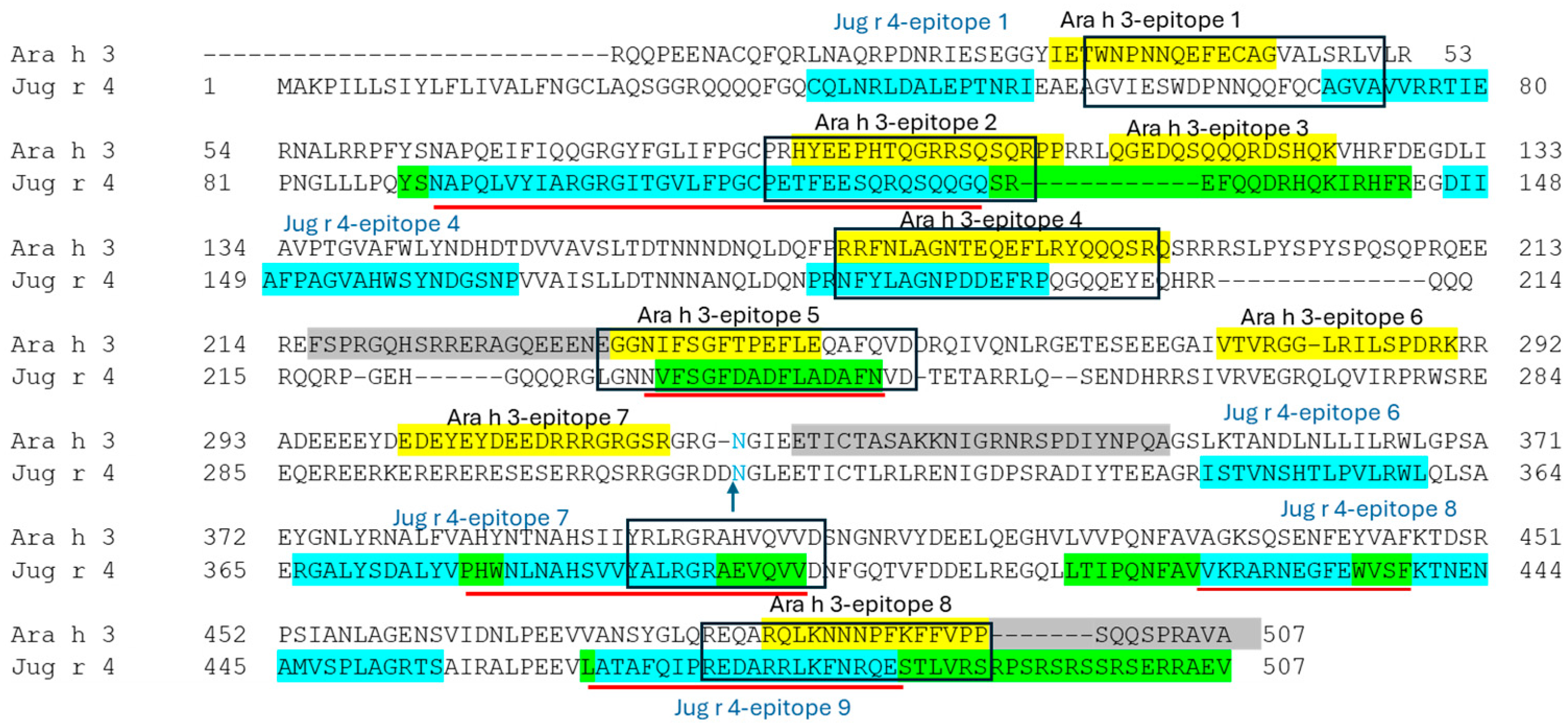
3.3. SDAP-Predicted Epitopes

ClustalW predicts a high level of homology between Ara h 3 and Jug r 4, and our anti-Ara h 3 antibody specifically recognized Jug r 4 in walnut extracts. We also used previously known and currently identified epitopes of both proteins and SDAP to predict potentially cross-reactive epitopes among these two glycinins. The results for these two proteins are summarized in Table 2. In this table, a peptide search is initiated in SDAP using epitopes identified in this study by microarray and from previously known epitopes of Ara h 3 or Jug r 4 (Figure 3) to identify potentially cross-reactive epitopes. Similar peptides are identified by their Property Distance (PD) similarity index or score, in which the smaller the number the more similar the peptides. In this table, we show the results and scores for Ara h 3 and Jug r 4, but have placed the more detailed search results that show cross-reactivity with multiple glycinins from other plant sources in Supplemental Table S1. The analysis indicates that Jug r 4 contains multiple regions with the potential for IgE binding, suggesting a strong allergenic potential. The presence of conserved epitopes along with similar physicochemical properties to those of well-known allergens supports the hypothesis that Jug r 4 may contribute to cross-reactivity with other nut allergens.

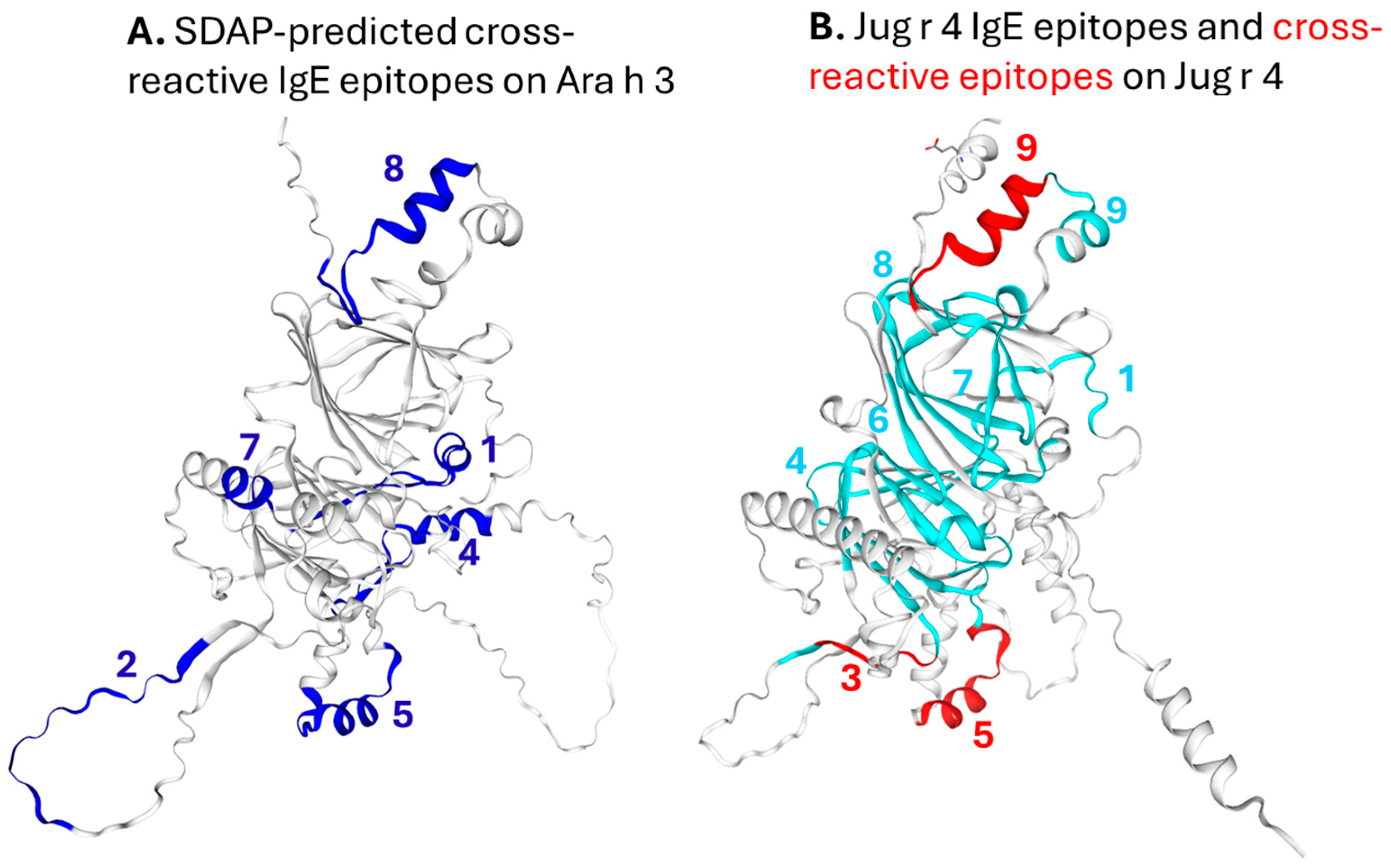
3.4. IgE Epitopes and Homology Modeling of Ara h 3 and Jug r 4

Our evaluation focused on the identification of the epitopes in Jug r 4 and their physicochemical properties and cross-reactivity with Ara h 3. The sequence of Jug r 4 and Ara h 3 were aligned using the ClustalW multiple protein alignment tool (Figure 3). The IgE epitope mapping of Ara h 3 and Jug r 4 was performed with PW-allergic subjects (Figure S1). Their IgE epitopes, previously identified epitopes, and SDAP-predicted as well actually IgE cross-reactive peptides are shown in Figure 3. Homology modeling confirms previous reports that Jug r 4 shares significant structural similarities with Ara h 3 (Figure 4). In Figure 4A, the known IgE binding sites of Ara h 3 that are predicted to cross-react with multiple glycinins (see Table S1), including Jug r 4, are shown in blue on a model of Ara h 3. The likelihood of cross-reactivity is predicted by the given PD score in Table 2. In Figure 4B, the epitopes of Jug r 4 identified by peptide microarray analysis with sera from PW-allergic individuals is shown in cyan and the three epitopes that are recognized in both Jug r 4 and Ara h 3 by these sera to be cross-reactive are shown in red. The comparative analysis, facilitated by the three-dimensional structure of Jug r 4, allowed us to identify potential cross-reactive epitopes. Several of the IgE binding regions in Jug r 4 exhibited physicochemical properties that are characteristics of major food allergens cataloged in the SDAP. While cross-reactivity among all homologs of Jug r 4 should be assessed, in this study we had access to unique sera from oral food challenge-positive dual peanut and walnut-allergic individuals, and since Ara h 3 was already crystalized, we chose to compare the previously identified epitopes from walnut-allergic individuals (for Jug r 4) or peanut-allergic individuals (for Ara h 3) with our dual-allergic subject sera and peptides that are computationally predicted to be similar (cross-reactive) to known epitopes (Figure 3). Other studies have discussed cross-reactivity among glycinins in multiple plants [9,11,12,19,20,21].

4. Discussion

Allergenic proteins from various environmental sources often share similar sequences and structures, which can explain cross-reactivity. This allows diverse plants and foods to provoke similar IgE-mediated reactions in certain individuals. Identifying common proteins and their structural features in these food sources through empirical and in silico methods may help improve diagnostic tools and enable personalized therapies for those suffering from food allergy. The prevalence of tree nut and peanut allergies is increasing, and cross-reactivity poses a significant barrier to creating reliable in vitro diagnostics and effective management strategies [19]. Our research aims to understand cross-reactivity at the molecular level using sera from individuals with confirmed single or dual allergies. Previous studies in both the U.S. and Europe have demonstrated that the 11S globulins or glycinins found in hazelnuts, cashews, sesame seeds, peanuts, and walnuts can be cross-reactive [4,20,22]. Importantly, allergic sera IgE from different countries recognize allergens at varying prevalence levels. For instance, one study in Europe indicates that while the English walnut protein, Jug r 4, is considered a minor allergen (recognized by <50% of allergic sera IgE), it has a high positive predictive diagnostic value of 90% for walnut allergy [22]. However, this does not imply that Jug r 4 is a minor allergen in other countries. In fact, Jug r 4 was classified as a major walnut allergen in the U.S. and is homologous to the major peanut allergen Ara h 3, suggesting its significance in cross-reactivity due to shared physicochemical properties [4,21,23,24,25]. It was shown that Jug r 4, like Ara h 3 in peanut, is one of the most abundant proteins in walnut and was recognized by 57–65% of walnut-allergic individuals sera in the U.S. [4,25]. IgE binding to Ara h 3 may also be more allergenic than generally thought, particularly in children [26]. This study seeks to identify potential immunoglobulin E (IgE) epitopes on Jug r 4 and Ara h 3 that may inform diagnosis and cross-reactivity with peanuts.
Juglans regia is the most commonly consumed walnut in the U.S. and in Europe [9]. Jug r 4 was purified from Juglans regia extract and the identity confirmed by LCMS and Western blot utilizing anti-Jug r 4 antibody. The purified Jug r 4 was originally used to confirm IgE binding to linearized Jug r 4 in SDS-PAGE by sera from PW-allergic individuals used in microarray analysis. The serum IgE binding of PW-allergic subjects that were shown to recognize Jug r 4 in walnut extracts was used to probe the microarrays for Jug r 4 and Ara h 3 epitope identification. We tested the potential cross-reactivity between Ara h 3 and Jug r 4 by Western blot analysis with antibodies against these allergens. The anti-Ara h 3 antibody was shown to bind mostly to the basic subunit of Jug r 4. All four tested PW-allergic individuals’ sera are shown by Western blot to recognize Jug r 4 (Figure 2). Sera from seven PW-allergic individuals were subjected to epitope mapping by microarray analysis.
Previous studies have identified the epitopes of Ara h 3 bound by peanut-allergic individuals [21,23,24,27]. We assess the IgE-binding epitopes and cross-reactivity between Jug r 4 and Ara h 3 with sera from PW dual-allergic individuals. This research confirms some previously identified epitopes and identifies novel Jug r 4 and Ara h 3 epitopes via empirical epitope mapping analysis. The actual cross-reactive linear epitopes are shown in Figure 3 and Figure 4. The computational prediction tool SDAP is also used to identify potentially cross-reactive epitopes (see Table 2 and Table S1). We found that dual-allergic PW-allergic subjects exhibit IgE binding to large portions of the Jug r 4 sequence (Figure 4) at similar frequencies as peanut- or walnut-only-allergic individuals. While Jug r 4 and Ara h 3 share over 70% amino acid identity and 84% homology, only three areas of high IgE binding to Jug r 4 were found to be bound by both Jug r 4 and the major Ara h 3 epitopes. These regions may be useful in diagnosing cross-reactivity.

5. Conclusions

Our interest is to understand the difference between clinically relevant and irrelevant IgE binding and cross-reactivity. In this study, we compare IgE binding to highly homologous walnut and peanut allergens in confirmed dual peanut and walnut-allergic subjects to that of previously identified IgE binding to Ara h 3 by peanut-allergic individuals and to Jug r 4 by walnut-allergic individuals with the goal of identifying potentially relevant linear IgE binding sites.
Of the nine epitopes of Jug r 4 and eight epitopes of Ara h 3 that were previously and currently identified, six were predicted to be cross-reactive and yet only three epitopes were bound most frequently by sera IgE from dual-allergic individuals. These epitopes, which are frequently recognized only by those confirmed allergic to peanut and walnut, may represent clinically relevant epitopes valuable in the diagnosis of cross-reactivity or development of multi-nut therapeutics.

Supplementary Materials

The following supporting information can be downloaded at https://www.mdpi.com/article/10.3390/allergies5010008/s1: Table S1: SDAP predicted similar peptides. Figure S1: Heat maps of Ara h 3 and Jug r 4 IgE epitopes.

Author Contributions

S.A.Y.G., J.B.N., H.C. and S.J.M. performed the experimental design, S.C.D., S.S.T. and S.S.M. supported the clinical characterization of allergic individuals and collection of sera. L.T.S. and H.C. performed the microarray analysis and screening. S.I.R. and H.C. performed the Western blot analysis; S.A.Y.G. conducted all other experiments. S.A.Y.G., J.B.N. and S.J.M. wrote the manuscript. All authors contributed to the critical review and preparation of the manuscript. All authors have read and agreed to the published version of the manuscript.

Funding

This work was funded by The United States Department of Agriculture (USDA), Agricultural Research Service (ARS project number 6054-43440-052-00D), and by Aimmune Nestlé Health Science, via a Cooperative Research and Development Agreement (CRADA agreement 358-6054-1-0011) (SJM), and the National Institute of Food and Agriculture (NIFA, 2017-67017-26518) (SJM).

Institutional Review Board Statement

This study was conducted in accordance with the Declaration of Helsinki and approved by the Institutional Review Boards of University of Colorado (protocol code 13-2102, approved 3 July 2024); Tulane School of Medicine (protocol code 14061, approved 3 December 2024); and Rochester Regional Health (protocol code 200210194-6, Approved 1 February 2024).

Informed Consent Statement

Informed consent was obtained from all subjects involved in this study.

Data Availability Statement

All the data relevant to this study can be found within this manuscript and submitted Supplementary Materials.

Acknowledgments

We would like to thank Mathew Lebar and Yuzhu Zheng for their critical review of this manuscript.

Conflicts of Interest

The authors declare no conflicts of interest.

References

  1. Gupta, R.S.; Warren, C.M.; Smith, B.M.; Jiang, J.; Blumenstock, J.A.; Davis, M.M.; Schleimer, R.P.; Nadeau, K.C. Prevalence and Severity of Food Allergies Among US Adults. JAMA Netw. Open 2019, 2, e185630. [Google Scholar] [CrossRef] [PubMed] [PubMed Central]
  2. Fleischer, D.M.; Conover-Walker, M.K.; Matsui, E.C.; Wood, R.A. The natural history of tree nut allergy. J. Allergy Clin. Immunol. 2005, 116, 1087–1093. [Google Scholar] [CrossRef] [PubMed]
  3. McWilliam, V.; Koplin, J.; Lodge, C.; Tang, M.; Dharmage, S.; Allen, K. The Prevalence of Tree Nut Allergy: A Systematic Review. Curr. Allergy Asthma Rep. 2015, 15, 54. [Google Scholar] [CrossRef] [PubMed]
  4. Wallowitz, M.; Peterson, W.R.; Uratsu, S.; Comstock, S.S.; Dandekar, A.M.; Teuber, S.S. Jug r 4, a legumin group food allergen from walnut (Juglans regia Cv. Chandler). J. Agric. Food Chem. 2006, 54, 8369–8375. [Google Scholar] [CrossRef] [PubMed]
  5. Sicherer, S.H.; Furlong, T.J.; Munoz-Furlong, A.; Burks, A.W.; Sampson, H.A. A voluntary registry for peanut and tree nut allergy: Characteristics of the first 5149 registrants. J. Allergy Clin. Immunol. 2001, 108, 128–132. [Google Scholar] [CrossRef] [PubMed]
  6. Burney, P.G.; Potts, J.; Kummeling, I.; Mills, E.N.; Clausen, M.; Dubakiene, R.; Barreales, L.; Fernandez-Perez, C.; Fernandez-Rivas, M.; Le, T.M.; et al. The prevalence and distribution of food sensitization in European adults. Allergy 2014, 69, 365–371. [Google Scholar] [CrossRef] [PubMed]
  7. Lee, J.; Jeong, K.; Jeon, S.A.; Lee, S. Component resolved diagnosis of walnut allergy in young children: Jug r 1 as a major walnut allergen. Asian Pac. J. Allergy Immunol. 2021, 39, 190–196. [Google Scholar] [CrossRef] [PubMed]
  8. Smits, M.; Verhoeckx, K.; Knulst, A.; Welsing, P.; de Jong, A.; Gaspari, M.; Ehlers, A.; Verhoeff, P.; Houben, G.; Le, T.M. Co-sensitization between legumes is frequently seen, but variable and not always clinically relevant. Front. Allergy 2023, 4, 1115022. [Google Scholar] [CrossRef] [PubMed] [PubMed Central]
  9. Ballmer-Weber, B.K.; Lidholm, J.; Lange, L.; Pascal, M.; Lang, C.; Gernert, S.; Lozano-Blasco, J.; Gräni, N.; Guillod, C.; Wangorsch, A.; et al. Allergen Recognition Patterns in Walnut Allergy Are Age Dependent and Correlate with the Severity of Allergic Reactions. J. Allergy Clin. Immunol. Pract. 2019, 7, 1560–1567.e6. [Google Scholar] [CrossRef] [PubMed]
  10. Verweij, M.M.; Hagendorens, M.M.; De Knop, K.J.; Bridts, C.H.; De Clerck, L.S.; Stevens, W.J.; Ebo, D.G. Young infants with atopic dermatitis can display sensitization to Cor a 9, an 11S legumin-like seed-storage protein from hazelnut (Corylus avellana). Pediatr. Allergy Immunol. 2011, 22, 196–201. [Google Scholar] [CrossRef] [PubMed]
  11. Konstantinou, G.N.; Baker, M.G.; Yu, J.; Ford, L.S.; Bencharitiwong, R.; Grishina, G.; Sampson, H.A.; Sicherer, S.; Nowak-Wegrzyn, A. Citrin: A novel food allergen in citrus seeds and citrus-derived pectin that shows cross-reactivity with cashew and pistachio. Ann. Allergy Asthma Immunol. 2023, 131, 759–765.e3. [Google Scholar] [CrossRef] [PubMed]
  12. Hu, J.; Zhu, L.P.; Wang, R.Q.; Zhu, L.; Chen, F.; Hou, Y.; Ni, K.; Deng, S.; Liu, S.; Ying, W.; et al. Identification, Characterization, Cloning, and Cross-Reactivity of Zan b 2, a Novel Pepper Allergen of 11S Legumin. J. Agric. Food Chem. 2024, 72, 8189–8199. [Google Scholar] [CrossRef] [PubMed] [PubMed Central]
  13. Thompson, J.D.; Higgins, D.G.; Gibson, T.J. CLUSTAL W: Improving the sensitivity of progressive multiple sequence alignment through sequence weighting, position-specific gap penalties and weight matrix choice. Nucleic Acids Res. 1994, 22, 4673–4680. [Google Scholar] [CrossRef] [PubMed] [PubMed Central]
  14. Hurlburt, B.K.; McBride, J.K.; Nesbit, J.B.; Ruan, S.; Maleki, S.J. Purification of Recombinant Peanut Allergen Ara h 1 and Comparison of IgE Binding to the Natural Protein. Foods 2014, 3, 642–657. [Google Scholar] [CrossRef] [PubMed] [PubMed Central]
  15. Waterhouse, A.M.; Studer, G.; Robin, X.; Bienert, S.; Tauriello, G.; Schwede, T. The structure assessment web server: For proteins, complexes and more. Nucleic Acids Res. 2024, 52, W318–W323. [Google Scholar] [CrossRef] [PubMed] [PubMed Central]
  16. Schein, C.H.; Ivanciuc, O.; Braun, W. Structural Database of Allergenic Proteins (SDAP). In Food Allergy; Maleki, S.J., Burks, A.W., Helm, R.M., Eds.; ASM Press: Washington, DC, USA, 2006; pp. 257–283. [Google Scholar]
  17. Ivanciuc, O.; Oezguen, N.; Mathura, V.S.; Schein, C.H.; Xu, Y.; Braun, W. Using property based sequence motifs and 3D modeling to determine structure and functional regions of proteins. Curr. Med. Chem. 2004, 11, 583–593. [Google Scholar] [CrossRef] [PubMed]
  18. Ivanciuc, O.; Schein, C.H.; Braun, W. SDAP: Database and computational tools for allergenic proteins. Nucleic Acids Res. 2003, 31, 359–362. [Google Scholar] [CrossRef] [PubMed] [PubMed Central]
  19. Sharma, E.; Vitte, J. A systematic review of allergen cross-reactivity: Translating basic concepts into clinical relevance. J. Allergy Clin. Immunol. Glob. 2024, 3, 100230. [Google Scholar] [CrossRef] [PubMed] [PubMed Central]
  20. Wallowitz, M.L.; Chen, R.J.Y.; Tzen, J.T.C.; Teuber, S.S. Ses i 6, the sesame 11S globulin, can activate basophils and shows cross-reactivity with walnut in vitro. Clin. Exp. Allergy 2007, 37, 929–938. [Google Scholar] [CrossRef] [PubMed]
  21. Barre, A.; Jacquet, G.; Sordet, C.; Culerrier, R.; Rougé, P. Homology modelling and conformational analysis of IgE-binding epitopes of Ara h 3 and other legumin allergens with a cupin fold from tree nuts. Mol. Immunol. 2007, 44, 3243–3255. [Google Scholar] [CrossRef] [PubMed]
  22. Blankestijn, M.A.; den Hartog Jager, C.F.; Blom, W.M.; Otten, H.G.; de Jong, G.A.H.; Gaspari, M.; Houben, G.F.; Knulst, A.C.; Verhoeckx, K.C.M. A subset of walnut allergic adults is sensitized to walnut 11S globulin Jug r 4. Clin. Exp. Allergy 2018, 48, 1206–1213. [Google Scholar] [CrossRef] [PubMed]
  23. Jin, T.; Guo, F.; Chen, Y.W.; Howard, A.; Zhang, Y.Z. Crystal structure of Ara h 3, a major allergen in peanut. Mol. Immunol. 2009, 46, 1796–1804. [Google Scholar] [CrossRef] [PubMed]
  24. Robotham, J.M.; Hoffman, G.G.; Teuber, S.S.; Beyer, K.; Sampson, H.A.; Sathe, S.K.; Roux, K.H. Linear IgE-epitope mapping and comparative structural homology modeling of hazelnut and English walnut 11S globulins. Mol. Immunol. 2009, 46, 2975–2984. [Google Scholar] [CrossRef] [PubMed]
  25. Teuber, S.; Peterson, W.; Uratsu, S.; Dandekar, A.; Roux, K.; Sathe, S. Identification and cloning of Jug r 4, a major food allergen from English walnut belonging to the legumin group. J. Allergy Clin. Immunol. 2003, 2, S248. [Google Scholar] [CrossRef]
  26. Restani, P.; Ballabio, C.; Corsini, E.; Fiocchi, A.; Isoardi, P.; Magni, C.; Poiesi, C.; Terracciano, L.; Duranti, M. Identification of the basic subunit of Ara h 3 as the major allergen in a group of children allergic to peanuts. Ann. Allergy Asthma Immunol. 2005, 94, 262–266. [Google Scholar] [CrossRef] [PubMed]
  27. Rabjohn, P.; Burks, A.W.; Sampson, H.A.; Bannon, G.A. Mutational analysis of the IgE-binding epitopes of the peanut allergen, Ara h 3: A member of the glycinin family of seed-storage proteins. J. Allergy Clin. Immunol. 1999, 103, S101. [Google Scholar]
Figure 1. SDS-PAGE of Jug r 4 purification profile from walnut extract. The molecular weight markers (MWMs) are shown in lane 1 and 3. Walnut (WN) extract and insoluble (insol) material are shown in lanes 2 and 4. The diluted walnut extract is loaded onto a High-S column (Load on S, lane 5), the washed and unbound material is shown in lane 6, and eluted with a NaCl gradient of 200–400 mM NaCl. The eluted fraction numbers (F15, F21, F25, F29, F55) are indicated in lanes 7–11 and correspond with fractions eluted from the S column in panel (B). Panel (B) shows a graph in which the optical density (O.D.) at 280 nM is shown on the y-axis and the fraction number for the samples eluted from the High-S cation exchange column are shown are shown on the x-axis. The red star (Ж) indicates the fractions selected for SDS-PAGE analysis in panel (A). The inset in panel (B) shows the pooled purified Jug r 4 fractions 21–29 assessed by SDS-PAGE. The inset in panel (B) shows the pooled purified fractions of Jug r 4.
Allergies 05 00008 g001
Figure 2. Western blot analysis of Jug r 4 binding to allergic serum IgE and anti-Jug r 3 and anti-Ara h 3 antibodies. Different amounts of walnut extracts, indicated as WN mg, on the top of each lane are subjected to incubation with anti-Jug r 4 antibody (indicated at top of the left lane), with 4 walnut-allergic subject sera (numbers 1–4 below each anti-IgE blot) and a non-allergic (NA) control, and anti-Ara h 3 antibody (right most panel).
Allergies 05 00008 g002
Figure 3. Alignment of Ara h 3 and Jug r 4 amino acid sequences. Ara h 3 (top sequence) and Jug r 4 (bottom sequence). The arrow and blue N residue indicate the residue cleavage site that separates the basic vs acidic (NGLEET start of acidic subunits). The yellow highlighted areas indicate the previously identified linear IgE epitopes of Ara h 3 (bound by peanut-allergic subject sera) and green highlighted area on Jug r 4 are epitopes previously identified using sera from walnut allergic individuals (https://www.uniprot.org/uniprotkb/Q2TPW5/entry, accessed on 3 November 2024). The cyan indicates IgE epitopes identified in this study with microarray analysis using sera from peanut and walnut (PW)-allergic subjects. The areas highlighted with cyan and green are overlapping and the red lines show the overlap in green and blue boxes. The gray highlighted areas are additional Ara h 3 IgE epitopes identified by microarray when using PW-allergic sera. The squares show areas predicted to be cross-reactive by SDAP in Table 1 and also in blue in the molecular model of Ara h 3 in Figure See also Supplemental Material Table S1 for more details on SDAP search results).
Allergies 05 00008 g003
Figure 4. Molecular models of Ara h 3 and Jug r 4 predicted and empirically identified IgE epitopes. The SDAP-predicted cross-reactive epitopes on Ara h 3 are shown in blue. The numbers in blue indicate the Ara h 3 epitope number. (panel A). Jug r 4 IgE epitopes identified by the microarray are shown in cyan and the IgE epitopes of peanut and walnut-allergic individuals that were shown to be cross-reactive with known Ara h 3 epitopes are shown in red. The numbers in red indicate the Jug r 4 epitope number (panel B).
Allergies 05 00008 g004
Table 1. Subject allergic characteristics.
SubjectAgeSexPeanutWalnutCashewAlmondHazelnutPecanPistachioOther
1.14MAx xxsesame, brazil nut, pine nut, garbanzo
2.19FAx x xxbrazil nut, pine nut, shellfish, fish
3.11Mxx sesame, milk
4.8?Axxxx pine nut, brazil nut
5.31Fxx xxx pine nut
6.21FAxxxxxxmacadamia, egg, milk
7.24Mxxxx x brazil nut
8.11Fxxxxxxxmacadamia nut, milk, egg, brazil, pine
9.21FLTLTx
10.25FLTLTLTLTLTnexbrazil nut
11.63FLTLTxxxLTnebrazil nut, potato, egg
12.nFxx sunflower
13.38Mxx x x coconut
14.42Fxxnenenex macadamia, brazil nut,
15.16Mxxxxxxxcrab, lobster, flax, soy
16.35Fxxxxxxxsoy
17.25Fxxxxxxxpoppy, coconut, fruits, shellfish
18.26Mxxxxxxxpine nut, crab, lobster
19.25 xxxnenex
20.46Mxxxxxxxbrazil, pine, macadamia, shellfish, fish
21.25Fxxxnexnexpea, soy, sesame
22.9M xxx
23.22M x
24.25FA soy
25.13MA x sesame
26.15Mx x xoutgrew egg
27.10Mx
28.35 x
29.19 x
30.16Mx soy
31.ch Chicken serum negative control
32.23F x x
33.9M x
A = anaphylactic, LT = life threatening, ne = never eaten.
Table 2. Ara h 3 and Jug r 4 linear epitopes predicted to be cross-reactive using SDAP Property Distance (PD) tool. PD score 0 = identical sequence, scores between 0 and 8 = potential IgE cross-reactivity, and scores >8 = not likely to be cross-reactive.
AllergenNCBIPD Sequence Similarity IndexStart ResidueMatching RegionEnd Residue
Ara h 3 Epitope 1
Ara h 3O825800.0028GYIETWNPNNQEFECAGVAL47
Jug r 4.0101Q2TPW52.9455GVIESWDPNNQQFQCAGVAV74
Ara h 3 Epitope 2—Cross-reactive with Jug r 4 Epitope 3
Ara h 337031070.0091HYEEPHTQGRRSQSQR106
Jug r 4.0101Q2TPW57.77115TFEESQRQSQQGQSRE130
Ara h 3 Epitope 3 QQQRDSHQKVHFDEGD—No low PD match to Jug r 4
Ara h 3 Epitope 4—Cross-reactive with Jug r 4 Epitope 5
Ara h 3O825800.00170PRRFNLAGNTEQEFLRYQQQSRQ192
Jug r 4.0101Q2TPW58.82185PRNFYLAGNPDDEFRPQGQQEYE207
Ara h 3 Epitope 5—Cross-reactive with previously identified Jug r 4 epitope
Ara h 337031070.00242IFSGFTPEFLEQAFQVD258
Jug r 4.0101Q2TPW55.13233VFSGFDADFLADAFNVD249
Ara h 3 Epitope 6 VTVRGGLRILSPDRK—No low PD match to Jug r 4
Ara h 3 Epitope 7—Similar peptide found in Jug r 4, but with low likelihood of being cross-reactive
Ara h 3O825800.00300DEDEYEYDEEDRRRGRGSR318
Jug r 4.0101Q2TPW59.40294RERERESESERRQSRRGGR312
Ara h 3 Epitope 8—Similar peptide found in Jug r 4 Epitope 9, but with low likelihood of being cross-reactive
Ara h 3O825800.00480REQARQLKNNNPFKFFVPP498
Jug r 4.0101Q2TPW59.87473REDARRLKFNRQESTLVRS491
Jug r 4 Epitope 1 CQLNRLDALEPTNRI—Similar peptide found in Ara h 3, but no IgE binding detected on microarray
Jug r 4 Epitope 3—Cross-reactive with Ara h 3 Epitope 2
Jug r 4.0101Q2TPW50.00111GCPETFEESQRQSQQGQSR129
Ara h 337031076.5487GCPRHYEEPHTQGRRSQSQ105
Jug r 4 Epitope 4—Cross-reactive with peptide found in Ara h 3
Jug r 4.0101Q2TPW50.00149AFPAGVAHWSYNDGSNP165
Ara h 337031077.55137AVPTGVAFWLYNDHDTD153
Jug r 4 Epitope 6 ISTVNSHTLPVLRWL—Similar peptide found in Ara h 3, but with low likelihood of being cross-reactive
Jug r 4 Epitope 9—Microarray-confirmed cross reactivity with Ara h 3 Epitope 8 despite elevated PD score.
Jug r 4.0101Q2TPW50.00465LATAFQIPREDARRLKFNRQESTLVR490
Ara h 3370310710.36475VANSYGLQREQARQLKNNNPFKFFVP500
Disclaimer/Publisher’s Note: The statements, opinions and data contained in all publications are solely those of the individual author(s) and contributor(s) and not of MDPI and/or the editor(s). MDPI and/or the editor(s) disclaim responsibility for any injury to people or property resulting from any ideas, methods, instructions or products referred to in the content.

