Abstract
Eriobotrya japonica (E. japonica) leaves have been used as an herbal traditional medicine in China and Japan owing to their anti-inflammatory and protective effects against skin conditions and allergy symptoms. These beneficial effects are likely mediated by the various triterpenoids present in E. japonica leaves. However, the efficacy of E. japonica leaves in the treatment of allergic rhinitis has not been evaluated in humans. Therefore, in the present study, a randomized, controlled, double-blind trial was performed on healthy adults of age >20 (n = 27) who were randomly assigned to receive either 2.5 g of placebo or E. japonica leaf supplements once daily for 4 weeks. The Japanese Allergic Rhinitis Quality of Life Standard Questionnaire (JRQLQ), dermatological allergy symptoms, Dermatology Life Quality Index, and skin condition parameters were assessed at baseline and after 4 weeks. Significant differences were observed in the variability of the itchy nose, itchy eyes, and eye symptoms between the E. japonica supplementation and placebo groups after 4 weeks. Arm skin transepidermal water loss was improved only in the E. japonica supplementation group. This study suggests that E. japonica leaves can be used as a functional food ingredient to relieve allergic symptoms.
1. Introduction
The leaves of loquat (Eriobotrya japonica) have long been used as herbal medicines in China and Japan [1], and they are still currently utilized as traditional medicine [2,3]. Various triterpenoids [4,5], sesquiterpenoids [6], flavonoids [7], and tannins [7,8] have been identified in E. japonica. Triterpenoids are structurally diverse natural products, constitute major components of numerous medicinal plants, and are expected to be potential agents in drug discovery [9,10]. Terpenoids derived from the leaves of E. japonica possess biological activities [11] and exhibit anti-inflammatory [4], antitumor [5,12], antioxidant [7], and antiviral properties [13]. In addition, E. japonica-derived triterpenoids have been suggested to possess protective effects on the skin melanin formation, promotion of collagen and hyaluronic acid production, and inhibition of acne growth and allergic substance production [14,15,16].
The leaves of E. japonica contain triterpenoids such as ursolic acid, corosolic acid, maslinic acid, and oleanolic acid. Ursolic acid, a major active component of the leaves of E. japonica [4,17] (Appendix A), inhibits skeletal muscle atrophy by regulating insulin/insulin-like growth factor-1 signaling [18] and enhances muscle strength during resistance training [19]. In addition, ursolic acid prevents osteoporosis [20] and alleviates diet-induced obesity, glucose intolerance, and fatty liver disease [21]. Ursolic acid is also predicted to be involved in the production of allergic substances; it suppresses allergy symptoms by inhibiting the production of immunoglobulin E (IgE) antibodies [22,23], reduces the release of β-hexosaminidase from IgE-stimulated RBL-2H3 mast cells, and relieves allergic symptoms [14]. Oleanolic acid promotes the recovery of epidermal permeability barrier function [24]. Moreover, maslinic acid and corosolic acid present in E. japonica are expected to exhibit protective effects against inflammatory diseases [25].
Allergic rhinitis (AR) and atopic dermatitis (AD) are substantial health concerns affecting humans worldwide. AD, the most common inflammatory skin disease in the industrialized world, has multiple underlying causes [26], affecting the quality of life (QOL) of adult patients in whom the condition can be severe and persistent [27]. AR is induced by IgE-mediated inflammation of the nasal membrane in response to allergen exposure [28]. Similar to AD, AR causes physical discomfort in patients and affects their QOL [29]. Patients with AR or AD are often prescribed symptomatic therapeutic drugs, which may cause secondary effects, such as drowsiness, mental fogginess, or asthenia [30]. Notably, herbal medicines, such as E. japonica leaves, are expected to exhibit fewer adverse effects, suggesting their potential as traditional medicines, suitable for use by children and older individuals.
As major active components of E. japonica leaves, the use of ursolic acid and triterpenoids as a complementary treatment is expected to alleviate the symptoms of AR or AD. Although the effects of ursolic acid on AR symptoms have been reported in rats [22,23], the efficacy of ursolic acid derived from E. japonica leaves on AR has not yet been evaluated in humans. Therefore, the present study aimed to evaluate the effects of supplements containing ingredients derived from the leaves of E. japonica on allergy symptoms and the skin quality of healthy adults.
2. Materials and Methods
2.1. Supplement Preparation
The leaves of E. japonica were obtained from Totsukawa Co., Ltd. (Kagoshima, Japan). E. japonica (product code: NBT83) leaf supplements were formulated for a daily intake of 250 mg, distributed across ten tablets. Each tablet of this supplement contained 83% E. japonica leaf powder and 17% excipients. Each placebo supplement tablet contained dextrin and cornstarch, which replaced E. japonica leaves, excipients, and natural pigments.
2.2. Clinical Study and Ethics
This randomized, double-blind, placebo-controlled clinical study was conducted from 12 November 2018 to 15 December 2018 in the Laboratory of Systematic Forest and Forest Products Sciences, Faculty of Agriculture, Kyushu University. The clinical study included two groups with a 1:1 allocation ratio of receiving placebo or E. japonica supplements. Stratified randomization was used to reduce bias.
This study was approved by the Ethics Committee of the Faculty of Humanity-Oriented Science and Engineering, Kindai University (4 March 2017) and registered in the University Hospital Medical Information Network Clinical Trials Registry (ID:000034859).
2.3. Participants and Settings
As no previous study has reported the statistical significance of the oral intake of E. japonica leaves, we referred to a previous investigation reporting significant differences after oral intake of another supplement [31]. The sample size was set at 22 participants in 2 groups (11 in each group) to ensure the detection of significant differences at p < 0.05, significance level (α) of 0.05, and statistical power (1−β) of 0.80 [31]. Furthermore, the final number of participants was set at 30 (15 in each group) to allow for a margin for dropouts and noncompliance with the protocol during the study period.
Healthy adults of age >20 years (n = 30) who fulfilled the specified inclusion and exclusion criteria (Table 1) were included and were evaluated by a staff member (not the investigator). All volunteers signed an informed consent form stating the purpose, method, compensation, confidentiality, and right to withdraw from the study. In collaboration with the two clinics, we were able to consult physicians in case of adverse events.

Table 1.
Inclusion and exclusion criteria.
2.4. Randomization
Randomization and allocation were performed by a staff member independent of the investigators and were centralized and performed based on a computer-generated list of random numbers. Randomization was performed based on stratified random sampling with age, body mass index (BMI), and sex (less than 40 years and BMI less than 21; less than 40 years and BMI 21 or more; 40 years or more and BMI less than 21; 40 years or more and BMI 21 or more), with adaptive randomization for an equal number in each arm. The investigators were not involved in the allocation, and the order of assignment was concealed until the assignment was completed. The sample assignment to each group was blinded to both the participants and investigators until the study was completed.
2.5. Study Schedule
All the participants took ten tablets (orally) of their assigned study formulation daily. To minimize the limitations of the study, all participants were required to refrain from consuming any similar dietary supplements, quasi-drugs, or medicines. They were also prohibited from using any skincare treatments and massages or from changing their daily skincare cosmetics from the start to the end of the study. Each participant visited the research laboratory for assessment twice: before intake of the study formulation at baseline (0 W) and after 4 weeks (4 W) of study formulation intake for efficacy measurements. The participants were requested to apply daily skincare products on the morning of the visit and remove the skincare products at each visit.
2.6. Outcomes
In the design of the clinical study, we set the outcomes for the response to the oral administration of supplements. The primary outcome was AR symptoms according to the Japanese Allergic Rhinitis Standard Quality of Life Questionnaire (JRQLQ) [32], dermatological allergy symptoms according to the Dermatology Life Quality Index (DLQI) [33], and skin condition according to the skin measurement devices.
2.7. Measurement of AR Symptoms and Skin Areas
The JRQLQ [32], commonly used in otorhinolaryngology in Japan, was used to evaluate AR symptoms. In this index, a higher score indicates more severe symptoms and a total of 23 items are included, and each item’s score ranges from zero to four. All items were divided into six domains, and the allergic symptoms and QOL of individuals within each domain were assessed. The total symptom score was calculated as the sum of the four nasal symptom scores and two eye symptom scores.
Dermatological allergy symptoms were assessed by DLQI [33], a dermatology-specific health-related quality of life (HRQOL) questionnaire. The DLQI consists of ten questions concerning symptoms and feelings, daily activities, leisure, work, school, personal relationships, and treatment. Skin hydration (arbitrary units; a.u.) and transepidermal water loss (TEWL) (g/h/m2) were measured using a Corneometer® CM 825 and Tewameter® TM 300, respectively (Courage and Khazaka, Cologne, Germany). Measurements were obtained on the left upper arm (inner side, 3 cm above the elbow). The skin region of interest was cleansed using a cleansing sheet (Bifesta Cleansing Sheet, Mandom Corporation, Osaka, Japan), wiped with cotton containing a cleansing liquid (Bifesta Face Wash, Mandom Corporation), rinsed with warm water, wiped, and dried for 20 min at stable temperature (23 ± 5 °C) and humidity conditions (50% ± 15%). Three intermediate values were used to calculate the mean values.
2.8. Statistical Analysis
SPSS (version 25.0, Chicago, IL, USA) was used to analyze the data. To compare the quantitative demographic variables between the two groups, a parametric test (normally distributed data), independent sample Student’s t-test or non-parametric test (non-normally distributed data), and Mann–Whitney U test was used. Changes in variables at the end of the study compared to those at the beginning were measured using the Wilcoxon signed-rank test. To compare changes in parameters between the two groups, the Mann–Whitney U test was used. In the presence of outliers—data points exceeding one and a half times the interquartile range (IQR)—the Moses test of extreme reaction was used in addition to the Mann–Whitney U test. Statistical significance was set at a p < 0.05. In this study, multiple adjustments were necessary to establish multiple primary outcomes. We adopted a closed testing procedure to avoid multiplexity; the analysis was pre-determined to be performed in the order of (1) AR, (2) skin quality evaluation, and (3) dermatological allergy symptoms. If the result indicated no significant difference between the groups, the analysis would be terminated.
3. Results
3.1. Demographic Information at Baseline
Participants were recruited from 19 October to 8 November and after excluding 3 participants who declined the study for personal reasons, 27 healthy adults assigned to their group on 10 November by a staff member (not an investigator). From 12 November 2018 to 15 December 2018, 27 participants completed the study and were analyzed (Figure 1). The background characteristics of each group are presented in Table 2. There were no significant differences in age, height, body weight, body mass index, AR, or skin quality between the two groups.
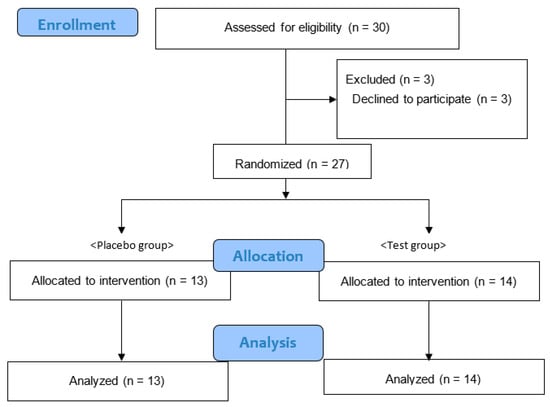
Figure 1.
Consolidated standards of reporting flow diagram.

Table 2.
Characteristics of the participants.
3.2. Effects of E. japonica Leaves on Allergic Symptoms
Evaluation of AR at the baseline revealed no significant differences in the items of the JRQLQ between the two groups. In addition, all domain scores of the JRQLQ in the test group showed no significant changes after four weeks. In the placebo group, one domain, emotional function, decreased after 4 weeks of supplementation (Table 3). However, there was no significant difference in the changes in the JRQLQ domain score during 4 weeks of supplementation between the placebo and test groups (Table 3). Regarding itching items and symptoms (itchy nose, nasal symptoms, itchy eyes, and eye symptoms), the itchy eye symptoms in the placebo group increased significantly, whereas it did not significantly change in the test group (Table 4). Because there were outliers in the changes in itching and symptoms items, the Moses test of extreme reactions was used. The analysis revealed significant differences in the variability between the two groups for itchy nose (p = 0.006), itchy eyes (p < 0.001), and eye symptoms (p < 0.001) over the 4-week supplementation period (Figure 2).

Table 3.
Comparison of the Japanese Allergic Rhinitis Quality of Life Standard Questionnaire (JRQLQ) domain scores within groups and changes between groups.

Table 4.
Comparison of itch and symptom of nose and eyes within groups and changes between groups.
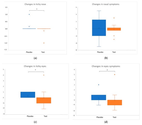
Figure 2.
Comparison of symptoms between the test and placebo groups. Dots beyond the whisker bounds denote outliers. Cross marks and horizontal lines indicate the means and medians of each group, respectively. Asterisks indicate significant differences between groups; * p < 0.01.
3.3. Effects of E. japonica Leaves on Skin Condition
Evaluation of skin condition at the baseline revealed no significant differences in the hydration and the TEWL of the arm skin between the two groups. The hydration of the arm skin in the test group was significantly decreased between baseline and after four weeks of supplementation (p = 0.019). The TEWL of the arm skin in the test group was significantly improved between baseline and after 4 weeks (p = 0.004). Moreover, no significant differences in the changes in either hydration or TEWL were observed between the groups (Table 5).

Table 5.
Comparison of the skin condition of arm scores pre-and post-intervention.
3.4. End of the Analysis and Safety Assessment
As a closed testing procedure was adopted to avoid multiplexity, the analysis was terminated after the results of the skin condition showed no significant difference between the groups. Notably, no side effect or adverse event was observed throughout the study period under the conditions of this study.
4. Discussion
In the present study, we investigated the effects of oral intake of E. japonica leaf supplements on the symptoms of AR and skin conditions in healthy adults. We observed significant variability between the placebo and test groups in AR symptoms, such as itchy nose and eyes, after supplement intake. Although we did not provide quantitative evidence of this effect, our results suggest that intake of E. japonica leaf supplements alleviates itchy eye and nose symptoms. To clarify the effect in the present study more accurately, additional clinical studies on the quantitative effects of E. japonica should be conducted in the future.
We conducted a qualitative analysis of the triterpenoids in the E. japonica leaves and supplements using high-performance liquid chromatography (HPLC) coupled with electrospray ionization and quadrupole time-of-flight mass spectrometry. We detected peaks corresponding to the four targeted triterpenoids, namely ursolic, corosolic, maslinic, and oleanolic acids (unpublished data). The results of HPLC coupled with an evaporative light-scattering detector, including the contents of the four main triterpenoids in E. japonica leaves collected each month of the year (Appendix A), are presented in Table A3. These results showed that the order of contents was as follows: ursolic acid > maslinic acid > corosolic acid > oleanolic acid. Moreover, we estimated the contents of the four triterpenoids in the supplement tablets administered to the participants in the present clinical trial. As the proportion of E. japonica leaf powder in the supplements was 83%, the content per gram of the leaves was calculated based on the value obtained from the quantitative analysis, and the results showed no difference between the leaf and the tablet samples (Table A4). This result indicates that the manufacturing process of the tablet sample did not affect the triterpenoid content.
The mechanisms underlying allergic itching have been previously reported; mast cells undergo degranulation following the administration of IgE antibody and allergenic conjugate stimulation, releasing histamines and transmitters, such as prostaglandins, chemokines, and cytokines, thus causing allergic reactions [34,35]. A previous study has demonstrated the effects of ursolic acid, a pentacyclic triterpenoid abundant in E. japonica leaves, on nasal symptoms in a rat model. Ursolic acid relieves nasal symptoms caused by PM2.5 exposure, possibly by inhibiting the expression of Th2 cytokine genes, eosinophilic infiltration, and specific IgE production [18,23]. Another study has reported the underlying mechanism of ursolic acid; it inhibits mast cell degranulation by reducing intracellular calcium levels and attenuating proinflammatory cytokine secretion. Moreover, the effects of ursolic acid were dependent on the inhibition of FcεRI-mediated signaling [36]. Ursolic acid and oleanolic acid have been reported to inhibit β−hexosaminidase release [16,24]. In the present study, several compounds were isolated from the leaves of E. japonica, and ursolic, oleanolic, maslinic, and corosolic acids (chemical structures shown in Appendix A) were identified as the main components. Moreover, we measured the release of β-hexosaminidase from RBL-2H3 cells (unpublished data, see Appendix B). We found that the methanol extract from E. japonica leaves and its ethyl-acetate-soluble and hexane-soluble fractions suppressed β-hexosaminidase release in RBL-2H3 cells (Appendix B, Figure A4). However, the residual water fraction did not exhibit any activity. This result indicates that hydrophobic compounds, in addition to water-soluble hydrophilic compounds, contribute to the observed anti-allergic activities. Therefore, the four hydrophobic compounds, ursolic, oleanolic, maslinic, and corosolic acids, were considered to partially contribute to the anti-allergic activities of the E. japonica leaf supplements in the present clinical trial (Appendix B, Figure A5). These results are consistent with previous studies in which triterpenoid compounds derived from the leaves of E. japonica and other plants exhibited anti-allergic and anti-inflammatory activities [15,23,25].
In the present study, analysis of the skin condition revealed that the placebo group showed maintained hydration of the arm skin, whereas the test group showed decreased hydration. In contrast, the TEWL of the test group improved significantly, whereas that of the placebo group did not change. Overall, no significant differences were observed between the two groups over the four-week supplementation. Although some triterpenoids have been reported to partially promote hyaluronic acid production [14], the results of the present study, which indicate both decreased hydration and retained skin barrier function in the test group, cannot be explained by the previous theory. This unexpected result might be attributed to the room humidity conditions during the dry winter season; however, this explanation alone does not sufficiently account for the observed results. Therefore, we inferred the corresponding mechanism of action based on previous studies. Lim et al. have reported, using in vivo and in vitro tests, that ursolic acid and oleanolic acid can improve the recovery of the skin barrier function and induce epidermal keratinocyte differentiation via a peroxisome proliferator-activated receptor (PPAR)-α [37]. In addition, Uehara et al. showed the feasibility of the qualitative evaluation of TEWL by measuring the thickness and water content of the stratum corneum in an environment in which the effect of perspiration is small [38]. These previous findings suggest that in the present study, the improved TEWL in the test group promoted the recovery of skin barrier function and induced epidermal keratinocyte differentiation and that these effects are likely mediated by ursolic acid and oleanolic acid present in the leaves of E. japonica. Therefore, epidermal keratinocyte differentiation was induced, promoting the thickening of the stratum corneum. As the observation period was short (4 weeks), although the stratum corneum became thicker, the hydration development was not yet fully established, resulting in a temporary decrease in hydration. Consequently, improved TEWL and decreased hydration were simultaneously observed in the test group. Nevertheless, further investigations are required to investigate this mechanism of action, as we did not conduct in vivo and in vitro experiments but rather relied on previous literature.
This study has some limitations. First, we did not investigate whether each allergy was caused by seasonal factors, such as pollen, other year-round factors, pollutants, or viral. Therefore, clinical studies on the effects of E. japonica leaves on participants with the identified causes of the allergy are required. Second, AR is a chronic disease that is difficult to cure, leading to a growing tendency to seek improvement of the QOL as a treatment goal. The JRQLQ is a QOL questionnaire developed by Okuda et al. for Japanese people [32] based on the RQLQ. Although it is intended to be used for both patients and healthy people, it only evaluates QOL related to rhinitis symptoms. Therefore, the presence of a significant difference from the JRQLQ does not indicate that the threshold for Minimal Clinical Important Difference (MCID) has been met, and we are cautious not to overemphasize this aspect. In addition, the JRQLQ has not undergone statistical validation, and its range of applicability is limited. In the present study, in the placebo group, the emotional functional area improved after the intervention period. Although the exact reason for this improvement is difficult to identify, as the participants of this study were healthy adults rather than patients, they did not experience severe symptoms. Therefore, respondents might have evaluated items, such as “irritability, depression, and dissatisfaction with life”, due to daily life stressors not directly related to AR symptoms. Third, the sample size was small, and the intake period was relatively short. Thus, these results should be cautiously interpreted when applied to clinical practice, and further studies with more participants, longer intake periods, different doses, and more comprehensive clinical evaluation methods are needed to evaluate the clinical effect of oral intake of E. japonica leaf supplements. Finally, this was an exploratory study of the leaves of E. japonica in human participants (healthy volunteers), and no side effects or adverse events were observed throughout the study period under the conditions of this study. Although there have been several reports of toxicity verification in vivo [39], reports on toxicity verification in humans are limited. As the safety assessment in this study was concise, more solid safety studies should be performed in the future.
In summary, this study investigated the effects of E. japonica leaves on healthy adults with common eye and nose-related allergic symptoms in everyday life. To the best of our knowledge, this is the first study to clinically evaluate the effect of the oral intake of E. japonica leaf supplements on allergy symptoms and skin conditions. The extract of the leaves of E. japonica is expected to be safely administrated to children and older individuals as a traditional medicine, with few side effects, to alleviate AR or maintain skin conditions. However, additional studies on the effects of E. japonica leaves as the traditional medicine on AR and skin conditions using an appropriate study design should be conducted in the future.
Author Contributions
Conceptualization, funding acquisition, methodology, project administration, and resources, K.S.; investigation, data curation, validation, formal analysis, and visualization, M.N. (Masumi Nagae), M.N. (Maki Nagata), M.M., N.T., Y.A., D.W. and Y.Y.; original draft preparation, M.N. (Masumi Nagae); writing—review and editing, K.S., M.N. (Masumi Nagae), M.N. (Maki Nagata), M.M., N.T., Y.A., D.W. and Y.Y. All authors have read and agreed to the published version of the manuscript.
Funding
This research and APC were funded by Totsukawa Co. Ltd., Kurume Research Park Co. Ltd. (2018–2019), and the Kagoshima Industry Support Center (2019–2020).
Institutional Review Board Statement
The study was conducted in accordance with the Declaration of Helsinki, and approved by the ethics committee of Kindai University Faculty of Humanity Oriented Science and Engineering (approval no. 4 March 2017).
Informed Consent Statement
Informed consent was obtained from all participants involved in the study.
Data Availability Statement
All available data in this study are presented in the article and Appendix A and Appendix B.
Acknowledgments
We would like to thank Koichiro Onuki of the Faculty of Humanity-Oriented Science and Engineering, Kindai University, and the students in the laboratory for their cooperation in carrying out this research.
Conflicts of Interest
The authors declare no conflict of interest.
Appendix A
Appendix A.1. Materials
The leaves of E. japonica, which are used today as traditional medicine, contain various triterpenoids, including ursolic acid, corosolic, maslinic, and oleanolic acids. In the present study, E. japonica leaves were collected between April 2020 and March 2021, and supplements containing these E. japonica leaves were manufactured by Totsukawa Farm (Kagoshima, Japan), an agricultural production corporation. The supplements were manufactured at a dose of 250 mg per tablet and comprised 83% E. japonica leaf powder, 5% reduced maltose, 5% calcium seaweed, 4.5% citrus fiber, and 2.5% calcium stearate.
Ursolic acid (98%) and maslinic acid (98%) standards were purchased from Funakoshi Co., Ltd. (Tokyo, Japan). Oleanolic acid (97%) and corosolic acid (98%) standards were purchased from Wako Pure Chemical Industries (Osaka, Japan). Liquid chromatography-mass spectrometry (LC-MS)-grade acetonitrile and methanol were purchased from Wako Pure Chemical Industries.
Appendix A.1.1. Sample Extraction and Fractionation
E. japonica leaves were freeze-dried and milled, and 5 kg of sample powder was extracted with methanol by immersion extraction at room temperature for seven days. This procedure was repeated three times.
The combined extract solutions were dried by evaporation under a vacuum to obtain a crude methanol extract (671 g). The HPLC-evaporative light-scattering detector (ELSD) chromatogram of the methanol extract is presented in Figure A1. The crude methanol extract was suspended in a 30% methanol solution and sequentially partitioned with n-hexane and ethyl acetate to obtain an n-hexane-soluble fraction (38 g), an ethyl-acetate-soluble fraction (200 g), and water phase residue (431 g). The obtained fractions were subjected to biological investigation.
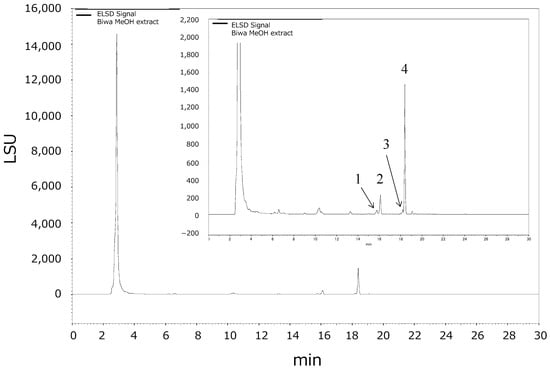
Figure A1.
A representative high-performance liquid chromatography-evaporative light-scattering detector chromatogram of the crude methanol extract (1. Maslinic acid, 2. corosolic acid, 3. oleanolic acid, and 4. ursolic acid).
Appendix A.1.2. Preparation of the Sample Solutions of the E. japonica Leaves and Supplements
The dried leaves or supplement powder (100 mg) was added to 5 mL of ethyl acetate, and ultrasonic extraction was performed at 45 °C for 15 min. The mixture was centrifuged at 2,330× g for 15 min, and the supernatant was collected. This procedure was repeated four times, and all the extracted supernatant solutions were mixed.
The supernatant was concentrated using a rotary evaporator (45 °C) under the vacuum to remove the solvent. The dried solid was redissolved in a methanol/chloroform (9:1, v/v) mixture, filtered through a 0.22-μm polytetrafluoroethylene filter, and used as a sample solution for HPLC analysis.
Appendix A.1.3. HPLC Analysis Conditions
An Agilent 1220 Infinity LC system (Agilent Technologies, Santa Clara, CA, USA) was used along with Phenomenex Prodigy (250 × 4.6 mm, 5 µm) with the mobile phase as 6 mM aqueous ammonium formate/methanol (10:90, v/v) eluting by isocratic conditions. An ELSD was used for detection, which was set to a nebulizer temperature of 30 °C and an evaporator temperature of 80 °C. Target compounds were identified by comparing their retention times with those of the standards.
Appendix A.2. Method Validation
Method precision was studied by performing repeatability (intra-day precision) and reproducibility (inter-day precision) studies. Repeatability was calculated as the within-day relative standard deviation (RSD)% of the peak areas of the four targeted compounds measured by HPLC–ELSD from six replicates of one E. japonica leaf sample obtained from independent sample solution preparation. For reproducibility measurements, E. japonica leaf sample solutions were prepared and analyzed in triplicates per day over three different days.
Accuracy was assessed by spiking the mixed standards into the supplement tablet powder sample at three different concentrations of the four standards in triplicates for each level, and the recovery rate of the spiked compounds was calculated.
As presented in Table A1 and Table A2, the quantitative analysis of the four targeted compounds in E. japonica leaves containing the supplement tablets performed in this study showed good precision and accuracy. Excellent results were obtained for all targets studied, with attained RSDs for intra-day and inter-day precisions lower than 2.01% and 4.00%, respectively (Table A1). Recoveries (%) and RSDs obtained for the accuracy validation of this method were between 93.84–105.93% and 0.64–3.79% for all four compounds, respectively (Table A2).

Table A1.
Validation of intra-day and inter-day precision.
Table A1.
Validation of intra-day and inter-day precision.
| Ursolic Acid | Maslinic Acid | Corosolic Acid | Oleanolic Acid | |
|---|---|---|---|---|
| Intra-day precision | ||||
| RSD% (n = 6) | 0.78 | 1.36 | 2.01 | 1.63 |
| Inter-day precision | ||||
| RSD% (n = 3) | 4.00 | 3.00 | 1.86 | 2.81 |

Table A2.
Recoveries (%) and RSD% obtained for the accuracy validation of the method.
Table A2.
Recoveries (%) and RSD% obtained for the accuracy validation of the method.
| Compounds | Recovery Rate (%) | ||||
|---|---|---|---|---|---|
| Low Conc. | Med. Conc. | High Conc. | Average | RSD (%) | |
| Ursolic acid | 97.84 | 96.67 | 96.42 | 96.98 | 0.64 |
| Maslinic acid | 104.02 | 101.87 | 99.37 | 101.75 | 1.87 |
| Corosolic acid | 96.68 | 97.72 | 93.84 | 96.08 | 1.71 |
| Oleanolic acid | 98.46 | 99.66 | 105.93 | 101.35 | 3.79 |
RSD: relative standard deviation; Recovery (%) = (amount in the added sample − amount in the non-added sample)/added amount × 100. Conc.: concentration, low concentration levels were set at 17%, 40%, 50%, and 60% concentrations of the spiked sample. Medium concentration levels were 100%. High concentration levels were set at 140%, 160%, 180%, and 200% for ursolic acid, maslinic acid, corosolic acid, and oleanolic acid, respectively.
Appendix A.2.1. Calibration Curves of the Targeted Compounds and Linear Range
A series of mixed standard solutions of ursolic acid (0.025–0.5 mg/mL), maslinic acid (0.0125–0.25 mg/mL), oleanolic acid (0.025–0.5 mg/mL), and corosolic acid (0.01–0.4 mg/mL) at seven concentration levels was prepared in methanol. Each point was evaluated three times. All calibration curves of the four targeted compounds showed good linearity (ursolic acid, R2 = 0.999; maslinic acid, R2 = 0.999; oleanolic acid, R2 = 0.999; and corosolic acid, R2 = 0.999).

Figure A2.
Calibration curves of the four standards according to high-performance liquid chromatography ((A): Ursolic acid, (B): Maslinic acid, (C): Corosolic acid, and (D): Oleanolic acid).
Appendix A.2.2. Quantification of the Four Triterpenoids in E. japonica-Containing Supplements and Leaves
Ten tablets were administered daily to the experimental participants in this clinical study at a dose of 250 mg/tablet. The following doses were estimated to be ingested by the subjects after consuming 10 tablets: 12.32 ± 0.10 mg of ursolic acid, 7.06 ± 0.08 mg of maslinic acid, 3.50 ± 0.038 mg of corosolic acid, and 2.23 ± 0.03 mg of oleanolic acid (Table A3).
Table A4 shows the contents of the four triterpenoids in E. japonica leaves collected each month. The results showed that the order of contents was as follows: ursolic acid > maslinic acid > corosolic acid > oleanolic acid. The content variations of the four compounds in the leaves of E. japonica collected in each month of the year were 9.31–13.86% RSD. This result suggests that the triterpenoid content in E. japonica leaves was relatively stable throughout the year and was not significantly affected by different seasons. These results are expected to be useful for quality control during the development of functional foods and other products containing E. japonica leaves.
Compared with the contents of the four triterpenoids in the supplement tablets administered to the participants in clinical trials, the content of E. japonica leaf powder was 83%. Thus, the content per gram of the leaves was calculated based on this value obtained from the quantitative analysis. The results showed no difference between the leaf and the tablet samples (Table A4), indicating that the manufacturing process of the tablets did not affect the triterpenoid contents.
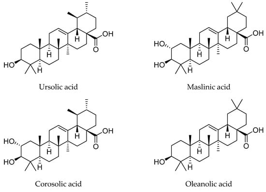
Figure A3.
Chemical structures of the main four triterpenoids in E. japonica leaves.

Table A3.
Contents of ursolic, maslinic, corosolic, and oleanolic acids in 10 supplement tablets of E. japonica leaves (mean ± standard deviation, n = 10).
Table A3.
Contents of ursolic, maslinic, corosolic, and oleanolic acids in 10 supplement tablets of E. japonica leaves (mean ± standard deviation, n = 10).
| Ursolic Acid | Maslinic Acid | Corosolic Acid | Oleanolic Acid |
|---|---|---|---|
| Content (mg/2.5 g) | |||
| 12.32 ± 0.10 | 7.06 ± 0.08 | 5.92 ± 0.004 | 2.23 ± 0.03 |

Table A4.
Contents of ursolic, maslinic, corosolic, and oleanolic acids in E. japonica leaves sampled during 12 months of collection (mean ± standard deviation, n = 3).
Table A4.
Contents of ursolic, maslinic, corosolic, and oleanolic acids in E. japonica leaves sampled during 12 months of collection (mean ± standard deviation, n = 3).
| Month | Ursolic Acid | Maslinic Acid | Corosolic Acid | Oleanolic Acid |
|---|---|---|---|---|
| Content (mg/g) | ||||
| Jan | 5.18 ± 0.06 | 2.95 ± 0.03 | 2.36 ± 0.03 | 1.04 ± 0.01 |
| Feb | 5.62 ± 0.12 | 4.43 ± 0.13 | 2.72 ± 0.07 | 1.23 ± 0.03 |
| Mar | 5.00 ± 0.06 | 3.66 ± 0.05 | 2.35 ± 0.02 | 1.07 ± 0.00 |
| Apr | 4.74 ± 0.33 | 3.31 ± 0.22 | 2.16 ± 0.12 | 1.01 ± 0.08 |
| May | 5.24 ± 0.13 | 2.64 ± 0.08 | 2.00 ± 0.08 | 0.98 ± 0.03 |
| Jun | 5.15 ± 0.24 | 3.58 ± 0.10 | 2.21 ± 0.06 | 1.07 ± 0.04 |
| Jul | 5.01 ± 0.10 | 3.50 ± 0.04 | 2.52 ± 0.03 | 1.00 ± 0.02 |
| Aug | 5.11 ± 0.18 | 3.27 ± 0.08 | 2.65 ± 0.06 | 0.97 ± 0.03 |
| Sep | 4.31 ± 0.22 | 2.93 ± 0.13 | 1.99 ± 0.10 | 0.81 ± 0.04 |
| Oct | 5.69 ± 0.28 | 3.71 ± 0.19 | 2.71 ± 0.13 | 1.11 ± 0.05 |
| Nov | 5.52 ± 0.11 | 3.01 ± 0.09 | 2.41 ± 0.01 | 1.07 ± 0.03 |
| Dec | 6.08 ± 0.09 | 3.50 ± 0.16 | 2.86 ± 0.08 | 1.21 ± 0.07 |
| Average | 5.21 ± 0.49 | 3.38 ± 0.47 | 2.41 ± 0.29 | 1.05 ± 0.12 |
| % RSD | 9.31 | 13.87 | 12.00 | 11.09 |
RSD: relative standard deviation.
Appendix B
Appendix B.1. Materials and Methods
Appendix B.1.1. β-Hexosaminidase Release Inhibition Activity Assay
To evaluate whether the leaves of E. japonica, their fractions, or the dominant compounds (ursolic, oleanolic, maslinic, and corosolic acids) exhibit anti-allergic activity, we measured the release of β-hexosaminidase from RBL-2H3 cells [1]. RBL-2H3 cells were seeded in a 96-well plate at a density of 5 × 10⁴ cells/well. After 24 h of incubation at 37 °C and 5% CO2, the cells were washed twice with Tyrode’s buffer (pH 7.2). The cells were treated for 1 h with Tyrode’s buffer containing a dimethyl sulfoxide (DMSO) solution of the tested samples at several concentrations. Tyrode’s buffer containing only DMSO was used as a negative control, whereas quercetin hydrate (final concentration: 100 μM, Sigma-Aldrich, St. Louis, MO, USA) was used as a positive control [40].
RBL-2H3 cells were stimulated with Tyrode’s buffer containing 5 µM A23187 (Sigma-Aldrich) for 1 h. The supernatant was transferred to a new 96-well plate, and 50 µL of a citric acid buffer (pH 5.2) containing 1 mM p-nitrophenyl-N-acetyl-β-D-glucosaminide was added. The β-hexosaminidase reaction was performed in the dark at room temperature for 3 h. The enzymatic reaction was stopped by adding 100 mM sodium bicarbonate (pH 10). Finally, the absorbance was measured at 405 nm using a microplate reader (MTP-310, Corona Electric, Hitachinaka, Japan).
Appendix B.1.2. Cell Viability Assay
To assess cell viability, RBL-2H3 cells were seeded in a 96-well plate at 37 °C and 5% CO2. After discarding the culture medium, the cells were treated with the E. japonica leaf extracts, their fractions, or the dominant compounds for 1 h. A total of 100 µL of fresh Eagle’s minimum essential medium and 20 µL of the 5 mg/mL MTT solution were added to each well. Incubation was continued for an additional 4 h. After the culture medium was removed, 40 mM of HCl-isopropanol was added to each well. The absorbance was measured at 570 nm using the microplate reader.
Appendix B.2. Results and Discussion
Inhibitory Effect of the Leaves of E. japonica on β-Hexosaminidase Release
We investigated the anti-allergic activity of the E. japonica leaf extracts by evaluating their ability to inhibit β-hexosaminidase release from RBL-2H3 cells. The tested samples derived from the E. japonica leaf extracts were methanol extract and hexane-soluble, ethyl-acetate-soluble, and water residue fractions. As shown in Figure A4, the methanol extract and ethyl acetate-soluble and hexane-soluble fractions from the leaves of E. japonica suppressed β-hexosaminidase release in RBL-2H3 cells. In contrast, β-hexosaminidase release in RBL-2H3 cells did not change upon the application of the water residue fraction. This result suggests that the hydrophobic compounds in the leaves of E. japonica exhibited strong anti-allergic activity.
To determine which compounds in the leaves of E. japonica exhibited strong anti-allergic activity, we isolated several compounds from the leaves of E. japonica and identified them. In this study, ursolic, oleanolic, maslinic, and corosolic acids were evaluated for their anti-allergic activities because they were the dominant compounds in the leaves of E. japonica. The results showed that ursolic acid (IC50 = 5.40 µM), oleanolic acid (IC50 = 21.20 µM), and maslinic acid (IC50 = 22.08 µM) strongly suppressed β-hexosaminidase release compared to the control. Additionally, corosolic acid showed a slight anti-allergic activity compared to the control (Figure A5). These results suggest that the leaves of E. japonica exhibit anti-allergic activity, and the main active compounds in the leaves of E. japonica are ursolic, oleanolic, maslinic, and corosolic acids. Therefore, the leaves of E. japonica may alleviate allergic symptoms.
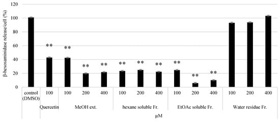
Figure A4.
Effect of the E. japonica leaf extracts on β-hexosaminidase activity. The inhibitory effect of each E. japonica leaf extract on β-hexosaminidase release in RBL-2H3 cells treated with each extract for 1 h. Black bars show β-hexosaminidase release (%) compared to cells treated with dimethyl sulfoxide as a control. Data are presented as the mean ± standard deviation (n = 3). Significant differences were determined using the Student’s t-test (** p < 0.01).
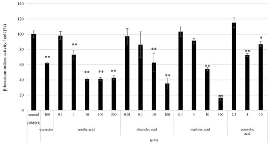
Figure A5.
Effect of the compounds on β-hexosaminidase activity. Inhibitory effect of ursolic, oleanolic, maslinic, and corosolic acids on β-hexosaminidase release in RBL-2H3 cells treated with each isolated compound for 1 h. Black bars show β-hexosaminidase release (%) compared to cells treated with dimethyl sulfoxide as a control. Data are presented as the mean ± standard deviation (n = 3). Significant differences were determined using the Student’s t-test (* p < 0.05, ** p < 0.01).
References
- Cha, D.S.; Eun, J.S.; Jeon, H. Anti-Inflammatory and Antinociceptive Properties of the Leaves of Eriobotrya japonica. J. Ethnopharmacol. 2011, 134, 305–312. [Google Scholar] [CrossRef]
- Noh, K.K.; Chung, K.W.; Sung, B.; Kim, M.J.; Park, C.H.; Yoon, C.; Choi, J.S.; Kim, M.K.; Kim, C.M.; Kim, N.D.; et al. Loquat (Eriobotrya japonica) Extract Prevents Dexamethasone-Induced Muscle Atrophy by Inhibiting the Muscle Degradation Pathway in Sprague Dawley Rats. Mol. Med. Rep. 2015, 12, 3607–3614. [Google Scholar] [CrossRef][Green Version]
- Liu, Y.; Zhang, W.; Xu, C.; Li, X. Biological Activities of Extracts from Loquat (Eriobotrya japonica Lindl.): A Review. Int. J. Mol. Sci. 2016, 17, 1983. [Google Scholar] [CrossRef]
- Shimizu, M.; Uemitsu, N.; Shirota, M.; Matsumoto, K.; Tezuka, Y. A New Triterpene Ester from Eriobotrya japonica. Chem. Pharm. Bull. 1996, 44, 2181–2182. [Google Scholar] [CrossRef]
- Taniguchi, S.; Imayoshi, Y.; Kobayashi, E.; Takamatsu, Y.; Ito, H.; Hatano, T.; Sakagami, H.; Tokuda, H.; Nishino, H.; Sugita, D.; et al. Production of Bioactive Triterpenes by Eriobotrya japonica Calli. Phytochemistry 2002, 59, 315–323. [Google Scholar] [CrossRef]
- De Tommasi, N.; De Simone, F.; Cirino, G.; Cicala, C.; Pizza, C. Hypoglycemic Effects of Sesquiterpene Glycosides and Polyhydroxylated Triterpenoids of Eriobotrya japonica. Planta Med. 1991, 57, 414–416. [Google Scholar] [CrossRef]
- Jung, H.A.; Park, J.C.; Chung, H.Y.; Kim, J.; Choi, J.S. Antioxidant Flavonoids and Chlorogenic Acid from the Leaves of Eriobotrya japonica. Arch. Pharm. Res. 1999, 22, 213–218. [Google Scholar] [CrossRef] [PubMed]
- Kwon, H.J.; Kang, M.J.; Kim, H.J.; Choi, J.S.; Paik, K.J.; Chung, H.Y. Inhibition of NF-κB by Methyl Chlorogenate from Eriobotrya japonica. Mol. Cells 2000, 10, 241–246. [Google Scholar] [PubMed]
- Madasu, C.; Karri, S.; Sangaraju, R.; Sistla, R.; Uppuluri, M.V. Synthesis and Biological Evaluation of Some Novel 1,2,3-Triazole Hybrids of Myrrhanone B Isolated from Commiphora mukul Gum Resin: Identification of Potent Antiproliferative Leads Active against Prostate Cancer Cells (PC-3). Eur. J. Med. Chem. 2020, 188, 111974. [Google Scholar] [CrossRef] [PubMed]
- Mallavadhani, U.V.; Chandrashekhar, M.; Shailaja, K.; Ramakrishna, S. Design, Synthesis, Anti-Inflammatory, Cytotoxic and Cell Based Studies of Some Novel Side Chain Analogues of Myrrhanones A & B Isolated from the Gum Resin of Commiphora mukul. Bioorg. Chem. 2019, 82, 306–323. [Google Scholar] [CrossRef]
- Banno, N.; Akihisa, T.; Tokuda, H.; Yasukawa, K.; Taguchi, Y.; Akazawa, H.; Ukiya, M.; Kimura, Y.; Suzuki, T.; Nishino, H. Anti-Inflammatory and Antitumor-Promoting Effects of the Triterpene Acids from the Leaves of Eriobotrya japonica. Biol. Pharm. Bull. 2005, 28, 1995–1999. [Google Scholar] [CrossRef] [PubMed]
- Ito, H.; Kobayashi, E.; Li, S.H.; Hatano, T.; Sugita, D.; Kubo, N.; Shimura, S.; Itoh, Y.; Tokuda, H.; Nishino, H.; et al. Antitumor Activity of Compounds Isolated from Leaves of Eriobotrya japonica. J. Agric. Food Chem. 2002, 50, 2400–2403. [Google Scholar] [CrossRef] [PubMed]
- De Tommasi, N.; De Simone, F.; Pizza, C.; Mahmood, N.; Moore, P.S.; Conti, C.; Orsi, N.; Stein, M.L. Constituents of Eriobotrya japonica. A Study of Their Antiviral Properties. J. Nat. Prod. 1992, 55, 1067–1073. [Google Scholar] [CrossRef] [PubMed]
- Tan, H.; Sonam, T.; Shimizu, K. The Potential of Triterpenoids from Loquat Leaves (Eriobotrya japonica) for Prevention and Treatment of Skin Disorder. Int. J. Mol. Sci. 2017, 18, 1030. [Google Scholar] [CrossRef]
- Nelson, A.T.; Camelio, A.M.; Claussen, K.R.; Cho, J.; Tremmel, L.; Digiovanni, J.; Siegel, D. Synthesis of Oxygenated Oleanolic and Ursolic Acid Derivatives with Anti-Inflammatory Properties. Bioorg. Med. Chem. Lett. 2015, 25, 4342–4346. [Google Scholar] [CrossRef]
- Sultana, N.; Ata, A. Oleanolic Acid and Related Derivatives as Medicinally Important Compounds. J. Enzym. Inhib. Med. Chem. 2008, 23, 739–756. [Google Scholar] [CrossRef] [PubMed]
- Chen, Q.; Zhang, Y.; Zhang, W.; Chen, Z. Identification and Quantification of Oleanolic Acid and Ursolic Acid in Chinese Herbs by Liquid Chromatography-Ion Trap Mass Spectrometry. Biomed. Chromatogr. 2011, 25, 1381–1388. [Google Scholar] [CrossRef]
- Kunkel, S.D.; Suneja, M.; Ebert, S.M.; Bongers, K.S.; Fox, D.K.; Malmberg, S.E.; Alipour, F.; Shields, R.K.; Adams, C.M. MRNA Expression Signatures of Human Skeletal Muscle Atrophy Identify a Natural Compound That Increases Muscle Mass. Cell Metab. 2011, 13, 627–638. [Google Scholar] [CrossRef]
- Bang, H.S.; Seo, D.Y.; Chung, Y.M.; Oh, K.M.; Park, J.J.; Arturo, F.; Jeong, S.H.; Kim, N.; Han, J. Ursolic Acid-Induced Elevation of Serum Irisin Augments Muscle Strength during Resistance Training in Men. Korean J. Physiol. Pharmacol. 2014, 18, 441–446. [Google Scholar] [CrossRef]
- Lee, S.U.; Park, S.J.; Kwak, H.B.; Oh, J.; Min, Y.K.; Kim, S.H. Anabolic Activity of Ursolic Acid in Bone: Stimulating Osteoblast Differentiation In Vitro and Inducing New Bone Formation In Vivo. Pharmacol. Res. 2008, 58, 290–296. [Google Scholar] [CrossRef]
- Kunkel, S.D.; Elmore, C.J.; Bongers, K.S.; Ebert, S.M.; Fox, D.K.; Dyle, M.C.; Bullard, S.A.; Adams, C.M. Ursolic Acid Increases Skeletal Muscle and Brown Fat and Decreases Diet-Induced Obesity, Glucose Intolerance and Fatty Liver Disease. PLoS ONE 2012, 7, e39332. [Google Scholar] [CrossRef] [PubMed]
- Sun, N.; Deng, C.; Zhao, Q.; Han, Z.; Guo, Z.; Wang, H.; Dong, W.; Duan, Y.; Zhuang, G.; Zhang, R. Ursolic Acid Alleviates Mucus Secretion and Tissue Remodeling in Rat Model of Allergic Rhinitis after PM2.5 Exposure. Am. J. Rhinol. Allergy 2021, 35, 272–279. [Google Scholar] [CrossRef] [PubMed]
- Sun, N.; Han, Z.; Wang, H.; Guo, Z.; Deng, C.; Dong, W.; Zhuang, G.; Zhang, R. Effects of Ursolic Acid on the Expression of Th1–Th2-Related Cytokines in a Rat Model of Allergic Rhinitis after PM2.5 Exposure. Am. J. Rhinol. Allergy 2020, 34, 587–596. [Google Scholar] [CrossRef] [PubMed]
- Matsuda, H.; Dai, Y.; Ido, Y.; Murakami, T.; Matsuda, H.; Yoshikawa, M.; Kubo, M. Studies on Kochiae Fructus. V. Antipruritic Effects of Oleanolic Acid Glycosides and the Structure-Requirement. Biol. Pharm. Bull. 1998, 21, 1231–1233. [Google Scholar] [CrossRef] [PubMed][Green Version]
- Yap, W.H.; Lim, Y.M. Mechanistic Perspectives of Maslinic Acid in Targeting Inflammation. Biochem. Res. Int. 2015, 2015, 279356. [Google Scholar] [CrossRef]
- Kim, J.; Kim, B.E.; Leung, D.Y.M. Pathophysiology of Atopic Dermatitis: Clinical Implications. Allergy Asthma Proc. 2019, 40, 84–92. [Google Scholar] [CrossRef]
- Birdi, G.; Cooke, R.; Knibb, R.C. Impact of Atopic Dermatitis on Quality of Life in Adults: A Systematic Review and Meta-Analysis. Int. J. Dermatol. 2020, 59, e75–e91. [Google Scholar] [CrossRef] [PubMed]
- Singh-Franco, D.; Ghin, H.L.; Robles, G.I.; Borja-Hart, N.; Perez, A. Levocetirizine for the Treatment of Allergic Rhinitis and Chronic Idiopathic Urticaria in Adults and Children. Clin. Ther. 2009, 31, 1664–1687. [Google Scholar] [CrossRef]
- Blaiss, M.S. Quality of Life in Allergic Rhinitis. Ann. Allergy Asthma Immunol. 1999, 83, 449–454. [Google Scholar] [CrossRef] [PubMed]
- Wei, C. The Efficacy and Safety of H1-Antihistamine versus Montelukast for Allergic Rhinitis: A Systematic Review and Meta-Analysis. Biomed. Pharmacother. 2016, 83, 989–997. [Google Scholar] [CrossRef]
- Nakano, M.; Kamimura, A.; Watanabe, F.; Kamiya, T.; Watanabe, D.; Yamamoto, E.; Fukagawa, M.; Hasumi, K.; Suzuki, E. Effects of Orally Administered Pyrroloquinoline Quinone Disodium Salt on Dry Skin Conditions in Mice and Healthy Female Subjects. J. Nutr. Sci. Vitaminol. 2015, 61, 241–246. [Google Scholar] [CrossRef] [PubMed]
- Okuda, M.; Ohkubo, K.; Goto, M.; Okamoto, H.; Konno, A.; Baba, K.; Ogino, S.; Enomoto, M.; Imai, T.; So, N.; et al. Comparative Study of Two Japanese Rhinoconjunctivitis Quality-of-Life Questionnaires. Acta Otolaryngol. 2005, 125, 736–744. [Google Scholar] [CrossRef] [PubMed]
- Finlay, A.Y.; Khan, G.K. Dermatology Life Quality Index (DLQI)—A Simple Practical Measure for Routine Clinical Use. Clin. Exp. Dermatol. 1994, 19, 210–216. [Google Scholar] [CrossRef] [PubMed]
- Janeway, C.A., Jr.; Travers, P.; Walport, M.; Shlomchik, M. Effector Mechanisms in Allergic Reactions. In Immunobiology: The Immune System in Health and Disease, 5th ed.; Penelope, A., Eleanor, L., Sarah, G., Eds.; Garland Science: New York, NY, USA, 2001; pp. 564–598. [Google Scholar]
- Larché, M.; Akdis, C.A.; Valenta, R. Immunological Mechanisms of Allergen-Specific Immunotherapy. Nat. Rev. Immunol. 2006, 6, 761–771. [Google Scholar] [CrossRef]
- Dhakal, H.; Kim, M.J.; Lee, S.; Choi, Y.A.; Kim, N.; Kwon, T.K.; Khang, D.; Kim, S.H. Ursolic Acid Inhibits FcεRI-Mediated Mast Cell Activation and Allergic Inflammation. Int. Immunopharmacol. 2021, 99, 107994. [Google Scholar] [CrossRef] [PubMed]
- Lim, S.W.; Hong, S.P.; Jeong, S.W.; Kim, B.; Bak, H.; Ryoo, H.C.; Lee, S.H.; Ahn, S.K. Simultaneous Effect of Ursolic Acid and Oleanolic Acid on Epidermal Permeability Barrier Function and Epidermal Keratinocyte Differentiation via Peroxisome Proliferator-Activated Receptor-α. J. Dermatol. 2007, 34, 625–634. [Google Scholar] [CrossRef]
- Uehara, O.; Kusuhara, T.; Nakamura, T. Transepidermal Water Loss Estimation Model for Evaluating Skin Barrier Function. Adv. Biomed. Eng. 2023, 12, 1–8. [Google Scholar] [CrossRef]
- Zhu, X.; Wang, L.; Zhao, T.; Jiang, Q. Traditional Uses, Phytochemistry, Pharmacology, and Toxicity of Eriobotrya Japonica Leaves: A Summary. J. Ethnopharmacol. 2022, 298, 115566. [Google Scholar] [CrossRef] [PubMed]
- Kishikawa, A.; Ashour, A.; Zhu, Q.; Yasuda, M.; Ishikawa, H.; Shimizu, K. Multiple biological effects of olive oil byproducts such as leaves, stems, flowers, olive milled waste, fruit pulp, and seeds of the olive plant on skin. Phytother. Res. 2015, 29, 877–886. [Google Scholar] [CrossRef]
Disclaimer/Publisher’s Note: The statements, opinions and data contained in all publications are solely those of the individual author(s) and contributor(s) and not of MDPI and/or the editor(s). MDPI and/or the editor(s) disclaim responsibility for any injury to people or property resulting from any ideas, methods, instructions or products referred to in the content. |
© 2023 by the authors. Licensee MDPI, Basel, Switzerland. This article is an open access article distributed under the terms and conditions of the Creative Commons Attribution (CC BY) license (https://creativecommons.org/licenses/by/4.0/).