Article

Effects of Syo-seiryu-to and Its Constituent Crude Drugs on Phorbol Ester-Induced Up-Regulation of IL-33 and Histamine H1 Receptor mRNAs in Swiss 3T3 and HeLa Cells

1
Departments of Otolaryngology, Institute of Biomedical Sciences, Tokushima University Graduate School, Tokushima 770-8503, Japan
2
Departments of Molecular Pharmacology, Institute of Biomedical Sciences, Tokushima University Graduate School, Tokushima 770-8503, Japan
3
Medical Corporation Kinshukai, Osaka 558-0011, Japan
4
Laboratory of Pharmacology, Faculty of Pharmacy, Osaka Ohtani University, Osaka 584-8540, Japan
*
Author to whom correspondence should be addressed.
Allergies 2021, 1(3), 163-175; https://doi.org/10.3390/allergies1030015
Submission received: 16 June 2021 / Revised: 3 August 2021 / Accepted: 3 August 2021 / Published: 9 August 2021
(This article belongs to the Section Physiopathology)

Abstract

:
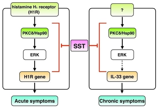
Syo-seiryu-to (SST) is a traditional herbal medicine that has been used clinically to treat allergic rhinitis (AR) in Japan. SST improves acute symptoms, such as sneezing and rhinorrhea, as well as chronic symptoms, such as nasal obstruction, in patients with AR. However, its therapeutic mechanisms remain unknown. We examined the effects of SST and eight constituent crude drugs on phorbol 12-myristate-13-acetate (PMA)-induced gene up-regulation of IL-33 and histamine H1 receptor (H1R), which are responsible for the pathogenesis of AR. We found that SST and its crude drugs, except for Pinellia tuber, significantly and dose-dependently suppressed PMA-induced both IL-33 and H1R mRNA up-regulation in vitro. The half-maximal inhibitory concentration values of the seven crude drugs to inhibit PMA-induced IL-33 mRNA up-regulation were correlated with those related to H1R mRNA up-regulation, suggesting that they act on a common signal molecule. These results suggest that SST improves nasal congestion that is induced by IL-33-related eosinophil infiltration and inhibits sneezing and rhinorrhea that are induced by H1R-mediated histamine signaling in the nasal mucosa of AR patients through its inhibition of a common molecule in the gene expression pathways of IL-33 and H1R. The results could explain the advantages of traditional herbal medicine, in which mixing various crude drugs not only acts on a common target to enhance its pharmacological action, similar to the effect of a high concentration of a single crude extract but also has the benefit of reducing the side effects of each crude drug.

Graphical Abstract

1. Introduction

Syo-seiryu-to (SST) is a traditional herbal medicine that has been used clinically in Asian countries including Japan for the treatment of allergic diseases such as allergic rhinitis (AR) and asthma [1]. SST improves acute symptoms, such as sneezing and rhinorrhea, as well as chronic symptoms, such as nasal obstruction, in patients with AR. The anti-allergic activities of SST include inhibition of histamine release from rat peritoneal mast cells [2], reduction of serum IgE levels in AR patients [3,4], modulation of Th1- and/or Th2-cytokines in CD4+ T cells in mice [5,6,7], and inhibition of airway eosinophil infiltration and blood eosinophil counts in mice [8,9]. Previously, we reported that SST suppressed nasal symptoms and gene expression of histamine H1 receptor (H1R), histidine decarboxylase (HDC), and Th2-cytokines including interleukin (IL)-4 and -5, in the nasal mucosa of AR model rats [10]. However, these pharmacological effects of SST reported are mainly for acute symptoms, and the underlying mechanisms for the chronic symptoms of SST remain to be elucidated.
The IL-33 gene was identified as a gene responsible for asthma in two large international joint studies [11,12]. IL-33 is expressed in Th2 cells, mast cells, basophils, and eosinophils. Since IL-33 stimulates these cells to produce Th2-cytokines, such as IL-5 and IL-13, it is suggested that IL-33 plays an important role in allergic reactions [13,14,15,16,17]. Recently, it was reported that levels of IL-33 in the blood and gene expression of IL-33 in the nasal mucosa of AR patients were elevated compared to those in healthy participants [18,19]. Therefore, it is suggested that IL-33 up-regulation is involved in the development of AR [20,21].
Histamine is a key chemical mediator of AR. IgE-mediated histamine release from mast cells in response to allergens stimulates H1R on nasal trigeminal nerve endings to cause nasal symptoms including sneezing and watery rhinorrhea in AR patients [22]. Levels of H1R mRNA have been reported to be up-regulated in the nasal mucosa of patients with AR [23,24]. In our previous study, we found that histamine increased the expression of H1R mRNA in HeLa cells [25,26] and that the gene expression levels of H1R in the nasal mucosa were correlated with the severity of nasal symptoms in AR patients [27,28]. Taken together, these results suggest that IL-33 and H1R gene up-regulation in the nasal mucosa leads to the exacerbation of nasal symptoms in patients with AR.
In the present study, to clarify the anti-allergic actions of SST, we first examined whether SST suppresses PMA-induced IL-33 and H1R mRNA up-regulation in Swiss 3T3 cells and HeLa cells, respectively, because phorbol 12-myristate-13-acetate (PMA) increases the expression levels of IL-33 mRNA in Swiss 3T3 cells [29] and H1R mRNA in HeLa cells [25]. Since SST is composed of eight crude drugs, we then examined whether each constituent crude drug of SST suppresses PMA-induced IL-33 and H1R mRNA up-regulation. We showed that SST and its crude drugs except for Pinellia tuber suppressed PMA-induced up-regulation of IL-33 mRNA and H1R mRNA. The half-maximal inhibitory concentration (IC50) values of the seven crude drugs to inhibit PMA-induced IL-33 mRNA up-regulation were correlated with PMA-induced H1R mRNA up-regulation.

2. Materials and Methods

2.1. Preparation of Hot Water Extracts from Syo-seiryu-to (SST) and Its Crude Drugs

SST was kindly gifted by Tsumura and Co., Tokyo, Japan. Its components: Ephedra herb, cinnamon bark, processed ginger, Asiasarum root, peony root, Schisandra fruit, Glycyrrhiza, and Pinellia tuber were purchased from Tsumura. SST or its crude drugs (27 g each) was added to 375 mL of distilled water and stand for 1 h at room temperature. After that, the mixture was boiled for 2 h and then filtered twice to remove insoluble materials. The supernatant was centrifuged and the obtained supernatant was freeze-dried and kept at −30 °C until used. The freeze-dried extract was re-dissolved in water on the day of the experiments. The yield of freeze-dried SST extract was 59% (w/w) and those of crude drugs were dependent on the crude drugs and around 50–80% (w/w).

2.2. Cell Culture

Swiss 3T3 cells that endogenously expressed IL-33 were cultured in Dulbecco’s Modified Eagle’s medium (Gibco, Grand Island, NY, USA) containing 10% fetal bovine serum (FBS) (Sigma, St. Louis, MO, USA) and antibiotics (10,000 Units/mL penicillin G sodium, 10 mg/mL streptomycin sulfate salt) at 37 °C under a humidified atmosphere of 5% CO2 and 95% air. After reaching approximately 90% confluence, the medium was replaced with starvation medium (FBS 0.5%) and cultured for an additional 24 h before mRNA determination. HeLa cells that endogenously expressed H1R were cultured in MEM-alpha medium (Gibco) containing 8% FBS (Sigma) and antibiotic-antimycotic (Gibco) at 37 °C and 5% CO2 incubator. After reaching about 90% confluence, the media was replaced with FBS-free media and cultured for an additional 24 h before being subject to the mRNA determination.

2.3. Reverse Transcription

Cells were pretreated with extracts from SST and each crude drug for 12 h before PMA treatment. After treatment with 100 nM PMA for 3 h, the cells were washed several times with Ca2+- and Mg2+-free PBS (PBS [–]) at 37 °C. The cells were harvested with 700 µL of RNAiso Plus (Takara Bio Inc., Kyoto, Japan), mixed with 210 µL of chloroform, and centrifuged at 15,000 rpm for 15 min at 4 °C. The aqueous phase, including RNA, was collected, and the RNA was precipitated by the addition of isopropanol. After centrifugation at 15,000 rpm for 15 min at 4 °C, the resulting RNA pellets were washed with 0.5 mL of ice-cold 75% ethanol at −20 °C. After centrifugation at 15,000 rpm for 15 min at 4 °C, diethyl pyrocarbonate (DEPC)-treated water (DEPC-water) was added to the resulting pellets to prepare RNA solutions. The total RNA concentrations and purity of each sample were measured using the spectrophotometer (Nanodrop ND-1000, Thermo Fisher Scientific, Waltham, MA, USA). The RNA solution and DEPC-water were added to the sample tube so that the total RNA was equivalent to 1.0 µg. The total volume of the solution was set to 5 µL. Using the PrimeScript® RT reagent kit (Takara Bio Inc.), a reverse transcription reaction with a thermal cycler (T3000 thermocycler, Biometra, Göettingen, Germany) was performed to obtain cDNA.

2.4. Real-Time Quantitative RT-PCR

Reagents containing Premix Taq (Probe qPCR) (Takara Bio Inc.) were mixed to prepare 20 μL of reaction solution per well of a MicroAmp Optical 96-well Reaction Plate (Applied Biosystems, Foster City, CA, USA). The PCR reaction was conducted using a Sequence Detector (Gene Amp 7300 Sequence Detection System, Applied Biosystems), the amplification curve of the PCR product was detected in real-time and analysis and quantification were performed using Sequence Detection software. The sequences of the primers and probe were as follows: a forward primer for human H1R, 5′-CAGAGGATCAGATGTTAGGTGATAGC-3′; a reverse primer for human H1R, 5′-AGCGGAGCCTCTTCCAAGTAA-3′; and probe, FAM-CTTCTCTCTCGAACGGACTCAGATACCACC-TAMRA. Mouse IL-33 primers and a probe kit (Mm00505403_m1, Applied Biosystems) were used for the determination of mouse IL-33 mRNA levels. The housekeeping gene glyceraldehyde-3-phosphate dehydrogenase (GAPDH) was used as an internal standard to correct for differences in RNA purity and reverse transcription efficiency, which are the main factors responsible for the fluctuation in quantitative RT-PCR. Theoretically, the expression of GAPDH does not change due to environmental conditions, such as cell activation or proliferation, and the expression level is considered to be always constant. The human GAPDH primer and probe kit (Pre-Developed TaqMan Assay Reagents Control Kit (human GAPDH), Applied Biosystems) was used to determine the human GAPDH mRNA levels. Mouse GAPDH primer and probe kit (TaqMan Rodent GAPDH Control Reagent, Applied Biosystems) was used to determine the mouse GAPDH mRNA levels. Several experiments were conducted using cells with different passage numbers and determined the suitable dose to calculate IC50 values. The final data were obtained from the latest experiments using the cells with the same passage number and used for statistical analysis.

2.5. Statistical Analysis

The results are shown as means ± SEM. Data were analyzed using GraphPad Prism software (GraphPad Software, Inc., San Diego, CA, USA). A one-way ANOVA followed by Dunnett’s multiple comparison test was used for statistical analysis. Statistical significance was set at p < 0.05.

3. Results

3.1. Effect of SST on PMA-Induced IL-33 and H1R Gene Up-Regulation

Stimulation with PMA significantly increased IL-33 mRNA levels in Swiss 3T3 cells (Figure 1A). The hot water extract of SST significantly and dose-dependently suppressed PMA-induced up-regulation of IL-33 mRNA in Swiss 3T3 cells (Figure 1A). H1R gene up-regulation in response to PMA was observed in HeLa cells (Figure 1B). The hot water extract of SST significantly and dose-dependently suppressed PMA-induced up-regulation of H1R mRNA in HeLa cells (Figure 1B).

3.2. Effect of Constituent Crude Drugs of SST on PMA-Induced Up-Regulation of IL-33 and H1R Gene Expression

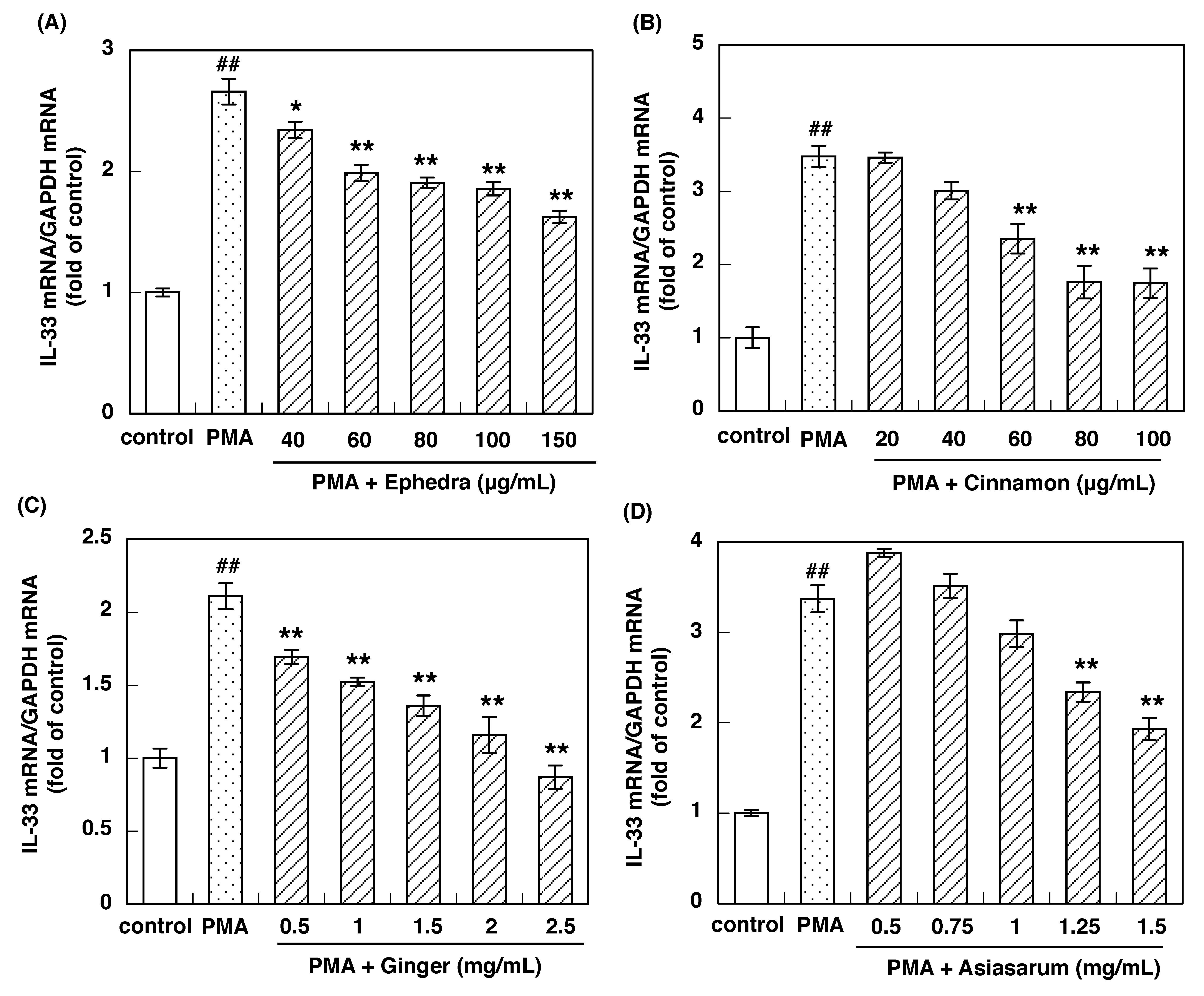
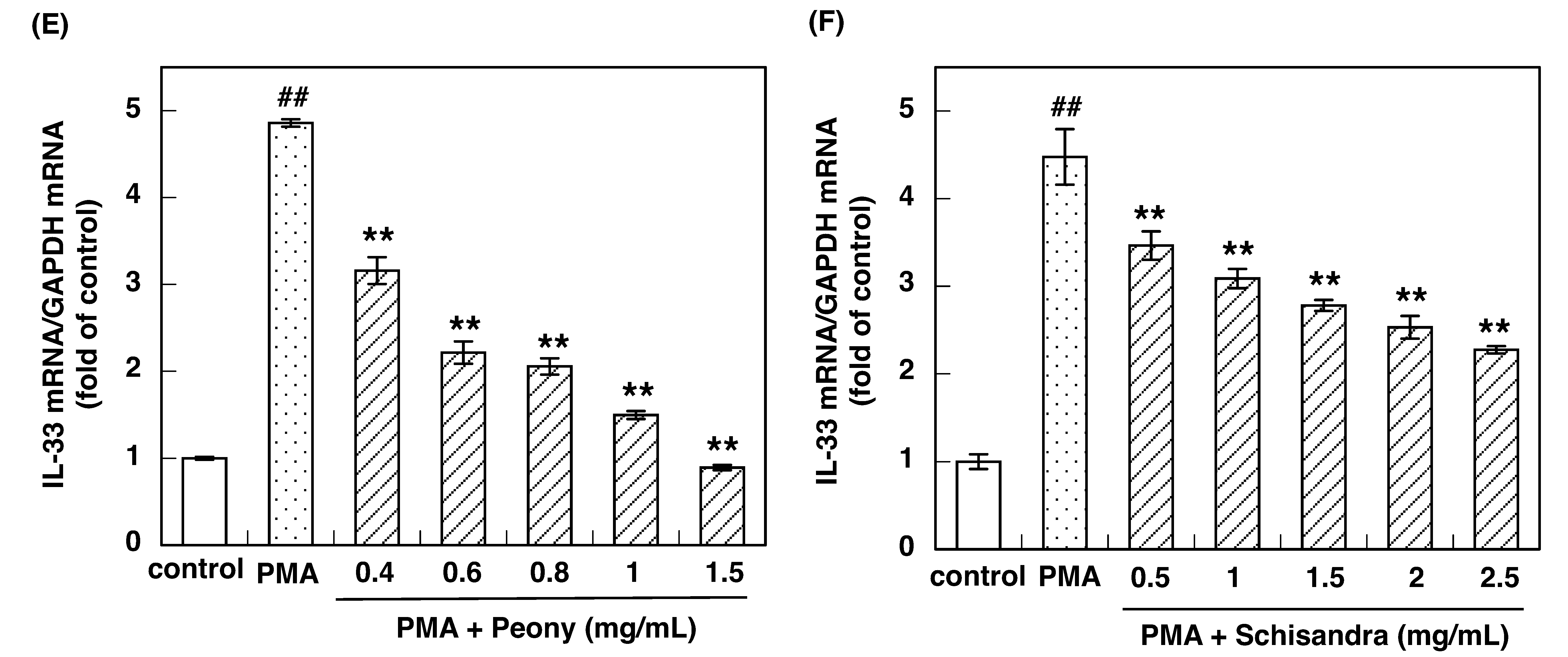
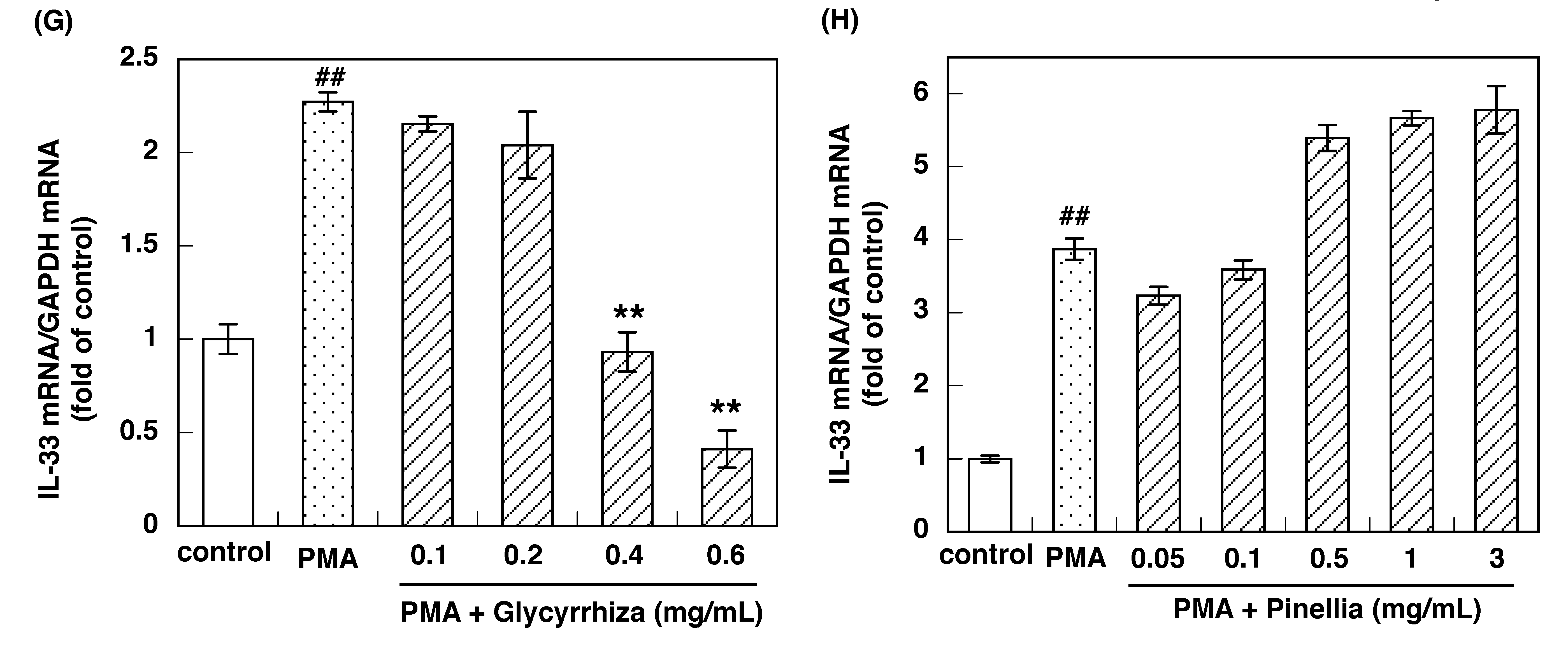
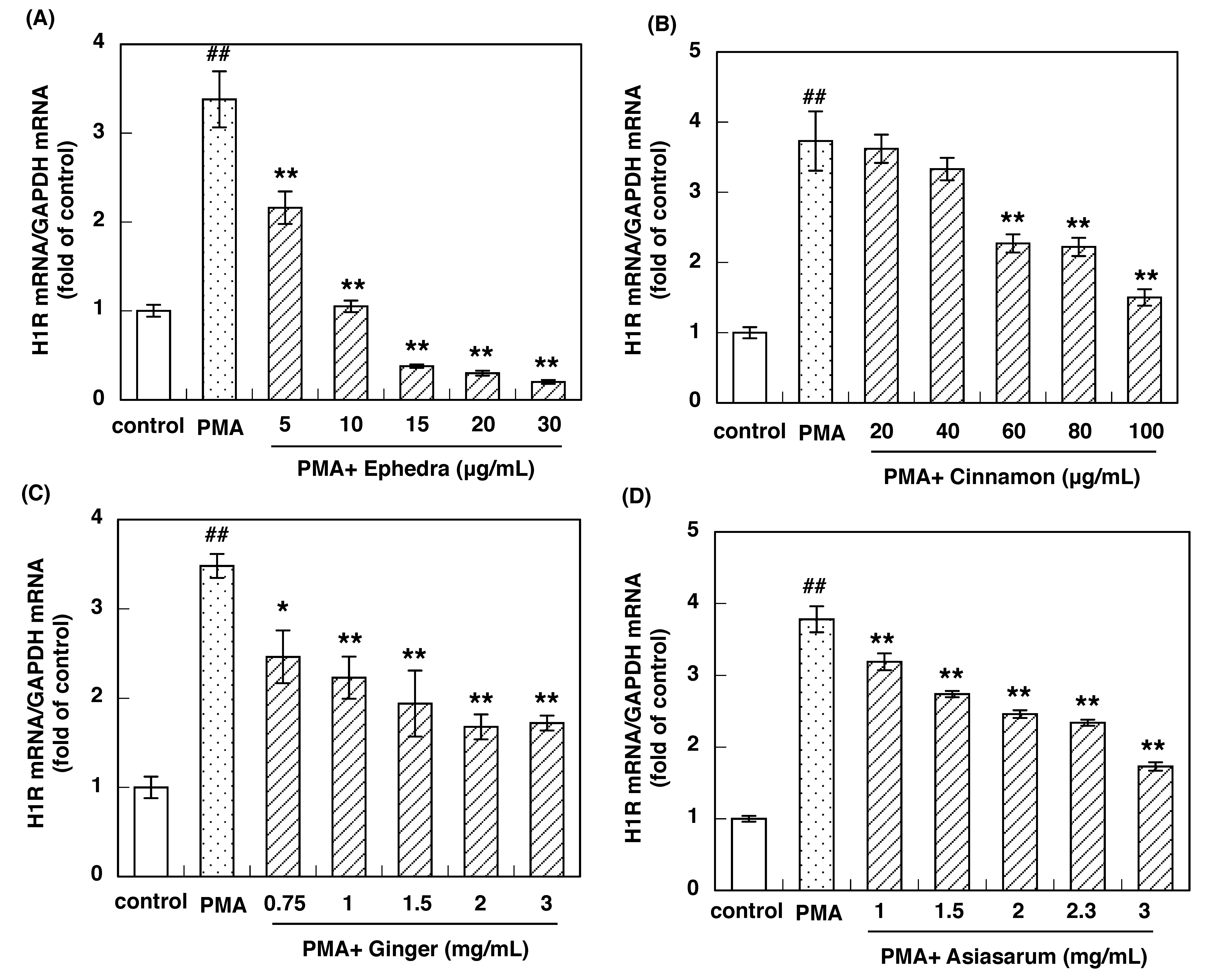
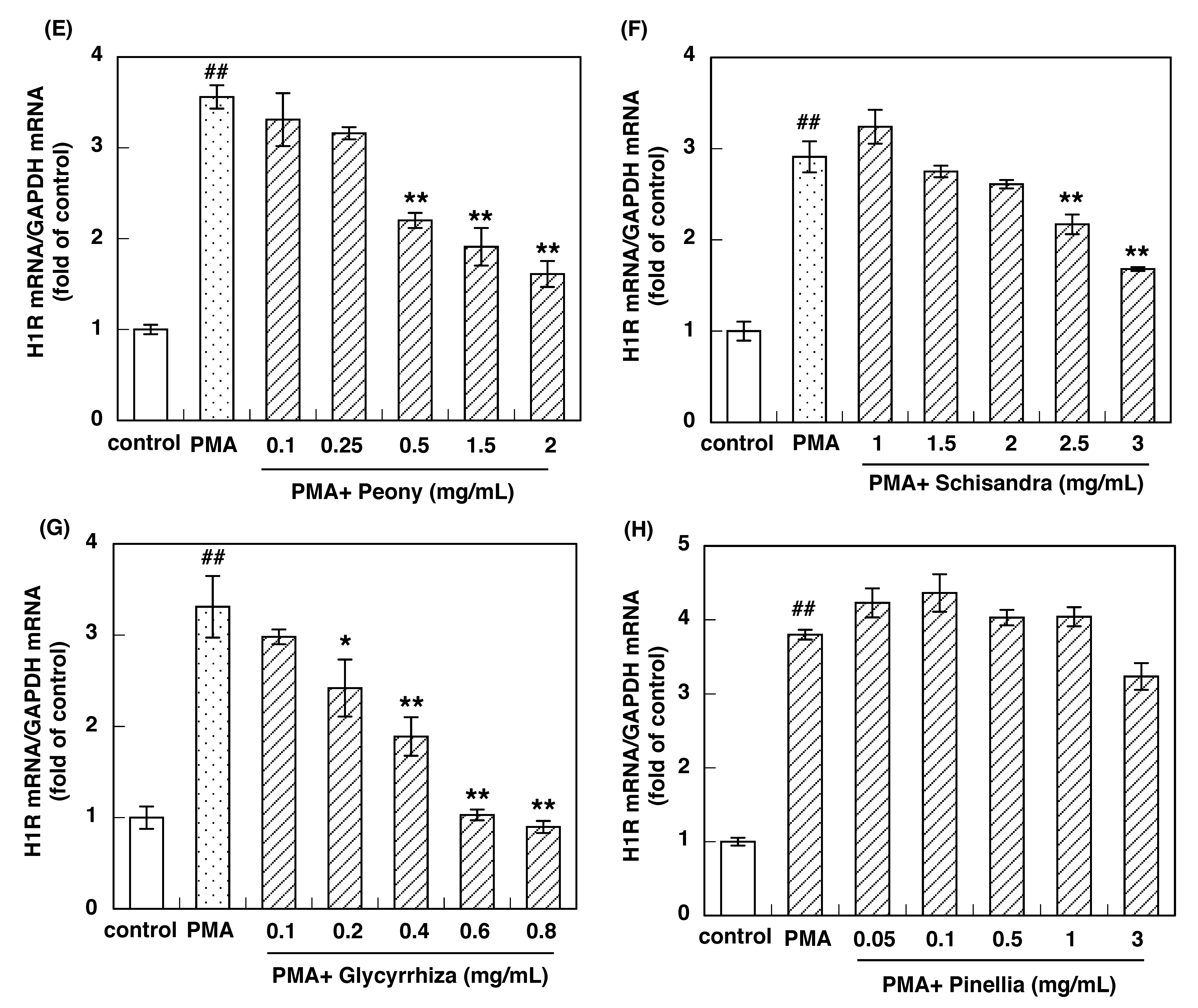
SST is composed of eight crude drugs: Ephedra herb, cinnamon bark, processed ginger, Asiasarum root, peony root, Schisandra fruit, Glycyrrhiza, and Pinellia tuber. Thus, we next investigated whether each constituent crude drug of SST suppresses PMA-induced IL-33 and H1R mRNA up-regulation in Swiss 3T3 cells and HeLa cells, respectively. The hot water extracts from Ephedra herb, cinnamon bark, processed ginger, Asiasarum root, peony root, Schisandra fruit, and Glycyrrhiza significantly and dose-dependently suppressed PMA-induced IL-33 mRNA up-regulation in Swiss 3T3 cells (Figure 2A–G). These extracts also significantly and dose-dependently suppressed PMA-induced H1R gene up-regulation in HeLa cells (Figure 3A–G). However, Pinellia tuber did not suppress PMA-induced up-regulation of IL-33 or H1R mRNAs (Figure 2H and Figure 3H).

3.3. IC50 of Constituent Crude Drugs of SST to Inhibit PMA-Induced Up-Regulation of IL-33 and H1R Gene Expression

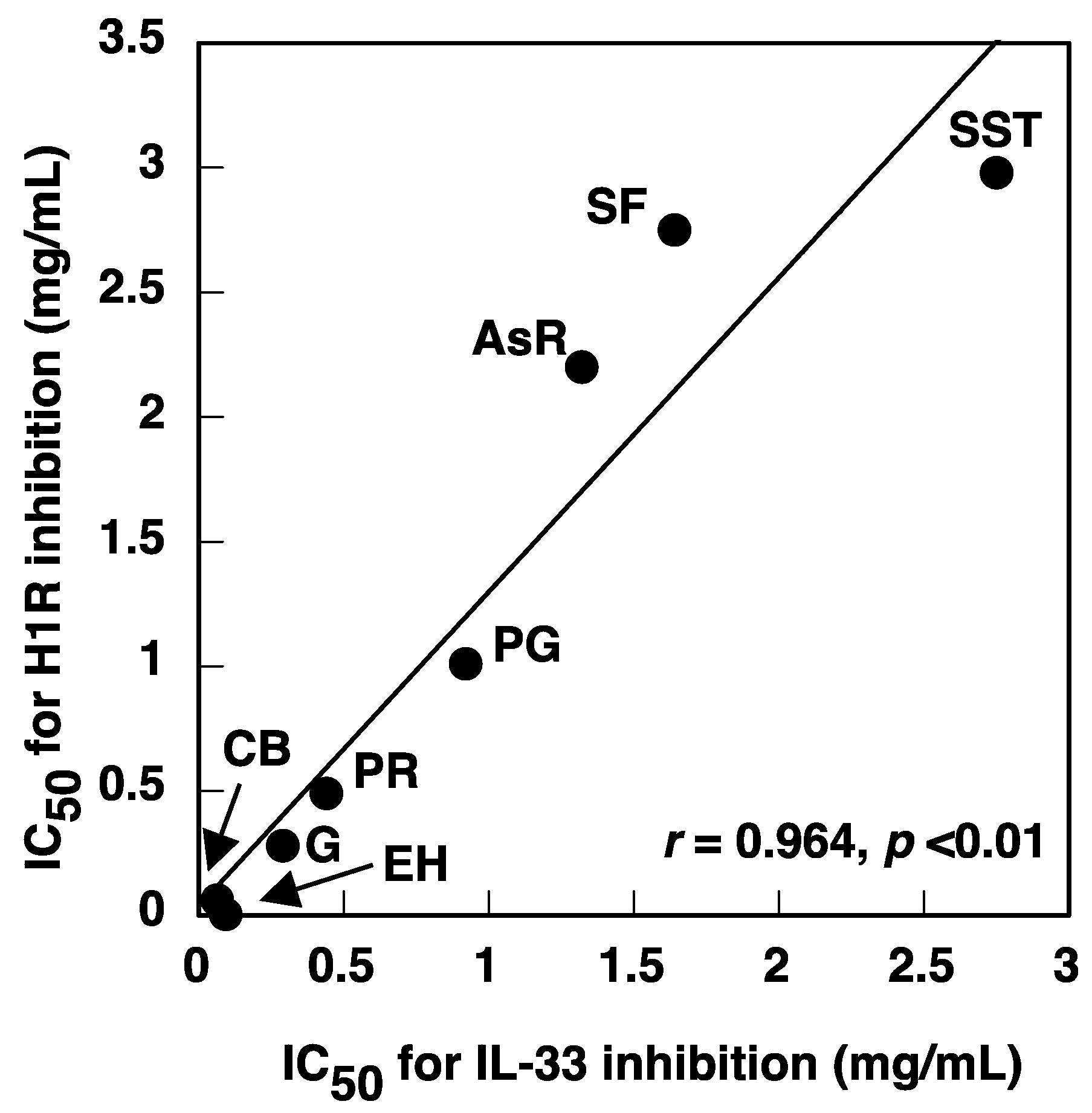
The IC50 values of Ephedra herb, cinnamon bark, processed ginger, Asiasarum root, peony root, Schisandra fruit, and Glycyrrhiza to inhibit PMA-induced IL-33 and H1R mRNA up-regulation were calculated (Table 1). The IC50 values of Ephedra herb, cinnamon bark, processed ginger, Asiasarum root, peony root, Schisandra fruit, and Glycyrrhiza to inhibit up-regulation of IL-33 mRNA were correlated with those related to up-regulation of H1R mRNA (r = 0.964, p < 0.01; Figure 4). These results suggest that the active compound in each crude drug acts on a common signal molecule involved in the pathways of IL-33 and H1R gene expression.

3.4. Effects of Inhibitors of the H1R Gene Expression Signaling Pathway on PMA-Induced IL-33 Gene Up-Regulation

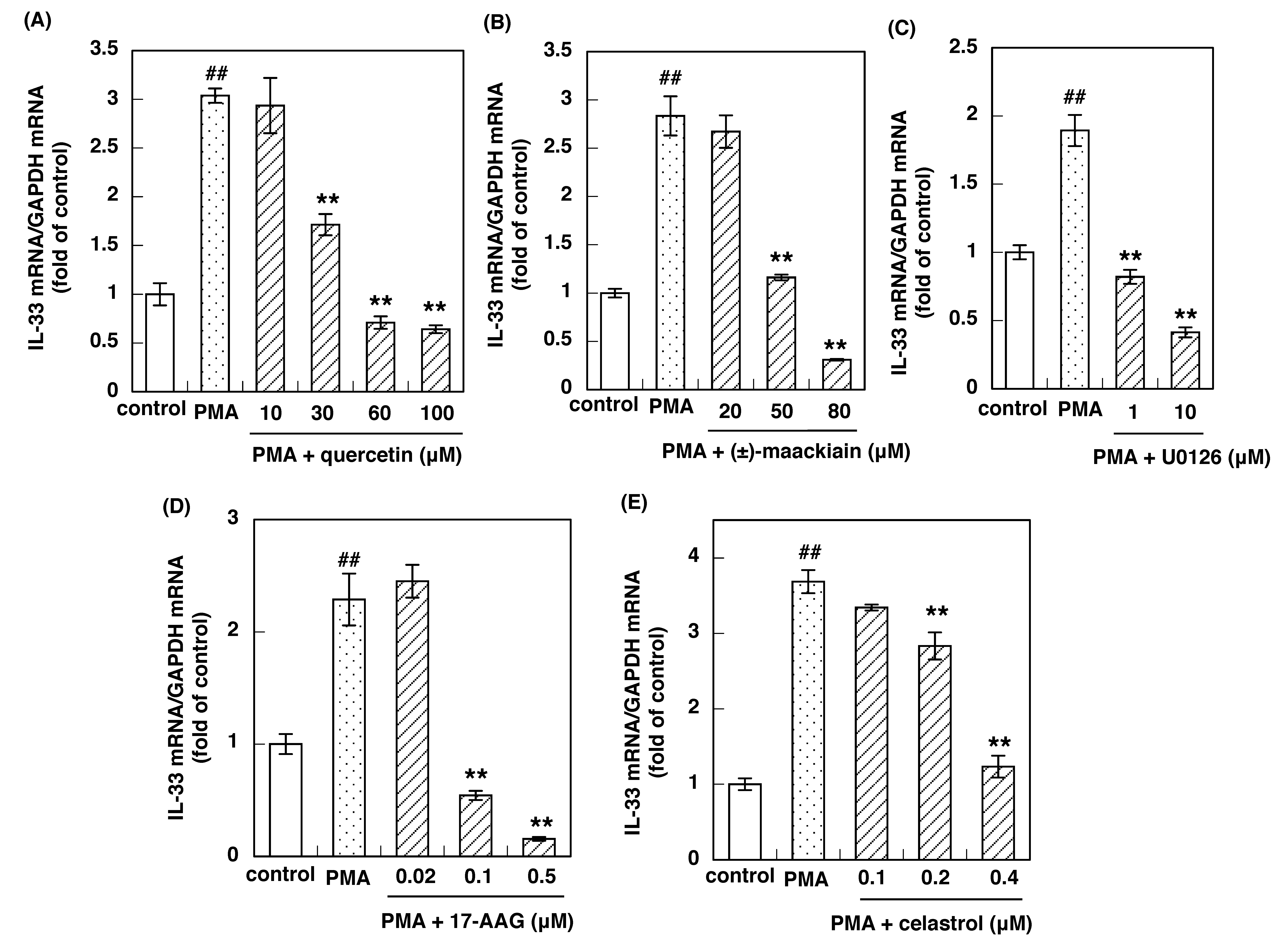
We have shown that the protein kinase Cδ (PKCδ)/heat shock protein 90 (Hsp90)/ERK/poly(ADP-ribose)polymerase-1 (PARP-1) signaling pathway is involved in PMA-induced H1R gene up-regulation in HeLa cells [26]. We also reported that quercetin, (±)-maackiain, U0126, 17-(allyl-amino)-17-demethoxygeldanamycin (17-AAG), and celastrol suppressed H1R gene expression via PKCδ inhibition, disruption of the PKCδ–Hsp90 interaction, MEK inhibition, and the inhibition of Hsp90 ATPase activity, respectively [26,30]. MEK inhibition subsequently inhibited ERK activation.
Since IL-33 mRNA was up-regulated in response to PMA, in the present study, we investigated the effect of these compounds on PMA-induced IL-33 gene up-regulation in Swiss 3T3 cells. These compounds suppressed PMA-induced up-regulation of IL-33 gene expression in Swiss 3T3 cells (Figure 5), suggesting that PKCδ, Hsp90, MEK, and ERK, which are involved in H1R gene expression signaling, are also involved in the IL-33 gene expression signal pathway.

4. Discussion

In the present study, we showed that SST suppressed PMA-induced IL-33 mRNA and H1R mRNA up-regulation in Swiss 3T3 cells and HeLa cells, respectively. We used Swiss3T3 cells and HeLa cells in this study. HeLa cells are not relevant cells to allergic reactions. We have not identified H1R-expressing cells in the nasal mucosa, however, it was reported that the expression level of H1R was marked in patients with nasal allergy than those with non-allergic rhinitis, and H1R-immunoreactivity was found in epithelial cells and vascular endothelial cells [31]. Therefore, HeLa cells are one of the candidates responsible for histamine-induced H1R gene up-regulation, because they are derived from cervical cancer cells arising in epithelial cells. We demonstrated that histamine/PMA stimulation up-regulated H1R gene expression and the PKCδ/ERK signaling pathway was involved in this gene up-regulation [25,26]. The involvement of PKC/ERK signaling in nasal epithelial cells was also reported [32,33]. From these results, it is considered that HeLa cells can be used for the mechanistic studies of H1R gene up-regulation. This is the reason why we used HeLa cells in this study although they are not representative of typical target cells involved in allergic reactions. It was reported that IL-33 was expressed in epithelial cells, endothelial cells, and fibroblasts [34]. Therefore, HeLa cells can be used for the investigation of IL-33 gene expression signaling. Indeed, IL-33 gene up-regulation was induced by PMA stimulation (data not shown). However, we have demonstrated that the expression level of IL-33 mRNA did not relate to that of H1R mRNA in the nasal mucosa of AR patients (29). Thus, it is likely that IL-33-expressing cells are different from the H1R-expressing cells. This is the reason why we used Swiss3T3 cells but not HeLa cells for the IL-33 study. We have shown that activation of the gene expression signaling pathways for H1R and IL-33 gene followed by the elevation of their mRNA levels are responsible for the pathogenesis of AR [27,28]. However, changes in mRNA levels are often not reflected at protein levels. We have reported that histamine or PMA stimulation increased H1R at both mRNA and protein levels in HeLa cells [25]. However, we have no data demonstrating the correlation between IL-33 mRNA level and IL-33 protein level. Further studies are required and are under investigation in our laboratory.
Reportedly, IL-33 induces the production of IL-5, which is involved in eosinophil infiltration [13,35]. We previously showed that the expression level of nasal mucosal IL-33 mRNA was correlated positively with the number of blood eosinophils in patients who suffered from Japanese cedar pollinosis [29]. Moreover, Haenuki et al. reported that the infiltration of eosinophils in the nasal mucosa induced by ragweed pollen stimulation was suppressed in IL-33-deficient sensitized mice [19]. Since we previously reported that SST suppressed IL-5 mRNA up-regulation in the nasal mucosa of AR model rats [10], these results suggest that SST suppresses IL-5 mRNA up-regulation through the suppression of IL-33 mRNA up-regulation, leading to inhibition of eosinophil infiltration in the nasal mucosa. Since eosinophil infiltration in the nasal mucosa is associated with nasal congestion symptoms [22], it is further suggested that SST inhibits eosinophil infiltration in the nasal mucosa to improve nasal obstruction in patients with AR [3].
SST also suppressed H1R gene up-regulation induced by PMA in HeLa cells. It was reported that nasal mucosal H1R gene expression was up-regulated in AR patients. Furthermore, we have shown that gene expression levels of H1R in the nasal mucosa were correlated positively with the severity of sneezing and rhinorrhea in patients with pollinosis [27]. We also reported that SST suppressed nasal mucosal H1R mRNA up-regulation as well as sneezing in AR model rats [10]. Since IgE-mediated histamine release from mast cells in response to the specific allergens stimulates H1R on nasal trigeminal nerve endings to cause nasal symptoms in AR patients [22], it is suggested that SST suppresses nasal mucosal H1R gene up-regulation to inhibit sneezing and watery rhinorrhea in patients with AR [3]. All these findings suggest that SST not only inhibits sneezing and rhinorrhea induced by histamine signaling in the acute phase reaction with its ability to inhibit the nasal mucosal H1R gene up-regulation but also improves nasal mucosal swelling induced by eosinophil infiltration in the chronic phase reaction with its ability to inhibit the nasal mucosal IL-33 gene up-regulation in AR patients. SST is composed of eight crude drugs, Ephedra herb, cinnamon bark, processed Ginger, Asiasarum root, peony root, Schisandra fruit, Glycyrrhiza, and Pinellia tuber. Among them, seven constituent crude drugs, except Pinellia tuber, suppressed PMA-induced both IL-33 and H1R mRNA up-regulation. As SST and its crude drugs target signaling molecules responsible for IL-33 or H1R gene expression signaling pathways, it is speculated that SST and its crude drugs suppress the basal mRNA level of these genes. In Figure 1A, SST (4.09 mg/mL) showed mRNA level below the control level, suggesting the suppression of basal mRNA expression. In crude drugs, Ephedra herb, cinnamon bark, processed ginger, peony root, Schisandra fruit, and Glycyrrhiza suppressed PMA-induced up-regulation of IL-33 and/or H1R mRNA expression below the control level (data not shown), suggesting these crude drugs also suppress the basal mRNA expression. We think that Asiasarum root also suppresses basal mRNA expression because 3 mg/mL of this crude drug suppressed PMA-induced up-regulation of IL-33 mRNA expression to the control level (1.17 ± 0.098 fold of the control) although we have no experimental data using over 3 mg/mL of extract from Asiasarum root. H1R and IL-33 genes were allergic-diseases sensitive genes and keeping these gene expression levels low are effective for improving nasal symptoms. In this context, this “inverse agonist-like activity” of SST and its crude drugs is very important to suppress basal mRNA levels of IL-33 and H1R.
The IC50 values of the seven crude drugs to inhibit the IL-33 mRNA up-regulation were correlated with those related to the H1R mRNA up-regulation. We previously demonstrated that PKCδ/Hsp90/ERK/PARP-1 signaling pathway is involved in the H1R mRNA up-regulation [24,25]. Studies using inhibitors of the H1R gene expression signaling pathway suggest that PKCδ, Hsp90, MEK, and ERK, which are involved in H1R gene expression signaling, are also involved in the up-regulation of IL-33 gene expression signaling. Thus, it is suggested that the suppressive effect of SST and its seven crude drugs was through the inhibition of common signaling molecules involved in both IL-33 and H1R gene up-regulation. All these findings suggest that SST was combined with seven multiple crude drugs with a strategy of mixing their common effects of the suppression of both IL-33 and H1R gene expression, thus leading to additive anti-allergic actions with fewer side effects. According to the data in Table 1, it seems that much higher concentrations of SST are needed to inhibit PMA-induced upregulation of IL-33 and H1R when compared to the individual crude drugs, especially Ephedra herb and cinnamon bark. As the proportion of 8 crude drugs in SST are Ephedra herb (11%), cinnamon bark (11%), processed ginger (11%), Asiasarum root (11%), peony root (11%), Schisandra fruit (11%), Glycyrrhiza (11%), and Pinellia tuber (22%), 2.5 mg of SST contains approximately 0.25 mg each of Ephedra herb, Cinnamon bark. These amounts were the in same order of IC50 values of these crude drugs, suggesting that the contribution of these crude drugs to the suppressive effect of SST is high. Since the IC50 values of other crude drugs are large, it is considered that the contribution of these crude drugs to the suppressive effect of SST is low. In addition, since the hot water extract of the crude drugs contains many compounds that positively or negatively affect IL-33 and H1R gene expression, it is considered that the sum of these complex effects shows the suppressive effect of SST. From these facts, it is difficult to simply compare the suppressive effects of SST and crude drugs using only IC50 values.
In addition, it was reported that Schisandra fruit, a crude drug of SST, that improves liver function and Glycyrrhiza, another crude drug, that has an anti-gastric ulcer effect can prevent the development of possible side effects from other crude drugs [36,37]. Pinellia tuber, the remaining crude drug of SST, did not affect the IL-33 and H1R gene up-regulation. However, it is possible that Pinellia tuber has other anti-allergic actions, because it was reported to suppress Th2 cytokines, such as IL-4, -5, and -13, as well as eosinophil infiltration, IgE, and histamine in the airway [38].

5. Conclusions

In conclusion, the present results suggest that SST and its crude drugs alleviate both acute and chronic symptoms of AR by inhibiting both H1R and IL-33 gene expressions, mainly through inhibition of the common molecule in their gene expression pathways responsible for the pathogenesis of AR. In general, traditional herbal medicines are prescribed as mixtures of multiple crude drugs. Although the chemical composition and pharmacological profiles of the crude drugs are still not fully defined at present and, therefore, require further investigations, the results could explain the advantages of traditional herbal medicine, in which mixing various crude drugs not only acts on a common target to enhance its pharmacological action, similar to the effect of a high concentration of a single crude extract but also has the benefit of reducing the side effects of each crude drug.

Author Contributions

Conceptualization, H.M., H.F. and N.T.; investigation, S.N. (Seiichi Nakano), S.Y., T.E., S.N. (Shiho Naniwa), Y.K. (Yuki Konishi), T.W. and S.K.; data curation, Y.K. (Yoshiaki Kitamura), T.F., H.M.; writing—original draft preparation, S.N. (Seiichi Nakano), Y.K. (Yoshiaki Kitamura), S.K.; writing—review and editing, S.N. (Seiichi Nakano), Y.K. (Yoshiaki Kitamura), N.T. and H.M.; supervision, H.F., N.T. and H.M.; project administration, H.M., H.F. and N.T. All authors have read and agreed to the published version of the manuscript.

Funding

This research received no external funding.

Institutional Review Board Statement

Not applicable.

Informed Consent Statement

Not applicable.

Data Availability Statement

Not applicable.

Conflicts of Interest

Conflicts of Interest: H.F. is an employee of Medical Corporation Kinshukai. The company has no role in the design of the study; in the collection, analyses, or interpretation of data; in the writing of the manuscript, or in the decision to publish the results. All other authors declare no conflict of interest.

References

  1. Kim, M.H.; Hong, S.U.; Kim, H.T.; Seo, H.S.; Kim, K.; Ko, S.G.; Choi, I. A multicenter study on the efficacy and safety of So-Cheong-Ryong-Tang for perennial allergic rhinitis. Complement. Ther. Med. 2019, 45, 50–56. [Google Scholar] [CrossRef] [PubMed]
  2. Sakaguchi, M.; Mase, A.; Iizuka, A.; Yuzurihara, M.; Ishige, A.; Amagaya, S.; Komatsu, Y.; Takeda, H.; Matsumiya, T. Further pharmacological study on Sho-seiryu-to as an antiallergic. Methods Find. Exp. Clin. Pharmacol. 1997, 19, 707–713. [Google Scholar]
  3. Tanaka, A.; Ohashi, Y.; Kakinoki, Y.; Washio, Y.; Yamada, K.; Nakai, Y.; Nakano, T.; Nakai, Y.; Ohmoto, Y. The herbal medicine shoseiryu-to inhibits allergen-induced synthesis of tumour necrosis factor alpha by peripheral blood mononuclear cells in patients with perennial allergic rhinitis. Acta Otolaryngol. Suppl. 1998, 538, 118–125. [Google Scholar] [PubMed]
  4. Yang, S.H.; Hong, C.Y.; Yu, C.L. Decreased serum IgE level, decreased IFN-gamma and IL-5 but increased IL-10 production, and suppressed cyclooxygenase 2 mRNA expression in patients with perennial allergic rhinitis after treatment with a new mixed formula of Chinese herbs. Int. Immunopharmacol. 2001, 1, 1173–1182. [Google Scholar] [CrossRef]
  5. Ikeda, Y.; Kaneko, A.; Yamamoto, M.; Ishige, A.; Sasaki, H. Possible involvement of suppression of Th2 differentiation in the anti-allergic effect of Sho-seiryu-to in mice. Jpn. J. Pharmacol. 2002, 90, 328–336. [Google Scholar] [CrossRef] [Green Version]
  6. Ko, E.; Rho, S.; Lee, E.J.; Seo, Y.H.; Cho, C.; Lee, Y.; Min, B.I.; Shin, M.K.; Hong, M.C.; Bae, H. Traditional Korean medicine (SCRT) modulate Th1 and/or Th2 specific cytokine production in mice CD4+ T cell. J. Ethnopharmacol. 2004, 92, 121–128. [Google Scholar] [CrossRef] [PubMed]
  7. Ko, E.; Rho, S.; Cho, C.; Choi, H.; Ko, S.; Lee, Y.; Hong, M.C.; Shin, M.K.; Jung, S.G.; Bae, H. So-Cheong-Ryong-Tang, traditional Korean medicine, suppresses Th2 lineage development. Biol. Pharm. Bull. 2004, 27, 739–743. [Google Scholar] [CrossRef] [PubMed] [Green Version]
  8. Nagai, T.; Nakao, M.; Shimizu, Y.; Kodera, Y.; Oh-Ishi, M.; Maeda, T.; Yamada, H. Proteomic Analysis of Anti-inflammatory Effects of a Kampo (Japanese Herbal) Medicine “Shoseiryuto (Xiao-Qing-Long-Tang)” on Airway Inflammation in a Mouse Model. Evid. Based Complement. Alternat. Med. 2011, 2011, 604196. [Google Scholar] [CrossRef] [Green Version]
  9. Kao, S.T.; Wang, S.D.; Wang, J.Y.; Yu, C.K.; Lei, H.Y. The effect of Chinese herbal medicine, xiao-qing-long tang (XQLT), on allergen-induced bronchial inflammation in mitesensitized mice. Allergy 2000, 55, 1127–1133. [Google Scholar] [CrossRef]
  10. Das, A.K.; Mizuguchi, H.; Kodama, M.; Dev, S.; Umehara, H.; Kitamura, Y.; Matsushita, C.; Takeda, N.; Fukui, H. Sho-seiryu-to suppresses histamine signaling at the transcriptional level in TDI-sensitized nasal allergy model rats. Allergol. Int. 2009, 58, 81–88. [Google Scholar] [CrossRef] [Green Version]
  11. Gudbjartsson, D.F.; Bjornsdottir, U.S.; Halapi, E.; Helgadottir, A.; Sulem, P.; Jonsdottir, G.M.; Thorleifsson, G.; Helgadottir, H.; Steinthorsdottir, V.; Stefansson, H.; et al. Sequence variants affecting eosinophil numbers associate with asthma and myocardinal infarction. Nat. Genet. 2009, 41, 342–347. [Google Scholar] [CrossRef]
  12. Moffatt, M.F.; Gut, I.G.; Demenais, F.; Strachan, D.P.; Bouzigon, E.; Heath, S.; von Mutius, E.; Farrall, M.; Lathrop, M.; Cookson, W.O. GABRIEL Consortium. A large-scale, consortium-based genome wide association study of asthma. N. Engl. J. Med. 2010, 363, 1211–1221. [Google Scholar] [CrossRef] [Green Version]
  13. Schmitz, J.; Owyang, A.; Oldham, E.; Song, Y.; Murphy, E.; McClanahan, T.K.; Zurawski, G.; Moshrefi, M.; Qin, J.; Li, X.; et al. IL-33, an interleukin-1-like cytokine that signals via the IL-1 receptor-related protein ST2 and induces T helper type 2-associated cytokines. Immunity 2005, 23, 479–490. [Google Scholar] [CrossRef] [Green Version]
  14. Ho, L.H.; Ohno, T.; Oboki, K.; Kajiwara, N.; Suto, H.; Iikura, M.; Okayama, Y.; Akira, S.; Saito, H.; Galli, S.J.; et al. IL-33 induces IL-13 production by mouse mast cells independently of IgE-FcepsilonRI signals. J. Leukoc. Biol. 2007, 82, 1481–1490. [Google Scholar] [CrossRef] [Green Version]
  15. Smithgall, M.D.; Comeau, M.R.; Yoon, B.R.; Kaufman, D.; Armitage, R.; Smith, D.E. IL-33 amplifies both Th1- and Th2-type responses through its activity on human basophils, allergen-reactive Th2 cells, iNKT and NK cells. Int. Immunol. 2008, 20, 1019–1030. [Google Scholar] [CrossRef] [PubMed]
  16. Stolarski, B.; Kurowska-Stolarska, M.; Kewin, P.; Xu, D.; Liew, F.Y. IL-33 exacerbates eosinophil-mediated airway inflammation. J. Immunol. 2010, 185, 3472–3480. [Google Scholar] [CrossRef] [PubMed] [Green Version]
  17. Smith, D.E. IL-33: A tissue derived cytokine pathway involved in allergic inflammation and asthma. Clin. Exp. Allergy 2010, 40, 200–208. [Google Scholar] [CrossRef] [PubMed]
  18. Sakashita, M.; Yoshimoto, T.; Hirota, T.; Harada, M.; Okubo, K.; Osawa, Y.; Fujieda, S.; Nakamura, Y.; Yasuda, K.; Nakanishi, K.; et al. Association of serum interleukin-33 level and the interleukin-33 genetic variant with Japanese cedar pollinosis. Clin. Exp. Allergy 2008, 38, 1875–1881. [Google Scholar] [CrossRef]
  19. Haenuki, Y.; Matsushita, K.; Futatsugi-Yumikura, S.; Ishii, K.J.; Kawagoe, T.; Imoto, Y.; Fujieda, S.; Yasuda, M.; Hisa, Y.; Akira, S.; et al. A critical role of IL-33 in experimental allergic rhinitis. J. Allergy Clin. Immunol. 2012, 130, 184–194. [Google Scholar] [CrossRef] [PubMed]
  20. Morita, H.; Arae, K.; Unno, H.; Miyauchi, K.; Toyama, S.; Nambu, A.; Oboki, K.; Ohno, T.; Motomura, K.; Matsuda, A.; et al. An Interleukin-33-Mast Cell-Interleukin-2 Axis Suppresses Papain-Induced Allergic Inflammation by Promoting Regulatory T Cell Numbers. Immunity 2015, 21, 175–186. [Google Scholar] [CrossRef] [PubMed] [Green Version]
  21. Morita, H.; Saito, H.; Matsumoto, K.; Nakae, S. Regulatory roles of mast cells in immune responses. Semin. Immunopathol. 2016, 38, 623–629. [Google Scholar] [CrossRef] [PubMed]
  22. Okubo, K.; Kurono, Y.; Ichimura, K.; Enomoto, T.; Okamoto, Y.; Kawauchi, H.; Suzaki, H.; Fujieda, S.; Masuyama, K. Japanese guidelines for allergic rhinitis 2020. Allergol. Int. 2020, 69, 331–345. [Google Scholar] [CrossRef]
  23. Iriyoshi, N.; Takeuchi, K.; Yuta, A.; Ukai, K.; Sakakura, Y. Increased expression of histamine H1 receptor mRNA in allergic rhinitis. Clin. Exp. Allergy 1996, 26, 379–385. [Google Scholar] [CrossRef]
  24. Dinh, Q.T.; Cryer, A.; Dinh, S.; Peiser, C.; Wu, S.; Springer, J.; Hamelmann, E.; Klapp, B.F.; Heppt, W.; Fischer, A. Transcriptional up-regulation of histamine receptor-1 in epithelial, mucus and inflammatory cells in perennial allergic rhinitis. Clin. Exp. Allergy 2005, 35, 1443–1448. [Google Scholar] [CrossRef] [PubMed]
  25. Das, A.K.; Yoshimura, S.; Mishima, R.; Fujimoto, K.; Mizuguchi, H.; Dev, S.; Wakayama, Y.; Kitamura, Y.; Horio, S.; Takeda, N.; et al. Stimulation of histamine H1 receptor up-regulates histamine H1 receptor itself through activation of receptor gene transcription. J. Pharmacol. Sci. 2007, 103, 374–382. [Google Scholar] [CrossRef] [PubMed] [Green Version]
  26. Mizuguchi, H.; Terao, T.; Kitai, M.; Ikeda, M.; Yoshimura, Y.; Das, A.K.; Kitamura, Y.; Takeda, N.; Fukui, H. Involvement of protein kinase Cdelta/extracellular signal-regulated kinase/poly(ADP-ribose) polymerase-1 (PARP-1) signaling pathway in histamine-induced up-regulation of histamine H1 receptor gene expression in HeLa cells. J. Biol. Chem. 2011, 286, 30542–30551. [Google Scholar] [CrossRef] [Green Version]
  27. Mizuguchi, H.; Kitamura, Y.; Kondo, Y.; Kuroda, W.; Yoshida, H.; Miyamoto, Y.; Hattori, M.; Fukui, H.; Takeda, N. Preseasonal prophylactic treatment with antihistamines suppresses nasal symptoms and expression of histamine H₁ receptor mRNA in the nasal mucosa of patients with pollinosis. Methods Find. Exp. Clin. Pharmacol. 2010, 32, 745–748. [Google Scholar] [CrossRef]
  28. Kitamura, Y.; Mizuguchi, H.; Ogishi, H.; Kuroda, W.; Hattori, M.; Fukui, H.; Takeda, N. Preseasonal prophylactic treatment with antihistamines suppresses IL-5 but not IL-33 mRNA expression in the nasal mucosa of patients with seasonal allergic rhinitis caused by Japanese cedar pollen. Acta Otolaryngol. 2012, 132, 434–438. [Google Scholar] [CrossRef]
  29. Islam, R.; Mizuguchi, H.; Shaha, A.; Nishida, K.; Yabumoto, M.; Ikeda, H.; Fujino, H.; Kitamura, Y.; Fukui, H.; Takeda, N. Effect of wild grape on the signaling of histamine H1 receptor gene expression responsible for the pathogenesis of allergic rhinitis. J. Med. Investig. 2018, 65, 242–250. [Google Scholar] [CrossRef]
  30. Nariai, Y.; Mizuguchi, H.; Ogasawara, T.; Nagai, H.; Sasaki, Y.; Okamoto, Y.; Yoshimura, Y.; Kitamura, Y.; Nemoto, H.; Takeda, N.; et al. Disruption of Heat Shock Protein 90 (Hsp90)-Protein Kinase Cδ (PKCδ) Interaction by (-)-Maackiain Suppresses Histamine H1 Receptor Gene Transcription in HeLa Cells. J. Biol. Chem. 2015, 290, 27393–27402. [Google Scholar] [CrossRef] [Green Version]
  31. Shirasaki, H.; Kanaizumi, E.; Seki, N.; Himi, T. Localization and upregulation of the nasal histamine H1 receptor in perennial allergic rhinitis. Mediator Inflam. 2012. [Google Scholar] [CrossRef] [PubMed] [Green Version]
  32. Matsubara, M.; Ohmori, K.; Hasegawa, K. Histamine H1 receptor-stimulated interleukin 8 and granulocyte macrophage colony-stimulating factor production by bronchial epithelial cells requires extracellular signal-regulated kinase signaling via protein kinase C. Int. Arch. Allergy Immunol. 2006, 139, 279–293. [Google Scholar] [CrossRef] [PubMed]
  33. Masaki, T.; Kojima, T.; Okabayashi, T.; Ogasawara, N.; Ohkuni, T.; Obata, K.; Takasawa, A.; Murata, M.; Tanaka, S.; Hirakawa, S.; et al. A nuclear factor-kB signaaling pathway via protein kinase Cd regulates replication of respiratory syncytical virus in polarized normal human nasal epithelial cells. Mol. Biol. Cell 2011, 22, 2144–2156. [Google Scholar] [CrossRef]
  34. Moussion, C.; Ortega, N.; Girard, J.P. The IL-1-like cytokine IL-33 is constitutively expressed in the nucleus of endothelial cells and epithelial cells in vivo: A novel ‘alarmin’? PLoS ONE 2008, 3, e3331. [Google Scholar] [CrossRef] [Green Version]
  35. Oboki, K.; Ohno, T.; Kajiwara, N.; Saito, H.; Nakae, S. IL-33 and IL-33 receptors in host defense and diseases. Allergol. Int. 2010, 59, 143–160. [Google Scholar] [CrossRef] [PubMed] [Green Version]
  36. Zhu, P.; Li, J.; Fu, X.; Yu, Z. Schisandra fruits for the management of drug-induced liver injury in China: A review. Phytomedicine 2019, 59, 152760. [Google Scholar] [CrossRef] [PubMed]
  37. Satoh, K.; Nagai, F.; Seto, T.; Yamauchi, H. The effects of kampo-formulation and the constituting crude drugs, prescribed for the treatment of peptic ulcer on H,K-ATPase activity. Yakugaku Zasshi 2001, 121, 173–178. [Google Scholar] [CrossRef] [Green Version]
  38. Ok, I.S.; Kim, S.H.; Kim, B.K.; Lee, J.C.; Lee, Y.C. Pinellia ternata, Citrus reticulata, and their combinational prescription inhibit eosinophil infiltration and airway hyperresponsiveness by suppressing CCR3+ and Th2 cytokines production in the ovalbumin-induced asthma model. Mediators Inflamm. 2009. [Google Scholar] [CrossRef] [PubMed] [Green Version]
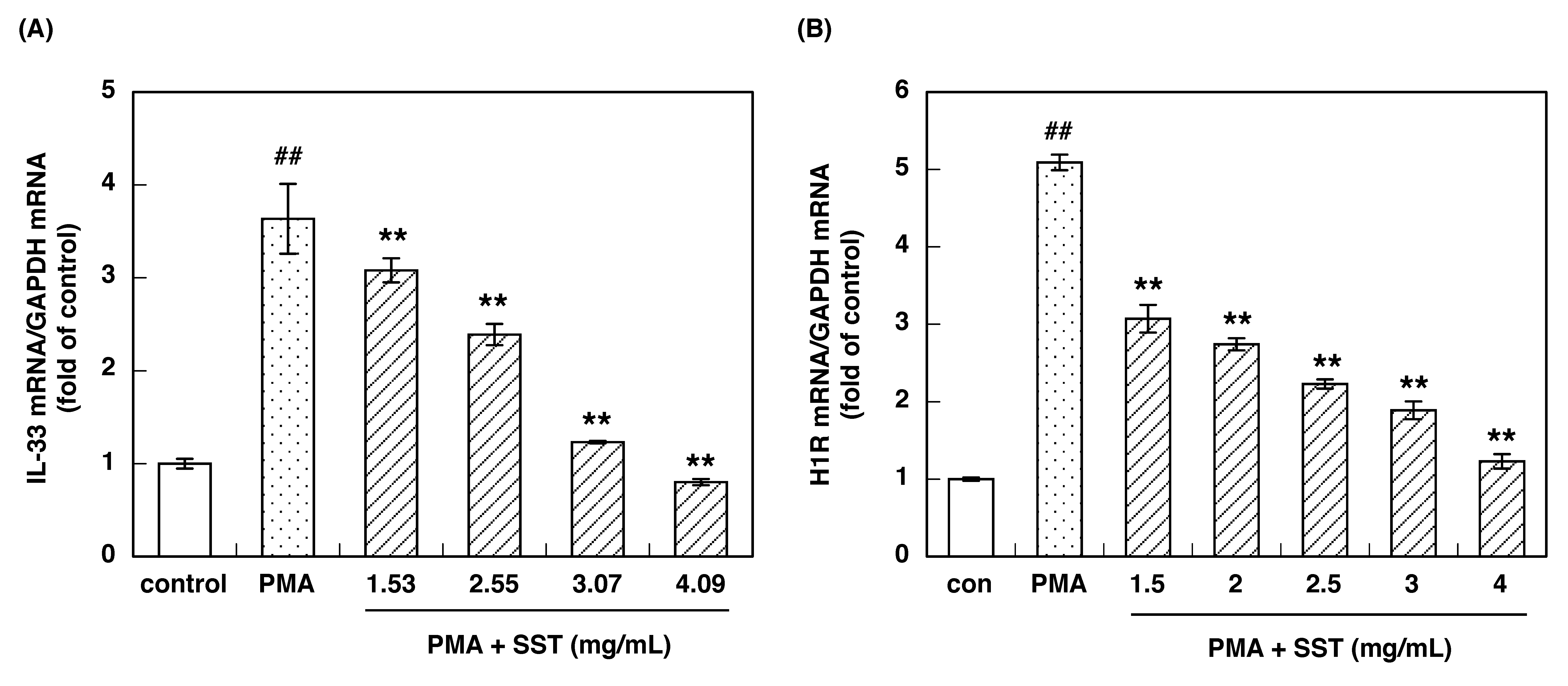
Figure 1. Effect of SST on PMA-induced up-regulations of IL-33 mRNA in Swiss 3T3 cells (A) and H1R mRNA in HeLa cells (B). The hot water extract of SST was incubated for 12 h before stimulation with 100 nM PMA. After stimulation with PMA for 3 h, the cells were harvested, and total RNA was prepared. IL-33 and H1R mRNA levels were determined using quantitative RT-PCR. Data are expressed as means ± SEM (n = 3–4). ## p < 0.01 vs. control; ** p < 0.01, vs. PMA.
Allergies 01 00015 g001
Figure 2. Effects of eight crude drugs of SST on PMA-induced up-regulation of IL-33 mRNA. Swiss 3T3 cells were treated with Ephedra herb (A), cinnamon bark (B), processed ginger (C), Asiasarum root (D), peony root (E), Schisandra fruit (F), Glycyrrhiza (G), and Pinellia tuber (H) for 12 h before stimulation with 100 nM PMA. After stimulation with PMA for 3 h, the cells were harvested, and total RNA was prepared. IL-33 mRNA levels were determined using quantitative RT-PCR. Data are expressed as means ± SEM (n = 3–4). ## p < 0.01 vs. control; * p < 0.05, ** p < 0.01 vs. PMA.
Allergies 01 00015 g002aAllergies 01 00015 g002bAllergies 01 00015 g002c
Figure 3. Effects of eight crude drugs of SST on PMA-induced up-regulation of H1R mRNA. HeLa cells were serum-starved for 24 h and treated with Ephedra herb (A), cinnamon bark (B), processed ginger (C), Asiasarum root (D), peony root (E), Schisandra fruit (F), Glycyrrhiza (G), and Pinellia tuber (H) for 12 h before stimulation with 100 nM PMA. After stimulation with PMA for 3 h, the cells were harvested, and total RNA was prepared. H1R mRNA levels were determined using quantitative RT-PCR. Data are expressed as means ± SEM (n = 3–4). ## p < 0.01 vs. control; * p < 0.05, ** p < 0.01 vs. PMA.
Allergies 01 00015 g003aAllergies 01 00015 g003b
Figure 4. The correlation between IC50 values of SST and its seven crude drugs to inhibit the IL-33 and H1R gene expression. A Spearman’s rank correlation test was used for the statistical analysis (r = 0.964, p < 0.01). SST: Syo-seiryu-to; EH: Ephedra herb; CB: cinnamon bark; PG: processed ginger; AsR: Asiasarum root; PR: peony root; SF: Schisandra fruit; G: Glycyrrhiza.
Allergies 01 00015 g004
Figure 5. Effects of inhibitors of the H1R gene expression signaling pathway on PMA-induced IL-33 gene up-regulation. Swiss 3T3 cells were treated with quercetin (A), (±)-maackiain (B), U0126 (C), 17-AAG (D), and celastrol (E) for 12 h before stimulation with 100 nM PMA. After stimulation with PMA for 3 h, the cells were harvested, and total RNA was extracted. IL-33 mRNA levels were determined using quantitative RT-PCR. Data are expressed as means ± SEM (n = 3–4). ## p < 0.01 vs. control; ** p < 0.01 vs. PMA.
Allergies 01 00015 g005
Table 1. Comparison of IC50 values of SST and its crude drugs to inhibit the IL-33 gene up-regulation with those related to H1R gene up-regulation.
Crude DrugsIC50 Values to Inhibit IL-33 Gene Expression (mg/mL)IC50 Values to Inhibit H1R Gene Expression (mg/mL)
SST2.752.98
Ephedra herb0.0930.0047
Cinnamon bark0.0640.062
Processed ginger0.921.01
Asiasarum root1.322.20
Peony root0.440.49
Schisandra fruit1.642.75
Glycyrrhiza0.290.28
Pinellia tuberNO *NO *
* no inhibition.
Publisher’s Note: MDPI stays neutral with regard to jurisdictional claims in published maps and institutional affiliations.