VGG16 Feature Extractor with Extreme Gradient Boost Classifier for Pancreas Cancer Prediction



Abstract
1. Introduction
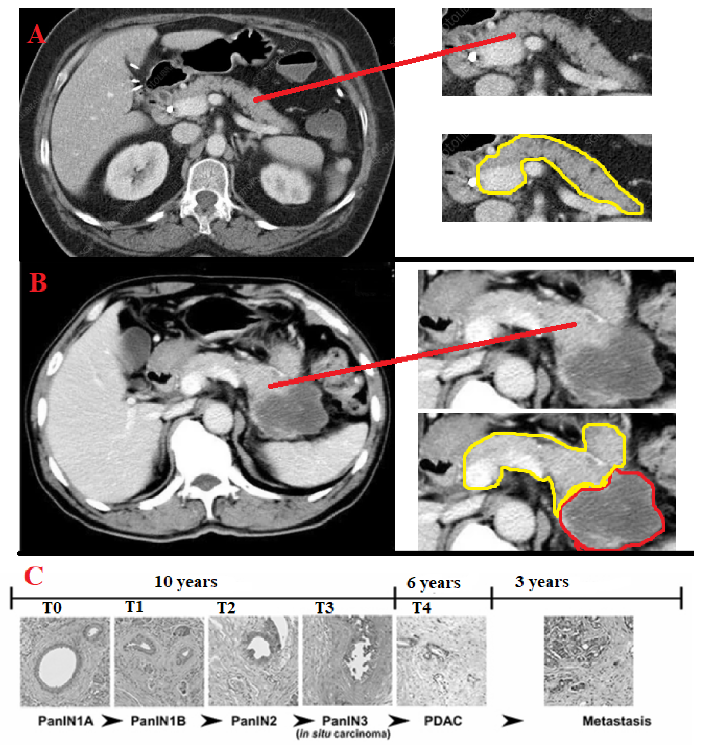
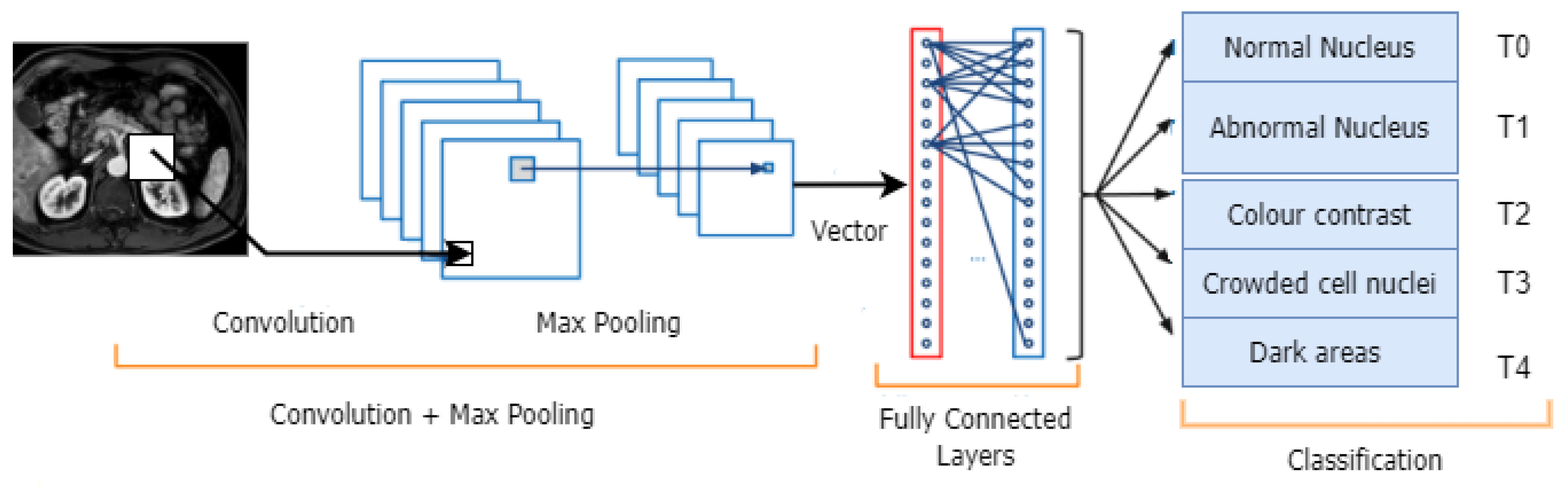
- T0 (time plus 0): There are no signs of cancer in the pancreas;
- T1: The tumour is solely in the pancreas and is no more than 2 cm in size (according to item T1). T1a, T1b, and T1c are other stages that can be identified based on the size of the tumour;
- T2: The tumour, which is only in the pancreas, is more than 2 cm but less than 4 cm in size;
- T3: The tumour is larger than 4 cm and extends past the pancreas. There is no involvement of the major arteries or veins near the pancreas;
- T4: The tumour spread affects the pancreas and nearby major arteries and veins. A T4 tumour cannot be completely removed during surgery.
- 1.
- 2.
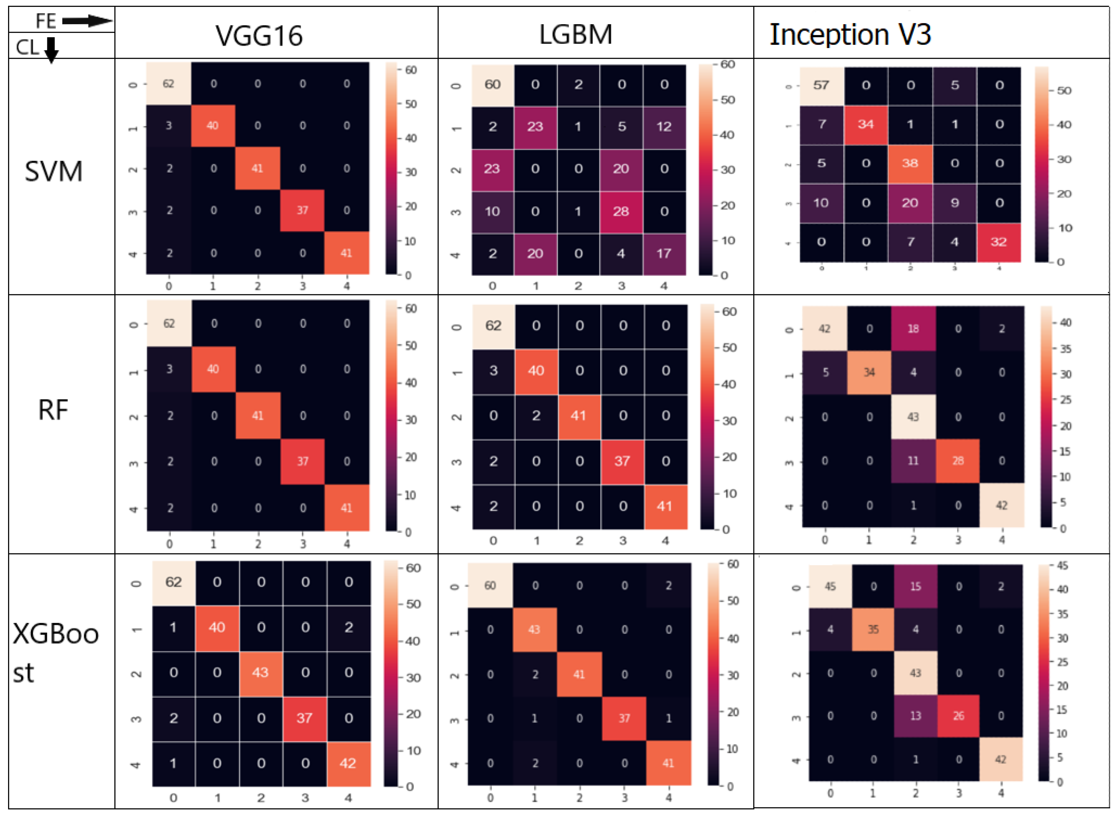
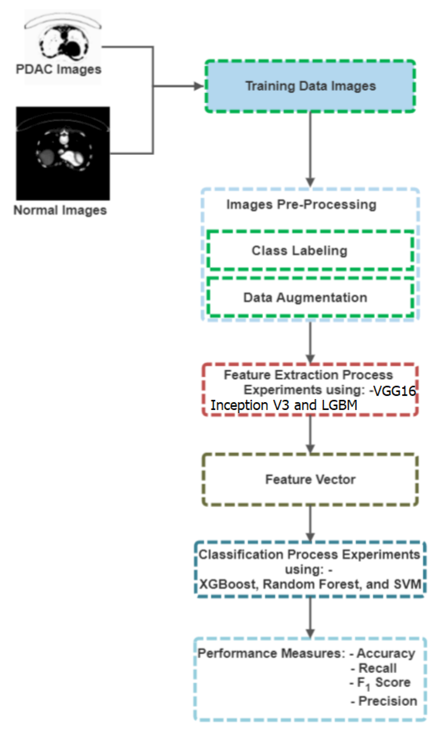
- Nine different combinations, including VGG16–XGBoost, VGG16–RF, and VGG16–SVM, Inception V3–XGBoost, Inception V3–RF, and Inception V3–SVM, as well as LGBM–XGBoost, LGBM–RF, and LGBM–SVM, were tested to identify PDAC in pancreas CT images. In these combinations, XGBoost, RF [13,14], and SVM [15] were employed as classifiers, while Inception V3, VGG16, and LGBM [16] were used as deep feature extractors;
- 3.
- To evaluate the effectiveness of the proposed framework in terms of accuracy, precision, recall, F1-score, and confusion matrix, a detailed experimental investigation was performed.
2. Related Work
3. Materials and Methods
3.1. CT Images Dataset
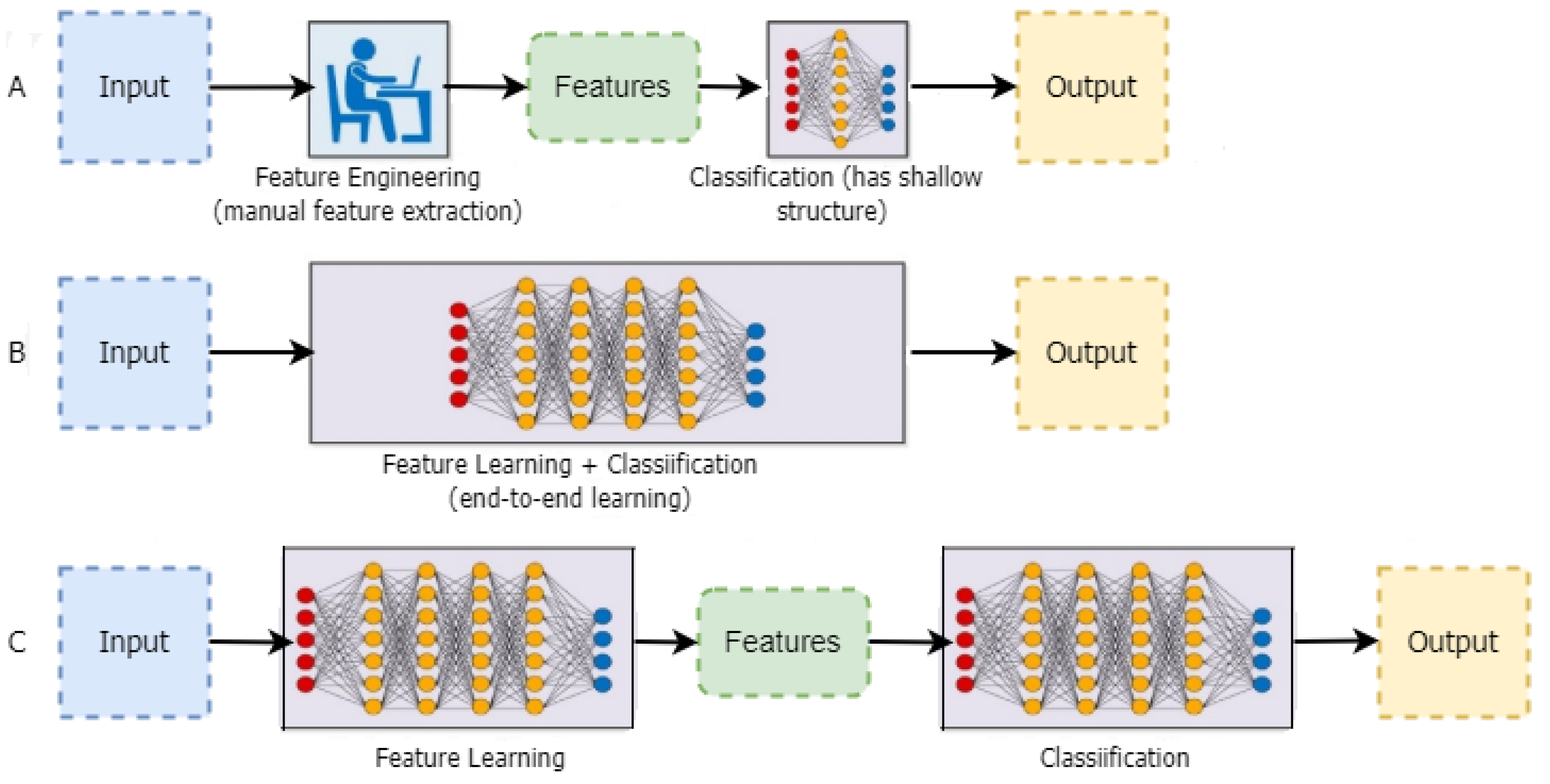
3.2. Feature Extractors (FE)
- Statistic Pixel-Level Features (SPLF) [35]: The pixels within a segmented region can be quantitatively described by these properties. The SPL features are as follows: mean, variance, a histogram of the grey values of the pixels in the region as well as the region’s area, details on the contrast of the pixels inside the region, and edge gradients of the pixels defining the region’s boundaries;
- Feature Shape Circularity: compactness, moments, chain codes, and the Hough transform are among the characteristics that reveal details about the shape of the region boundary [36]. To describe shapes, morphological processing techniques have also been employed;
- Texture characteristics [37]: determined using the second-order statistical histogram or co-occurrence matrices, these provide information on the local texture within the region or related area of the image. Additionally, wavelet processing describes local texture information in spatial frequency analysis;
- Relational characteristics [37]: these reveal the relational and hierarchical organisation of the regions connected to a single object or a collection of objects.
3.2.1. LGBM Feature Extractor
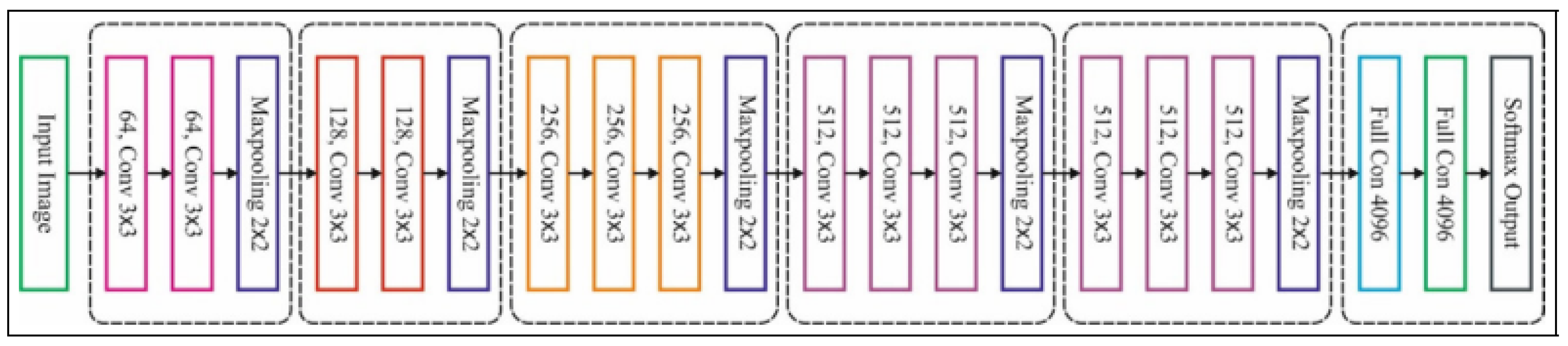
3.2.2. VGG16 Feature Extractor
Convolution Layer
Pooling Layer
Max Pooling
Softmax Layer
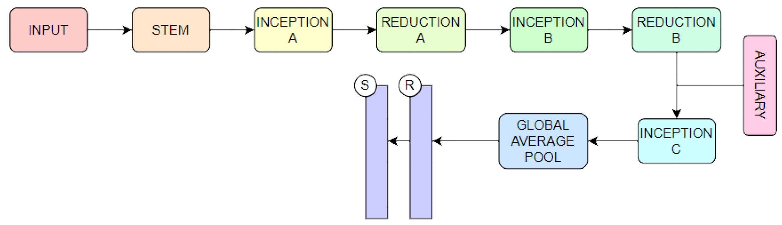
3.2.3. Inception V3
3.3. Classifiers
3.3.1. SVM Classifier
3.3.2. RF Classifier
3.3.3. XGBoost Classifier
3.4. Proposed Model
4. Experiments
| Algorithm 1: Model algorithm: Implementing LGBM to Classifiers |
|
| Algorithm 2: Model algorithm: Implementing VGG16 to Classifiers |
|
5. Experimental Results
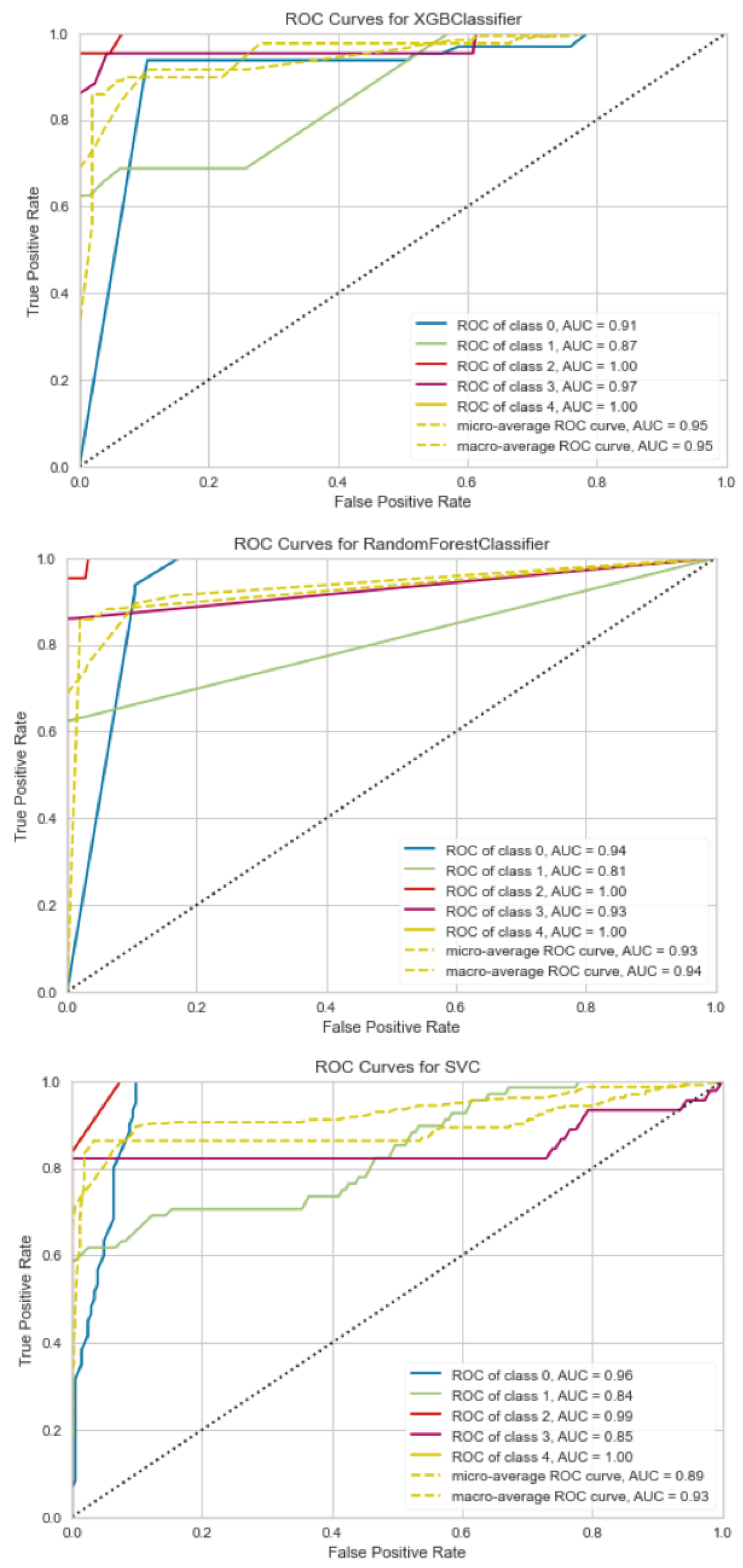
ROC
- ≤0.5 = There is no prejudice;
- 0.5–0.7 = Inadequate discrimination;
- 0.7–0.8 = Acceptable discrimination;
- 0.8–0.9 = Good discrimination;
- 0.9 = Excellent discrimination.
6. Discussion
7. Conclusions
Author Contributions
Funding
Institutional Review Board Statement
Informed Consent Statement
Data Availability Statement
Conflicts of Interest
References
- Ghojogh, B.; Samad, M.N.; Mashhadi, S.A.; Kapoor, T.; Ali, W.; Karray, F.; Crowley, M. Feature selection and feature extraction in pattern analysis: A literature review. arXiv 2019, arXiv:1905.02845. [Google Scholar]
- Waring, J.; Lindvall, C.; Umeton, R. Automated machine learning: Review of the state-of-the-art and opportunities for healthcare. Artif. Intell. Med. 2020, 104, 101822. [Google Scholar] [CrossRef] [Scilit] [PubMed]
- Maghdid, H.S.; Asaad, A.T.; Ghafoor, K.Z.; Sadiq, A.S.; Mirjalili, S.; Khan, M.K. Diagnosing COVID-19 pneumonia from X-ray and CT images using deep learning and transfer learning algorithms. In Multimodal Image Exploitation and Learning 2021; SPIE: Bellingham, WA, USA, 2021; Volume 11734, pp. 99–110. [Google Scholar]
- Gupta, A.; Gupta, K.; Gupta, K.; Gupta, K. A survey on human activity recognition and classification. In Proceedings of the 2020 International Conference on Communication and Signal Processing (ICCSP), Chennai, India, 28–30 July 2020; pp. 915–919. [Google Scholar]
- Rosen, R.D.; Sapra, A. TNM classification. In StatPearls [Internet]; StatPearls Publishing: Tapa, FL, USA, 2021. [Google Scholar]
- Rosen, R.D.; Sapra, A. TNM classification. In StatPearls [Internet]; StatPearls Publishing: Tapa, FL, USA, 2022. [Google Scholar]
- Kelleher, J.D. Deep Learning; MIT Press: Cambridge, MA, USA, 2019. [Google Scholar]
- Anaya-Isaza, A.; Mera-Jiménez, L.; Zequera-Diaz, M. An overview of deep learning in medical imaging. Inform. Med. Unlocked 2021, 26, 100723. [Google Scholar] [CrossRef] [Scilit]
- Chan, H.P.; Samala, R.K.; Hadjiiski, L.M.; Zhou, C. Deep learning in medical image analysis. Adv. Exp. Med. Biol. 2020, 1213, 3–21. [Google Scholar]
- Paleczek, A.; Grochala, D.; Rydosz, A. Artificial breath classification using XGBoost algorithm for diabetes detection. Sensors 2021, 21, 4187. [Google Scholar] [CrossRef] [Scilit]
- Singh, D.; Taspinar, Y.S.; Kursun, R.; Cinar, I.; Koklu, M.; Ozkan, I.A.; Lee, H.N. Classification and analysis of pistachio species with pre-trained deep learning models. Electronics 2022, 11, 981. [Google Scholar] [CrossRef] [Scilit]
- Albashish, D.; Al-Sayyed, R.; Abdullah, A.; Ryalat, M.H.; Almansour, N.A. Deep CNN model based on VGG16 for breast cancer classification. In Proceedings of the 2021 International Conference on Information Technology (ICIT), Amman, Jordan, 14–15 July 2021; pp. 805–810. [Google Scholar]
- Chowdhury, A.R.; Chatterjee, T.; Banerjee, S. A Random Forest classifier-based approach in the detection of abnormalities in the retina. Med. Biol. Eng. Comput. 2019, 57, 193–203. [Google Scholar] [CrossRef] [Scilit]
- Guo, Z.; Li, X.; Huang, H.; Guo, N.; Li, Q. Deep learning-based image segmentation on multimodal medical imaging. IEEE Trans. Radiat. Plasma Med. Sci. 2019, 3, 162–169. [Google Scholar] [CrossRef] [Scilit]
- Yadav, S.S.; Jadhav, S.M. Deep convolutional neural network based medical image classification for disease diagnosis. J. Big Data 2019, 6, 113. [Google Scholar] [CrossRef] [Scilit]
- Ono, Y.; Mitani, Y. Evaluation of feature extraction methods with ensemble learning for breast cancer classification. In Proceedings of the 2022 IEEE 4th Global Conference on Life Sciences and Technologies (LifeTech), Osaka, Japan, 7–9 March 2022; pp. 194–195. [Google Scholar]
- Battineni, G.; Sagaro, G.G.; Chinatalapudi, N.; Amenta, F. Applications of machine learning predictive models in the chronic disease diagnosis. J. Pers. Med. 2020, 10, 21. [Google Scholar] [CrossRef] [Scilit]
- Arabahmadi, M.; Farahbakhsh, R.; Rezazadeh, J. Deep learning for smart Healthcare—A survey on brain tumor detection from medical imaging. Sensors 2022, 22, 1960. [Google Scholar] [CrossRef] [Scilit]
- Liu, X.; Song, L.; Liu, S.; Zhang, Y. A review of deep-learning-based medical image segmentation methods. Sustainability 2021, 13, 1224. [Google Scholar] [CrossRef] [Scilit]
- Ahsan, M.; Based, M.A.; Haider, J.; Kowalski, M. COVID-19 detection from chest X-ray images using feature fusion and deep learning. Sensors 2021, 21, 1480. [Google Scholar]
- Masud, M.; Sikder, N.; Nahid, A.A.; Bairagi, A.K.; AlZain, M.A. A machine learning approach to diagnosing lung and colon cancer using a deep learning-based classification framework. Sensors 2021, 21, 748. [Google Scholar] [CrossRef] [Scilit]
- Nguyen, D.T.; Lee, M.B.; Pham, T.D.; Batchuluun, G.; Arsalan, M.; Park, K.R. Enhanced image-based endoscopic pathological site classification using an ensemble of deep learning models. Sensors 2020, 20, 5982. [Google Scholar] [CrossRef] [Scilit] [PubMed]
- Malini, M.; Gopalakrishnan, B.; Dhivya, K.; Naveena, S. Diabetic Patient Prediction using Machine Learning Algorithm. In Proceedings of the 2021 Smart Technologies, Communication and Robotics (STCR), Sathyamangalam, India, 9–10 October 2021; pp. 1–5. [Google Scholar]
- Huang, S.; Cai, N.; Pacheco, P.P.; Narrandes, S.; Wang, Y.; Xu, W. Applications of support vector machine (SVM) learning in cancer genomics. Cancer Genom. Proteom. 2018, 15, 41–51. [Google Scholar]
- Iwendi, C.; Bashir, A.K.; Peshkar, A.; Sujatha, R.; Chatterjee, J.M.; Pasupuleti, S.; Mishra, R.; Pillai, S.; Jo, O. COVID-19 patient health prediction using boosted random forest algorithm. Front. Public Health 2020, 8, 357. [Google Scholar] [CrossRef] [Scilit]
- Toğaçar, M.; Ergen, B.; Cömert, Z. BrainMRNet: Brain tumor detection using magnetic resonance images with a novel convolutional neural network model. Med. Hypotheses 2020, 134, 109531. [Google Scholar] [CrossRef] [Scilit]
- Ghimire, D.; Kil, D.; Kim, S.-h. A survey on efficient convolutional neural networks and hardware acceleration. Electronics 2022, 11, 945. [Google Scholar] [CrossRef] [Scilit]
- Florkowski, M. Anomaly Detection, Trend Evolution, and Feature Extraction in Partial Discharge Patterns. Energies 2021, 14, 3886. [Google Scholar] [CrossRef] [Scilit]
- Amerikanos, P.; Maglogiannis, I. Image Analysis in Digital Pathology Utilizing Machine Learning and Deep Neural Networks. J. Pers. Med. 2022, 12, 1444. [Google Scholar] [CrossRef] [Scilit] [PubMed]
- Polap, D.; Wlodarczyk-Sielicka, M. Classification of non-conventional ships using a neural bag-of-words mechanism. Sensors 2020, 20, 1608. [Google Scholar] [CrossRef] [Scilit] [PubMed]
- El-Rashidy, N.; El-Sappagh, S.; Islam, S.R.; El-Bakry, H.M.; Abdelrazek, S. End-to-end deep learning framework for coronavirus (COVID-19) detection and monitoring. Electronics 2020, 9, 1439. [Google Scholar] [CrossRef] [Scilit]
- Naranjo-Torres, J.; Mora, M.; Hernández-García, R.; Barrientos, R.J.; Fredes, C.; Valenzuela, A. A review of convolutional neural network applied to fruit image processing. Appl. Sci. 2020, 10, 3443. [Google Scholar] [CrossRef] [Scilit]
- Roth, F.; Turkbey, L.; Liu, S. The Cancer Imaging Archive. Data From Pancreas-CT (Version 2) [Data Set]. 2016. Available online: https://www.cancerimagingarchive.net/ (accessed on 20 May 2023).
- Chang, C.C.; Li, Y.Z.; Wu, H.C.; Tseng, M.H. Melanoma Detection Using XGB Classifier Combined with Feature Extraction and K-Means SMOTE Techniques. Diagnostics 2022, 12, 1747. [Google Scholar] [CrossRef] [Scilit] [PubMed]
- Augustauskas, R.; Lipnickas, A. Improved pixel-level pavement-defect segmentation using a deep autoencoder. Sensors 2020, 20, 2557. [Google Scholar] [CrossRef] [Scilit]
- Damian, F.A.; Moldovanu, S.; Dey, N.; Ashour, A.S.; Moraru, L. Feature selection of non-dermoscopic skin lesion images for nevus and melanoma classification. Computation 2020, 8, 41. [Google Scholar] [CrossRef] [Scilit]
- Wang, Y.; Qi, Q.; Shen, X. Image segmentation of brain mri based on ltridp and superpixels of improved slic. Brain Sci. 2020, 10, 116. [Google Scholar] [CrossRef] [Scilit]
- Alzubaidi, L.; Fadhel, M.A.; Al-Shamma, O.; Zhang, J.; Duan, Y. Deep learning models for classification of red blood cells in microscopy images to aid in sickle cell anemia diagnosis. Electronics 2020, 9, 427. [Google Scholar] [CrossRef] [Scilit]
- Tamang, L.D.; Kim, B.W. Deep learning approaches to colorectal cancer diagnosis: A review. Appl. Sci. 2021, 11, 10982. [Google Scholar] [CrossRef] [Scilit]
- Bhatt, D.; Patel, C.; Talsania, H.; Patel, J.; Vaghela, R.; Pandya, S.; Modi, K.; Ghayvat, H. CNN variants for computer vision: History, architecture, application, challenges and future scope. Electronics 2021, 10, 2470. [Google Scholar] [CrossRef] [Scilit]
- Ali, M.; Ali, R. Multi-input dual-stream capsule network for improved lung and colon cancer classification. Diagnostics 2021, 11, 1485. [Google Scholar] [CrossRef] [Scilit] [PubMed]
- Capra, M.; Bussolino, B.; Marchisio, A.; Shafique, M.; Masera, G.; Martina, M. An updated survey of efficient hardware architectures for accelerating deep convolutional neural networks. Future Internet 2020, 12, 113. [Google Scholar] [CrossRef] [Scilit]
- Wang, Y.; Li, Y.; Song, Y.; Rong, X. The influence of the activation function in a convolution neural network model of facial expression recognition. Appl. Sci. 2020, 10, 1897. [Google Scholar] [CrossRef] [Scilit]
- Solé, R.; Seoane, L.F. Evolution of brains and computers: The roads not taken. Entropy 2022, 24, 665. [Google Scholar] [CrossRef] [Scilit]
- Rao, H.; Shi, X.; Rodrigue, A.K.; Feng, J.; Xia, Y.; Elhoseny, M.; Yuan, X.; Gu, L. Feature selection based on artificial bee colony and gradient boosting decision tree. Appl. Soft Comput. 2019, 74, 634–642. [Google Scholar] [CrossRef] [Scilit]
- Zhang, D.; Gong, Y. The comparison of LightGBM and XGBoost coupling factor analysis and prediagnosis of acute liver failure. IEEE Access 2020, 8, 220990–221003. [Google Scholar] [CrossRef] [Scilit]
- Fang, M.; Chen, Y.; Xue, R.; Wang, H.; Chakraborty, N.; Su, T.; Dai, Y. A hybrid machine learning approach for hypertension risk prediction. Neural Comput. Appl. 2021, 35, 14487–14497. [Google Scholar] [CrossRef] [Scilit]
- Chen, R.C.; Caraka, R.E.; Arnita, N.E.G.; Pomalingo, S.; Rachman, A.; Toharudin, T.; Tai, S.K.; Pardamean, B. An end to end of scalable tree boosting system. Sylwan 2020, 165, 1–11. [Google Scholar]
- Xu, G.; Shen, X.; Chen, S.; Zong, Y.; Zhang, C.; Yue, H.; Liu, M.; Chen, F.; Che, W. A deep transfer convolutional neural network framework for EEG signal classification. IEEE Access 2019, 7, 112767–112776. [Google Scholar] [CrossRef] [Scilit]
- Theckedath, D.; Sedamkar, R. Detecting affect states using VGG16, ResNet50 and SE-ResNet50 networks. SN Comput. Sci. 2020, 1, 1–7. [Google Scholar] [CrossRef] [Scilit]
- Sajja, V.R.; Kalluri, H.K. Classification of Brain tumors using Fuzzy C-means and VGG16. Turk. J. Comput. Math. Educ. 2021, 12, 2103–2113. [Google Scholar]
- Pardede, J.; Sitohang, B.; Akbar, S.; Khodra, M.L. Implementation of transfer learning using VGG16 on fruit ripeness detection. Int. J. Intell. Syst. Appl. 2021, 13, 52–61. [Google Scholar] [CrossRef] [Scilit]
- Nahata, H.; Singh, S.P. Deep learning solutions for skin cancer detection and diagnosis. In Machine Learning with Health Care Perspective: Machine Learning and Healthcare; Springer: Cham, Switzerland, 2020; pp. 159–182. [Google Scholar]
- Wang, C.; Chen, D.; Hao, L.; Liu, X.; Zeng, Y.; Chen, J.; Zhang, G. Pulmonary image classification based on inception-v3 transfer learning model. IEEE Access 2019, 7, 146533–146541. [Google Scholar] [CrossRef] [Scilit]
- Giger, M.L. Machine learning in medical imaging. J. Am. Coll. Radiol. 2018, 15, 512–520. [Google Scholar] [CrossRef] [Scilit]
- Scholkopf, B.; Smola, A.J. Learning with Kernels: Support Vector Machines, Regularization, Optimization, and Beyond; MIT Press: Cambridge, MA, USA, 2018. [Google Scholar]
- Hsu, C.W.; Chang, C.C.; Lin, C.J. A Practical Guide to Support Vector Classification. 2003. Available online: https://www.csie.ntu.edu.tw/~cjlin/ (accessed on 20 May 2023).
- Asselman, A.; Nasseh, A.; Aammou, S. Revealing strengths, weaknesses and prospects of intelligent collaborative e-learning systems. Adv. Sci. Technol. Eng. Syst. J. 2018, 3, 67–79. [Google Scholar] [CrossRef] [Scilit]
- Cutler, A.; Cutler, D.R.; Stevens, J.R. Random forests. In Ensemble Machine Learning; Springer: Berlin/Heidelberg, Germany, 2012; pp. 157–175. [Google Scholar]
- He, K.; Zhang, X.; Ren, S.; Sun, J. Deep residual learning for image recognition. In Proceedings of the IEEE Conference on Computer Vision and Pattern Recognition, Las Vegas, NV, USA, 27–30 June 2016; pp. 770–778. [Google Scholar]
- Ren, B.; Liu, X.; Suriawinata, A.A. Pancreatic ductal adenocarcinoma and its precursor lesions: Histopathology, cytopathology, and molecular pathology. Am. J. Pathol. 2019, 189, 9–21. [Google Scholar] [CrossRef] [Scilit] [PubMed]
- Javadi, S.; Menias, C.O.; Korivi, B.R.; Shaaban, A.M.; Patnana, M.; Alhalabi, K.; Elsayes, K.M. Pancreatic calcifications and calcified pancreatic masses: Pattern recognition approach on CT. Am. J. Roentgenol. 2017, 209, 77–87. [Google Scholar] [CrossRef] [Scilit]
- Lu, D.; Reber, H.A.; Krasny, R.M.; Kadell, B.M.; Sayre, J. Local staging of pancreatic cancer: Criteria for unresectability of major vessels as revealed by pancreatic-phase, thin-section helical CT. AJR Am. J. Roentgenol. 1997, 168, 1439–1443. [Google Scholar] [CrossRef] [Scilit] [PubMed]
- DeVita, V.T., Jr.; Rosenberg, S.A.; Lawrence, T.S. DeVita, Hellman, and Rosenberg’s Cancer: Short Title; Lippincott Williams & Wilkins: Philadelphia, PA, USA, 2022. [Google Scholar]
- Lee, T.C.; Angelina, C.L.; Kongkam, P.; Wang, H.P.; Rerknimitr, R.; Han, M.L.; Chang, H.T. Deep-Learning-Enabled Computer-Aided Diagnosis in the Classification of Pancreatic Cystic Lesions on Confocal Laser Endomicroscopy. Diagnostics 2023, 13, 1289. [Google Scholar] [CrossRef] [Scilit] [PubMed]
- Sadewo, W.; Rustam, Z.; Hamidah, H.; Chusmarsyah, A.R. Pancreatic cancer early detection using twin support vector machine based on kernel. Symmetry 2020, 12, 667. [Google Scholar] [CrossRef] [Scilit]
- Gouda, W.; Sama, N.U.; Al-Waakid, G.; Humayun, M.; Jhanjhi, N.Z. Detection of skin cancer based on skin lesion images using deep learning. Healthcare 2022, 10, 1183. [Google Scholar] [CrossRef] [Scilit] [PubMed]
- Karim, A.M.; Kaya, H.; Güzel, M.S.; Tolun, M.R.; Çelebi, F.V.; Mishra, A. A novel framework using deep auto-encoders based linear model for data classification. Sensors 2020, 20, 6378. [Google Scholar] [CrossRef] [Scilit]
- Caroprese, L.; Vocaturo, E.; Zumpano, E. Features for melanoma lesions: Extraction and classification. In Proceedings of the IEEE/WIC/ACM International Conference on Web Intelligence-Companion Volume, Thessaloniki, Greece, 14–17 October 2019; pp. 238–243. [Google Scholar]
- Almeida, M.A.; Santos, I.A. Classification models for skin tumor detection using texture analysis in medical images. J. Imaging 2020, 6, 51. [Google Scholar] [CrossRef] [Scilit]
- Yan, Y.; Yao, X.J.; Wang, S.H.; Zhang, Y.D. A survey of computer-aided tumor diagnosis based on convolutional neural network. Biology 2021, 10, 1084. [Google Scholar] [CrossRef] [Scilit] [PubMed]
- Hameed, Z.; Zahia, S.; Garcia-Zapirain, B.; Javier Aguirre, J.; Maria Vanegas, A. Breast cancer histopathology image classification using an ensemble of deep learning models. Sensors 2020, 20, 4373. [Google Scholar] [CrossRef] [Scilit] [PubMed]







| CF | ACC | WP | WFS | CL | AV | PR | RE | FS |
|---|---|---|---|---|---|---|---|---|
| SVM | 0.56 | 0.46 | 0.49 | |||||
| MAV | 0.45 | 0.52 | 0.47 | |||||
| WAV | 0.46 | 0.56 | 0.49 | |||||
| T0 | 0.62 | 0.97 | 0.75 | |||||
| T1 | 0.53 | 0.53 | 0.53 | |||||
| T2 | 0.57 | 0.60 | 0.57 | |||||
| T3 | 0.49 | 0.72 | 0.58 | |||||
| T4 | 0.59 | 0.40 | 0.47 | |||||
| RF | 0.92 | 0.91 | 0.93 | |||||
| MAV | 0.97 | 0.96 | 0.96 | |||||
| WAV | 0.96 | 0.96 | 0.96 | |||||
| T0 | 0.90 | 1.00 | 0.95 | |||||
| T1 | 0.95 | 0.93 | 0.94 | |||||
| T2 | 0.97 | 0.95 | 0.98 | |||||
| T3 | 0.97 | 0.95 | 0.97 | |||||
| T4 | 0.97 | 0.95 | 0.98 | |||||
| XGBoost | 0.94 | 0.93 | 0.93 | |||||
| MAV | 0.97 | 0.96 | 0.96 | |||||
| WAV | 0.96 | 0.96 | 0.96 | |||||
| T0 | 0.90 | 1.00 | 0.95 | |||||
| T1 | 0.95 | 0.93 | 0.94 | |||||
| T2 | 1.0 | 0.95 | 0.98 | |||||
| T3 | 0.95 | 0.95 | 0.97 | |||||
| T4 | 0.94 | 0.95 | 0.97 |
| CF | ACC | WP | WFS | CL | AV | PR | RE | FS |
|---|---|---|---|---|---|---|---|---|
| SVM | 0.95 | 0.96 | 0.94 | |||||
| MAV | 0.97 | 0.96 | 0.96 | |||||
| WAV | 0.97 | 0.96 | 0.96 | |||||
| T0 | 0.87 | 1.00 | 0.93 | |||||
| T1 | 1.00 | 0.93 | 0.96 | |||||
| T2 | 1.00 | 0.95 | 0.98 | |||||
| T3 | 1.00 | 0.95 | 0.97 | |||||
| T4 | 1.00 | 0.95 | 0.96 | |||||
| RF | 0.96 | 0.95 | 0.95 | |||||
| MAV | 0.97 | 0.96 | 0.96 | |||||
| WAV | 0.97 | 0.96 | 0.96 | |||||
| T0 | 0.87 | 1.00 | 0.93 | |||||
| T1 | 1.00 | 0.93 | 0.96 | |||||
| T2 | 1.00 | 0.95 | 0.98 | |||||
| T3 | 1.00 | 0.95 | 0.97 | |||||
| T4 | 1.00 | 0.95 | 0.98 | |||||
| XGBoost | 0.98 | 0.98 | 0.97 | |||||
| MAV | 0.98 | 0.97 | 0.97 | |||||
| WAV | 0.98 | 0.97 | 0.97 | |||||
| T0 | 0.94 | 1.00 | 0.97 | |||||
| T1 | 1.00 | 0.94 | 0.96 | |||||
| T2 | 1.0 | 1.00 | 1.00 | |||||
| T3 | 1.00 | 0.95 | 0.98 | |||||
| T4 | 0.96 | 0.98 | 0.99 |
| CF | ACC | WP | WFS | CL | AV | PR | RE | FS |
|---|---|---|---|---|---|---|---|---|
| SVM | 0.63 | 0.60 | 0.61 | |||||
| MAV | 0.57 | 0.62 | 0.58 | |||||
| WAV | 0.55 | 0.60 | 0.58 | |||||
| T0 | 0.68 | 0.94 | 0.62 | |||||
| T1 | 0.58 | 0.62 | 0.60 | |||||
| T2 | 0.59 | 0.59 | 0.67 | |||||
| T3 | 0.60 | 0.69 | 0.61 | |||||
| T4 | 0.68 | 0.68 | 0.51 | |||||
| RF | 0.86 | 0.87 | 0.89 | |||||
| MAV | 0.89 | 0.90 | 0.92 | |||||
| WAV | 0.87 | 0.91 | 0.90 | |||||
| T0 | 0.90 | 0.90 | 0.92 | |||||
| T1 | 0.91 | 0.89 | 0.89 | |||||
| T2 | 0.89 | 0.89 | 0.89 | |||||
| T3 | 0.89 | 0.88 | 0.91 | |||||
| T4 | 0.87 | 0.91 | 0.91 | |||||
| XGBoost | 0.95 | 0.95 | 0.95 | |||||
| MAV | 0.92 | 0.93 | 0.94 | |||||
| WAV | 0.91 | 0.91 | 0.93 | |||||
| T0 | 0.90 | 0.96 | 0.93 | |||||
| T1 | 0.91 | 0.95 | 0.95 | |||||
| T2 | 0.92 | 0.95 | 0.95 | |||||
| T3 | 0.93 | 0.95 | 0.94 | |||||
| T4 | 0.93 | 0.94 | 0.95 |
| Paper | Application | Model | Accuracy |
|---|---|---|---|
| [23] | Diabetes Prediction | K-Nearest Neighbour, SVM, Random Forest, LGBM, AdaBoost, and Decision Tree | 89.85% |
| [24] | Identify Colon Cancer | SVM | 94.51% |
| [25] | Identify COVID-19 | Random Forest + AdaBoost | 94% |
| [65] | Pancreatic Cystic Lesion | CNN of VGG16 | 93.11% |
| [66] | Pancreatic Cancer | Twin SVM | 98% |
| [67] | Skin Cancer | ||
| CNN | 83.2% | ||
| ResNet50 | 83.7% | ||
| Inception V3 | 85.8% | ||
| Inception ResNet | 84% |
Disclaimer/Publisher’s Note: The statements, opinions and data contained in all publications are solely those of the individual author(s) and contributor(s) and not of MDPI and/or the editor(s). MDPI and/or the editor(s) disclaim responsibility for any injury to people or property resulting from any ideas, methods, instructions or products referred to in the content. |
© 2023 by the authors. Licensee MDPI, Basel, Switzerland. This article is an open access article distributed under the terms and conditions of the Creative Commons Attribution (CC BY) license (https://creativecommons.org/licenses/by/4.0/).
Share and Cite
Bakasa, W.; Viriri, S. VGG16 Feature Extractor with Extreme Gradient Boost Classifier for Pancreas Cancer Prediction. J. Imaging 2023, 9, 138. https://doi.org/10.3390/jimaging9070138
Bakasa W, Viriri S. VGG16 Feature Extractor with Extreme Gradient Boost Classifier for Pancreas Cancer Prediction. Journal of Imaging. 2023; 9(7):138. https://doi.org/10.3390/jimaging9070138
Chicago/Turabian StyleBakasa, Wilson, and Serestina Viriri. 2023. "VGG16 Feature Extractor with Extreme Gradient Boost Classifier for Pancreas Cancer Prediction" Journal of Imaging 9, no. 7: 138. https://doi.org/10.3390/jimaging9070138
APA StyleBakasa, W., & Viriri, S. (2023). VGG16 Feature Extractor with Extreme Gradient Boost Classifier for Pancreas Cancer Prediction. Journal of Imaging, 9(7), 138. https://doi.org/10.3390/jimaging9070138