Article

Embedded Quantitative MRI T Mapping Using Non-Linear Primal-Dual Proximal Splitting
Department of Applied Physics, University of Eastern Finland, 70211 Kuopio, Finland
*
Author to whom correspondence should be addressed.
J. Imaging 2022, 8(6), 157; https://doi.org/10.3390/jimaging8060157

Submission received: 25 March 2022 / Revised: 13 May 2022 / Accepted: 26 May 2022 / Published: 31 May 2022
(This article belongs to the Special Issue The Present and the Future of Imaging)

Abstract

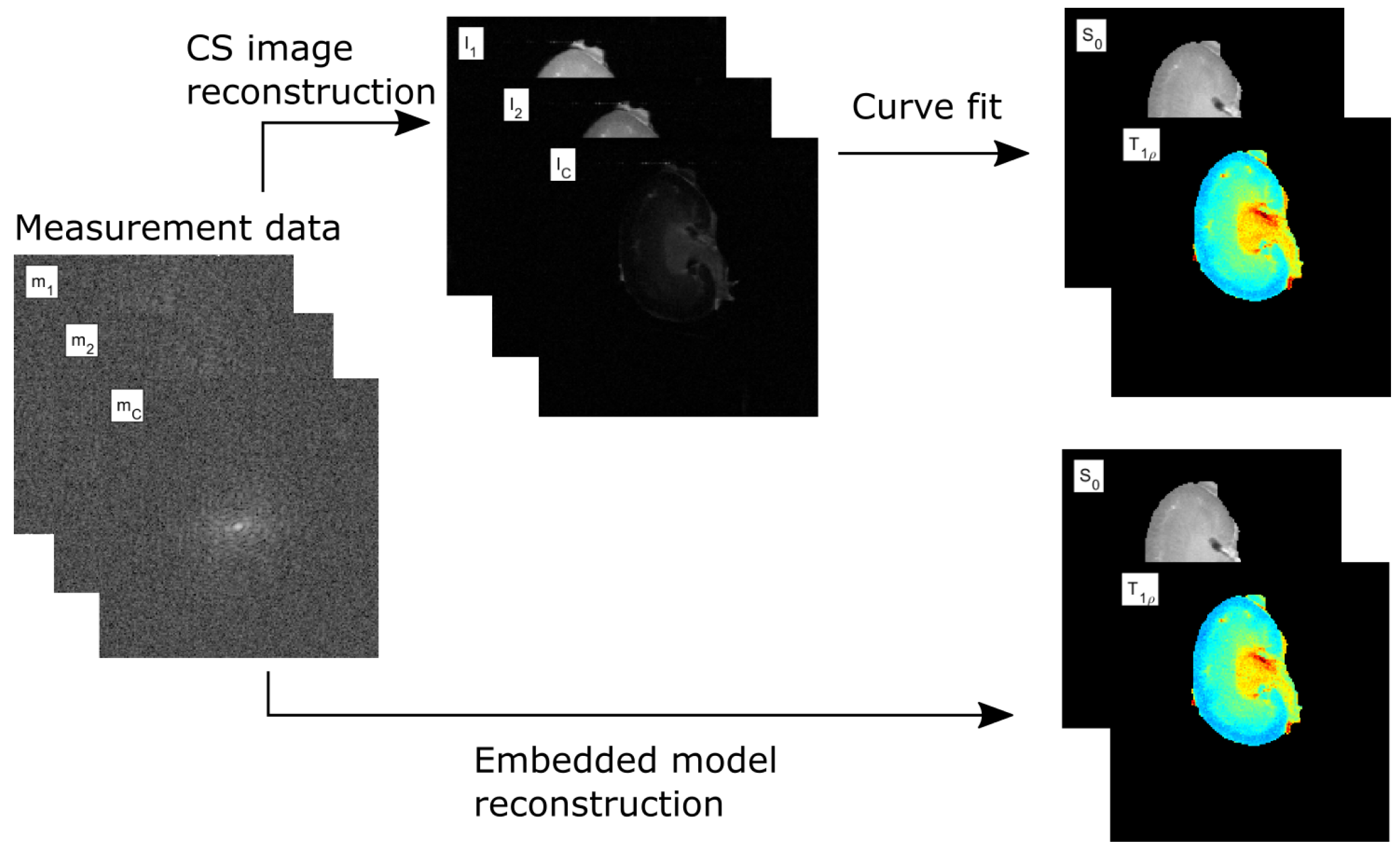
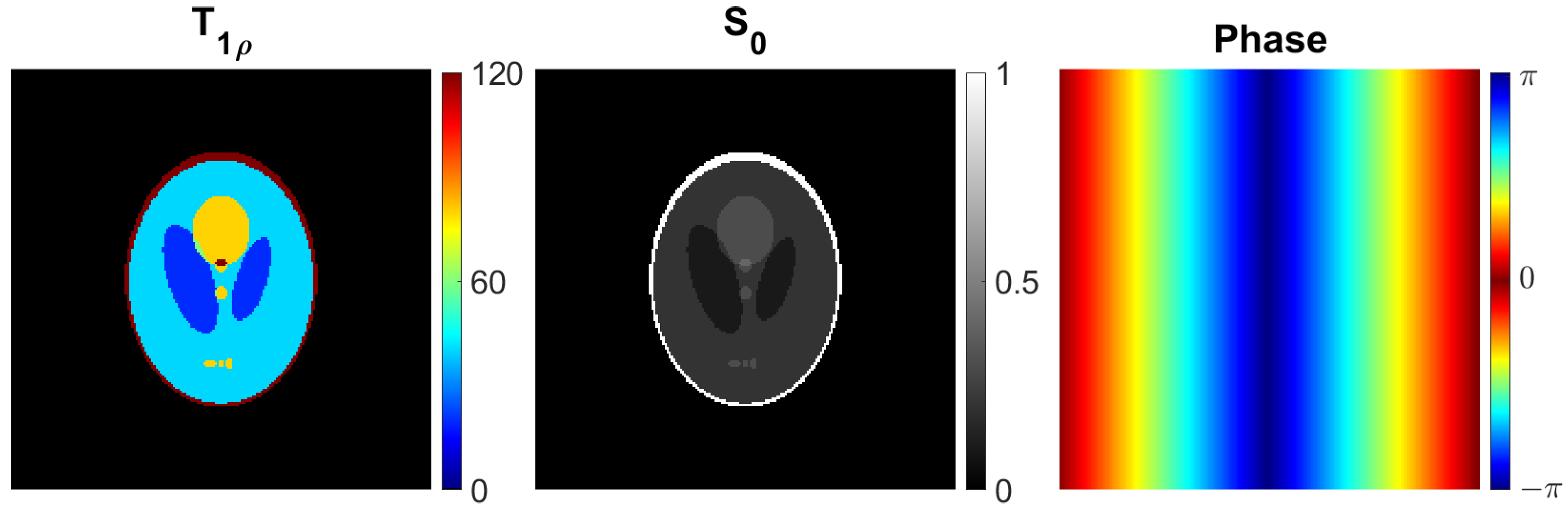
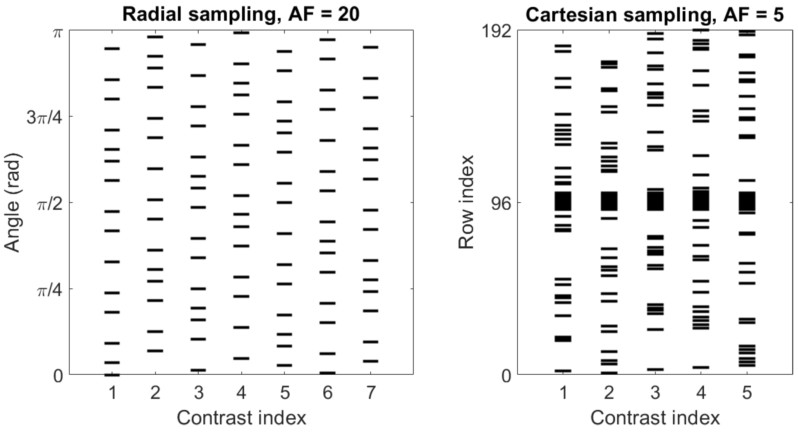
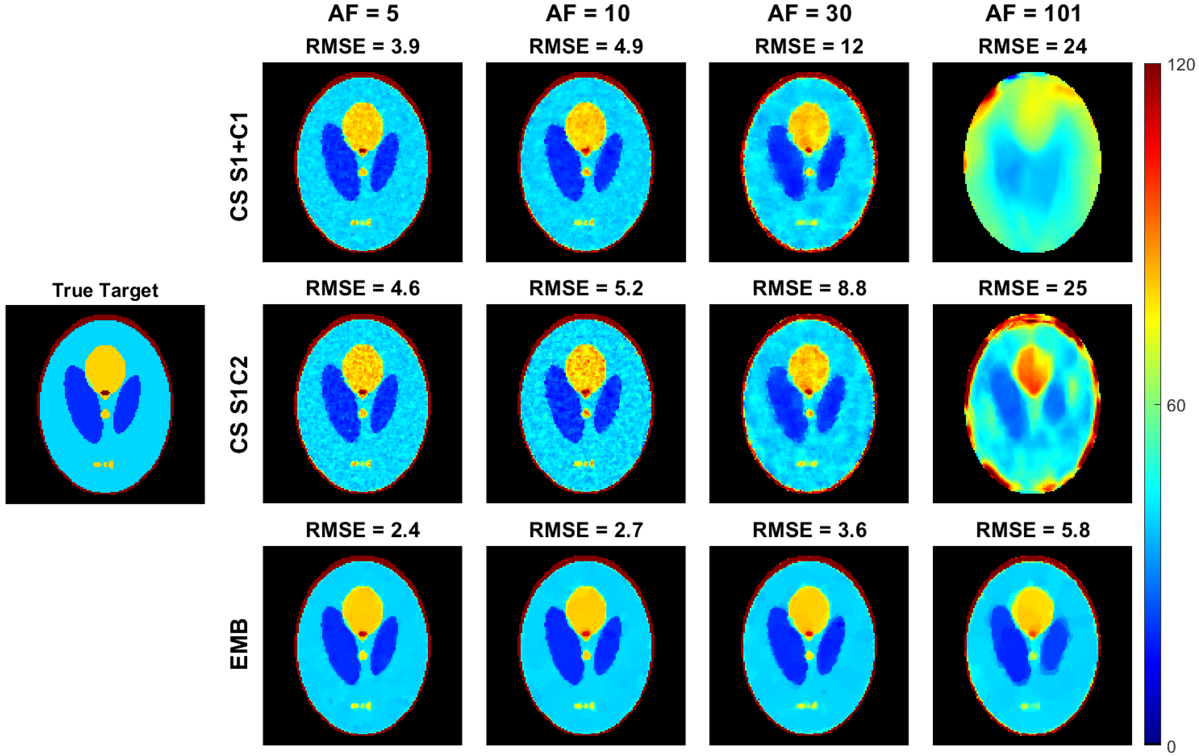
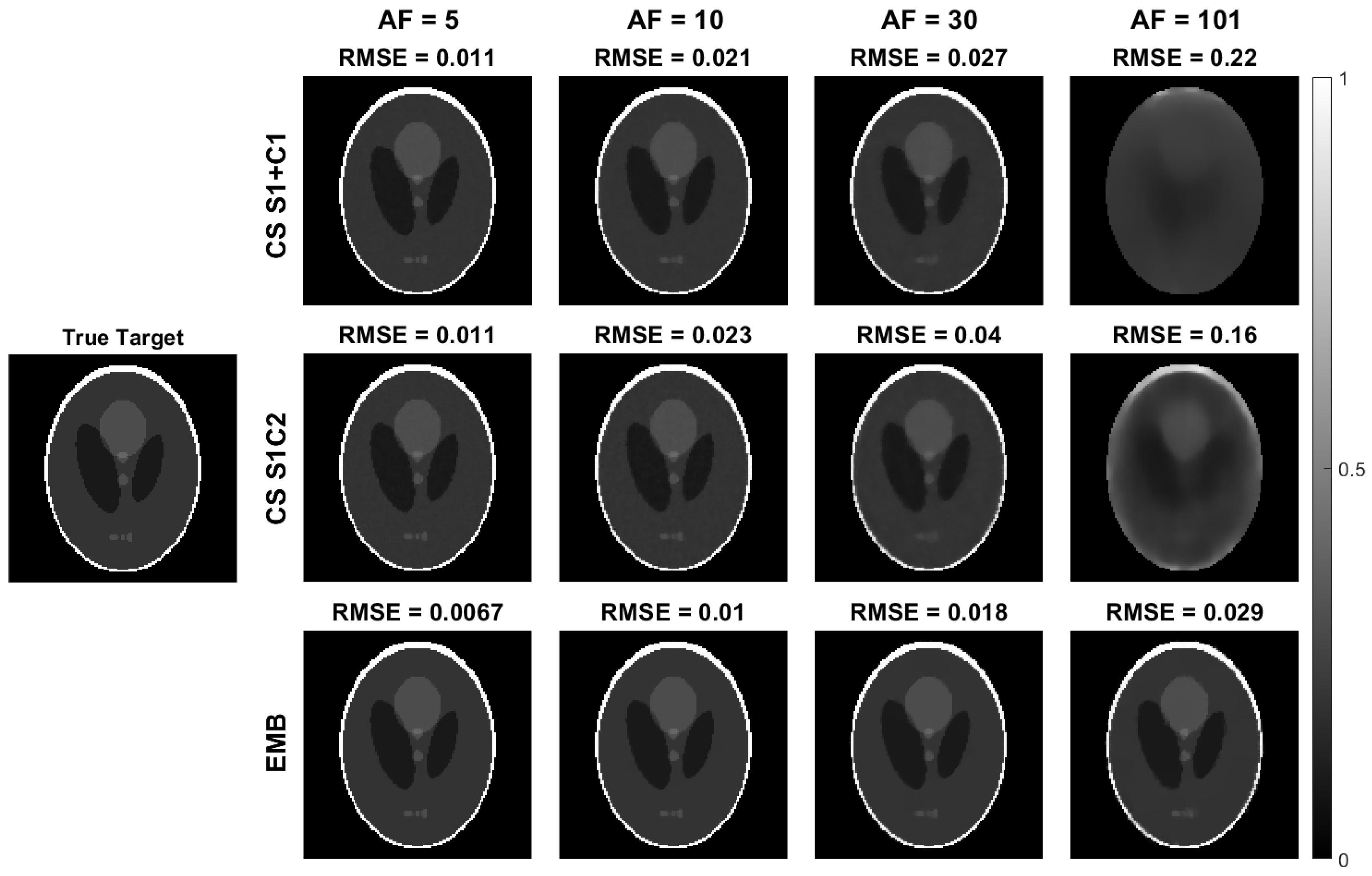
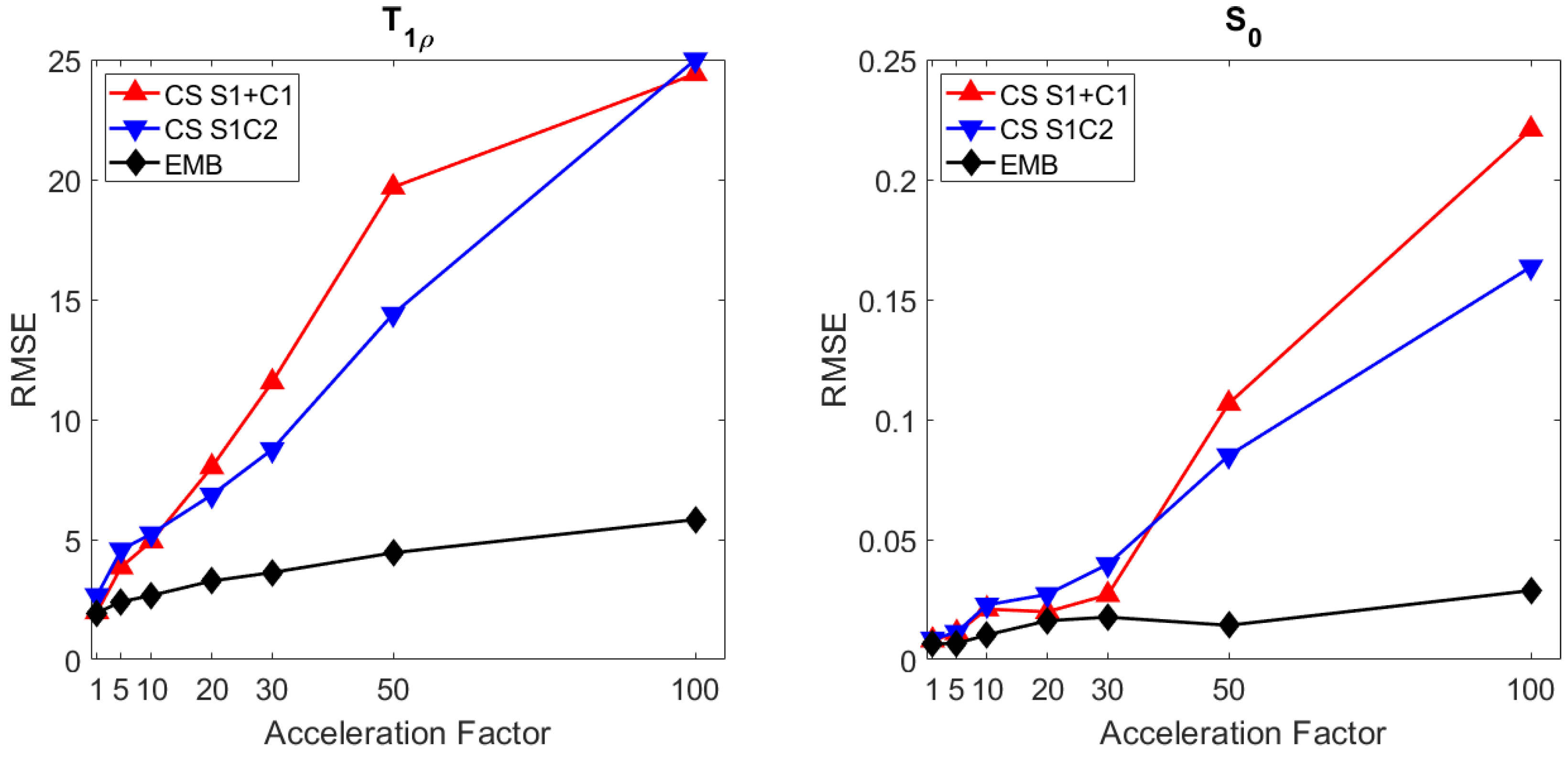
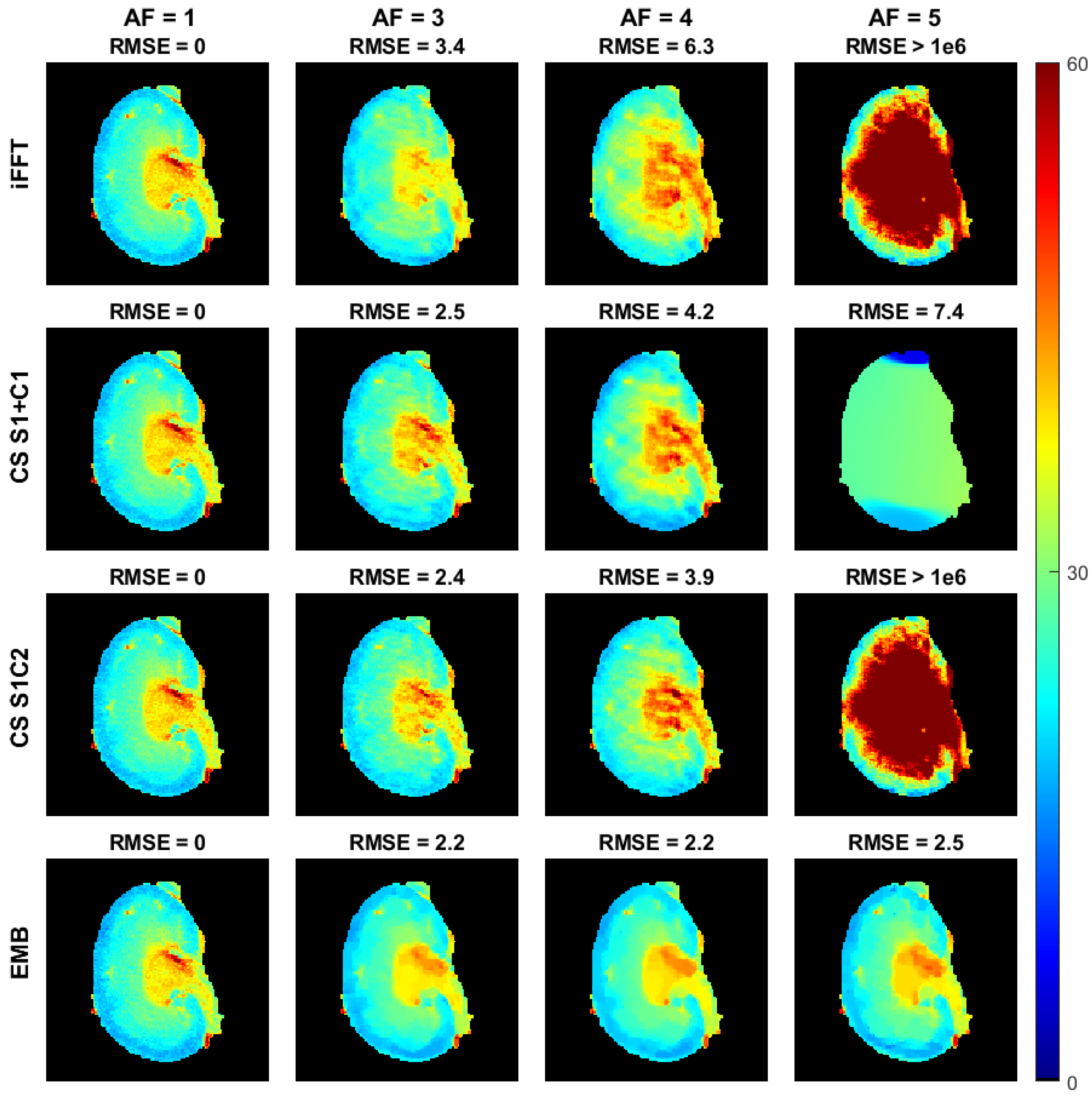
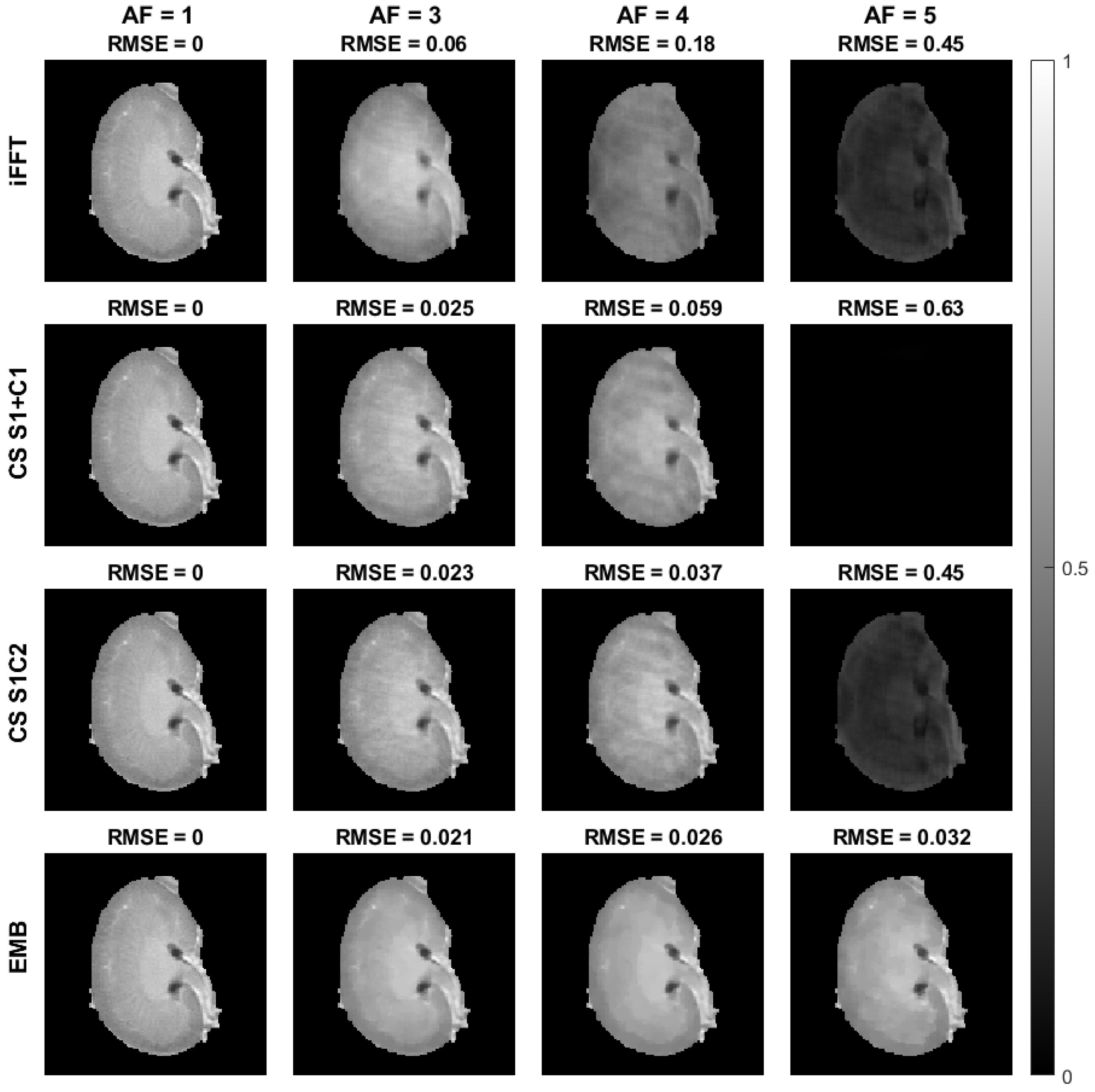
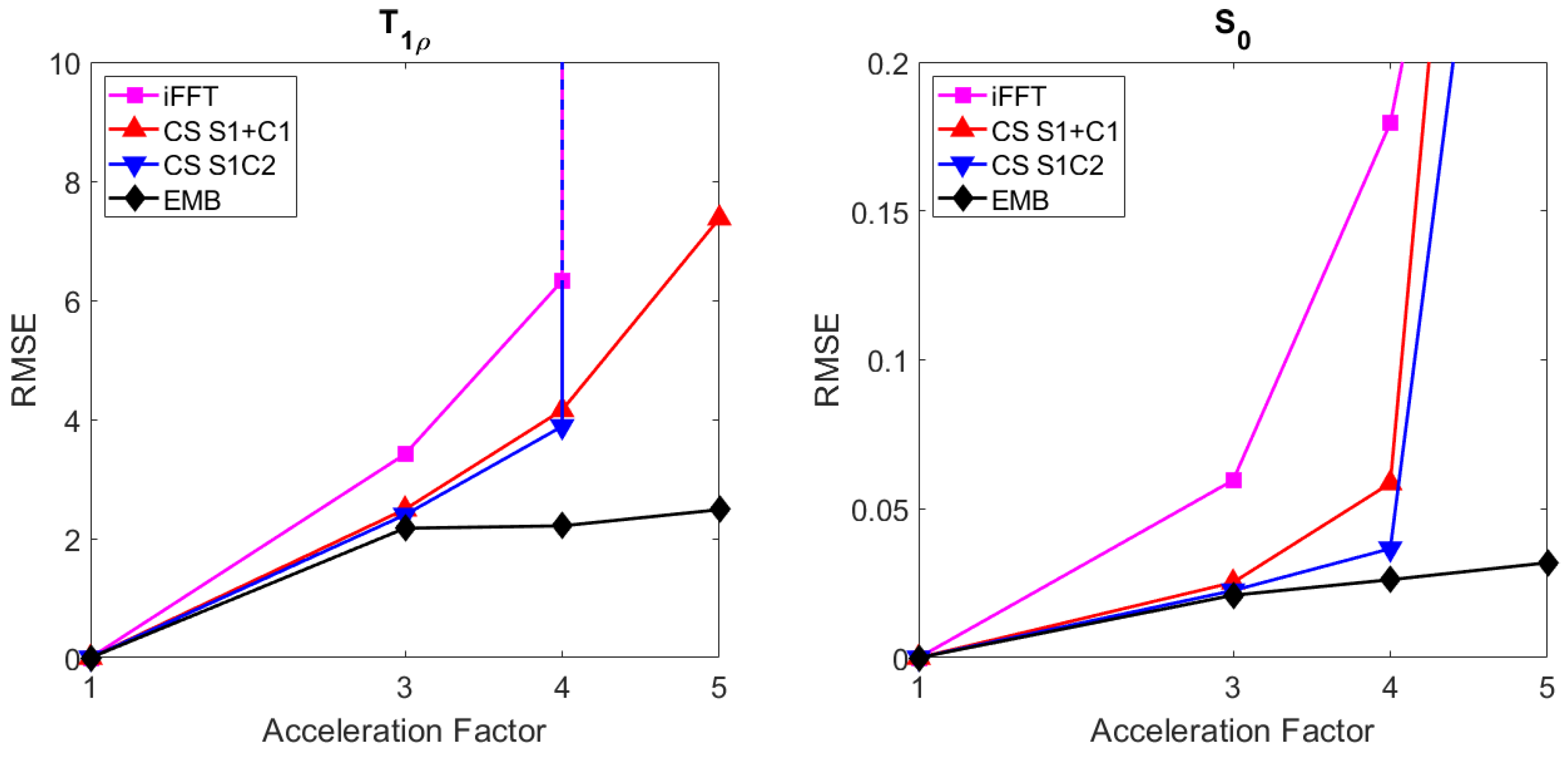
Quantitative MRI (qMRI) methods allow reducing the subjectivity of clinical MRI by providing numerical values on which diagnostic assessment or predictions of tissue properties can be based. However, qMRI measurements typically take more time than anatomical imaging due to requiring multiple measurements with varying contrasts for, e.g., relaxation time mapping. To reduce the scanning time, undersampled data may be combined with compressed sensing (CS) reconstruction techniques. Typical CS reconstructions first reconstruct a complex-valued set of images corresponding to the varying contrasts, followed by a non-linear signal model fit to obtain the parameter maps. We propose a direct, embedded reconstruction method for T1ρ mapping. The proposed method capitalizes on a known signal model to directly reconstruct the desired parameter map using a non-linear optimization model. The proposed reconstruction method also allows directly regularizing the parameter map of interest and greatly reduces the number of unknowns in the reconstruction, which are key factors in the performance of the reconstruction method. We test the proposed model using simulated radially sampled data from a 2D phantom and 2D cartesian ex vivo measurements of a mouse kidney specimen. We compare the embedded reconstruction model to two CS reconstruction models and in the cartesian test case also the direct inverse fast Fourier transform. The T1ρ RMSE of the embedded reconstructions was reduced by 37–76% compared to the CS reconstructions when using undersampled simulated data with the reduction growing with larger acceleration factors. The proposed, embedded model outperformed the reference methods on the experimental test case as well, especially providing robustness with higher acceleration factors.
Keywords: compressed sensing; embedded reconstruction; model-based reconstruction; quantitative MRI; T1rho mapping compressed sensing; embedded reconstruction; model-based reconstruction; quantitative MRI; T1rho mapping