Currently, a-Si detectors are widely used for digital radiography, conventional intraoperative fluoroscopy and cone beam CT (CBCT) [
6,
13,
14,
15,
16,
17]. However, a-Si detectors have demonstrated reduced performance parameters, especially under low-dose conditions [
5,
9]. Previous studies have reported technical advantages of CMOS and IGZO detectors over a-Si detectors in terms of reduced electronic noise and increased DQE [
5,
7,
9,
18]. However, a direct comparison of CMOS and IGZO detectors is lacking. The present study demonstrated a higher observer preference and improved subjective image quality ratings for C-arm systems equipped with CMOS and IGZO detectors compared with the a-Si-based system. Final image quality is influenced by several factors and cannot be attributed to detector technology alone. Although comparable acquisition presets and identical temporal-filter settings were used, not all image acquisition and post-processing parameters could be standardized. Automatic exposure control determines parameters such as tube voltage and tube current-time product according to object attenuation and the selected acquisition preset. Although automatic exposure control was enabled for all acquisitions and similar dose presets were used, the underlying control algorithms were proprietary and could not be standardized. Similarly, the post-processing algorithms were system-specific and may have affected perceived noise, sharpness, contrast and overall image appearance. Therefore, the findings should be interpreted as a comparison of complete C-arm systems rather than an isolated comparison of detector technologies.
4.1. CMOS/a-Si Comparison
Sheth et al. performed a technical analysis and subjective image quality comparison of CMOS and a-Si detectors in mobile C-arms. Electronic noise was reduced in CMOS compared to a-Si detectors. Additionally, DQE was higher for CMOS compared to a-Si at low-dose levels while a-Si showed a higher DQE at high-dose levels and low frequencies [
5].
Despite the technical advantages, no perceived difference was observed for tasks in orthopedic surgery, although the visibility of fine details in low-dose fluoroscopy was rated superior for CMOS compared to a-Si [
5].
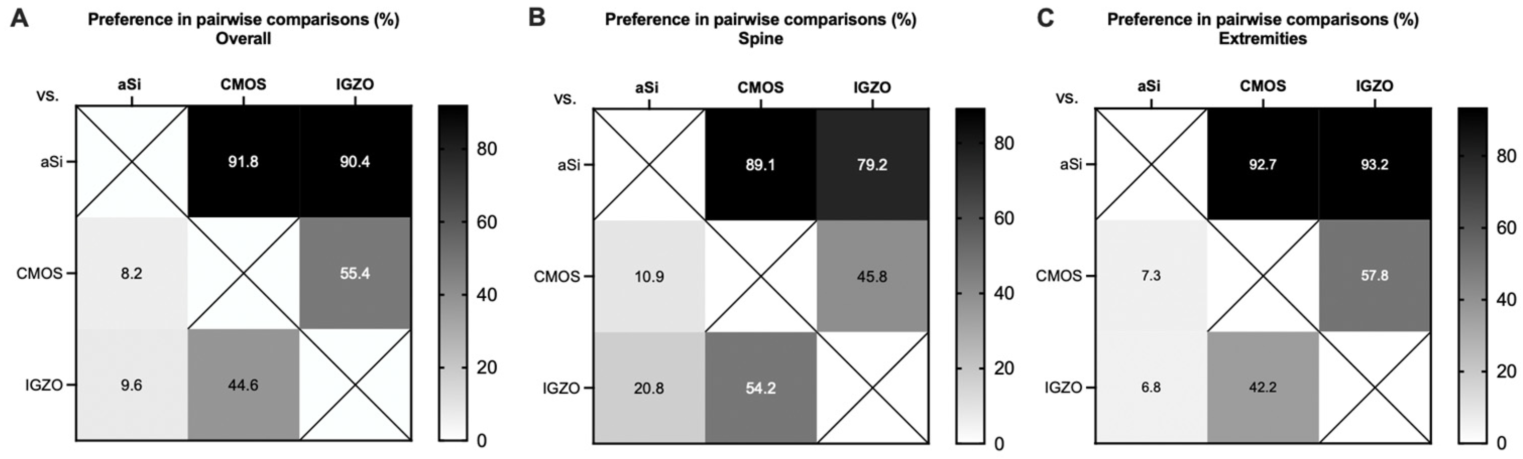
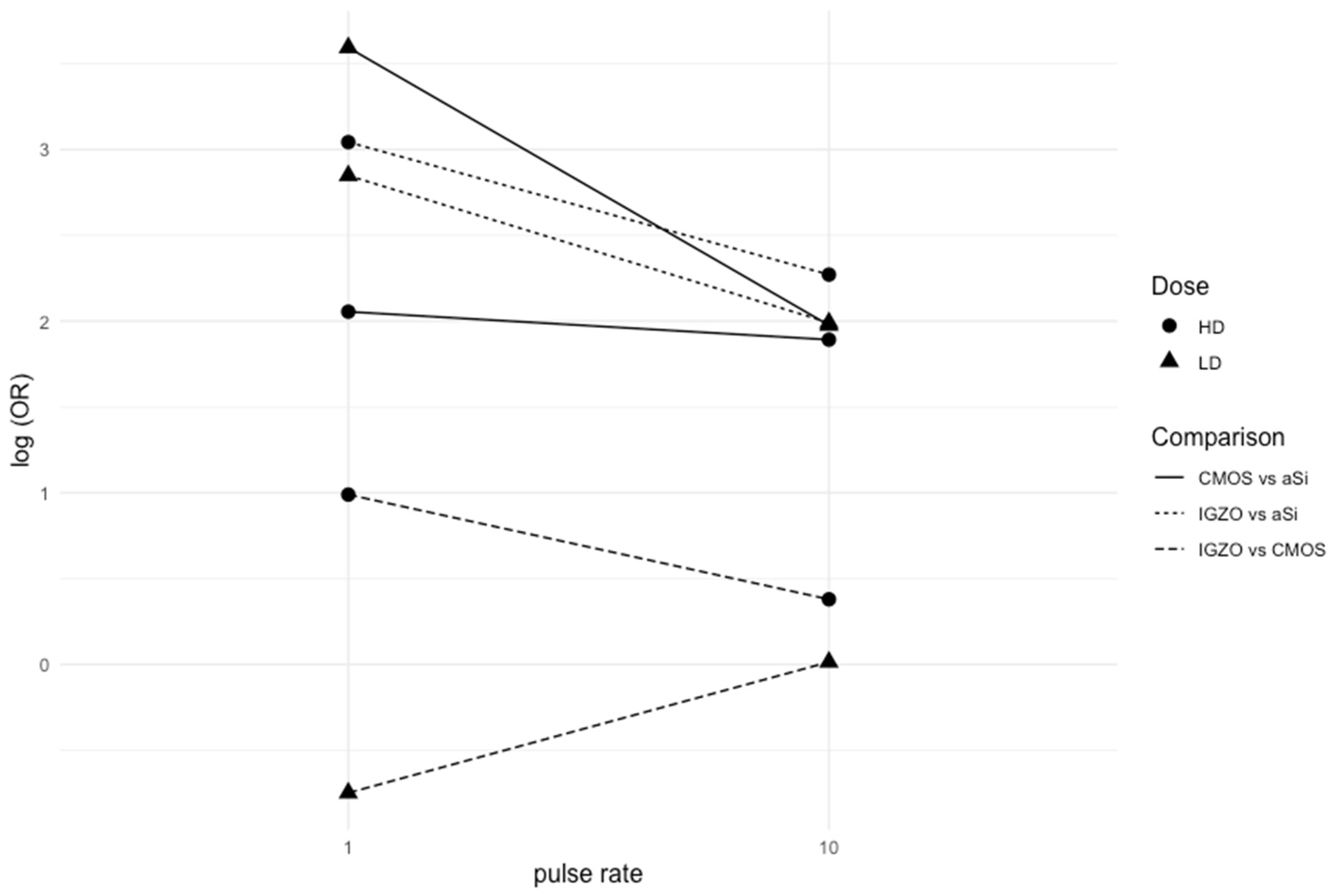
Abiola et al. compared the preference of interventional radiologists for specific tasks between systems equipped with CMOS and a-Si detectors for 2D and 3D imaging. Fluoroscopy images acquired using a CMOS detector were preferred over a-Si, especially in low-dose settings, while no significant difference was observed with high-dose settings [
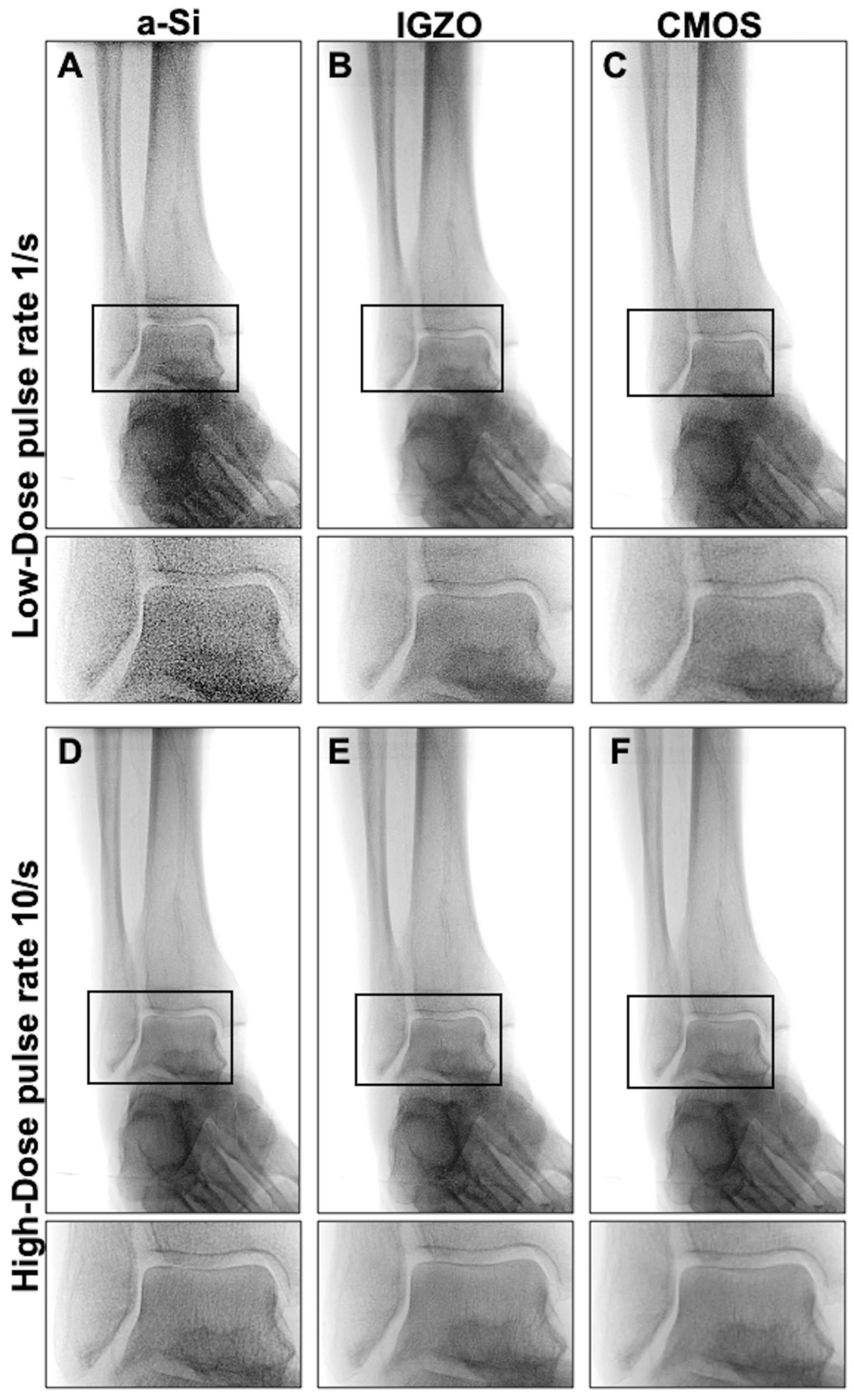
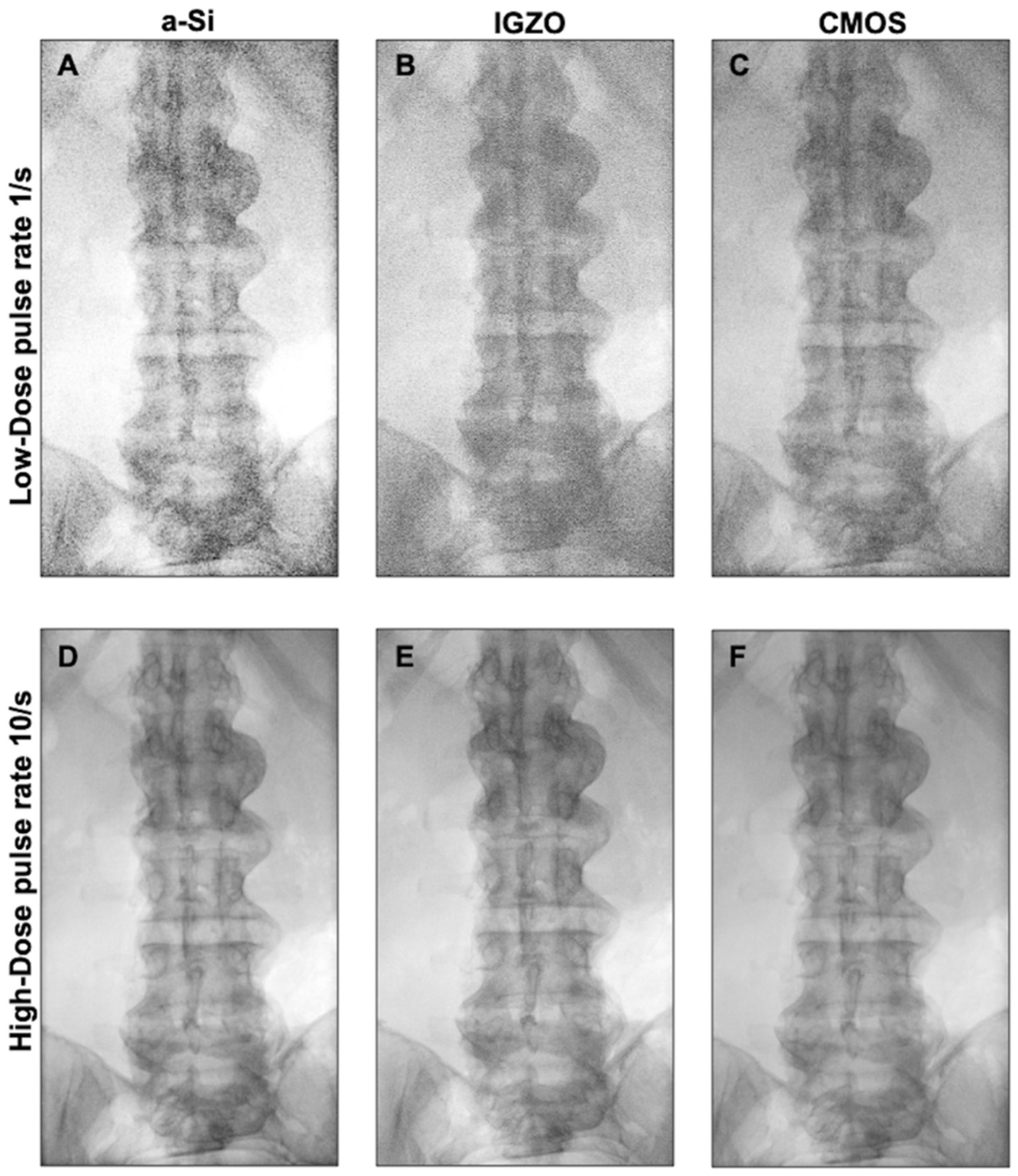
10]. The present study also demonstrated a stronger preference for CMOS- over a-Si-based systems in low-dose compared to high-dose settings, especially at low pulse rates. At higher pulse rates, this difference was reduced, possibly due to the influence of temporal filtering, which may decrease perceived image noise [
19]. Since only frames acquired one second after the initiation of fluoroscopy were analyzed, the number of frames available for temporal filtering differed among the pulse rate settings. At a pulse rate of 1/s, temporal noise reduction may not have reached a steady state, possibly contributing to higher perceived image noise at low pulse rates [
19,
20,
21]. Accordingly, subjective ratings of image noise for individual images in this study were also lower at higher pulse rates. Overall, these results suggest that systems equipped with CMOS detectors might be preferred over a-Si-based systems in low-dose settings, consistent with previously reported technical findings [
5].
4.3. IGZO/CMOS Comparison
Direct comparisons of IGZO and CMOS detectors are currently lacking. Previous studies indicate that the electronic noise performance of IGZO detectors is superior to that of a-Si detectors but inferior to that of CMOS detectors [
9]. However, at higher exposure levels, CMOS detectors have demonstrated reduced DQE compared to a-Si, while IGZO detectors showed equivalent or increased DQE under these conditions [
5,
9]. A-Si detectors exhibit a lower charge mobility compared to CMOS and IGZO FPDs, contributing to higher electronic noise and slow readout rates [
5,
6,
7].
No significant overall preference was observed between the IGZO- and CMOS-equipped systems in the present study. For extremity images, the IGZO-based system was preferred over the CMOS-based system, however the effect was modest (OR 1.33, 95% CI 1.02–1.74).
According to manufacturer specifications, the IGZO-based system evaluated in the present study provides a higher spatial resolution in overview fluoroscopy mode (2.2 lp/mm) compared with the CMOS-based system (1.8 lp/mm), which may partly relate to differences in effective pixel size. In addition, manufacturer-reported DQE values are higher for the IGZO detector (approximately 80%) than for the CMOS detector (approximately 72%). These technical characteristics may have contributed to the observed preference for IGZO over CMOS in extremity imaging, where the visibility of small details may be particularly important. However, this explanation remains speculative because the systems also differed in image processing and the independent contributions of spatial resolution, DQE and other system-specific factors could not be determined.
4.4. Clinical Implications
In intraoperative fluoroscopy, minimizing radiation exposure is important, as both patients and operating room staff are exposed. Previous studies have shown an increased prevalence of malignancies among orthopedic surgeons [
22,
23,
24]. Therefore, optimizing image quality under low-dose conditions represents an important objective in the development of C-arm systems. Radiation exposure to patients and medical staff can be reduced by using pulsed fluoroscopy and lower pulse rates [
25,
26]. Pulse rates between 5 and 15/s have been reported for orthopedic procedures [
27,
28].
Median clinical-assessability ratings indicated that images were sufficient for intraoperative assessment under all evaluated conditions except for the a-Si- and IGZO-equipped systems at a pulse rate of 1/s under low-dose conditions. This may suggest that differences between the systems become more clinically apparent under low-dose conditions at low pulse rates. The reduced subjective image quality observed for the C-arm equipped with an a-Si detector in low-dose settings in this study is consistent with the higher susceptibility of a-Si detectors to electronic noise at low exposure levels reported in prior technical evaluations [
5,
9]. However, the present study did not evaluate whether these differences affected diagnostic accuracy or intraoperative decision-making.
It should be noted that additional factors, such as system cost, long-term detector durability, and CBCT performance, may also influence the choice of C-arm system. CMOS detectors have been reported to exhibit lower radiation resistance compared with a-Si detectors [
6].
This study only evaluated 2D imaging. Previous studies have demonstrated advantages of CMOS FPDs over a-Si FPDs for CBCT in terms of reduced noise and improved detectability of small features [
5,
15,
29]. IGZO FPDs have also demonstrated improved 3D imaging performance compared to a-Si FPDs [
9].
4.5. Limitations
This study was conducted using human specimens to allow for repeated imaging without radiation exposure to patients. However, this experimental setup does not fully reflect the clinical use of fluoroscopy in orthopedic and trauma surgery and further studies evaluating the effect on surgical decision making based on assessment of fracture reduction and implant positions are needed.
Image quality and observer preference were compared between three commercially available C-arm systems equipped with different detectors. Although the systems share a similar design and comparable presets were used, observed differences in image quality may reflect not only detector characteristics but also additional system-specific factors, including automatic exposure control and image post-processing. Therefore, the findings of this study represent a comparison of C-arm systems rather than an isolated assessment of detector performance. Additionally, all evaluated C-arms were produced by a single manufacturer, which limits the generalizability of the findings to systems from other manufacturers because of potential differences in system design, automatic exposure control and image-processing algorithms.
Pixel size differed across the three systems and may have contributed to differences in perceived image quality. The effective pixel size of the IGZO detector was smaller (0.218 mm) compared to the effective pixel sizes of the CMOS (0.304 mm) and a-Si (0.388 mm) detectors. These variations may have contributed to differences in spatial resolution and perceived image noise between the C-arm systems. However, their independent contribution could not be determined.
This study evaluated only subjective image quality. No objective image-quality metrics, such as modulation transfer function, DQE or noise power spectrum, were obtained as independent references for the observed differences between imaging conditions, limiting direct comparison with previous technical assessments.
Image assessment was performed by two trauma surgery residents, experienced in interpreting intraoperative fluoroscopic images and subjective image assessment. Although the observers represented the intended clinical users, the limited number and experience range of observers restrict the generalizability of the findings.
Observers agreed on the preferred image in pairwise comparisons in 77.5% of cases.
Inter-rater reliability for the Likert-scale ratings ranged from moderate to substantial, likely reflecting individual preferences in subjective image-quality assessment. Still, this variability should be considered when interpreting the results, particularly given the limited number of observers.
No formal sample-size or power calculation was performed because of the exploratory nature of the study and the absence of sufficient preliminary data. The findings should therefore be considered exploratory and require confirmation in larger studies involving additional specimens and observers with different levels of experience.
Bradley–Terry models have been applied in radiological studies to analyze forced-choice pairwise comparisons of image quality [
30,
31,
32,
33]. Previous studies have suggested that forced-choice comparisons are superior in detecting small differences in perceived image quality and may provide better inter- and intraobserver agreement compared to Likert scales [
12,
34,
35]. Still, Likert scales are frequently used to assess subjective image quality of fluoroscopic images [
36,
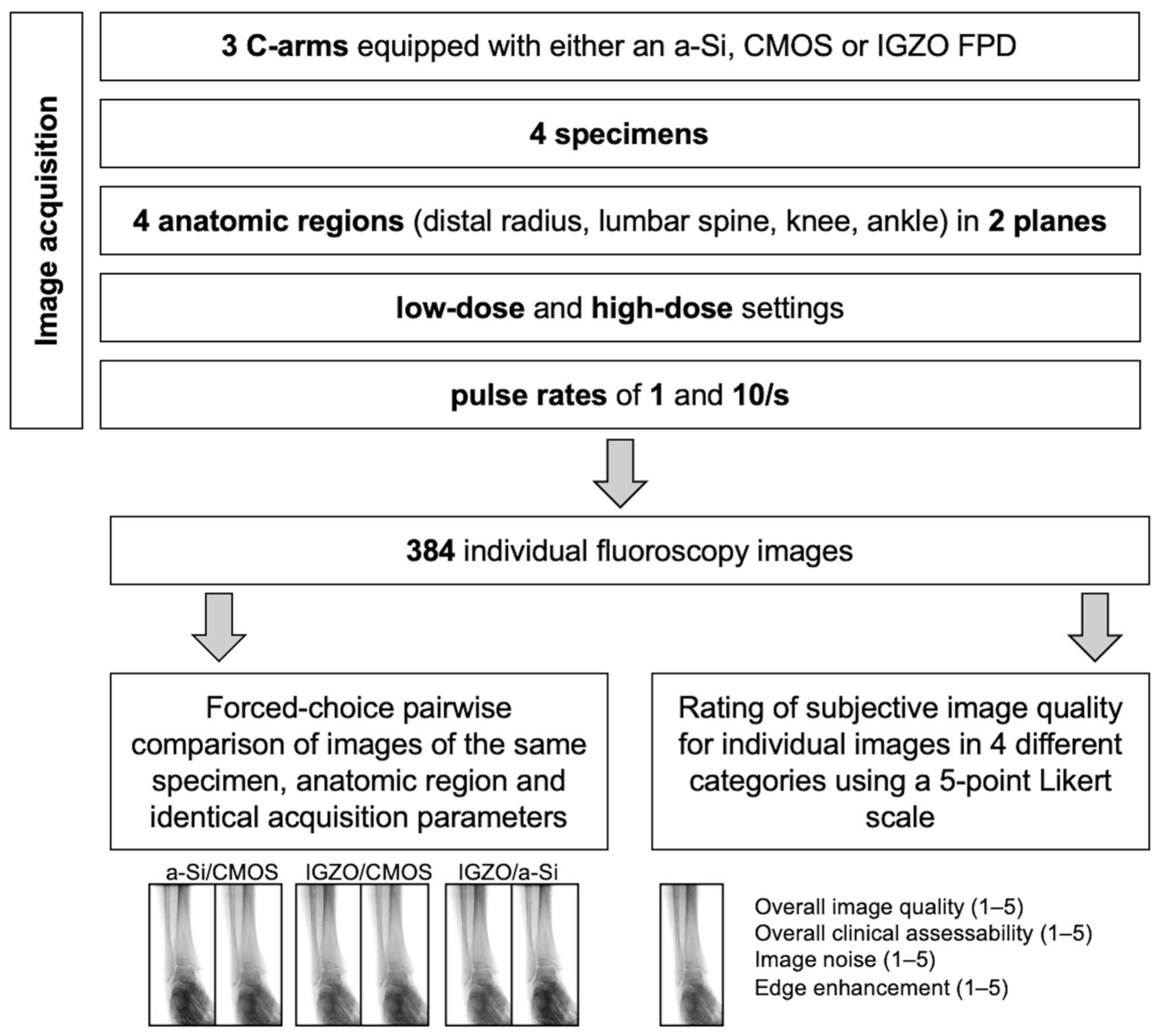
37]. Therefore, pairwise forced-choice comparisons were used to assess preference in this study, while Likert scale ratings were used to evaluate specific image quality categories.




 ,
,