Article

Enriched Synthesis of Magnetosomes by Expanding the Magnetospirillum magneticum AMB-1 Culture at Optimal Iron Concentration

1
College of Chemical Engineering, Huaqiao University, Xiamen 361021, China
2
Fujian Provincial Key Laboratory of Biochemical Technology, Huaqiao University, Xiamen 361021, China
3
Institute of Pharmaceutical Engineering, Huaqiao University, Xiamen 361021, China
*
Author to whom correspondence should be addressed.
Magnetochemistry 2021, 7(8), 115; https://doi.org/10.3390/magnetochemistry7080115
Submission received: 15 June 2021 / Revised: 5 August 2021 / Accepted: 9 August 2021 / Published: 11 August 2021

Abstract

:
The Magnetospirillum magneticum AMB-1 species is one of the most widely used magnetotactic bacterial strains for producing magnetosomes under laboratory conditions. Nevertheless, there exist several challenges in expanding and purifying the AMB-1 culture due to the restricted culture conditions. In an attempt to enrich the production of magnetosomes, this study reports the utilization of fermenter culture, which substantially promotes the cell densities at different concentrations of iron content. The experimental results confirmed magnetosomes’ high yield (production rate of 21.1 mg L−1) at the iron content of 0.2 μmol L−1. Moreover, different characterization techniques systematically confirmed the coated lipid membrane, particle size, dispersity, stability, and elemental composition of magnetosomes. Notably, the fermenter culture-based process resulted in highly discrete, dispersed, and stable magnetosomes with an average particle diameter of 50 nm and presented the integrated lipid membrane around the surface. The chemical composition by EDS of magnetosomes represented the presence of various elements, i.e., C, O, Na, P, and Fe, at appropriate proportions. In conclusion, the culture method in our study effectively provides a promising approach towards the culture of the magnetotactic bacterium for the enriched production of magnetosomes.

Graphical Abstract

1. Introduction

Since the first report by Blakemore in 1975 [1], magnetotactic bacteria, aquatic prokaryotes that respond to the Earth’s geomagnetic field, have garnered enormous interest due to their ability to synthesize intracellular organelles magnetosomes [2,3]. These subcellular membrane-bound constituents are composed of nanometer-sized, iron-based magnetite (Fe3O4) or greigite (Fe3S4) [4,5] crystals, arranged as chains [6]. Notably, the magnetotactic bacteria are predominantly distributed in the freshwater sediment, predominantly in the suboxic zone [7]. Moreover, the biodiversity of magnetotactic bacteria in the tropical marine environment and volcanic lake sediments has been reported [8,9]. Owing to these specialized magnetosome-based architectures, magnetotactic bacteria have been used for widespread applications, including but not limited to environmental pollution [10], the development of magnetic materials, iron biomineralization, and potential applications in modern biomedicine [6,11,12,13].
Despite the success as a promising material in different fields, most of the research studies on magnetotactic bacteria have been centered on the structural elucidation of the magnetic nanoparticles, synthesis mechanism, biocompatibility, and other physicochemical attributes. Contrarily, several attributes relevant to synthesis with fewer steps and the enrichment of the production of magnetosomes by expanding the cultivation and product extraction, especially on AMB-1, yet remain to be resolved. In general, Magnetospirillum gryphiswaldense (MSR-1), often selected as a magnetotactic model, can be grown in simple liquid media containing short organic acids as a carbon source. Several efforts have been dedicated to improving the yields of magnetosomes by optimizing the culture media and culture conditions [14]. In one instance, the mass cultivation of the microaerophilic M. gryphiswaldense in flasks and a fermenter was reported to establish the optimum condit321ions [15]. In another case, Heyen and Schuler demonstrated that the production rate of magnetosomes using different strains followed the order of MSR-1 > AMB-1 > Magnetospirillum magnetotacticum (MS-1) under the same conditions [16]. Matsunaga and colleagues’ studies indicated that the generation of bacterial magnetic particles (BMPs) by AMB-1 cultivated in the fed-batch culture was seven-fold higher than those cultivated in the batch culture [17]. Further, it was observed that the continuous feeding of ferric quinate as an iron source significantly enhanced the culture growth and, subsequently, improved the magnetosomes production. The magnetosomes yield by fed-batch culture of AMB-1 was about three-fold higher than those obtained by batch culture [18]. Similarly, Yang and coworkers conducted an optimization study to improve the yield of BMPs by adjusting the addition of ferric iron content. The experimental results indicated that feeding ferric quinate at a rate of 15.4 μg/min resulted in the highest BMPs yield of 7.5 mg/L [19]. Marcano and colleagues designed two time-resolved experiments to demonstrate that biogenesis during the exponential growth phase enhanced the production of magnetosomes in M. gryphiswaldense [20]. Interestingly, Yan and colleagues employed the response surface methodology (RSM) integrated with a desirability function approach for concurrent expansion of cell and magnetosome yield by Acidithiobacillus ferrooxidans [21].
Motivated by these aspects, in this study, we demonstrate a promising approach for the enrichment of magnetosomes’ synthesis by expanding the culture of magnetotactic bacterium AMB-1. Further, the effect of iron source on the yield of magnetosomes was investigated in the fermenter-mode culture and then compared the results with the flask culture. We believe that the improved product will undoubtedly be beneficial for the future applications of magnetosomes.

2. Materials and Methods

2.1. Materials

Magnetospirillum magneticum (ATCC® 700264™) AMB-1 strain was purchased from the American type culture collection (ATCC, Manassas, VA 20108, USA).

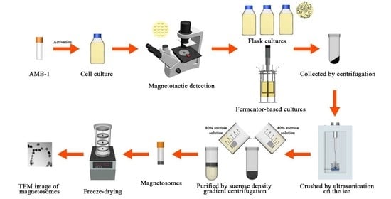
2.2. Culture of the AMB-1 Magnetotactic Bacteria

After activation, the bacteria strain was cultivated in microaerobic conditions by diluting it in a volume of 100 mL of the sterile ATCC® Medium 1653 (For detailed composition, please see the ATCC® website at www.atcc.org (accessed on 8 September 2015)) and incubated at 30 °C for 48 h. It should be noted that the culture medium has not been degassed but is closed to avoid contact with oxygen [22]. Further, the cells were inoculated into a glass vessel containing 250 mL of sterile culture medium. After 48 h, the mixture (25 mL) was transferred to the sterilized culture medium (500 mL) at a 5% (v/v) inoculation rate. When the bacteria entered the logarithmic growth phase, it could be used as an initial seed culture for flask and fermenter cultures. Magnetotactic detection of strains was confirmed by applying an external magnetic field. The growth curve and magnetic response (Cmag, coefficient of magnetically induced differential light scattering [23]) of the cells were then recorded to determine the optimal culture time of AMB-1 [24].

2.3. Comparison of AMB-1 in the Flask and Fermenter-Based Cultures

The production rates of magnetosomes in the flask and fermenter-based cultures were compared. In this experiment, the seed culture (5%, v/v) was inoculated into 1 L of medium (pH 6.75) in the flask-mode and 3 L of medium (pH 6.75) in the fermenter-mode cultures (BIOSTAT A plus, Germany, the maximum volume of 5 L), and incubated under similar conditions for further experiments. During the fermenter culture, 3 mL of the bacteria suspension was sampled from the fermenter tank every 2 h. The changes in the bacterial growth density were determined (data not shown). Bacterial suspensions were collected using centrifugation after entering the stationary phase. Further, the magnetosomes were crushed using ultrasonication and purified using the sucrose density gradient centrifugation approach [25,26]. The purified magnetic nanoparticles were dehydrated, and the dry weight of magnetosomes was recorded after freeze-drying.

2.4. Optimization of Iron Content in the Fermenter Culture

Herein, considering the ferriferous oxide as the main component of the magnetosome, the iron content in the fermenter culture was optimized. Ferric quinate, selected as the iron source, was added into the bacteria solution at the iron concentrations of 0.02, 0.2, and 2 μmol L−1 after the AMB-1 strain was inoculated into the culture medium in the fermenter at 30 °C for 48 h, respectively. Bacterial suspensions were collected, and magnetosomes were crushed and purified as mentioned above (see Section 2.3). Finally, the cumulative yield of magnetosomes was calculated after purity assessment using an ultraviolet spectrophotometer at 260 and 280 nm.

2.5. Characterization of Magnetosomes

The colloidal stability and dispersibility attributes of magnetosomes were investigated using the zeta potential tests (zetaPALS). The purified magnetic nanoparticles were dispersed in ultra-pure water, and 1 mL was used for measurement. Morphological attributes of AMB-1 cells, the intracellular arrangement and the integrity of lipid membrane, and particle size of the magnetosomes, were detected using transmission electron microscope (TEM). To determine the elemental composition, the purified magnetic nanoparticles were analyzed using energy dispersive spectroscopy (EDS)-assisted field emission scanning electron microscopy (FE-SEM).

3. Results and Discussion

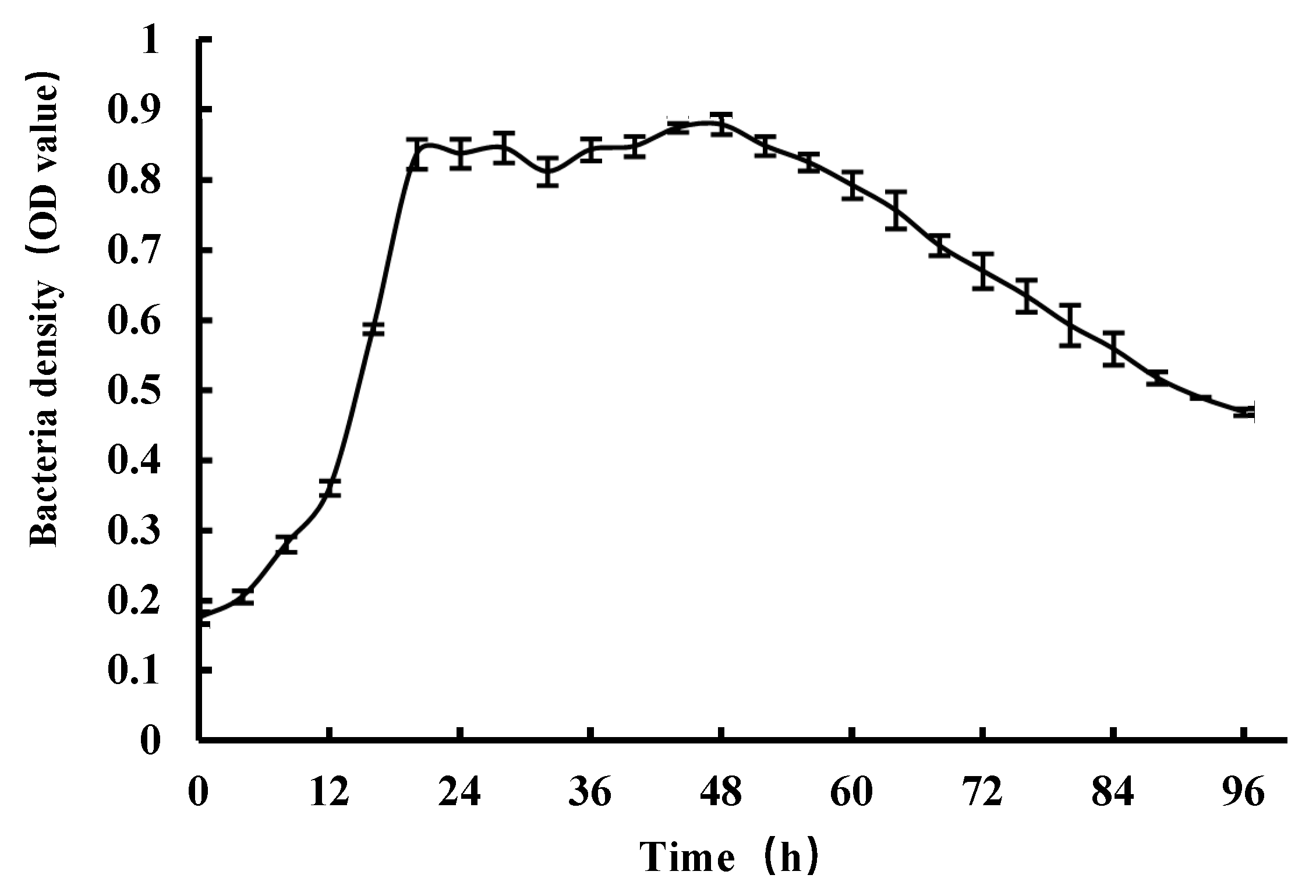
3.1. The Culture Conditions of the AMB-1 Magnetotactic Bacteria

The microscopic observations elucidated that the Magnetospirillum magneticum bacteria were whirling and certainly self-restrained in the direction of the applied magnetic field. Moreover, it was observed that most of the AMB-1 bacteria used in this study could synthesize magnetic nanoparticles with excellent magnetotactic properties. As shown in Figure 1, the growth curve of magnetotactic bacteria showed various phases with increasing time, including the initial lag phase, logarithmic growth phase, and the growth plateau in the time ranges of 0–4, 4–18, and 18–48 h, respectively. After 48 h, we observed that the density of cells commenced declining, representing the aging phase of growth. In this vein, the experimental results showing the magnetic sensitivity detection also represented that the value of Cmag decreased noticeably after 48 h (Figure 2). In general, magnetosomes are generated in the AMB-1 bacteria during the logarithmic growth phase under anaerobic conditions [17]. Therefore, the optimal culture time was set as 48 h to ensure the maximum density of cell bodies and the subsequent high yields of magnetosomes. The magnetotactic detection and magnetic sensitivity are provided in Supplementary Material Video S1, indicating the excellent growth trend of Magnetospirillum magneticum bacteria.

3.2. Yields of Magnetosomes by Flask and Fermenter Culture Approaches

After freeze-drying, the resultant yields of magnetosomes by flask and fermenter cultures were calculated by recording the wet and dry weights of the magnetosomes. It was observed from the experimental results that the yield of magnetosomes by the fermenter culture mode was two-fold more than that of the flask culture approach (Table 1). Therefore, we believe that it is feasible to improve the yield of magnetosomes using the fermenting tank method.

3.3. Optimization of Iron Source Content

Further, the yields of magnetosomes under different iron concentrations were enumerated. The wet weights of the magnetic nanoparticles obtained from the experiments by adding 0.02 and 0.2 μmol L−1 of iron content were around 190.1 and 235.0 mg, respectively, while the corresponding dry weights were 22.7 and 63.3 mg, respectively. The resultant concentrations of such samples were 7.6 and 21.1 mg L−1, respectively. However, the yield of magnetosomes was extremely low at the iron source concentration of 2 μmol L−1, which could not be weighed, indicating that the addition of an excessive iron source might not be absorbed by AMB-1 cells and cause slight toxic side effects. The experimental results in this study are in agreement with a report by Yang et al. In a previous study, the growth inhibition began to appear at iron feeding rates of 25.7 μg min−1, and significant inhibition was observed at 35.9 and 46.2 μg min−1. However, the growth inhibition at a lower iron concentration yet remained to be addressed [19].
Accordingly, the iron concentration of 0.2 μmol L−1 was selected as the optimal iron source content in the fermenter culture, which had resulted in the best yield of 21.1 mg L−1 among the three experiments (Table 2). Moreover, the obtained magnetosomes’ yield of 21.1 mg L−1 was higher than that of the previously reported yield values for AMB-1. To improve the AMB-1 magnetosomes’ yield using the fed-batch culture, Matsunaga and colleagues [17] developed an efficient method by adding nitric acids and succinate as nitrogen and carbon sources, respectively. The maximum magnetosomes’ yield of 4.5 mg L−1 was achieved at room temperature and a neutral pH. To further improve the yield, an advancement in the synthesis of continuously feeding ferric quinate was proposed, resulting in a significantly enhanced growth and a total magnetosome production of 6 mg L−1 [18]. In another case, after one day of culture (24 h), the production yield of AMB-1 magnetosomes was 3.3 mg L−1 under aerobic (0.25 mbar oxygen, pH 7) and strict anaerobic conditions in a five-liter batch bioreactor [16]. In another case, the synthesis was performed in a 10-liter jar, fed-batch, AMB-1 culture fermenter under microaerobic conditions, resulting in a maximum magnetosome yield of 7.5 mg L−1 at a ferric quinate feeding rate of 15.4 μg min−1 [19].

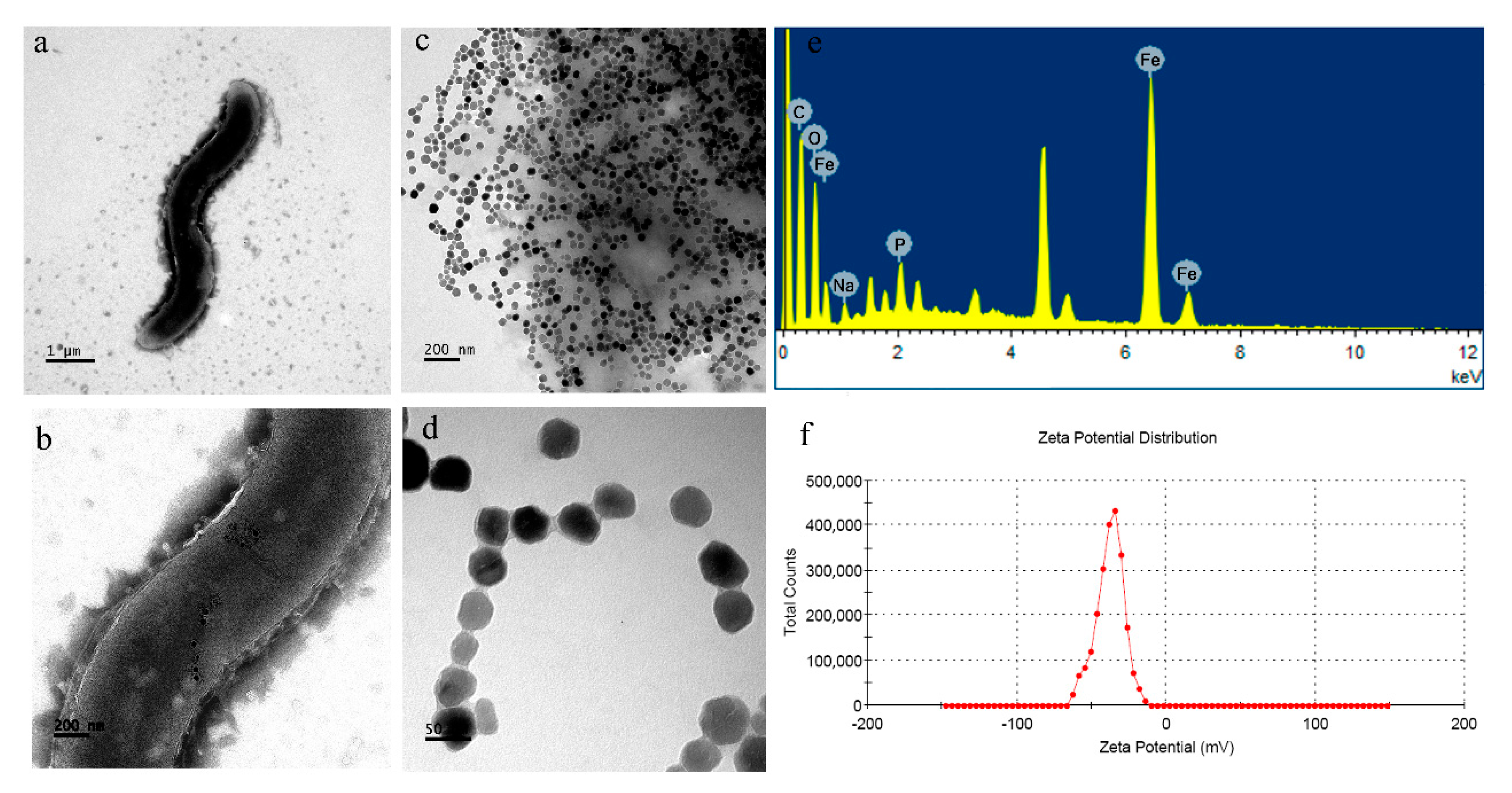
3.4. Characterizations

Furthermore, it was observed from the TEM images that the AMB-1 cells were spiral, with the length and width of the cell body at around 4 and 0.4–0.6 μm, respectively, and a flagellum at each end (Figure 3a). The TEM images presented that the resultant magnetosomes were aligned in a chain in the direction of the long axis intracellularly (Figure 3b), with an integrated lipid membrane on the surface. The average particle size was around 50 nm, with uniform particle size distribution and regular shape (Figure 3c,d), consistent with the reported literature [22,27].
FE-SEM and EDS observations demonstrated that the chemical composition of magnetosomes contained C, O, Na, P, and Fe elements (Figure 3e). However, the resulted composition in this study is not precisely the same as the magnetosome composition of the wild type magnetotactic bacteria, which could be attributed to pure cultivation [28]. Further, the zeta potential results showed that the resultant magnetosomes dispersed in water resulted in the negative zeta potential of −37.8 mV. The negative zeta potential value of the magnetosomes could improve their colloidal stability in water (Figure 3f) [22].

4. Conclusions

In conclusion, Magnetospirillum magneticum AMB-1 was successfully cultured. The experimental results showed that the magnetosomes’ yield of 21.1 mg/L in the fermenter culture at the iron concentration of 0.2 μmol L−1 was higher than that of the flask-mode culture. Moreover, the physical characterization results using TEM, EDS, and the zeta potential test showed that the magnetosomes were uniformly distributed and coated with a lipid membrane, with an average particle size of about 50 nm. These nanocomposites offered good dispersibility and stability, containing five elements, C, O, Na, P, Fe. Considering the experimental data, we believe that the culture method is a practically possible and convenient approach to culturing the magnetotactic bacterium for the enriched synthesis of magnetosomes.

Supplementary Materials

The following are available online at https://www.mdpi.com/article/10.3390/magnetochemistry7080115/s1, Video S1: Magnetosomes after sonification.

Author Contributions

Conceptualization, Y.H.; data curation, R.Y. and H.L.; funding acquisition, Y.L.; investigation, R.Y. and H.L.; methodology, Y.H. and R.Y.; project administration, Y.L.; supervision, R.K.K., and Y.L.; writing—original draft, Y.H.; writing—review and editing, Y.H. and R.K.K. All authors have read and agreed to the published version of the manuscript.

Funding

This work was supported by the National Marine Economic Innovation and Development Project (16PYY007SF17) and Program for Innovative Research Team in Science and Technology in Fujian Province University.

Data Availability Statement

The authors confirm that the data supporting the findings of this study are available within the article and supplementary material.

Conflicts of Interest

The authors declare no conflict of interest.

References

  1. Blakemore, R.P. Magnetotactic bacteria. Science 1975, 190, 377–379. [Google Scholar] [CrossRef]
  2. Bazylinski, D.A.; Frankel, R.B. Magnetosome formation in prokaryotes. Nat. Rev. Microbiol. 2004, 2, 217–230. [Google Scholar] [CrossRef] [Green Version]
  3. Jacob, J.J.; Suthindhiran, K. Magnetotactic bacteria and magnetosomes-scope and challenges. Mater. Sci. Eng. C 2016, 68, 919–928. [Google Scholar] [CrossRef] [PubMed]
  4. Bazylinski, D.A.; Frankel, R.B.; Heywood, B.R.; Mann, S.; King, J.W.; Donaghay, P.L.; Hanson, A.K. Controlled biomineralization of magnetite (Fe3O4) and greigite (Fe3S4) in a magnetotactic bacterium. Appl. Environ. Microbiol. 1995, 61, 3232–3239. [Google Scholar] [CrossRef] [Green Version]
  5. Farina, M.; Kachar, B.; Lins, U.; Broderick, R.; de Barros, H.L. The observation of large magnetite (Fe3O4) crystals from magnetotactic bacteria by electron and atomic force microscopy. J. Microsc. 1994, 173, 1–8. [Google Scholar] [CrossRef]
  6. Le Nagard, L.; Zhu, X.; Yuan, H.; Benzerara, K.; Bazylinski, D.A.; Fradin, C.; Besson, A.; Swaraj, S.; Stanescu, S.; Belkhou, R.; et al. Magnetite Magnetosome Biomineralization in Magnetospirillum magneticum strain AMB-1: A Time Course Study. Chem. Geol. 2019, 530, 119348. [Google Scholar] [CrossRef]
  7. Flies, C.B.; Jonkers, H.M.; de Beer, D.; Bosselmann, K.; Böttcher, M.E.; Schüler, D. Diversity and vertical distribution of magnetotactic bacteria along chemical gradients in freshwater microcosms. FEMS Microbiol. Ecol. 2005, 52, 185–195. [Google Scholar] [CrossRef]
  8. Tan, S.M.; Ismail, M.H.; Cao, B. Biodiversity of magnetotactic bacteria in the tropical marine environment of Singapore revealed by metagenomic analysis. Environ. Res. 2021, 194, 110714. [Google Scholar] [CrossRef]
  9. Xing, W.; Hu, H.; Zhang, Y.; Zhao, D.; Wang, W.; Pan, H.; Zhang, S.; Yan, L. Magnetotactic bacteria diversity of and magnetism contribution to sediment in Wudalianchi volcanic barrier lakes, NE China. Sci. Total Environ. 2020, 718, 137348. [Google Scholar] [CrossRef] [PubMed]
  10. Qu, Y.; Zhang, X.; Xu, J.; Zhang, W.; Guo, Y. Removal of hexavalent chromium from wastewater using magnetotactic bacteria. Sep. Purif. Technol. 2014, 136, 10–17. [Google Scholar] [CrossRef]
  11. Yan, L.; Da, H.; Zhang, S.; López, V.M.; Wang, W. Bacterial magnetosome and its potential application. Microbiol. Res. 2017, 203, 19–28. [Google Scholar] [CrossRef]
  12. Hafsi, M.; Preveral, S.; Hoog, C.; Herault, J.; Perrier, G.; Lefèvre, C.; Michel, H.; Pignol, D.; Doyen, J.; Pourcher, T.; et al. RGD-functionalized magnetosomes are efficient tumor radioenhancers for X-rays and protons. Nanomedicine 2019, 23, 102084. [Google Scholar] [CrossRef]
  13. Yadav, A.; Gerislioglu, B.; Ahmadivand, A.; Kaushik, A.; Cheng, G.J.; Ouyang, Z.; Wang, Q.; Yadav, V.S.; Mishra, Y.K.; Wu, Y.; et al. Controlled self-assembly of plasmon-based photonic nanocrystals for high performance photonic technologies. Nano Today 2021, 37, 101072. [Google Scholar] [CrossRef]
  14. Ali, I.; Peng, C.; Khan, Z.M.; Naz, I. Yield cultivation of magnetotactic bacteria and magnetosomes: A review. J. Basic Microbiol. 2017, 57, 643–652. [Google Scholar] [CrossRef]
  15. Claus, L.; Schüler, D. Biogenic nanoparticles: Production, characterization, and application of bacterial magnetosomes. J. Phys. Condens. Matter. 2006, 18, S2815. [Google Scholar]
  16. Heyen, U.; Schüler, D. Growth and magnetosome formation by microaerophilic Magnetospirillum strains in an oxygen-controlled fermentor. Appl. Microbiol. Biotechnol. 2003, 61, 536–544. [Google Scholar] [CrossRef]
  17. Matsunaga, T.; Tsujimura, N.; Kamiya, S. Enhancement of magnetic particle production by nitrate and succinate fed-batch culture of Magnetospirillum sp. AMB-1. Biotechnol. Tech. 1996, 10, 495–500. [Google Scholar] [CrossRef]
  18. Matsunaga, T.; Togo, H.; Kikuchi, T.; Tanaka, T. Production of luciferase magnetic particle complex by recombinant Magnetospirillum sp. AMB-1. Biotechnol. Bioeng. 2000, 70, 704–709. [Google Scholar] [CrossRef]
  19. Yang, C.; Takeyama, H.; Matsunaga, T. Iron feeding optimization and plasmid stability in production of recombinant bacterial magnetic particles by Magnetospirillum magneticum AMB-1 in fed-batch culture. J. Biosci. Bioeng. 2001, 91, 213–216. [Google Scholar] [CrossRef]
  20. Marcano, L.; García-Prieto, A.; Muñoz, D.; Barquin, L.F.; Orue, I.; Alonso, J.; Muela, A.; Fdez-Gubieda, M.L. Influence of the bacterial growth phase on the magnetic properties of magnetosomes synthesized by Magnetospirillum gryphiswaldense. Biochim. Biophys. Acta. Gen. Subj. 2017, 1861, 1507–1514. [Google Scholar] [CrossRef] [PubMed]
  21. Yan, L.; Zhang, S.; Liu, H.; Wang, W.; Chen, P.; Li, H. Optimization of magnetosome production by Acidithiobacillus ferrooxidans using desirability function approach. Mat. Sci. Eng. C 2016, 59, 731–739. [Google Scholar] [CrossRef]
  22. Alphandéry, E.; Guyot, F.; Chebbi, I. Preparation of chains of magnetosomes, isolated from Magnetospirillum magneticum strain AMB-1 magnetotactic bacteria, yielding efficient treatment of tumors using magnetic hyperthermia. Int. J. Pharm. 2012, 434, 444–452. [Google Scholar] [CrossRef] [Green Version]
  23. Schüler, D.; Uhl, R.; Bäuerlein, E. A simple light scattering method to assay magnetism in Magnetospirillum gryphiswaldense. FEMS Microbiol. Lett. 1995, 132, 139–145. [Google Scholar] [CrossRef]
  24. Lefevre, C.T.; Song, T.; Yonnet, J.P.; Wu, L.F. Characterization of Bacterial Magnetotactic Behaviors by Using a Magnetospectrophotometry Assay. Appl. Environ. Microbiol. 2009, 75, 3835–3841. [Google Scholar] [CrossRef] [PubMed] [Green Version]
  25. Xie, J.; Liu, X.; Liu, W.; Qiu, G. Extraction of Magnetosome from Acidithiobacillus ferrooxidans. Biomagnetism 2005, 5, 7–10. [Google Scholar]
  26. Fu, G.; Jiang, W.; Li, Y.; Sun, J.; Wang, Z.; Zhang, Y.; Pan, F.; Liu, W.; Luo, Y. Electron microscopic observation of magnetosome formation in magnetospirillum gryphiswaldense and its purification. China J. Mod. Med. 2004, 14, 45–49. [Google Scholar]
  27. Alphandéry, E.; Idbaih, A.; Adam, C.; Delattre, J.-Y.; Schmitt, C.; Guyot, F.; Chebbi, I. Chains of magnetosomes with controlled endotoxin release and partial tumor occupation induce full destruction of intracranial U87-Luc glioma in mice under the application of an alternating magnetic field. J. Control. Release 2017, 262, 259–272. [Google Scholar] [CrossRef] [PubMed]
  28. Liu, Y.; Gao, M.; Dai, S.; Peng, K.; Jia, R. Characterization of magnetotactic bacteria and their magnetosomes isolated from Tieshan iron ore in Hubei Province of China. Mater. Sci. Eng. C 2006, 26, 597–601. [Google Scholar] [CrossRef]
Figure 1. The growth curve of magnetotactic bacteria AMB-1.
Magnetochemistry 07 00115 g001
Figure 2. The magnetic response (Cmag) of magnetotactic bacteria AMB-1.
Magnetochemistry 07 00115 g002
Figure 3. Characterization of magnetosomes. (a,b) TEM images of Magnetospirillum magneticum, scale bars of (a) 1 μm, and (b) 200 nm, (c,d) TEM images of purified magnetosomes, scale bars of (c) 200 nm, and (d) 50 nm, (e) EDS spectra showing the elemental composition of magnetosomes, and (f) zeta potential distribution of magnetosomes dispersed in water.
Magnetochemistry 07 00115 g003
Table 1. Yields of magnetosomes by flask and fermenter cultures.
Flask-TypeFermenter-Type
Wet weight/mg37.178.8
Dry weight/mg0.32.2
Yield/mg L−10.30.7
Table 2. Yields of magnetosomes by fermenter culture under different iron concentrations.
0.02 μmol L−10.2 μmol L−12 μmol L−1
Wet weight/mg190.1235.0low
Dry weight/mg22.763.3low
Yield/mg L−17.621.1-
Publisher’s Note: MDPI stays neutral with regard to jurisdictional claims in published maps and institutional affiliations.

