Article

Hydrogen Peroxide-Assisted Hydrothermal Synthesis of BiFeO3 Microspheres and Their Dielectric Behavior

1
Department of Botany and Microbiology, College of Science, King Saud University, P.O. Box 2455, Riyadh 11451, Saudi Arabia
2
Department of Chemistry, School of Engineering, DayanandaSagar University, Bangalore 560 068, Karnataka, India
3
School of Chemical Engineering and Light Industry, Guangzhou 510006, China
4
Energy Materials Research Laboratory, Department of Chemistry, Siddaganga Institute of Technology, Tumkur 572103, India
*
Author to whom correspondence should be addressed.
Magnetochemistry 2020, 6(3), 42; https://doi.org/10.3390/magnetochemistry6030042
Submission received: 5 August 2020 / Revised: 2 September 2020 / Accepted: 4 September 2020 / Published: 9 September 2020
(This article belongs to the Special Issue Magnetic Nanoparticles 2020)

Abstract

:
Despite considerable efforts undertaken in a rapidly developing area of multiferroic research, synthesis of phase pure BiFeO3 is still a matter of intensive research. In this work, we report the shape-controlled synthesis of pure BiFeO3 microspheres via a facile hydrothermal route. The prepared BiFeO3 powder has been characterized using powder X-ray Diffraction (XRD), Differential Thermal analysis (DTA), Scanning Electron microscopy (SEM), and impedance spectroscopy. Powder XRD analysis confirms the formation of pure rhombohedrally distorted perovskite with R3c space group. Scanning electron micrograph revealed that the prepared BiFeO3 microspheres are nearly spherical in shape with uniform size distribution. The BiFeO3 microspheres exhibit a dielectric constant value of ~110 at 1000 KHz, which is higher than the BiFeO3 prepared by conventional solid-state reaction and sol–gel method. Variation of dielectric constant with temperature at different frequencies shows that the BiFeO3 has a dielectric anomaly of ferroelectric to paraelectric type at 1093 K and this phenomenon is well supported by TGA results.

1. Introduction

Nowadays, multiferroic (particularly BiFeO3) materials are being widely investigated for spintronics, data-storage, sensors and multiple-state memory applications [1] because of their dual ordering (i.e., both magnetic and polar) characteristics at room temperature. BiFeO3 crystallizes in a rhombohedral structure with R3c space group, and exhibits ferroelectric and antiferromagnetic properties at ~1100 and 630 K, respectively [2]. BiFeO3 generates spontaneous polarization, ~100 μC/cm2, from the lone pair of the Bi3+ ions, while the magnetic ordering originates from the superexchange interaction between the 3d electrons of the Fe3+ ions [3]. In recent years, BiFeO3 has attracted significant interest owing to its high Curie temperature (1083 K) [4] and high Neel temperature (625 K) and its potential applications, such as water splitting, organic pollutants degradation (2.2 eV), solar cells and visible light optoelectronic devices [5,6,7,8]. However, BiFeO3 finds limited application due to the high electric loss via leakage, existence of impurities, high electric coercive field, and weak magnetoelectric coupling due to the significant difference between the Curie temperature (1083 K) and the high Neel temperature (625 K), which leads to inadequate ferroelectric and magnetic properties in bulk BiFeO3. Different methods have been adopted in order to overcome the above limitations [2,3], including but not limited to different synthesis techniques, producing solid solution via rare earth ions doping at both Bi and Fe sites [2]. These methods can improve the stability, multiferroic features and magnetoelectric coupling effect of BiFeO3. The magnetoelectric coupling effect can be effectively altered by tuning Curie temperature and Neel temperature. It was reported that the incorporation of rare earth ion at Bi site resulted in the modification of the ferroelectric properties, whereas the rare earth ion at the Fe site alters the magnetic properties [3]. The preparation of doped and un-doped BiFeO3 via sol–gel and solid-state [1] reactions requires elevated temperature of 600–900 °C and complex/expensive reagents like citric acid and urea [9].
Therefore, there is an urgent need to develop new synthesis processes for obtaining pure BiFeO3, which is a challenging task even today due to the appearance of secondary phases in BiFeO3. It is very difficult to obtain single phase BiFeO3 because of non-stoichiometric oxygen in the structure, which leads to the formation of undesirable impurity phases such as BiFe4O9, Bi25FeO39 and Bi25FeO40 [10]. The presence of BiFe4O9, Bi25FeO39 and Bi25FeO40 impurities significantly reduces the thermal stability of BiFeO3, and they also increase the leakage current [11]. The synthesis of pure BiFeO3 is quite subtle, because it is necessary to take both kinetic and thermodynamic properties into account. In this view, considerable effort has been made in the past for the controlled synthesis of BiFeO3 owing to its size- and morphology-dependent magnetic, electrical, and optical properties [5,12,13,14,15,16]. However, the successive modulation of size and morphology of BiFeO3 has yet to become a reality.
In the present paper, as the synthesis technique and reaction parameters play a crucial role in controlling the material properties, we successfully applied the hydrothermal synthesis route for the fabrication of single phase BiFeO3 microspheres without any additional thermal treatment. The hydrothermal synthesis route possessed the potential to produce well-crystallized BiFeO3 with controlled morphology and narrow distribution of particle size. The BiFeO3 microspheres prepared by the hydrothermal method show higher dielectric constant value (~110 at 1000 KHz) than the BiFeO3 prepared by conventional solid-state reaction (~20 at 100 KHz) and sol–gel method (~68 at 1M Hz). Additionally, BiFeO3 has shown dielectric anomaly of ferroelectric to paraelectric type at 1093 K.

2. Experimental

2.1. Materials

The chemical reagents used in the work were bismuth nitrate (Bi(NO3)3·5H2O, 99%), Ferric nitrate (Fe(NO3)3·9H2O, 99%), Sodium hydroxide (NaOH, 90%), Potassium hydroxide (KOH, 90%), and Hydrogen peroxide (30% H2O2). All these analytical-grade purity chemicals were purchased from S D Fine-Chem Limited, India, and used without further purification.

2.2. Method

Amounts of 0.01 mol Bi(NO3)3·5H2O, 0.01 mol Fe(NO3)3·9H2O, 0.051 mol NaOH, 0.049 mol KOH were mixed with 12 mL acetone and stirred for 10 min. The reason for using high concentrations of NaOH and KOH was to sufficiently maintain the basic condition. During this, an exothermic reaction took place, and all the acetone evaporated. After thorough mixing, 4 mL H2O2 was added to provide an oxygen-enriched atmosphere to keep the Fe ion in Fe3+ form and convert hydroxide into oxide. The obtained resin-like suspension was poured into the Teflon-lined stainless-steel autoclave for hydrothermal treatment. The autoclave was sealed and maintained at 200 °C ± 5 for 24 h. After the completion of the reaction, the product was removed from the autoclave and washed with water and acetone several times and dried in a laboratory oven at 80 °C ± 5 for several hours.

2.3. Characterization

The structure and crystallinity of the prepared BiFeO3 microspheres were characterized by PANalytical X′pert X-ray diffractometer (XRD) equipped with graphite monochromatized CuKa radiation (Malvern Panalytical, Lelyweg, The Netherlands). Differential thermal analysis of BiFeO3 microspheres was carried out from room temperature to 900 °C in pure oxygen atmosphere at a scan rate of 10 °C/min using Q600. Scanning electron microscope (SEM) images were taken with a JEOL. The SEM images were recorded by adding a small amount of sample onto the carbon conductive tape, and then gold was spurred for 9 s. Then, the gold-spurred samples were examined using SEM. The densities of the BiFeO3 pellets (made with 2% polyvinyl alcohol as a binder) were measured using the Archimedes principle. The sintered BiFeO3 ceramic specimens were polished and then electroded with high-quality silver paint on both sides of the disk-shaped samples. The dielectric and electrical properties of the samples were measured using a Hewlett Packard 4194A impedance gain phase analyzer (Commonwealth of Massachusetts, Woburn) over wide ranges of frequency 100 Hz–10 MHz and temperature room temperature to 830 °C.

3. Results and Discussion

The phase composition and structure of the as-prepared BiFeO3 sintered at different temperatures were examined by powder XRD technique, and the observed XRD patterns are displayed in Figure 1. All the diffraction peaks in the XRD pattern of as-prepared BiFeO3 (prepared at 200 °C/24 h by hydrothermal method) were consistent with the standard Joint Committee on Powder Diffraction Standards No. 86-1518 of pure BiFeO3 having a rhombohedral crystal structure and R3c (161) space group. Additionally, the diffraction (012) peaks (2 theta = 22.34) of the as-prepared BiFeO3 are intense and sharp, indicating the formation of well-crystallized single phase BiFeO3. To investigate the effect of heat treatment on the BiFeO3 samples, the powders were pressed into pellets and heat-treated at different temperatures (at 600, 700 and 800 °C), as shown in Figure 1. It is observed that the sintered BiFeO3 pellets retain the rhombohedral structure with space group R3c at higher sintering temperature. The secondary phases associated with the BiFeO3 such as Bi2Fe4O9 and Bi25FeO40 are absent in the present study. This suggests the formation of crystalline single phase BiFeO3 with good thermal stability. The crystallite size (D) of BiFeO3 samples was calculated by following the Scherrer Formula.
D = (0.9 λ)/(β cosθ)
where D is the crystallite size, λ is the wavelength of radiation, β is the full width at half maximum (FWHM) of the diffraction peak in radian and θ is Braggs angle. The crystallite sizes are calculated by considering the prominent diffraction peaks peaked at 2θ = 22.34 and 45.73, corresponding to the (012) and (024) planes, respectively. The average crystallite size of the as-prepared, 600, 700, and 800 °C samples was 34 ± 1.24, 41 ± 0.47, 43 ± 1.67, and 47 ± 0.94 nm, respectively.
The formation of BiFeO3 under the present experimental conditions can be explained as follows. Bismuth hydroxide and iron hydroxides were formed when bismuth nitrate and iron nitrate were mixed with sodium hydroxide and potassium hydroxide at room temperature (Equations (2) and (3)). Then, under hydrothermal conditions, the formed Fe(OH)3 and Bi(OH)3 undergo condensation to form BiFeO3 microspheres (Equation (4)).
Bi 3 + ( aq )   +   3 OH ( aq )     Bi ( OH ) 3
Fe 3 + ( aq )   +   3 OH ( aq )     Fe ( OH ) 3
Bi ( OH ) 3 ( s )   +   Fe ( OH ) 3 ( s )     BiFeO 3 +   3 H 2 O ( 1 )
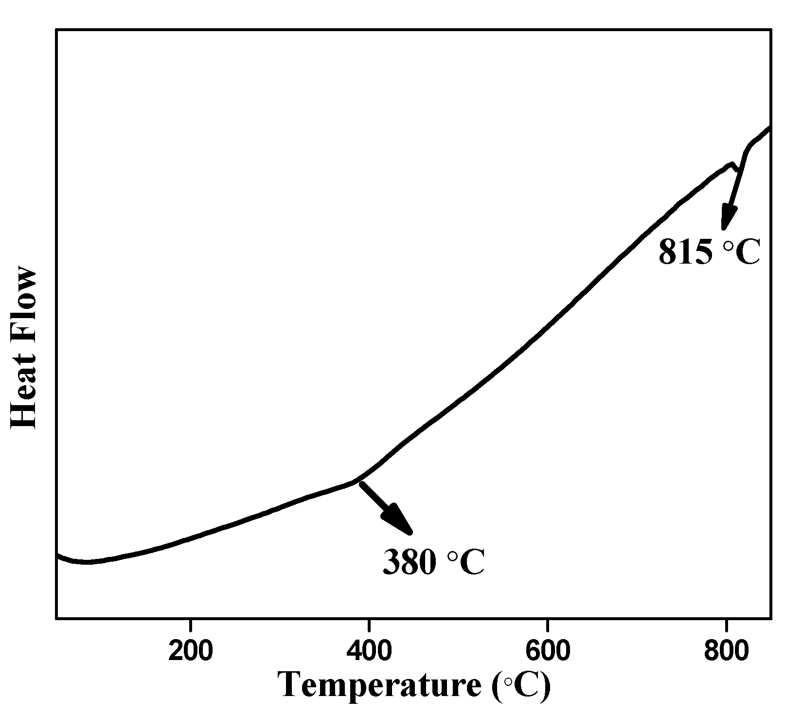
Figure 2 presents the DTA curve of the BiFeO3 microspheres recorded between the temperatures 25 to 850 °C. The endothermic peak appearing at around 1088 K is attributed to the phase transformation ferroelectric to paraelectric, i.e., Curie temperature [17]. This observed Curie temperature is in agreement with the reported values for BiFeO3 [18,19]. Furthermore, weak transition at 380 °C (653K) corresponds to antiferromagnetic phase transition.
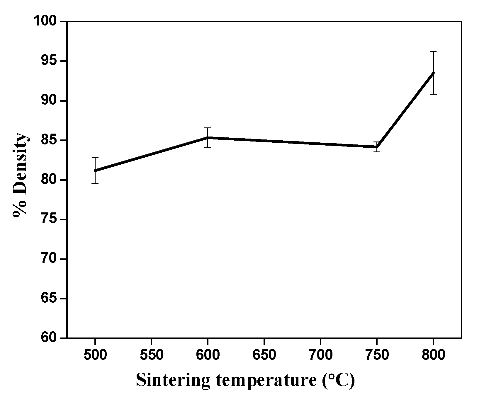
To study the dielectric permittivity, the density of the BiFeO3 was measured. The BiFeO3 powders were pressed into a pellet of diameter 9 mm. The pressed BiFeO3 pellets were submerged in water. The effective mass under water was determined by subtracting actual mass from the mass of the water displaced. Then, the volume of the BiFeO3 pellets was determined. Finally, the mass was divided by the volume to estimate the average density [20]. The effect of temperature on the density of the pellets can be clearly seen in Figure 3. The density of the prepared BiFeO3 pellets sintered at 500 °C, 600 °C, 750 °C and 800 °C was shown to be 81%, 85%, 83% and 94%, respectively. Therefore, with an increase in the sintering temperature, the density of the BiFeO3 increases, with the maximum being found to be ~94% at 800 °C. The observed density is h when the sample is sintered at 800 °C. The observed density, ~94%, is close to the BiFeO3 density (96%) reported by spark plasma sintering technique [21]. The lower density compared to the theoretical density may be due to the presence of little porosity in the pellets.
A typical scanning electron micrograph of the BiFeO3 powder prepared by the present method is shown in Figure 4. It was observed that the BiFeO3 powder exhibited a spherical structure with a rough surface. The average size of the BiFeO3 spherical structures was found to be in the range of 20–30 μm. Each sphere is composed of significantly smaller agglomerated particles. The spherical structures are formed due to the high oxygen partial pressure [22]. The formation of high oxygen partial pressure is due to the decomposition of molten nitrate during heat treatment in a closed container according to Equation (5). Similar results were reported by Xiaobo He et al. for the synthesis of BiFeO3 in presence of potassium and sodium nitrate [23].
2 NO 3 ( aq )     2 NO 2 ( g )   +   1 / 2 O 2 ( g )   +   O 2
The mixture KOH and NaOH was used for precipitation in our synthesis with the aim to carry out the reaction similar to molten metal flux method. Generally, melts can be made more acidic (H2O-rich) or basic (O2− rich) by controlling the water content of the melt, based on the product formation requirement. In our synthesis, both iron nitrate and bismuth nitrate dissolves and heating at 200 °C in air-tight autoclave resulted in slow loss of water as vapors, thereby making the melt more basic and the product insoluble [24]. In addition, the excess added NaOH and KOH maintain a basic pH.
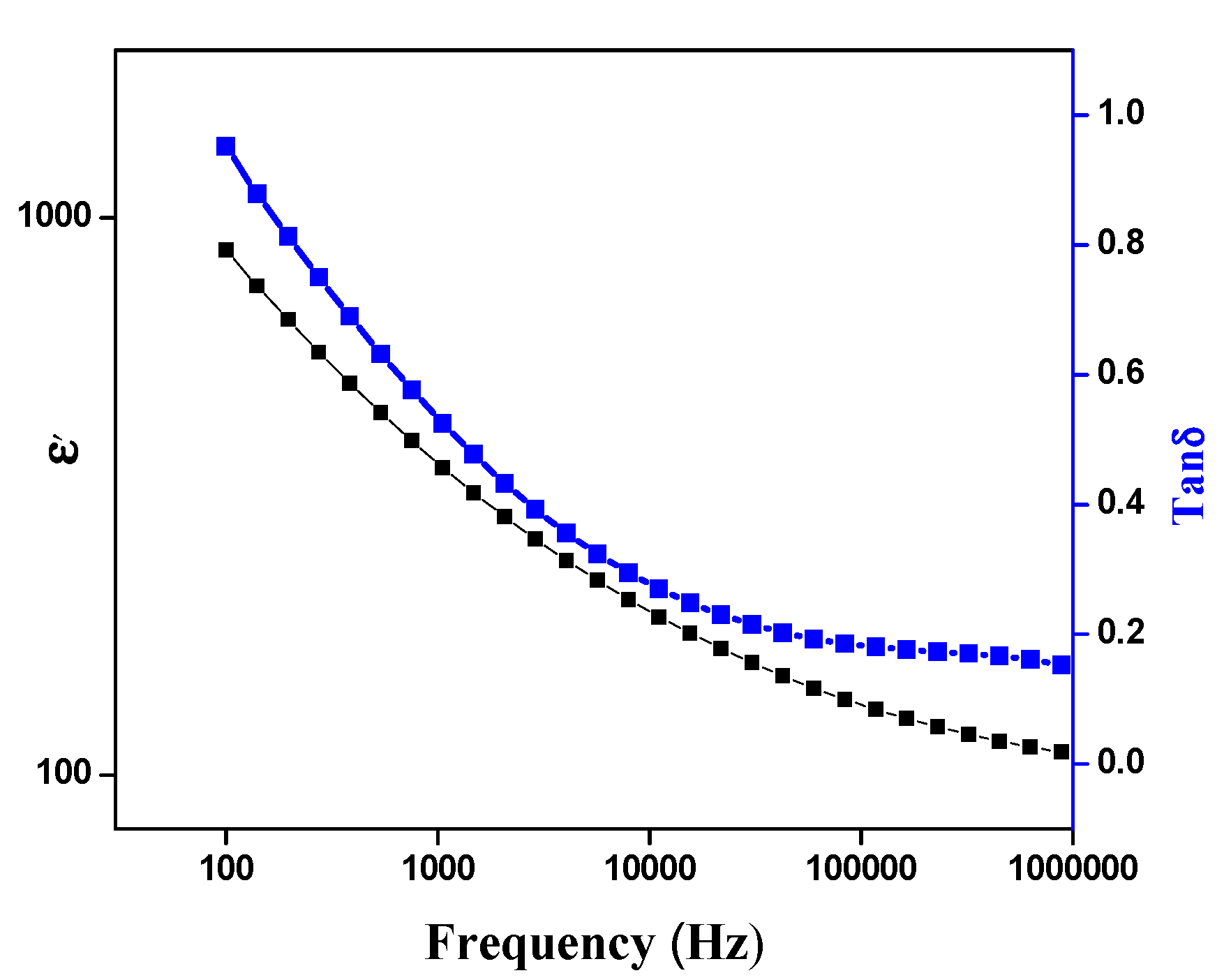
The effect of frequency on the dielectric constant and the dielectric loss factor of BiFeO3 are presented in Figure 5. The dielectric constant is found to decrease slowly when frequency increases from 100 Hz to 100 kHz, and then becomes almost constant, showing the usual dielectric dispersion [25]. It is observed from the figure that the prepared BiFeO3 microsphere exhibit a high dielectric constant value of ~113 ± 4.64 at 1000 KHz, which is significantly higher than the hydrothermally derived Cr-substituted BiFeO3 (50 at 0.2–1.2 MHz), and BiFeO3 prepared by conventional solid-state (~20 at 100 KHz) and sol–gel reaction (~68 at 1M Hz) [25,26,27]. Generally, the dielectric properties of the relaxors depend on the microstructural characteristics such as grain size, grain-boundary character and pores. Therefore, microstructural control with an appropriate choice of additive can be critical in the applications. Better powder characteristics, such as a relatively uniform distribution of particle size and less particle agglomeration, obtained by the present method are believed to contribute to the resultant dielectric properties.
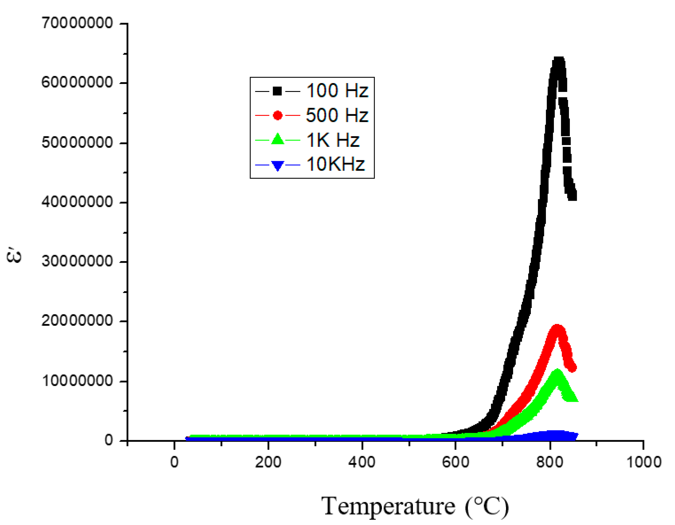
The effect of temperature on the dielectric constant is shown in Figure 6. The dielectric constant of the specimen was measured from room temperature to 850 °C at frequencies of 100, 500, 1000, and 10,000 kHz. It was found that the BiFeO3 has a dielectric anomaly at 1093 K (the Curie temperature, Tc) suggesting the occurrence of ferroelectric–paraelectric phase transition.

4. Conclusions

BiFeO3 microspheres were synthesized by direct precipitation followed by hydrothermal route from the mixture of iron nitrate and bismuth nitrate in the molten KOH/NaOH solution at temperature as low as 200 °C. The prepared BiFeO3 microspheres exhibit higher thermal stability up to 800 °C, and show larger dielectric constant values at different frequencies. The proposed method may be extended for the synthesis of other perovskite materials.

Author Contributions

Conceptualization, G.N. and A.S. (Asad Syed); methodology, A.S. (Ashoka Siddaramanna); software, A.S. (Ashoka Siddaramanna) and A.M.E.; formal analysis, A.S. (Ashoka Siddaramanna) and D.A.H.; investigation, A.S. (Ashoka Siddaramanna) and A.S. (Asad Syed); resources, G.N.; data curation, A.S. (Ashoka Siddaramanna), A.S. (Asad Syed) and G.N.; writing—original draft preparation, A.S. (Ashoka Siddaramanna), G.N.; writing—review and editing, A.S. (Ashoka Siddaramanna), A.S. (Asad Syed) and G.N.; supervision, A.S. (Asad Syed), and G.N.; project administration, G.N.; funding acquisition, A.M.E. All authors have read and agreed to the published version of the manuscript.

Funding

Researchers supporting project number (RSP-2020/56) King Saud University, Riyadh, Saudi Arabia.

Acknowledgments

The authors extend their appreciation to the researchers supporting project number (RSP-2020/56) King Saud University, Riyadh, Saudi Arabia.

Conflicts of Interest

The authors declare no conflict of interest.

References

  1. Ma, Y.; Lv, P.; Duan, F.; Sheng, J.; Lu, S.; Zhu, H.; Du, M.; Chen, M. Direct Z-scheme Bi2S3/BiFeO3 heterojunction nanofibers with enhanced photocatalytic activity. J. Alloys Compd. 2020, 834, 155158. [Google Scholar] [CrossRef]
  2. Carvalho, T.T.; Manjunath, B.; Pérez de la Cruz, J.; Amaral, V.S.; Fernandes, J.R.A.; Almeida, A.; Moreira, J.A.; Vilarinho, R.; Tavares, P.B. Enhancement of resistivity and magnetization of Bi1-xLaxFe1-yMnyO3 ceramics by composition optimization. J. Alloys Compd. 2020, 835, 155404. [Google Scholar] [CrossRef]
  3. Liu, J.; Niu, M.; Wang, L.; Peng, C.; Xu, D. Effect of tuning A/B substitutions on multiferroic characteristics of BiFeO3-based ternary system ceramics. J. Mag. Mag. Mater. 2020, 510, 166928–166937. [Google Scholar] [CrossRef]
  4. Wang, J.; Neaton, J.B.; Zheng, H.; Nagarajan, V.; Ogale, S.B.; Liu, B.; Viehland, D.; Vaithyanathan, V.; Schlom, D.G.; Waghmare, U.V.; et al. Epitaxial BiFeO3 Multiferroic Thin Film Heterostructures. Science 2003, 299, 1719–1722. [Google Scholar] [CrossRef] [PubMed]
  5. Lou, J.; Maggard, P.A. Hydrothermal Synthesis and Photocatalytic Activities of SrTiO3-Coated Fe2O3 and BiFeO3. Adv. Mater. 2006, 18, 514–517. [Google Scholar]
  6. Li, S.; Lin, Y.H.; Zhang, B.P.; Li, J.F.; Nan, C.W.J. BiFeO3/TiO2 core-shell structured nanocomposites as visible-active photocatalysts and their optical response mechanism. Appl. Phys. 2009, 105, 054310. [Google Scholar] [CrossRef]
  7. Gao, F.; Yuan, Y.; Wang, K.F.; Chen, X.Y.; Chen, F.; Liu, J.M.; Ren, Z.F. Preparation and photoabsorption characterization of nanowires. Appl. Phys. Lett. 2006, 89, 102506. [Google Scholar] [CrossRef]
  8. Joshi, U.A.; Jang, J.S.; Borse, P.H.; Lee, J.S. Microwave synthesis of single-crystalline perovskite BiFeO3 nanocubes for photoelectrode and photocatalytic applications. Appl, Phys. Lett. 2008, 92, 242106. [Google Scholar] [CrossRef]
  9. Tahir, M.; Riaz, S.; Khan, U.; Hussain, S.S.; Nairan, A.; Akbar, A.; Saleem, M.; Atiq, S.; Naseem, S. Enhanced structural and magnetic ordering in as-synthesized Ca doped bismuth iron oxide nanoceramics. J. Alloys Compd. 2020, 832, 154725. [Google Scholar] [CrossRef]
  10. Auromun, K.; Choudhary, R.N.P. Structural, Dielectric and Electrical investigation of Zirconium and Tin modified 0.5BFO-0.5BST. Mater. Chem. Phys. 2020, 250, 123033. [Google Scholar] [CrossRef]
  11. Selbach, M.; Einarsrud, M.-A.; Grande, T. On the Thermodynamic Stability of BiFeO3. Chem. Mater. 2009, 21, 169–173. [Google Scholar] [CrossRef]
  12. Achenbach, G.D.; James, W.J.; Gerson, R. Preparation of Single-Phase Polycrystalline BiFeO3. J. Am. Ceram. Soc. 1967, 50, 437. [Google Scholar] [CrossRef]
  13. Valant, M.; Axelsson, A.K.; Alford, N. Peculiarities of a Solid-State Synthesis of Multiferroic Polycrystalline BiFeO3. Chem. Mater. 2007, 19, 5431–5436. [Google Scholar] [CrossRef]
  14. Shetty, S.; Palkar, V.R.; Pinto, R. Size effect study in magnetoelectric BiFeO3 system. J. Phys. 2002, 58, 1027–1030. [Google Scholar] [CrossRef]
  15. Kim, J.K.; Kim, S.S.; Kim, W.J. Sol–Gel synthesis and properties of multiferroic BiFeO3. Mater. Lett. 2005, 59, 4006–4009. [Google Scholar] [CrossRef]
  16. Chen, C.; Cheng, J.; Yu, S.; Che, L.; Meng, Z. Hydrothermal synthesis of perovskite bismuth ferrite crystallites. J. Cryst. Growth 2006, 291, 135–139. [Google Scholar] [CrossRef]
  17. Tabares-Munoz, C.; Rivera, J.P.; Monnier, A.; Schmid, H. Measurement of the quadratic magnetoelectric effect on single crystalline BiFeO3. J. Appl. Phys. Suppl. 1985, 24, 1051–1053. [Google Scholar] [CrossRef] [Green Version]
  18. Palkar, V.R.; John, J.; Pinto, R. Observation of saturated polarization and dielectric anomaly in magnetoelectric thin films. Appl. Phys. Lett. 2002, 80, 1628–1631. [Google Scholar] [CrossRef]
  19. Iakovlev, S.; Solterbeck, C.H.; Kuhnke, M.; Es-Souni, M. Multiferroic thin films processed via chemical solution deposition: Structural and electrical characterization. J. Appl. Phys. 2005, 97, 094901. [Google Scholar] [CrossRef]
  20. Fabienne, R.; Michael, T.; Thorsten, B.; Hans, H.; Peter, D.J.; Josef, R.; Juan, C.; Miguel, F. Particle density determination of pellets and briquettes. Biomass Bioenergy 2006, 30, 954–963. [Google Scholar]
  21. Mazumder, R.; Chakravarty, D.; Bhattacharya, D.; Sen, A. Spark plasma sintering of BiFeO3. Mater. Res. Bull. 2009, 44, 555–559. [Google Scholar] [CrossRef]
  22. Fang, T.T.; Ting, C.C.; Miao, J.H. A Template-Free Synthesis of the One-Dimensional Nanostructure of Multiferroic BiFeO3. J. Am. Ceram. Soc. 2009, 92, 3065–3069. [Google Scholar] [CrossRef]
  23. Xiaobo, H.; Lian, G. Synthesis of pure phase BiFeO3 powders in molten alkali metal nitrates. Ceram. Int. 2009, 35, 975–978. [Google Scholar]
  24. Bondioli, F.; Bonamartini, C.A.; Leonelli, C.; Manfredini, T. Nanosized CeO2 powders obtained by flux method. Mater. Res. Bull. 1999, 34, 2159–2166. [Google Scholar] [CrossRef]
  25. Du, Y.; Cheng, Z.X.; Dou, S.X.; Shahbazi, M.; Wang, X.L. Enhancement of magnetization and dielectric properties of chromium-doped BiFeO3 with tunable morphologies. Thin Solid Film. 2010, 518, e5–e8. [Google Scholar] [CrossRef]
  26. Chang, F.; Zhang, N.; Yang, F.; Wang, S.; Song, G. Effect of Cr substitution on the structure and electrical properties of BiFeO3 ceramics. J. Phys. D 2007, 40, 7799–7803. [Google Scholar] [CrossRef]
  27. Liu, H.; Liu, Z.; Liu, Q.; Yao, K. Ferroelectric properties of BiFeO3 films grown by sol–gel process. Thin Solid Films 2006, 500, 105–109. [Google Scholar] [CrossRef]
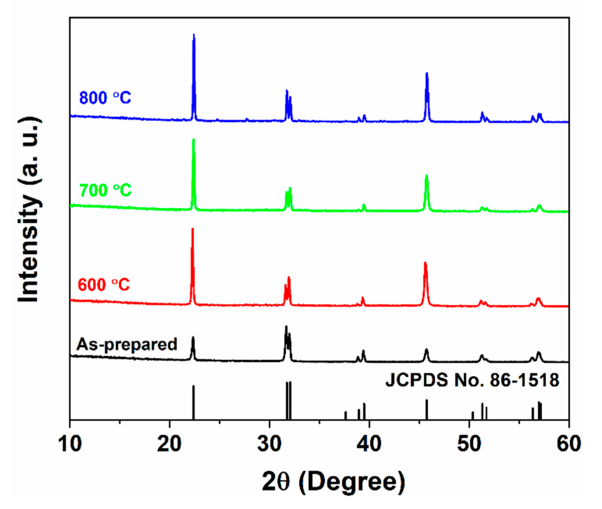
Figure 1. Powder XRD patterns of as-prepared BiFeO3 microspheres and sintered at 600, 700, and 800 °C.
Magnetochemistry 06 00042 g001
Figure 2. Dependence of heat flow with temperature for as-prepared BiFeO3 microspheres.
Magnetochemistry 06 00042 g002
Figure 3. The variation of the density (%) of the BiFeO3 against sintering temperature.
Magnetochemistry 06 00042 g003
Figure 4. Scanning electron micrograph of the BiFeO3 powder.
Magnetochemistry 06 00042 g004
Figure 5. Variation of dielectric constant (ɛ) and dielectric loss (έ) with frequency.
Magnetochemistry 06 00042 g005
Figure 6. The effect of temperature on the dielectric constant of BiFeO3.
Magnetochemistry 06 00042 g006