Article

Enhancing Magneto-Optical Performance in LaFeO3 Thin Films via Cubic-Phase Transition Induced by Ce3+/Ti4+ Co-Doping


1
College of Chemistry and Materials Science, Fujian Normal University, Fuzhou 350117, China
2
Key Laboratory of Non-Destructive Testing Technology, Fujian Polytechnic Normal University, Fuqing 350300, China
*
Authors to whom correspondence should be addressed.
Magnetochemistry 2026, 12(4), 46; https://doi.org/10.3390/magnetochemistry12040046

Submission received: 23 February 2026 / Revised: 24 March 2026 / Accepted: 30 March 2026 / Published: 7 April 2026
(This article belongs to the Section Magnetic Materials)

Abstract

Birefringence, arising from the low-symmetry structure in orthorhombic LaFeO3, limits the observation and utilization of magneto-optical effects. In this study, the pure-phase perovskite-typed La1−xCexFe1−xTixO3/SiO2 thin films were successfully fabricated via radio-frequency magnetron sputtering, where the co-doping of Ce3+ and Ti4+ ions effectively induced a structure transition from orthorhombic to a highly symmetric cubic phase, eliminating birefringence effect and thus reducing optical transmission loss. At the same time, the doped Ce3+ ions also effectively enhanced the magnetic and magneto-optical effects of the system due to their strong spin coupling effect and superexchange interaction with Fe3+ ions. The results show that the cubic-phase La0.5Ce0.5Fe0.5Ti0.5O3/SiO2 thin film exhibits excellent magnetic and magneto-optical performance. Their saturation magnetization reaches 180 emu/cm3 with an in-plane easy magnetic axis. And their magnetic circular dichroic ellipticity |ψF| reaches 3054 degrees/cm.

1. Introduction

Magneto-optical materials are crucial functional materials for optical information technology. Devices such as magneto-optical isolators, circulators, switches, and modulators, which are based on magneto-optical materials, are widely employed in optical fiber communications, computer technology, national defense, lasers, and other fields [1,2]. Common magneto-optical materials can be divided into glass, crystal, thin film, and ceramic [3]. Among them, magneto-optical glasses exhibit high absorption, low thermal conductivity, and severe thermal distortion, which can degrade the beam quality of lasers [4,5]. Magneto-optical crystals have high internal quality and low optical loss, which have great advantages in the application of high-performance devices. However, the cost of high-quality magneto-optical crystals is expensive, and the growth process is quite complex [6]. Although the preparation process of magneto-optical ceramics is relatively simple, their performance is not as good as that of crystals. In addition, with the rapid development of magneto-optical devices in the direction of miniaturization and monolithic integration, it is an irreversible trend that bulk crystals are replaced by thin films [7]. Therefore, the magneto-optical thin films with smaller size and easier integration are expected to be the next-generation core materials for magneto-optical devices [8,9,10].
Rare-earth iron garnet thin films, particularly Ce3+:YIG (Ce3+:Y3Fe5O12), have emerged as a research hotspot for next-generation magneto-optical materials due to their large Faraday rotation angle [11,12,13]. This is primarily attributed to the doping of Ce3+ ions, which can enhance the spin–orbit splitting of the 3d excited state of Fe3+ ions, thereby improving the magneto-optical performance of the material [14,15,16]. However, the difference in lattice constants and thermal expansion coefficients between garnet type YIG and commonly used semiconductor substrates is huge, which leads to poor film quality and a dramatic increase in magneto-optical and optical losses. In addition, the structure of garnet ferrite is complex, and the dodecahedral position of cations is sensitive to the change in ion radius. The replacement of Y3+ (1.08 Å) by Ce3+ (1.34 Å) introduces high stress.
In contrast, the structure of perovskite ferrite magneto-optical material REFeO3 (RE = rare earths) is simpler, and the A-site can accommodate a range of rare-earth ions with large radius differences from La3+ to Lu3+, thus allowing higher concentrations of Ce3+ doping. In addition, REFeO3 thin film has better lattice match with the mainstream semiconductor and oxide substrates, which can enable high-quality growth [17,18]. However, REFeO3 belongs to the orthorhombic crystal system, with low structural symmetry and birefringence effect, which limits the observation and application of its magneto-optical properties. Therefore, adjusting the structure to a high-symmetry cubic phase without birefringence effect is the key to improving the magneto-optical performance application of REFeO3. Related studies have demonstrated that the structure of ABO3-type perovskite materials can be designed and optimized by introducing suitable ions at the A and B sites. According to the tolerance factor formula R A + R O = t 2 R B + R O , the tolerance factor t closer to 1.0 favors the stabilization of the cubic structure [19,20,21]. Increasing the tolerance factor t can essentially improve the size matching between the A-site ion and the BO6 octahedral framework, thereby suppressing octahedral tilting and promoting a structural transition from a low-symmetry distorted phase (e.g., orthorhombic) to a high-symmetry cubic phase. In theory, the t value can be improved by introducing ions with a large radius at the A site or introducing ions with a smaller radius at the B site.
LaFeO3 possesses a high Néel temperature and good chemical stability, making it an attractive host material. And the ionic radius of La3+ (1.36 Å) is nearly identical to that of Ce3+ (1.34 Å), which allows high-concentration Ce3+ doping at the A-site. Furthermore, substituting Ti4+ (0.605 Å) for Fe3+ (0.645 Å) at the B site can increase the tolerance factor, thereby promoting the formation of a cubic phase. Therefore, we chose to incorporate Ce3+ into LaFeO3 at the A site and Ti4+ at the B site to adjust its structure to a cubic phase. Recently, very few related studies on cubic LaFeO3 have been reported, and numerous studies have focused on the structure, catalysis, and magnetism of LaFeO3, while research on its magneto-optical properties is extremely scarce [22,23,24].
In this paper, the series of La1−xCexFe1−xTixO3 (x = 0.3~0.5) (LCFTO) thin films were prepared on SiO2 quartz glass substrate. The crystal phase structure, film-forming quality, elemental valence state, optical transmittance, and magnetic and magneto-optical properties of the thin films were comprehensively characterized and analyzed, providing a theoretical foundation for the development of new magneto-optical materials and their integrated device applications.

2. Materials and Methods

LaFeO3 thin films with different Ce3+ and Ti4+ ion doping concentrations were prepared by using the radio frequency magnetron sputtering method. The series of LCFTO sputtering targets were synthesized via a high-temperature solid-state reaction. The raw materials, including CeO2 (99.99%) (Aladdin, Shanghai, China), Fe2O3 (AR) (Aladdin, Shanghai, China), Fe (AR) (Aladdin, Shanghai, China), La2O3 (99.99%) (Aladdin, Shanghai, China), and TiO2 (99.0%) (Aladdin, Shanghai, China), were weighed according to the stoichiometric ratio, mixed by ball milling, pressed into shape (20 MPa, 20 min), and then sintered in a vacuum environment at 800 °C for 6 h. To optimize the density, the initially sintered targets were ground with 5 mL of 10% (CH2CHOH)n (PVA), followed by a secondary vacuum sintering at 850 °C to obtain the targets containing La0.7Ce0.3Fe0.7Ti0.3O3 (LCFTO-0.3), La0.6Ce0.4Fe0.6Ti0.4O3 (LCFTO-0.4) and La0.5Ce0.5Fe0.5Ti0.5O3 (LCFTO-0.5). The thin films were deposited on SiO2 substrates using the prepared targets (specific parameters are shown in Table 1). To prevent the oxidation of Ce3+ to non-magnetic Ce4+, the as-deposited thin films were annealed at 600~750 °C under vacuum conditions for 4 h, with a controlled ramp rate of 1 °C/min to suppress cracking and ensure the structural integrity of the thin films.
The crystalline structure of the thin films was analyzed by using an X-ray diffractometer (D/max-3c, Rigaku Corporation, Tokyo, Japan). Transmission spectra of the thin films were measured in the 200~3000 nm wavelength range using a Perkin-Elmer Lambda 900 UV-Vis-NIR spectrophotometer (Waltham, MA, USA). The elemental valence states of the films were characterized by X-ray photoelectron spectroscopy (XPS, Escalab 250Xi, Thermo Fisher Scientific, Waltham, MA, USA) using a 500 μm spot size, constant analyzer energy mode, 30.0 eV pass energy, and 0.05 eV energy step size. The thickness and fault morphology of the thin films were analyzed by scanning electron microscope (SEM) (SU8000, Hitachi, Tokyo, Japan). Magnetic measurements were performed at room temperature using a vibrating sample magnetometer (VSM, LakeShore-7407, Westerville, OH, USA) with a maximum applied magnetic field of ±18000 Oe. Magneto-circular dichroism (MCD) spectra were acquired using a circular dichroism spectrometer (BioLogic MOS-450, Claix, France) equipped with a parallel magnetic field of 5000 Oe parallel to the direction of probe light propagation.

3. Results and Discussion

3.1. Crystalline Phase and Structure

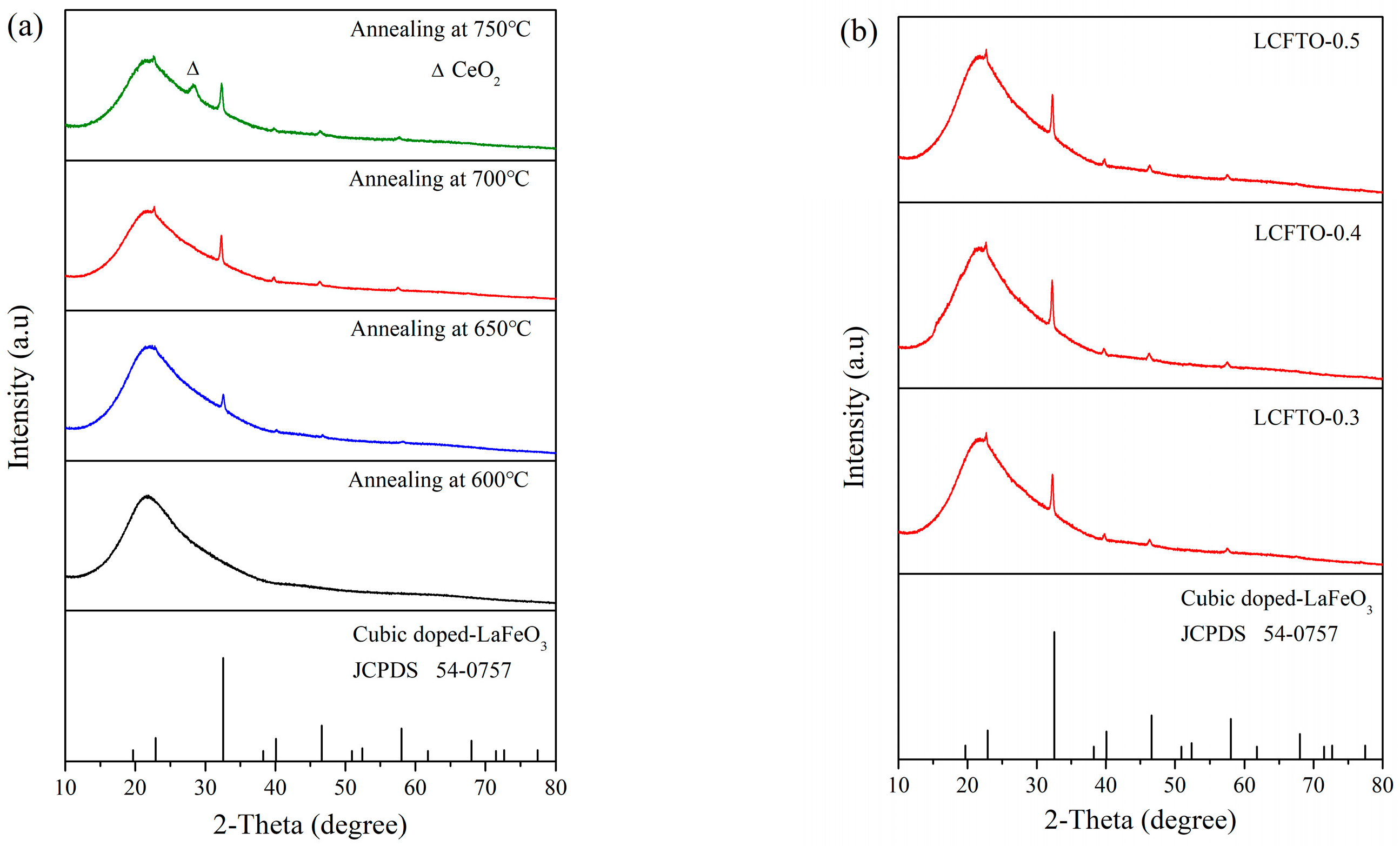
The optimal annealing temperature of LCFTO thin films was explored through X-ray diffraction (XRD) analysis. As shown in Figure 1a, the LCFTO-0.5 thin film annealed at 600 °C exhibits only the broadening peak of the amorphous SiO2 quartz glass substrate. It can also be seen from the figure that when the annealing temperature was increased to 650 °C and 700 °C, the thin film exhibited a series of sharp diffraction peaks at 2θ = 23.1°, 32.8°, 40.5°, 47.2°, 58.6° and 68.9°, which are highly consistent with the standard diffraction pattern for cubic perovskite-phase which used LaSrFeVO6 [25] standard cards (JCPDS 54-0757) as a reference. In particular, the intensity of the diffraction peaks of the thin film annealed at 700 °C was significantly enhanced, indicating a higher degree of crystallinity and a more complete crystallization process. When the annealing temperature was further increased to 750 °C, a diffraction peak at 2θ = 28.6° attributed to the CeO2 impurity phase (JCPDS 34-394) appeared, indicating that LCFTO thin film partially decomposes at high temperatures, and the cerium oxide heterophase was produced. These results demonstrate that 700 °C is the optimal crystallization temperature for LCFTO thin films. Figure 1b shows the XRD pattern of the series of LCFTO thin films annealing at 700 °C. It can be seen that multiple diffraction peaks appearing in these thin films all belong to the standard card of the cubic perovskite phase, and no other impurity phases exist. This indicates that the prepared LCFTO-0.3, LCFTO-0.4, and LCFTO-0.5 thin films are all pure-phase cubic perovskite.
In summary, by introducing Ce3+ and Ti4+ ion doping, the perovskite tolerance factor t of La1−xCexFe1−xTixO3 increased in the stable range of the cubic phase (t = 0.9~1.0). This modification successfully induced a structural transition from the orthorhombic to the cubic perovskite phase, laying a structural foundation for subsequent research on magneto-optical properties.

3.2. Morphological Analysis

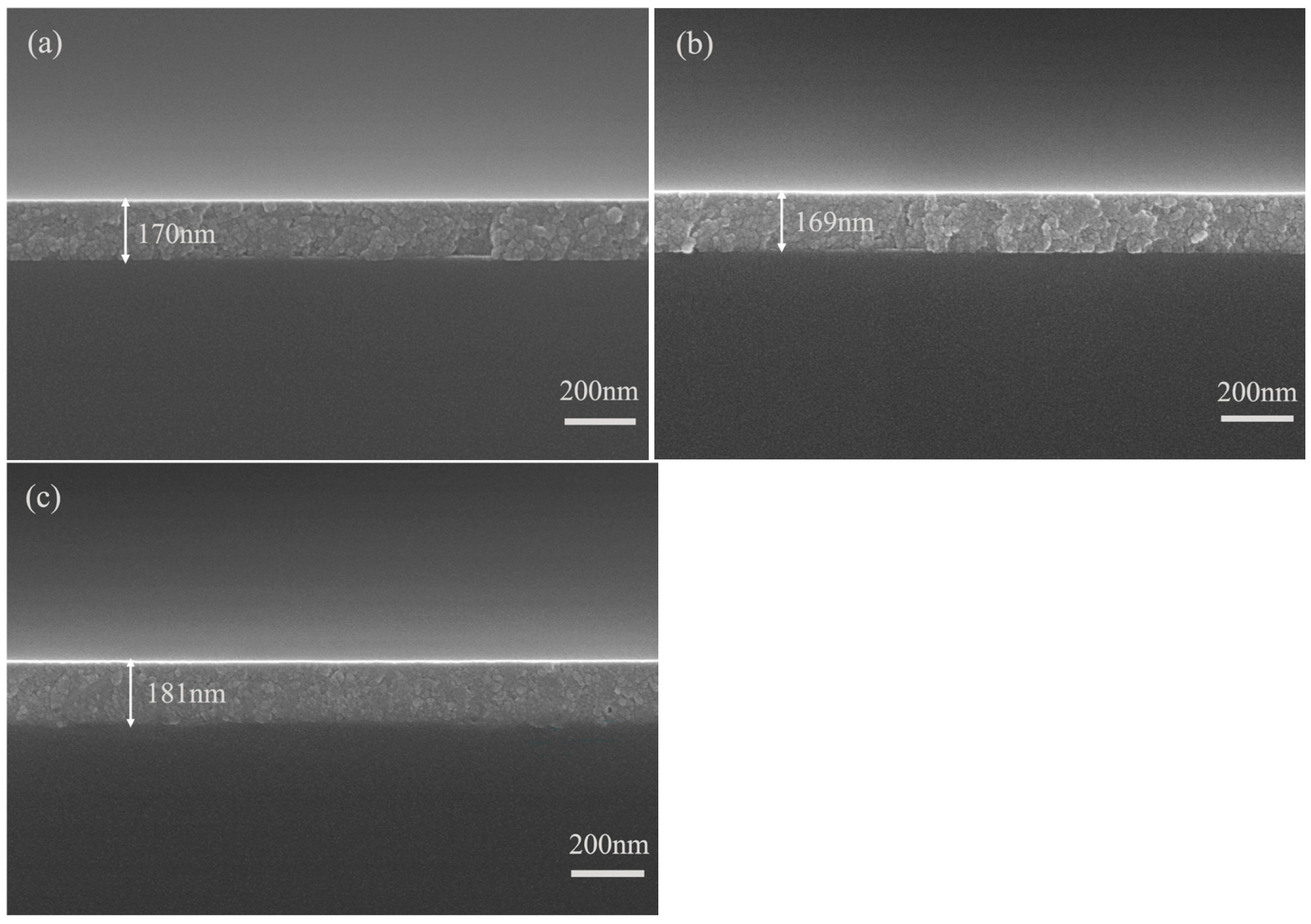
The microscopic morphology of thin films greatly influences their optical properties. Rough grains and excessive grain boundaries increase light scattering and reduce transmission, while the pores trigger multiple reflections of light, resulting in light loss. We characterized the cross-section morphology of the series LCFTO thin films by scanning electron microscopy (SEM), as shown in Figure 2. It can be seen from Figure 2a,b that the LCFTO-0.3 and LCFTO-0.4 thin films appear continuous with relatively uniform grain distribution, although a few local grain boundaries and small pores are present. When the doping concentration increases to x = 0.5 (as shown in Figure 2c), the grains of the LCFTO-0.5 thin film are closely and uniformly packed; almost no pores and rough grain boundaries are generated. This may be because the ratios of La/Ce and Fe/Ti are both 1:1, forming a more stable double perovskite structure, thus having better uniformity in the microstructure and defect control ability. In addition, we can also obtain the thicknesses of LCFTO-0.3, LCFTO-0.4, and LCFTO-0.5 thin films which are 170 nm, 169 nm and 181 nm, respectively. Overall, the three prepared thin films all exhibit excellent film-forming quality, with high structural integrity and surface uniformity.

3.3. Elemental Analysis

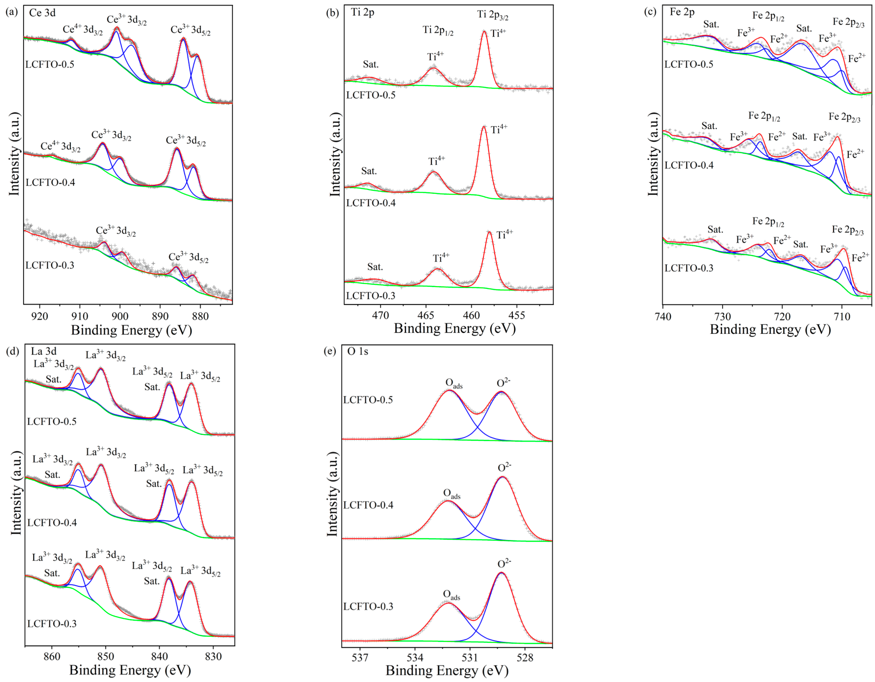
The Ce3+ ion is established as the critical contributor to enhanced magneto-optical response in rare-earth ferrites because of its distinctive 4f→5d electronic transition [26]. However, Ce3+ is thermodynamically metastable and readily oxidizes to nonmagnetic Ce4+ during synthesis, causing a marked loss of magneto-optical performance. Accordingly, this paper examines the valence state of Ce ions in the series of LCFTO thin films, as depicted in Figure 3a. The XPS spectrum of Ce 3d reveals complex multiple splitting characteristics. According to the existing literature [27], the peaks at 881.1 eV, 885.2 eV, 895.3 eV, 899.3 eV, and 903.4 eV are attributed to the Ce3+ ion. Notably, a relatively weak Ce4+ peak appears around 916.5 eV [28]. By calculating the area of each peak, it was found that as the doping concentration of cerium ions increased from low to high, the content of Ce4+ accounted for 0%, 2.26%, and 4.08% of the total cerium content, respectively. This finding indicates that the majority of cerium ions are in the +3 valence state, showing magneto-optical activity, while the trace amount of Ce4+ has negligible effects on the magnetic and magneto-optical properties of the material.
The valence state of doped titanium is crucial for modulating the electronic structure of materials. In the Ti 2p XPS spectrum (Figure 3b), the core-level binding energies for 2p3/2 and 2p1/2 are at 458.5 eV and 464.2 eV, respectively, with a spin–orbit splitting energy of 5.7 eV. These values align with the standard characteristic for Ti4+ [29]. This finding confirms that Ti ions in the LCFTO thin films are exclusively in the +4 valence state.
The valence state distribution of Fe ions, as the primary carriers of magnetism, is crucial. The superexchange interaction between Fe3+ ions forms antiferromagnetic order, and its spin canting is the source of weak ferromagnetism. However, the double exchange interaction formed by Fe2+ and Fe3+ can significantly change the net magnetic moment. The ratio of Fe2+/Fe3+ directly determines the competition and synergy between antiferromagnetic and ferromagnetic interactions and is the main switch for regulating saturation magnetization, coercivity and magneto-optical response. The XPS spectra of Fe 2p of the series of LCFTO thin films demonstrate the mixed valence state characteristics, as shown in Figure 3c. The peaks at binding energies of 709.5 eV and 710.9 eV correspond to Fe2+ 2p3/2 and Fe3+ 2p3/2, respectively, with their satellite peaks at 718.8 eV [30,31]. Additionally, Fe2+ 2p1/2 and Fe3+ 2p1/2 peaks appear at 722 eV and 724.2 eV, respectively, with a satellite peak at 731.8 eV [32]. The presence of Fe2+ is due to a charge compensation mechanism, where B-site Fe3+ (ion radius ~0.645 Å) is substituted by high-valent Ti4+ (ion radius ~0.605 Å) [33,34]. To maintain local electrical neutrality, the Ti4+ ion introduction reduces a part of Fe3+ to Fe2+. The results of quantitative fitting show that the ratios of Fe2+ to Fe3+ in these thin films are 0.38, 0.41, and 0.55, respectively, as the Ce3+/Ti4+ co-doping amount moves from low to high. It suggests that Fe3+ remains the dominant valence state, which upholds the material’s basic magnetic framework. However, with the increase in Ce/Ti ion doping, the Fe2+/Fe3+ ratio also increases.
Additionally, the analysis of matrix elements and oxygen states reveals the following: The La 3d spectra (Figure 3d) exhibit primary peaks at 834.2 eV (3d5/2) and 851.0 eV (3d3/2), along with characteristic satellite peaks at approximately 838.2 eV and 855.0 eV, which are typical XPS features of La3+ [35]. The O 1s spectra (Figure 3e) can be divided into two components: the peak at a lower binding energy (529.3 eV) arises from lattice oxygen (O2−), while a higher binding energy (532.6 eV) likely corresponds to surface-adsorbed hydroxyl or carbonate species. Additionally, as the doping concentration of Ti4+ increases; the characteristic peak at 532.6 eV gradually intensifies, indicating an intrinsic mechanism in the LaFeO3 system where charge compensation is achieved through the formation of oxygen vacancies by the substitution of high-valent ions. This mechanism may lead to a significant increase in the density of defect states near the Fermi level, thereby enhancing the probability of valence band electron transitions and ultimately effectively strengthening the magneto-optical effect [36,37].

3.4. Transmittance

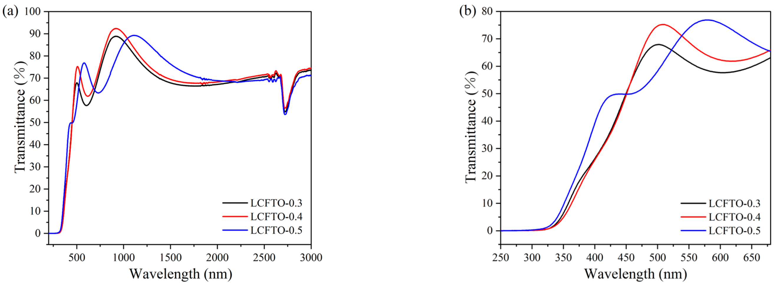
The excellent magneto-optical materials need to have high transmittance, so that reflection and scattering can be reduced and optical loss can be reduced in the device. Figure 4a shows that the series of LCFTO films exhibit high optical transmittance performance. The transmittance of these films reaches up to 78% in the visible region and can reach more than 90% in the near-infrared region. The absorption peak observed at 2700 nm is mainly caused by the absorption of the silica substrate (quartz glass has an OH impurity absorption band near 2.7 μm) and the interface effect between the film and the substrate [38,39]. In addition, the observed oscillatory behavior in the transmission spectra arises from the interference between light reflected from the upper and lower interfaces of the thin films. Since the interference phenomenon occurs only in the wavelength region where the transparency of the thin film is high, a rough surface or interface can cause diffuse reflection of light and destroy the coherence of light, thereby weakening the interference effect. Therefore, it can be concluded that the prepared LCFTO thin films have good thickness uniformity and very low surface and interface roughness on the optical scale.
In view of the spectral range relevant to the magneto-optical effect in this study, we focus on the analysis of the transmission spectra below 680 nm. Figure 4b shows transmission spectra of LCFTO thin films in the range of 250~680 nm. It can be seen from the figure that as the Ce3+/Ti4+ doping concentration (x) increases, the optical band edge of the thin film undergoes a slightly blue shift, from 320 nm at x = 0.3 to 280 nm at x = 0.5, indicating a gradual widening of the optical band gap (Eg). The blue shift of the absorption edge can reduce the absorption in the near-UV region and reduces the potential interference of this band to visible light operation, while expanding the transparent window to deep UV. This blue-shift phenomenon is mainly due to the lattice distortion and band structure change caused by the doping of Ce3+ and Ti4+ ions. Moreover, in magnetic semiconductors, the establishment of magnetic order can introduce exchange splitting, which may influence the band gap. Therefore, the co-doping of Ce3+ and Ti4+ ions may increase the magnetization and enhance the magnetic ordering of LaFeO3, thereby driving the blue shift of the absorption edge. This phenomenon can be further verified in the subsequent magnetic analysis. In addition, at the critical wavelength corresponding to the enhanced magneto-optical response (433 nm), the transmittance is observed to be approximately 50%. Detailed magneto-optical characterization and analysis are presented in subsequent sections.

3.5. Magnetism

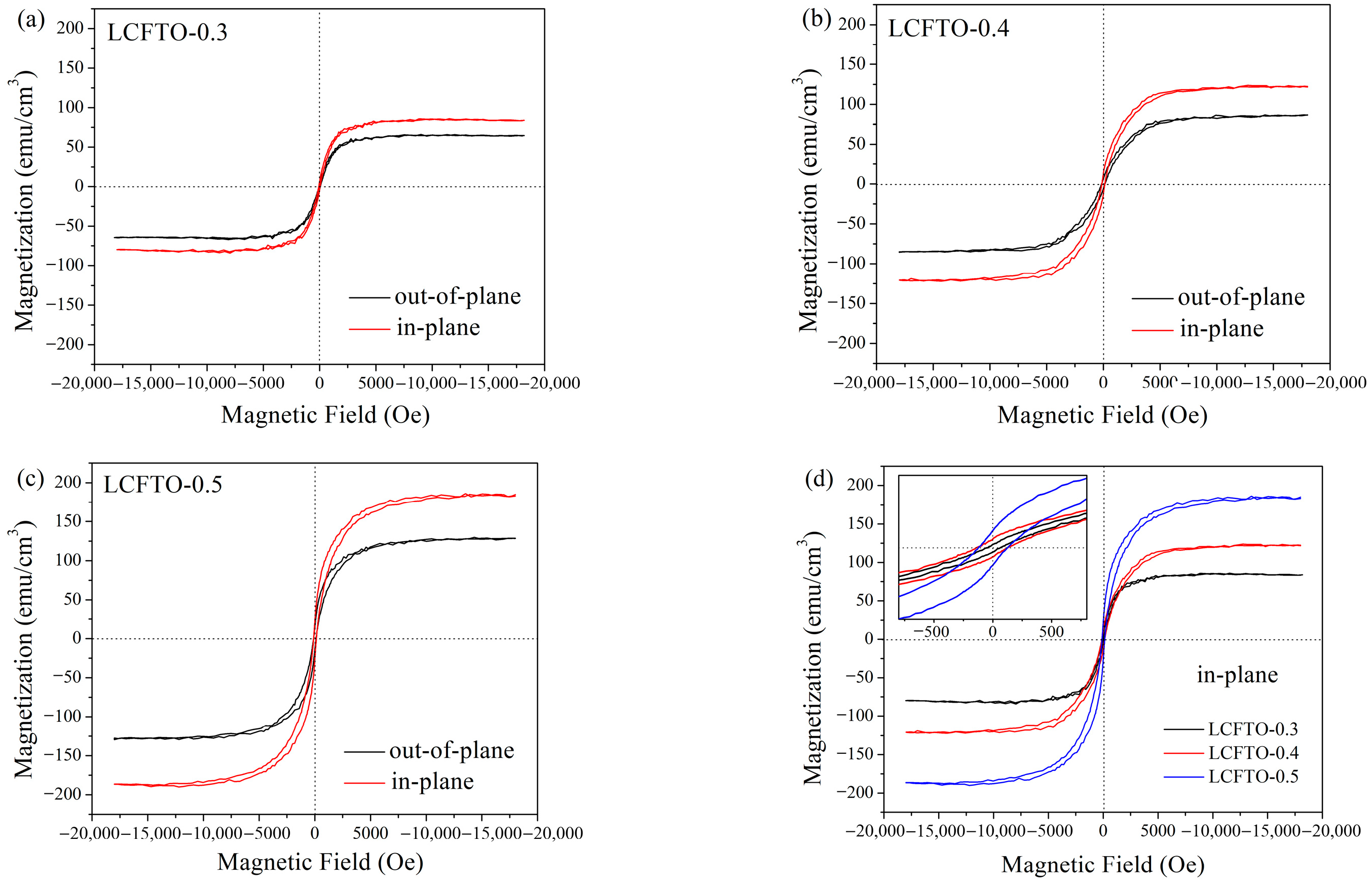
The room temperature saturated hysteresis loops of the series of LCFTO thin films are shown in Figure 5. All measurements were performed under a maximum applied magnetic field of ±18000 Oe. The field directions were the applied magnetic field perpendicular to the film surface (in-plane) and parallel to the film surface (out-of-plane).
It can be seen from Figure 5a–c that the series thin films have good magnetic properties and significant magnetic anisotropy. The easy magnetization axis lies on the surface of film. This is mainly caused by shape anisotropy. The in-plane dimensions of the film are much larger than its thickness, resulting in the demagnetization factor in the in-plane direction being much smaller than that in the perpendicular direction. This makes the in-plane direction more energetically favorable for the magnetic moment (lower demagnetization energy), while out-of-plane magnetization is hindered by a strong demagnetizing field. Therefore, the easy axis lies in the plane [40,41]. As shown in Figure 5d, the in-plane saturation magnetization (Ms) of LCFTO-0.3, LCFTO-0.4, and LCFTO-0.5 thin films are 85, 123, and 180 emu/cm3, respectively. In particular, the LCFTO-0.5 thin film demonstrates the most superior magnetic properties, which are much higher than un-doped LaFeO3 thin film (30 emu/cm3) and similar rare earth ferrites [42,43,44], indicating that co-doping of Ce3+ and Ti4+ ions play a substantial role in modulating the magnetic properties of the material. This phenomenon of enhanced magnetism mainly stems from the charge compensation effect may cause by heterovalent substitution and the double-exchange ferromagnetic interaction it induces. Specifically, after both Ce3+ and Ti4+ ions entered into the LaFeO3 lattice, a significant positive charge excess is introduced into the material system. In order to maintain overall electrical neutrality, the lattice spontaneously balances the excess positive charges by reducing part of Fe3+ to Fe2+, a charge compensation pathway, which is consistent with the result in XPS analysis that the Fe2+ content increases with the increase in Ti4+ doping concentration. The emergence of Fe2+ in the antiferromagnetic network originally dominated by the superexchange interaction of Fe3+-O-Fe3+ additionally establishes the double exchange interaction of Fe3+ and Fe2+. This interaction is essentially ferromagnetic and competes with the original antiferromagnetic superexchange interaction in the system [45,46]. As the doping concentration increased from 0.3 to 0.5, the Fe2+ content continued to increase, indicating that the ferromagnetic coupling network composed of Fe3+-O-Fe2+ double-exchange channels continued to expand and strengthen, which finally manifested as a systematic increase in the saturation magnetization at the macroscopic level. Furthermore, the lattice distortion introduced by doping may also further weaken the intrinsic antiferromagnetic superexchange strength by changing the Fe-O-Fe bond angle, providing favorable conditions for the emergence of ferromagnetic components. In addition, it can be seen from Figure 5d that the coercive forces (Hc) of these three thin films are 55, 135 and 132 Oe, respectively, and the overall values are relatively low. This characteristic indicates that the material is easily magnetized and can achieve rapid and low-energy magnetic domain switching. Combined with its in-plane anisotropy, it provides an important material basis for the development of low-power consumption and high-response-speed integrated non-reciprocal photonic devices.
In summary, the series LCFTO thin films have high saturation magnetization to ensure strong magneto-optical interaction, and their in-plane magnetic anisotropy is naturally compatible with planar optical waveguide structures for easy integration. This makes LCFTO thin films show significant application advantages in the field of low-power, integrable planar optical waveguide non-reciprocal devices (such as optical isolators and modulators) and high-sensitivity magneto-optical sensors.

3.6. Magneto-Optical Properties

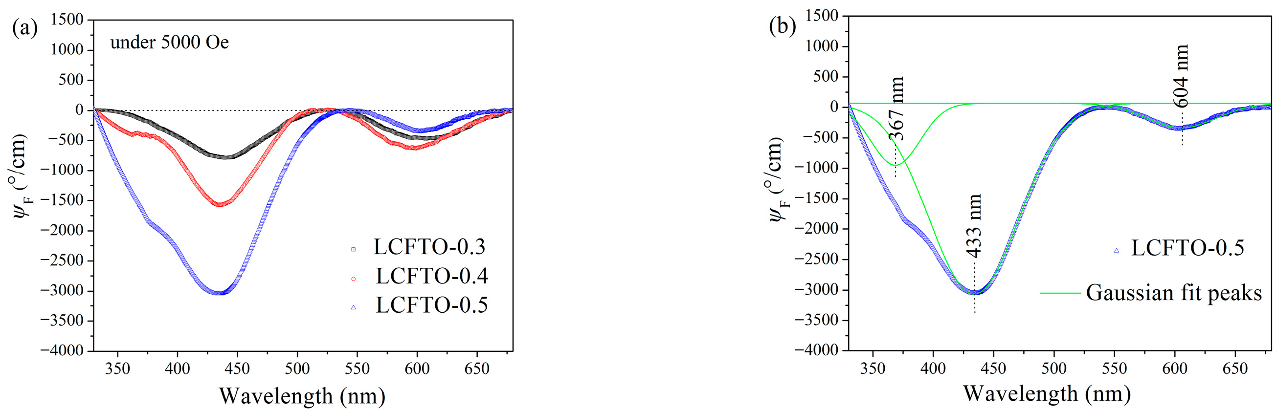
Magnetic circular dichroism (MCD), a powerful technique for characterizing the difference in absorption of left- and right-handed circularly polarized light in a material under a magnetic field, is a core tool for studying the properties of magneto-optical films. While current research on magneto-optical materials primarily focuses on garnet-type rare-earth ferrites, the magneto-optical properties and MCD effects of perovskite-type Ce3+-doped REFeO3 thin films remain relatively few. Figure 6a presents the MCD spectra of the prepared LCFTO thin films. It can be seen from the figure that the thin films exhibit an obvious MCD signal. As the Ce3+ and Ti4+ ion doping concentration (x) increased from 0.3 to 0.5, the MCD ellipticity ψF was significantly enhanced. On one hand, this is because the enhanced saturation magnetization (Ms) significantly improves the ability to modulate the polarization of the light, which manifests itself macroscopically as a stronger magneto-optical effect. On the other hand, it is due to the 5d-4f transition of Ce3+ ions in the visible region, which leads to high magneto-optical activity and large transition oscillation intensity [47]. The maximum MCD ellipticity value |ψF| of the LCFTO-0.5 thin film reaches 3054 degrees/cm at 433 nm under an applied magnetic field of 0.5 T. Although this value is lower than that of state-of-the-art Ce3+:YIG (x = 1.0) films (40,000~60,000 degrees/cm) [48], it remains competitive with other garnet-based magneto-optical materials, such as ~5000 degrees/cm for Gd2Ce1Fe5O12/SiO2 films under 0.7 T [49]. Notably, unlike garnet-based materials that typically require lattice-matched single-crystal garnet substrates for epitaxial growth, cubic perovskite films can be directly deposited on semiconductor substrates [50,51], offering superior compatibility with Si-based photonic integration platforms. Combined with its competitive magneto-optical performance and the effective elimination of birefringence achieved through Ce3+ and Ti4+ co-doping, the LCFTO system presents a promising material platform for integrated magneto-optical devices.
Gaussian fitting of the MCD spectrum of the LCFTO-0.5 thin film was performed, as shown in Figure 6b. The fitted spectrum revealed three distinct peaks at 367, 433, and 604 nm, with MCD ellipticity values |ψF| of 960, 3054 and 375 degrees/cm, respectively. As shown in the transmission spectrum of the LCFTO-0.5 thin film (Figure 4b), the transmittances corresponding to these three wavelengths are 15%, 50% and 75%, respectively. The strong absorption at 367 nm combined with an obvious MCD signal suggests a strong electronic transition with significant spin polarization, which usually corresponds to a charge transfer transition, such as O 2p → Fe 3d. The most pronounced MCD response occurs at 433 nm, where the transmittance is moderate. The large MCD ellipticity at this wavelength indicates that the difference in absorption between left- and right-handed circularly polarized light reaches a maximum, pointing to a charge-transfer transition as the dominant mechanism. In contrast to spin-forbidden d-d crystal field transitions, charge-transfer transitions are spin-allowed and typically exhibit both stronger absorption and larger magneto-optical activity. This assignment is consistent with the well-established understanding of magneto-optical spectra in rare-earth orthoferrites, where the prominent features in the visible region are attributed to charge-transfer transitions involving Fe 3d and O 2p orbitals [52,53,54]. In the present Ce and Ti co-doped system, the presence of Ti4+ at the B site and the mixed Fe2+/Fe3+ valences may further modulate the charge-transfer energy and enhance the magneto-optical response. At 604 nm, the high transmittance and relatively weak MCD signal are typical of the spin-forbidden d-d transitions of Fe3+ (6A1g4T1g; 4T2g), which have low transition probabilities but remain detectable due to the high-spin state and magnetic ordering of Fe3+ [49,55]. Overall, the magneto-optical response of the LCFTO-0.5 thin film at multiple wavelengths of visible light means that it has a wide spectrum of non-reciprocal optical activity. This property significantly broadens the application window of this material in integrated photonics, enabling it to be adapted to different wavelength light source requirements.

4. Conclusions

In this paper, the series LCFTO thin films were prepared by using radio frequency magnetron sputtering. Structural and morphology characterization revealed that the co-doping of Ce3+ and Ti4+ ions successfully transformed the crystal structure of LaFeO3 from an orthorhombic phase to a cubic phase without birefringence, and the prepared thin films are of high quality with tightly packed grains and uniform thickness. In addition, the thin films exhibit good transmission properties, with transmittance reaching up to 78% in the visible light band. In terms of magnetism, the thin films also exhibit excellent magnetic properties, particularly when x = 0.5, with a saturation magnetization reaching 180 emu/cm3. They also achieve a magnetic circular dichroism ellipticity |ψF| of up to 3054 degrees/cm at a wavelength of 433 nm. This performance is competitive with that of some garnet-based magneto-optical materials. With its excellent optical and magnetic properties, combined with superior compatibility with semiconductor substrates, the thin films are promising candidates for applications such as non-reciprocal optical devices and magneto-optical isolators and open up a new path for the design of a new generation of high-performance magneto-optical functional materials.

Author Contributions

Conceptualization, N.L.; methodology, Z.X. and Y.S.; validation, Z.X.; formal analysis, N.L. and Z.X.; investigation, C.X. and Y.S.; resources, Q.T. and N.L.; data curation, Z.X.; writing, original draft preparation, Z.X. and N.L.; writing—review, and editing, Q.T.; visualization, C.X.; supervision, Q.T.; project administration, N.L. and Q.T.; funding acquisition, N.L. and Q.T. All authors have read and agreed to the published version of the manuscript.

Funding

This research was funded by the National Natural Science Foundation of China (62105063), the Natural Science Foundation of Fujian Province, China (2023J011126) and the Innovation Team of Photoelectric Materials and Devices of Fujian Polytechnic Normal University, China (CXTD202405).

Institutional Review Board Statement

Not applicable.

Informed Consent Statement

Not applicable.

Data Availability Statement

The original contributions presented in the study are included in the article; further inquiries can be directed to the corresponding authors.

Conflicts of Interest

The authors declare no conflicts of interest.

References

  1. Zhang, T.; Zhou, W.; Li, Z.; Tang, Y.; Xu, F.; Wu, H.; Zhang, H.; Tang, J.-S.; Ruan, Y.-P.; Xia, K. Reversible optical isolators and quasi-circulators using a magneto-optical fabry-pérot cavity. Chin. Phys. Lett. 2024, 41, 044205. [Google Scholar] [CrossRef]
  2. Albino, L.V.; Roque, N.G.; Marcondes, L.M.; Orives, J.R.; Franco, D.F.; Manzan, R.S.; Aragón, F.H.; Felix, J.F.; Nalin, M. Magnetic and magneto-optical properties of manganese-containing antimony phosphate glasses-An alternative to Faraday rotators. Opt. Mater. 2024, 147, 114674. [Google Scholar] [CrossRef]
  3. Li, J.; Dai, J.-W.; Pan, Y.-B. Research progress on magneto-optical transparent ceramics. J. Inorg. Mater. 2018, 33, 1–8. [Google Scholar] [CrossRef]
  4. Mu, H.; Wang, P.; Shi, Y.; Zhang, Z.; Wu, A.; Su, L. Large-size CeF3 crystal growth by heat exchanger-bridgman method: Thermal field design and optimization. J. Inorg. Mater. 2023, 38, 288–295. [Google Scholar] [CrossRef]
  5. Snetkov, I.; Li, J. Selection of magneto-optical material for a faraday isolator operating in high-power laser radiation. Magnetochemistry 2022, 8, 168. [Google Scholar] [CrossRef]
  6. Zhang, L.; Hu, D.; Snetkov, I.L.; Balabanov, S.; Palashov, O.; Li, J. A review on magneto-optical ceramics for Faraday isolators. J. Adv. Ceram. 2023, 12, 873–915. [Google Scholar] [CrossRef]
  7. Askarzadeh, N.; Shokrollahi, H. Magneto-optical garnets in photonic integration. Results Phys. 2025, 78, 108473. [Google Scholar] [CrossRef]
  8. Kuila, M.; Sagdeo, A.; Longchar, L.A.; Choudhary, R.J.; Srinath, S.; Reddy, V.R. Robust perpendicular magnetic anisotropy in Ce substituted yttrium iron garnet epitaxial thin films. J. Appl. Phys. 2022, 131, 203901. [Google Scholar] [CrossRef]
  9. Ghising, P.; Hossain, Z.; Budhani, R.C. Stripe magnetic domains in CeY2Fe5O12 (Ce:YIG) epitaxial films. Appl. Phys. Lett. 2017, 110, 012406. [Google Scholar] [CrossRef]
  10. Shen, T.; Dai, H.; Song, M.; Liu, H.; Wei, X. Magnetic and magneto-optical properties of CeYIG thin films grown by pulsed laser deposition on quartz substrates. J. Supercond. Novel Magn. 2017, 30, 1915–1919. [Google Scholar] [CrossRef]
  11. Hasan, M.S.; Khan, M.I.; Ali, S.S.; Brahmia, A.; Ihtisham ul, H. Influence of gallium on structural, optical and magnetic properties of Bi-YIG thin films. Mater. Sci. Eng. B 2024, 301, 117180. [Google Scholar] [CrossRef]
  12. Halagačka, L.; Postava, K.; Vanwolleghem, M.; Vaurette, F.; Ben Youssef, J.; Dagens, B.; Pištora, J. Mueller matrix optical and magneto-optical characterization of Bi-substituted gadolinium iron garnet for application in magnetoplasmonic structures. Opt. Mater. Express 2014, 4, 1903–1919. [Google Scholar] [CrossRef]
  13. Zhou, T.; Pan, L.; Ma, Y.; Zhu, S.; Tan, G.; Man, Q. Origin of high-frequency magnetic loss of Y3Fe5O12 single crystal thin films prepared with high-throughput screening by magnetron sputtering. Vacuum 2023, 207, 111644. [Google Scholar] [CrossRef]
  14. Teterin, Y.A.; Smirnova, M.N.; Maslakov, K.I.; Teterin, A.Y.; Nikiforova, G.E.; Glazkova, Y.S.; Sobolev, A.N.; Presnyakov, I.A.; Ketsko, V.A. Synthesis, ionic, and phase compositions of ferrogarnet Y2.5Ce0.5Fe2.5Ga2.5O12. Russ. J. Inorg. Chem. 2023, 68, 829–837. [Google Scholar] [CrossRef]
  15. Murai, T.; Shoji, Y.; Nishiyama, N.; Mizumoto, T. Nonvolatile magneto-optical switches integrated with a magnet stripe array. Opt. Express 2020, 28, 31675–31685. [Google Scholar] [CrossRef]
  16. Lin, N.; Wang, Y.; Zhao, Y.; Huo, L.; Shi, W.; Chen, X.; Zhang, Y.; Hu, X.; Zhuang, N. High magneto-optical performance of GdFeO3 thin film with high orientation and heavy Ce3+ doping. Ceram. Int. 2019, 45, 14928–14933. [Google Scholar] [CrossRef]
  17. Zhang, X.; Song, H.; Tan, C.; Yang, S.; Xue, Y.; Wang, J.; Zhong, X. Epitaxial growth and magnetic properties of h-LuFeO3 thin films. J. Mater. Sci. 2017, 52, 13879–13885. [Google Scholar] [CrossRef]
  18. Li, X.; Yun, Y.; Xu, X. Recent progress on multiferroic hexagonal rare-earth ferrites (h-RFeO3, R = Y, Dy-Lu). J. Phys. D Appl. Phys. 2025, 58, 073003. [Google Scholar] [CrossRef]
  19. Tariq, S.; Alrashdi, A.O.; Al Bahir, A.; Gilani, S.S.; Hamioud, F.; Mubarak, A.; Ahmed, A.; Saad, H.-E.M.M. DFT insights into LaFeO3 with Mn substitution: A promising path to energy-efficient magneto-optical applications. J. Comput. Chem. 2024, 45, 843–854. [Google Scholar] [CrossRef] [PubMed]
  20. Li, Z.; Yang, M.; Park, J.-S.; Wei, S.-H.; Berry, J.J.; Zhu, K. Stabilizing perovskite structures by tuning tolerance factor: Formation of formamidinium and cesium lead iodide solid-state alloys. Chem. Mater. 2016, 28, 284–292. [Google Scholar] [CrossRef]
  21. Chauhan, S.; Kumar, M.; Pal, P. Substitution driven structural and magnetic properties and evidence of spin phonon coupling in Sr-doped BiFeO3 nanoparticles. RSC Adv. 2016, 6, 68028–68040. [Google Scholar] [CrossRef]
  22. Al-Mamari, R.T.; Widatallah, H.M.; Elzain, M.E.; Gismelseed, A.M.; Al-Rawas, A.D.; Al-Harthi, S.H.; Myint, M.T.Z.; Al-Saqri, N.; Al-Abri, M. Core and surface structure and magnetic properties of mechano-synthesized LaFeO3 nanoparticles and their Eu3+-doped and Eu3+/Cr3+-co-doped variants. Sci. Rep. 2024, 14, 14770. [Google Scholar] [CrossRef]
  23. Arman, M.M. The influence of rare earth doping on the structure, magnetic properties, and application of LaFeO3 nanoparticles. Eur. Phys. J. Plus 2025, 140, 212. [Google Scholar] [CrossRef]
  24. Gan, T.; Tao, L.; Zhang, Z.; Zhou, A.; Chen, Y.; Li, J.; Zhang, S.; Du, S.; Li, Y. Defect-Engineered LaFeO3 Stabilizing Oxidized Pt Single-Atom Sites for Low-Temperature CO Oxidation. J. Am. Chem. Soc. 2025, 147, 32729–32736. [Google Scholar] [CrossRef]
  25. Ramesha, K.; Gopalakrishnan, J.; Smolyaninova, V.; Greene, R.L. ALaFeVO6 (A=Ca, Sr): New double-perovskite oxides. J. Solid State Chem. 2001, 162, 250–253. [Google Scholar] [CrossRef]
  26. Goto, T.; Koguchi, T.; Yoshihara, Y.; Miyashita, H.; Mori, K.; Watanabe, T.; Kaczmarek, A.C.; Lim, P.B.; Inoue, M.; Ross, C.A. Magnetotaxial Perpendicular Magnetic Anisotropy and Enhanced Faraday Rotation in Ion Beam Sputtered Cerium-Substituted Yttrium Iron Garnet. ACS Appl. Opt. Mater. 2025, 3, 2923–2934. [Google Scholar] [CrossRef]
  27. Paparazzo, E. On the number, binding energies, and mutual intensities of Ce3d peaks in the XPS analysis of cerium oxide systems: A response to Murugan et al., Superlatt. Microstruct. 85 (2015) 321. Superlattices Microstruct. 2017, 105, 216–220. [Google Scholar] [CrossRef]
  28. Bêche, E.; Charvin, P.; Perarnau, D.; Abanades, S.; Flamant, G. Ce 3d XPS investigation of cerium oxides and mixed cerium oxide (CexTiyOz). Surf. Interface Anal. 2008, 40, 264–267. [Google Scholar] [CrossRef]
  29. Biesinger, M.C.; Lau, L.W.M.; Gerson, A.R.; Smart, R.S.C. Resolving surface chemical states in XPS analysis of first row transition metals, oxides and hydroxides: Sc, Ti, V, Cu and Zn. Appl. Surf. Sci. 2010, 257, 887–898. [Google Scholar] [CrossRef]
  30. Grosvenor, A.; Kobe, B.; Biesinger, M.C.; Mcintyre, N. Investigation of multiplet splitting of Fe 2p XPS spectra and bonding in iron compounds. Surf. Interface Anal. 2004, 36, 1564–1574. [Google Scholar] [CrossRef]
  31. Omran, M.; Fabritius, T.; Elmahdy, A.M.; Abdel-Khalek, N.A.; El-Aref, M.; Elmanawi, A.E.-H. XPS and FTIR spectroscopic study on microwave treated high phosphorus iron ore. Appl. Surf. Sci. 2015, 345, 127–140. [Google Scholar] [CrossRef]
  32. Wang, J.; Tang, Y.; Yang, Q.; Zhao, K.; Li, X.; Wang, Y.; Zhu, Y.; Zhu, Q.; Yu, F.; Wang, C. Enhanced photocatalytic nitrogen fixation via Pt-induced active hydrogen supply over Pt@NM-101(Fe). Appl. Catal. B-Environ. Energy 2025, 373, 125364. [Google Scholar] [CrossRef]
  33. Cui, P.; Li, G.-T.; Zhang, P.-P.; Wan, T.; Li, M.-Q.; Chen, X.-L.; Zhou, Y.; Guo, R.-Q.; Su, M.-R.; Liu, Y.-J.; et al. Arranging cation mixing and charge compensation of TiNb2O7 with W6+ doping for high lithium storage performance. Rare Met. 2023, 42, 3364–3377. [Google Scholar] [CrossRef]
  34. Peng, W.; Li, L.; Yu, S.; Yang, P.; Xu, K. Dielectric properties, microstructure and charge compensation of MnO2-doped BaTiO3-based ceramics in a reducing atmosphere. Ceram. Int. 2021, 47, 29191–29196. [Google Scholar] [CrossRef]
  35. Nedelko, N.; Lewinska, S.; Pashchenko, A.; Radelytskyi, I.; Diduszko, R.; Zubov, E.; Lisowski, W.; Sobczak, J.W.; Dyakonov, K.; Ślawska-Waniewska, A.; et al. Magnetic properties and magnetocaloric effect in La0.7Sr0.3−xBixMnO3 manganites. J. Alloys Compd. 2015, 640, 433–439. [Google Scholar] [CrossRef]
  36. Thomas, B.; Skariah, B. Spray deposited Mg-doped SnO2 thin film LPG sensor: XPS and EDX analysis in relation to deposition temperature and doping. J. Alloys Compd. 2015, 625, 231–240. [Google Scholar] [CrossRef]
  37. Pawlak, D.A.; Ito, M.; Oku, M.; Shimamura, K.; Fukuda, T. Interpretation of XPS O (1s) in Mixed Oxides Proved on Mixed Perovskite Crystals. J. Phys. Chem. B 2002, 106, 504–507. [Google Scholar] [CrossRef]
  38. Guo, S.; Ai, G.; Zhao, J.; Yang, S.; Wei, K.; Ma, W. Removal of hydroxyl impurities in vein quartz by vacuum roasting. Miner. Eng. 2024, 200, 108474. [Google Scholar] [CrossRef]
  39. Liu, S.; Xie, Z.; Liu, J.; Li, Y.; Li, Y.; Wang, H. A low-hydroxyl quartz glass prepared by using a plasma heat source as the flame and high-purity quartz sand as the raw material. Appl. Sci. 2024, 14, 9137. [Google Scholar] [CrossRef]
  40. Van de Putte, M.W.; Huijben, M. Control of Magnetic Shape Anisotropy by Nanopillar Dimensionality in Vertically Aligned Nanocomposites. ACS Appl. Electron. Mater. 2024, 6, 3695–3703. [Google Scholar] [CrossRef]
  41. Fang, W.; Lai, M.; Lin, S. On the influence of magnetic flux guide location to the out-of-plane magnetic field sensing of chip-scale AMR sensor. Sens. Actuators A-Phys. 2024, 375, 115517. [Google Scholar] [CrossRef]
  42. Arndt, N.D.; Hershkovitz, E.; Shah, L.; Kjærnes, K.; Yang, C.-Y.; Balakrishnan, P.P.; Shariff, M.S.; Tauro, S.; Gopman, D.B.; Kirby, B.J.; et al. Reduction-Induced Magnetic Behavior in LaFeO3−δ Thin Films. Materials 2024, 17, 1188. [Google Scholar] [CrossRef]
  43. Shen, H.; Xu, J.; Wu, A. Preparation and characterization of perovskite REFeO3 nanocrystalline powders. J. Rare Earths 2010, 28, 416–419. [Google Scholar] [CrossRef]
  44. Baghizadeh, A.; Vaghefi, P.M.; Huang, X.; Borme, J.; Almeida, B.; Salak, A.N.; Willinger, M.G.; Amaral, V.B.; Vieira, J.M. Interplay of Magnetic Properties and Doping in Epitaxial Films of h-REFeO3 Multiferroic Oxides. Small 2021, 17, 2005700. [Google Scholar] [CrossRef]
  45. Tang, A.S.; Pelliciari, J.; Song, Q.; Song, Q.; Ning, S.; Freeland, J.W.; Comin, R.; Ross, C.A. XMCD study of magnetism and valence state in iron-substituted strontium titanate. Phys. Rev. Mater. 2019, 3, 054408. [Google Scholar] [CrossRef]
  46. Li, Y.-L.; Zheng*, Y.-F.; Fu, B. Mossbauer spectroscopy of omphacite and garnet pairs from eclogites: Application to geothermobarometry. Am. Mineral. 2005, 90, 90–100. [Google Scholar] [CrossRef]
  47. Durach, D.; Neudert, L.; Schmidt, P.J.; Oeckler, O.; Schnick, W. La3BaSi5N9O2:Ce3+—A Yellow Phosphor with an Unprecedented Tetrahedra Network Structure Investigated by Combination of Electron Microscopy and Synchrotron X-ray Diffraction. Chem. Mater. 2015, 27, 4832–4838. [Google Scholar] [CrossRef]
  48. Onbasli, M.C.; Beran, L.; Zahradník, M.; Kučera, M.; Antoš, R.; Mistrik, J.; Dionne, G.F.; Veis, M.; Ross, C.A. Optical and magneto-optical behavior of Cerium Yttrium Iron Garnet thin films at wavelengths of 200–1770 nm. Sci. Rep. 2016, 6, 23640. [Google Scholar] [CrossRef]
  49. Fu, Q.; Xu, Q.; Zhao, Z.; Liu, X.; Huang, Y.; Hu, X.; Zhuang, N.; Chen, J. New magneto-optical film of Ce, Ga: GIG with high performance. J. Am. Ceram. Soc. 2016, 99, 234–240. [Google Scholar] [CrossRef]
  50. Atanova, A.V.; Seregin, D.S.; Zhigalina, O.M.; Khmelenin, D.N.; Orlov, G.A.; Turkina, D.I.; Sigov, A.S.; Vorotilov, K.A. Control of columnar grain microstructure in CSD LaNiO3 films. Molecules 2023, 28, 1938. [Google Scholar] [CrossRef]
  51. Posadas, A.; Berg, M.; Seo, H.; Smith, D.J.; Kirk, A.P.; Zhernokletov, D.; Wallace, R.M.; De Lozanne, A.; Demkov, A.A. Strain-induced ferromagnetism in LaCoO3: Theory and growth on Si (100). Microelectron. Eng. 2011, 88, 1444–1447. [Google Scholar] [CrossRef]
  52. Kahn, F.J.; Pershan, P.S.; Remeika, J.P. Ultraviolet Magneto-Optical Properties of Single-Crystal Orthoferrites, Garnets, and Other Ferric Oxide Compounds. Phys. Rev. 1969, 186, 891–918. [Google Scholar] [CrossRef]
  53. Moskvin, A.S. Charge transfer transitions and circular magnetooptics in ferrites. Magnetochemistry 2024, 8, 81. [Google Scholar] [CrossRef]
  54. Keller, N.; Mistrík, J.; Višňovský, Š. Magneto-optical Faraday and Kerr effect of orthoferrite thin films at high temperatures. Eur. Phys. J. B 2001, 21, 67–73. [Google Scholar] [CrossRef]
  55. Wettling, W. Magneto-optics of ferrites. J. Magn. Magn. Mater. 1976, 3, 147–160. [Google Scholar] [CrossRef]
Figure 1. (a) XRD patterns of LCFTO-0.5 thin films annealed at 600~750 °C, (b) XRD patterns of the series of LCFTO thin films annealed at 700 °C.
Magnetochemistry 12 00046 g001
Figure 2. Cross-sectional SEM morphology of annealed (a) LCFTO-0.3, (b) LCFTO-0.4, and (c) LCFTO-0.5 thin films.
Magnetochemistry 12 00046 g002
Figure 3. XPS spectra of the series of LCFTO thin films for the elements (a) Ce, (b) Ti, (c) Fe, (d) La, and (e) O. (Gray scatter point—XPS spectra, red line—fitted curves, blue line—fitted peaks, green curves—background.)
Magnetochemistry 12 00046 g003
Figure 4. (a) Transmission spectra of the series of LCFTO thin films deposited on SiO2 substrates. (b) Transmission spectra of LCFTO thin films in the range of 250~680 nm.
Magnetochemistry 12 00046 g004
Figure 5. Saturation hysteresis loops of (a) LCFTO-0.3, (b) LCFTO-0.4, (c) LCFTO-0.5 thin films, and (d) in-plane magnetic hysteresis loops of the series of LCFTO thin films.
Magnetochemistry 12 00046 g005
Figure 6. (a) MCD spectra of the series of LCFTO thin film; (b) MCD fitting spectrum of LCFTO-0.5 thin film.
Magnetochemistry 12 00046 g006
Table 1. Radio frequency magnetron sputtering parameters for depositing thin films.
Sputtering ParametersSpecific Conditions
Target materialLCFTO-0.3, LCFTO-0.4, LCFTO-0.5
SubstrateSiO2 quartz glass
Sputter gasAr
Sputter pressure/Pa1.0
Deposition time/h3
RF power/W70
Sputter gas flow/Sccm30
Substrate temperature/°C25
Background pressure/Pa<3.6 × 10−4
Substrate-target distance/cm10
Disclaimer/Publisher’s Note: The statements, opinions and data contained in all publications are solely those of the individual author(s) and contributor(s) and not of MDPI and/or the editor(s). MDPI and/or the editor(s) disclaim responsibility for any injury to people or property resulting from any ideas, methods, instructions or products referred to in the content.