Article

Enhanced Magnetocaloric Effect and Single-Molecule Magnet Behavior in a Series of Sulfur-Containing Ligand-Based Ln9 Clusters (Ln = Gd, Tb, and Dy)

School of Chemistry, Xi’an Jiaotong University, Xi’an 710049, China
*
Author to whom correspondence should be addressed.
These authors contributed equally to this work.
Magnetochemistry 2025, 11(9), 70; https://doi.org/10.3390/magnetochemistry11090070
Submission received: 31 July 2025 / Revised: 16 August 2025 / Accepted: 18 August 2025 / Published: 22 August 2025

Abstract

As an important branch of lanthanide-based complexes, clusters show unique properties in magnetocaloric effect (MCE) and single-molecule magnets (SMMs) using O/N ligands, while research on heavy p-block elements (e.g., S atom) with larger atomic radii and more diffuse p valence orbitals as coordinating atoms remains relatively scarce. Herein, using the sulfur-containing ligand of 2-pyridinethiol 1-oxide (HL), we successfully synthesized a series of hourglass-like Ln9 clusters [Ln9(L)17(μ3-OH)9(μ4-OH)]·nH2O (1: Ln = Gd, n = 3; 2: Ln = Tb, n = 3; 3: Ln = Dy, n = 1). Magnetic data analysis reveals that cluster 1 shows a significant MCE, with the entropy change (−ΔSm) reaching a maximum of 34.41 J kg−1 K−1 at 2 K under ΔH = 7 T. Cluster 3, meanwhile, exhibits distinct frequency- and temperature-dependent behavior, indicating its SMM characteristics. Interestingly, despite possessing the highest molar mass among reported Gd9 clusters with MCE, 1 exhibits a competitive −ΔSm value, highlighting the critical role of sulfur-containing ligand on the structure and even exchange interactions. This work offers new insights into synthesizing high-performance MCE materials and understanding magneto-structural relationships.

1. Introduction

From household refrigerators to space probes, refrigeration technology is deeply integrated into every facet of modern society [1]. While current 3He gas compression technology can meet ultra-low-temperature environmental demands, it faces two long-term challenges: scarcity of 3He resources [2] and the low efficiency of traditional vapor-compression refrigeration technology (5–10% of the theoretical Carnot cycle value) [3]. Therefore, developing alternatives to 3He has become an urgent need to support the development of future technologies. Magnetic refrigeration [4,5,6] has emerged as a research focus in the cooling field due to its dual advantages of high energy efficiency and environmental friendliness. The technology’s core principle relies on the magnetocaloric effect (MCE), a phenomenon where magnetic materials’ temperature changes with varying magnetic fields (H), enabling cooling with 30−60% Carnot cycle efficiency while eliminating ozone-damaging emissions [7].
To achieve high magnetic entropy changes (−ΔSm), a central parameter in magnetocaloric performance, metal centers with negligible magnetic anisotropy, large spin ground state, and quenched orbital angular momentum are essential [4,8,9,10,11]. The Gd3+ ion, with its isotropic 8S7/2 ground state, exemplifies these criteria and serves as a benchmark in molecular refrigerant design [4]. Efforts to enhance −ΔSm have primarily focused on two strategies: increasing the magnetic density and engineering weak magnetic interactions [8,12]. On the one hand, the theoretical upper limit of −ΔSm is Rln(2S + 1) (R is the gas constant and S is the spin state), typically expressed in molar units (J mol−1 K−1) [13]. However, experimental data are often normalized to mass units (J kg−1 K−1) or volume units (mJ cm−3 K−1), where higher magnetic density directly correlates with larger −ΔSm. Thus, most current high-performance molecular-based magnetic refrigerants employ light ligands such as inorganic anions [14] and carboxylate ligands [15,16]. On the other hand, when magnetic interactions between metal centers are weak, −ΔSm is a linear superposition of each ion, giving −ΔSmmax = nRln(2S + 1) (n is the number of paramagnetic centers). In contrast, strong antiferromagnetic coupling reduces −ΔSm due to the cancellation of adjacent magnetic moments, unless spin frustration [17] exists in the system; meanwhile, strong ferromagnetic coupling leads to a large ground-state spin that decreases entropy change to Rln(2nS + 1) [12,13]. The MCE performance of Gd3+-based clusters depends on a delicate balance between magnetic ion density and exchange coupling. For example, Gd140 (MW = 46730.00 g mol−1, magnetic density NGd/MW = 0.00300, Curie−Weiss constant θ = −2.4 K) [15] shows −ΔSm = 38.0 J kg−1 K−1 at 2 K for ΔH = 7 T, which is lower than Gd60’s −ΔSm of 48.0 J kg−1 K−1 (θ = −3.71 K, NGd/MW = 0.00302, MW = 19871.97 g mol−1) despite weaker antiferromagnetic coupling [16]. Conversely, Gd24 (MW = 8169.48 g mol−1, NGd/MW = 0.00294, θ = −0.16 K) [18], with the lowest magnetic ion density and weakest coupling, delivers −ΔSm of 46.12 J kg−1 K−1 at 2.5 K and ΔH = 7 T, outperforming Gd140.
Given the oxophilicity of lanthanide ions [19], employing oxygen-containing bidentate or multidentate ligands with controlled hydrolysis to generate μ2/3/4/5-O/OH groups promotes the aggregation of metal cores, which is one of the effective strategies for synthesizing structurally unique clusters [12,20,21]. However, synthesis involving heavier p-block elements, such as S atoms, is usually more challenging, primarily due to their reduced coordination affinity for lanthanide ions and the inherently less predictable/controllable nature of their coordination modes [22,23,24,25,26]. Notably, the larger atomic radii and diffuse p valence orbitals of these elements impart unique physicochemical properties to the resulting clusters [27,28], enabling structures ideal for studying magneto-structural relationships [29,30] and entropy change mechanisms. In complex cluster structures, when ligands containing heavy p-block elements coordinate in a terminal mode, they are likely to increase certain key parameters—such as intermetallic distances and metal–ligand bond lengths/angles—that directly influence magnetic coupling [31]. A substantial increase in the distance between Gd3+ ions, for example, may promote magnetic decoupling and reduce exchange pathways [32], ultimately leading to a significantly enhanced magnetic entropy change. Conversely, when ligands containing heavy p-block elements coordinate in a bridging mode, the diffuse p orbitals can facilitate stronger magnetic exchange coupling, thereby improving the relaxation dynamics in single-molecule magnets (SMMs) [33]. Additionally, strategic placement of soft S donors in the equatorial plane effectively weakens the transverse component of the crystal field, which contributes to enhanced SMM performance [27,28,29,30].
To this end, we employed 2-pyridinethiol 1-oxide (HL), a rarely explored ligand in lanthanide chemistry [34,35,36,37,38,39,40] that features both O and S donor sites, to construct lanthanide-based clusters. Using solvothermal methods, we successfully synthesized a new family of Ln9 clusters [Ln9(L)17(μ3-OH)9(μ4-OH)]·nH2O (1: Ln = Gd, n = 3; 2: Ln = Tb, n = 3; 3: Ln = Dy, n = 1). Single-crystal X-ray diffraction analysis reveals that the nine metal centers adopt an hourglass geometric configuration in space. Magnetic studies of clusters 13 show distinct behaviors: cluster 1 features dominant antiferromagnetic interactions and a significant MCE with −ΔSm = 34.41 J kg−1 K−1 at ΔH = 7 T and 2 K, while cluster 3 exhibits SMM behavior. This family enables comparison with previously reported MCE of Ln9 clusters supported by O/N donor ligands [41,42,43] and helps to understand the influence of S coordination on cluster assembly and magnetic properties.

2. Materials and Methods

2.1. Materials and Characterization

All the starting materials were commercially available reagents of analytical grade and were used without further purification. 2-pyridinethiol 1-oxide was purchased from Aladdin, Shanghai, China; LnCl3·6H2O (Ln = Gd, Tb, Dy) were purchased from 3A Material, Anqing, Anhui, China; triethylamine, and all solvents were purchased from Energy Chemical, Shanghai, China. Elemental analyses (C, N, H, and S) were carried out on a EUROVECTOR EA3000 elemental analyzer (Euro Vector, Sesena Comasco, MI, Italy). The powder X-ray diffraction (PXRD) measurements were recorded on a Bruker D8 ADVANCE X-ray diffractometer (Bruker, Karlsruhe, BW, Germany). Direct-current (dc) magnetic susceptibilities were measured on a Quantum Design MPMS-squid VSM magnetometer (Quantum Design, San Diego, CA, USA). Diamagnetic corrections were made with Pascal’s constants for all the constituent atoms and the sample holder. Alternating-current (ac) magnetic susceptibility data were collected on the same instrument employing a 1.5 Oe oscillating field at frequencies up to 1000 Hz.

2.2. Syntheses of Complexes 13

[Gd9(L)17(μ3-OH)9(μ4-OH)]·3H2O (1): A mixture of GdCl3·6H2O (185.85 mg, 0.5 mmol), ligand HL (127.16 mg, 1.0 mmol; HL = 2-pyridinethiol 1-oxide), 278.0 μL of triethylamine (2.0 mmol), and 8 mL of CH3OH was sealed in a Teflon-lined autoclave, which was heated at 130 °C for 24 h. After cooling to room temperature in 48 h, light-yellow block crystals of 1 suitable for single-crystal X-ray diffraction analysis were obtained. The crystals were washed with methanol (5 × 4.0 mL) and subsequently examined under an optical microscope to separate the white flocculent impurities from the target crystals. Drying the crystals under air at room temperature afforded a desolvated and hydrated species that was analyzed as 1. Yield: 49.6% (based on Gd). Elemental analysis calcd (%) for 1 (C85H84N17S17Gd9O30): C 26.98, N 6.29, H 2.24, S 14.41; found: C 26.69, N 6.30, H 2.36, S 13.92.
[Tb9(L)17(μ3-OH)9(μ4-OH)]·3H2O (2): This compound was synthesized by the same method as 1, except that GdCl3·6H2O was replaced with TbCl3·6H2O, affording light-yellow block crystals. The crystals were washed with methanol (5 × 4.0 mL) and subsequently examined under an optical microscope to separate the white flocculent impurities from the target crystals. Drying the crystals under air at room temperature afforded a desolvated and hydrated species that was analyzed as 2. Yield: 38.7% (based on Tb). Elemental analysis calcd (%) for 2 (C85H84N17S17Tb9O30): C 26.87, N 6.27, H 2.23, S 14.35; found: C 26.73, N 6.26, H 2.20, S 13.92.
[Dy9(L)17(μ3-OH)9(μ4-OH)]·H2O (3): This compound was synthesized by the same method as 1, except that GdCl3·6H2O was replaced with DyCl3·6H2O, affording light-yellow block crystals. The crystals were washed with methanol (5 × 4.0 mL) and subsequently examined under an optical microscope to separate the white flocculent impurities from the target crystals. Drying the crystals under air at room temperature afforded a desolvated species that was analyzed as 3. Yield: 42.8% (based on Dy). Elemental analysis calcd (%) for 3 (C85H80N17S17Dy9O28): C 26.90, N 6.27, H 2.12, S 14.36; found: C 26.54, N 6.76, H 2.24, S 14.22.

2.3. Single-Crystal Structure Determination

Single-crystal X-ray diffraction data were recorded on a Bruker D8 Venture diffractometer (Bruker, Karlsruhe, BW, Germany) with MoKα radiation. Diffraction data for crystals of 1, 2, and 3 were collected at 150 K using the APEX4 program. The structures were solved by the SHELXT-2024 software package and refined using a full-matrix least-squares method on F2 with anisotropic thermal parameters for all nonhydrogen atoms. Hydrogen atoms were located geometrically and refined with a riding model with relative isotropic displacement parameters. Structural refinement revealed disorder and pronounced thermal motion in some lattice solvent molecules, triggering CheckCIF alerts (e.g., missing O–H acceptors). These effects are common in highly solvated crystals and do not compromise the integrity of the main framework. Details can be found in the Cambridge Crystallographic Data Centre using the CCDC numbers 2475935 (1), 2475936 (2), and 2475937 (3).

3. Results and Discussion

3.1. Crystal Structure

Single-crystal X-ray diffraction analyses reveal that complexes 1 and 2 are isostructural, both crystallizing in the triclinic P-1 space group, whereas complex 3 crystallizes in the monoclinic P21/c space group (Table S1). Thus, the structures of complexes 1 and 3 are described in detail.
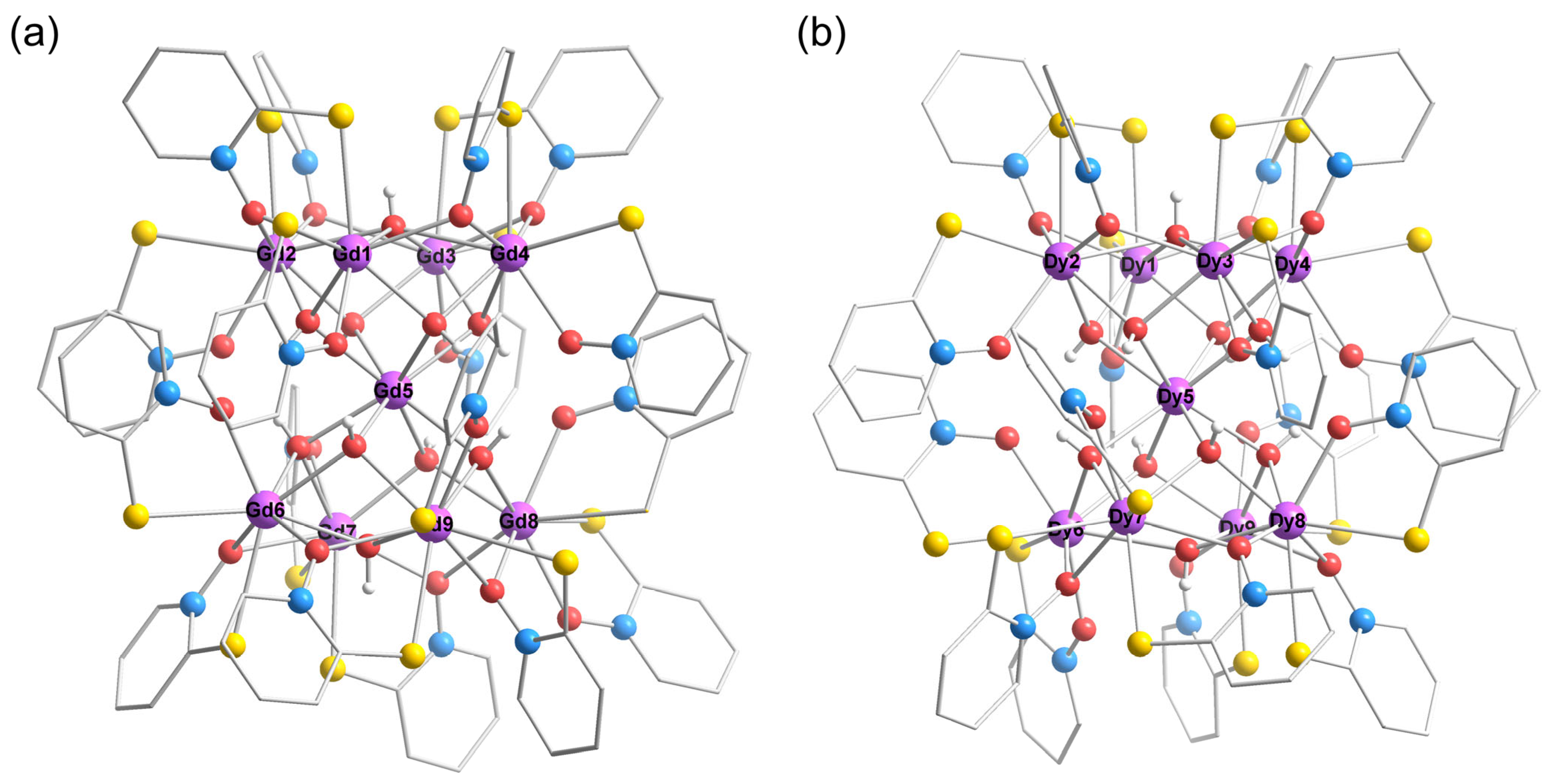
The asymmetric units of both complexes 1 and 3 feature nine metal ions, seventeen L ligands, nine μ3-OH and one μ4-OH groups, and some free solvent molecules. The eight Ln3+ (Ln1–Ln4 and Ln6–Ln9, Ln = Gd, Dy) ions occupy the vertices of a square antiprism, and the ninth (Ln5) resides at its geometric center, forming a canonical hourglass geometric configuration. This configuration consists of two quadrilateral base planes and eight triangular planes, resulting in an [Ln9(μ3-OH)9(μ4-OH)]17+ core encapsulated by seventeen bidentate L ligands. To facilitate the description of ligand coordination modes, three types are identified: (1) μ111: terminal bidentate coordination through both S and O donors to a single lanthanide center; (2) μ212: monodentate S-coordination to one lanthanide ion coupled with μ2-O bridging to a neighboring metal center; and (3) μ212: monodentate O-coordination to one lanthanide ion accompanied by μ2-S bridging to an adjacent metal ion (Figure 1).
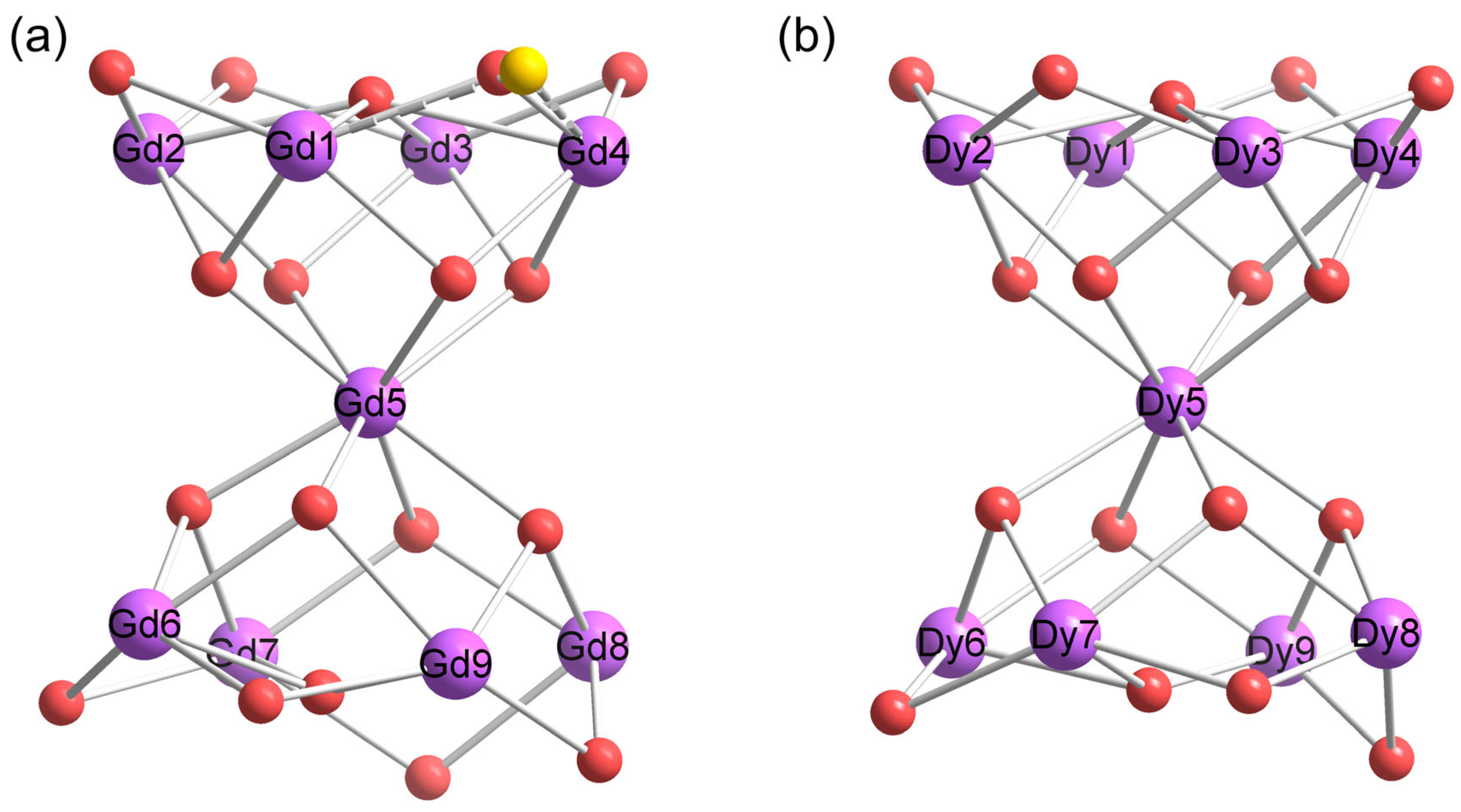
In complex 1, a μ4-OH group bridges the four Gd3+ ions (Gd1–Gd4) of the upper quadrilateral base plane (O atom 0.57 Å above the plane (Figure 2a); adjacent Gd···Gd distances in the range of 3.6107(6)–3.6700(6) Å). Adjacent Gd3+ ions in this plane are further interconnected by μ212 bridges from ligand L (Mode II: μ2-O and monodentate S), with the exception of the Gd1–Gd4 edge, where ligand disorder results in mixed μ212 bridging (approximately 60% Mode II (μ2-O) and 40% Mode III (μ2-S)) (Figure 3a). Four μ3-OH groups connect the upper quadrilateral plane to Gd5, forming four triangular planes. The Gd1–Gd4 ions are all eight-coordinate (Figure S1), adopting a triangular dodecahedron (D2d) local geometric configuration as determined by the SHAPE 2.1 software [44] (Table S2), with each coordination sphere containing two μ212 ligands, one μ111 ligand, one μ4-OH, and two μ3-OH groups. The central Gd5 ion occupies the geometric center, coordinated to eight μ3-OH groups that connect it to both the upper (Gd1–Gd4) and lower (Gd6–Gd9) quadrilateral planes, adopting a square antiprism (D4d) local geometry. The centers of the two quadrilateral base planes are separated by 5.5476 (2) Å with an interplanar torsion angle of 47.318(105)°. The lower quadrilateral base plane (Gd6–Gd9) exhibits distinct structural features (Figure S1). A μ3-OH group (O atom 0.53 Å below the plane) bridges Gd6, Gd7, and Gd9, while the plane displays significant distortion from ideal square geometry (adjacent Gd···Gd distances ranging from 3.5658(7) to 3.9598(7) Å and the bond lengths of Gd–μ3-O varying between 2.413(4) and 2.540(1) Å; Tables S3 and S4). This geometric asymmetry results in varied coordination environments for Gd6–Gd9 compared to the more symmetric upper Gd1–Gd4 plane, highlighting the overall molecular asymmetry. While adjacent Gd3+ ions in the lower plane are similarly interconnected by μ212 bridges from ligand L (Mode II: μ2-O and monodentate S), their coordination environments diverge significantly. Gd6 and Gd7 maintain eight-coordinate triangular dodecahedron (D2d) geometries, each binding to two μ212 ligands (combing one S atom and two μ2-O), one μ111 L ligand, and three μ3-OH groups. In contrast, Gd9 expands to nine-coordination through two μ212 modes (two S atoms and two μ2-O), one μ111 L ligand, and three μ3-OH groups, resulting in a spherical capped square antiprism (C4v) local geometry. Gd8 remains eight-coordinate with a triangular dodecahedron (D2d) local geometry but exhibits a distinct arrangement, coordinating to two μ2-O donors (from two μ212 L), two μ111 L ligands, and two μ3-OH groups.
According to the packing diagram (Figure S2), the shortest intermolecular Gd···Gd distance is 9.0353(7) Å. The presence of hydroxyl groups and solvent molecules within the cluster forms intra- and intermolecular hydrogen bonds (Table S5). Further analysis reveals that the pyridine rings mediate intra- and intermolecular π···π stacking (dihedral angle < 20°) (Table S6) and intermolecular C–H···π interactions, with distances between the hydrogen atoms and the ring centroids ranging from 2.68 to 2.98 Å (Table S7). These noncovalent interactions notably enhance the thermal and structural stability of the air-stable metal clusters.
In complex 3, the spatial arrangement of metal ions (Dy1–Dy9) and their linkage pattern across the eight triangular planes are identical to those in complex 1, forming a [Dy9(μ3-OH)9(μ4-OH)]17+ core structure (Figure 2b and Figure 3b). Notably, all Dy3+ ions are eight-coordinate. Dy1−Dy5 display coordination geometries that closely resemble those of their Gd3+ analogs (Gd1–Gd5), with Dy1–Dy4 adopting D2d local geometries and Dy5 exhibiting a D4d geometry (Table S8). Dy9 shows identical coordination to Gd6 and Gd7 (Figure S3), also featuring a D2d local geometry. However, Dy6 and Dy7 share a distinct coordination sphere comprising one μ3-OH from the lower plane, two μ3-OH bridges to central Dy5, one μ2-O donor (from one μ212 L), and two μ111 L ligands (Figure S4). Despite this similarity, their local geometries differ slightly, adopting D4d and D2d configurations, respectively. Dy8 adopts a unique environment, coordinated by two μ3-OH groups, two μ212 L ligands (containing two S and two μ2-O), and one μ111 L ligand, exhibiting a D2d local geometry. The adjacent Dy···Dy distances for Dy6–Dy9 range from 3.5824(7) to 3.8408(7) Å, and the Dy−μ3-O bond lengths display a wider variation (2.470(5)–2.715(5) Å) than those found in 1 (Tables S9 and S10). The packing analysis reveals that the shortest intermolecular Dy···Dy distance is 9.8618(6) Å (Figure S5), longer than that in 1. The structure also features an enhanced hydrogen bonding network (Table S5). Notably, the pyridine rings are only engaged in intramolecular π···π stacking (Table S6) and intermolecular C–H···π interactions, with distances between the hydrogen atom and the ring centroid ranging from 2.66 to 2.85 Å (Table S7).
Overall, in contrast to previously reported symmetric hourglass-like Ln9 clusters [24,41,42,43,45,46,47,48,49,50,51,52,53,54,55,56,57,58,59,60,61,62,63,64,65,66,67,68,69], the present Ln9 family exhibits significant structural asymmetry: Ln1–Ln4 and Ln6–Ln9 differ entirely in coordination environments, while the upper and lower basal planes feature different bridging modes (μ4-OH and μ3-OH, respectively). To facilitate subsequent studies on magneto-structural relationships, we compared cluster 1 with four previously reported Gd9 clusters that exhibit MCE [41,42,43]. Gd1–Gd5 share similar bridging modes: adjacent Gd1–Gd4 ions are connected by μ2-O bridges (Gd···Gd distance ≈ 3.6 Å) with a central μ4-OH group (diagonal Gd···Gd distance in the upper plane ≈ 5.2 Å), while Gd5 links to eight Gd3+ ions via eight μ3-OH bridges (Gd1–4···Gd5 distance ≈ 3.8 Å). Despite these similarities in connectivity and distances, cluster 1 exhibits distinct bond angles compared to the four cases: its adjacent Gd–μ2-O–Gd angles for Gd1–Gd4 (96.1–100.6°) are in a medium range (Table S11), its diagonal Gd–μ4-O–Gd angles in the upper plane (155.2–156.2°) are smaller, and its Gd1–4μ3-O–Gd5 angles (107.0–108.1°) show a larger average (Table S11). These differences become even more pronounced in Gd6–Gd9, which display greater variation in adjacent Gd···Gd distances (3.6–4.0 Å) with Gd–μ2-O–Gd angles (93.3–107.1°, Table S11). Here, the diagonal Gd6···Gd8 and Gd7···Gd9 distances are 5.7 Å and 4.8 Å, respectively, and the Gd6–9–O–Gd5 angles range from 102.4 to 109.9°. Considering that the key difference in cluster 1 lies in the partial replacement of conventional N/O donors by S atoms from the ligands, with a disordered segment containing μ2-S bridging Gd1 and Gd4, we propose that the asymmetric structure and variations in bond angles and Gd···Gd distances likely arise from the coordination and bridging of sulfur-containing ligands.
The powder X-ray diffraction patterns of polycrystalline samples of 1 and 2 are distinctly different from that of 3, matching well with their respective single-crystal X-ray diffraction simulations (Figure S6). This agreement confirms the high phase purity of all samples.

3.2. Magnetic Properties

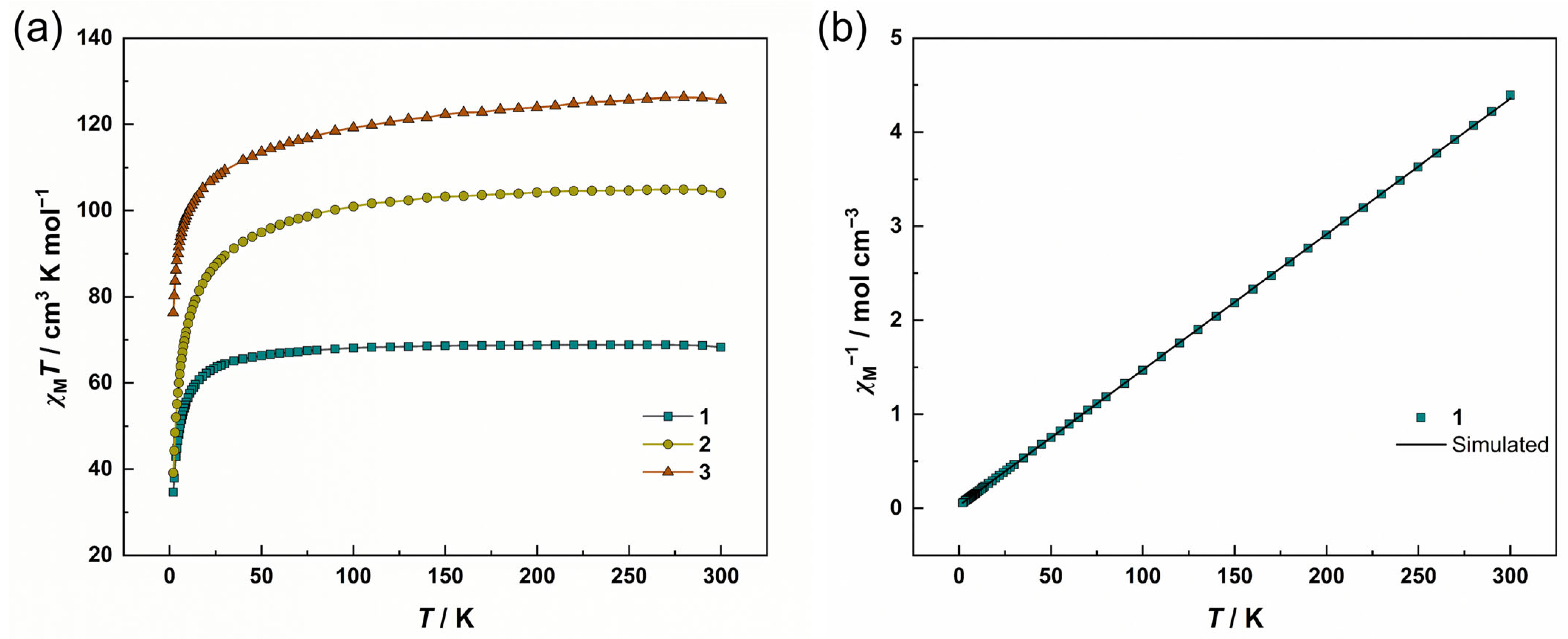
Variable-temperature magnetic susceptibility measurements of complexes 13 were performed on polycrystalline samples under a 1 kOe direct current (dc) magnetic field over the temperature range of 2–300 K. At 300 K, the χMT products for complexes 13 are 68.26, 104.02, and 125.64 cm3 K mol−1, respectively (Figure 4a), in close agreement with the theoretical values calculated for non-interacting magnetic centers: 70.92 cm3·K·mol−1 for nine independent Gd3+ ions (S = 7/2, g = 2.0), 106.38 cm3·K·mol−1 for nine isolated Tb3+ ions (S = 3, g = 3/2), and 127.53 cm3 K mol−1 for nine non-coupled Dy3+ ions (S = 5/2, g = 4/3). Upon cooling, complex 1 exhibits nearly constant χMT values above 80 K, reflecting the dominant paramagnetic behavior of non-interacting Gd3+ ions; below 80 K, χMT decreases gradually before undergoing a steep decline to 34.66 cm3 K mol−1 at 2 K, indicating the onset of antiferromagnetic interactions. Curie–Weiss analysis (2–300 K) yields Curie constant C = 69.35 cm3 K mol−1 and Curie temperature θ = −2.15 K, with the negative θ confirming antiferromagnetic interactions between metal ions (Figure 4b). Complex 2 exhibits a subtle decrease in χMT above 100 K, while complex 3 shows a more pronounced reduction in this regime. Both systems undergo progressive declines followed by steep reductions below 50 K, reaching minima of 39.12 and 76.30 cm3 K mol−1 at 2 K for 2 and 3, respectively. This behavior originates from thermal depopulation of Stark sublevels, with potential contributions from weak antiferromagnetic exchange.
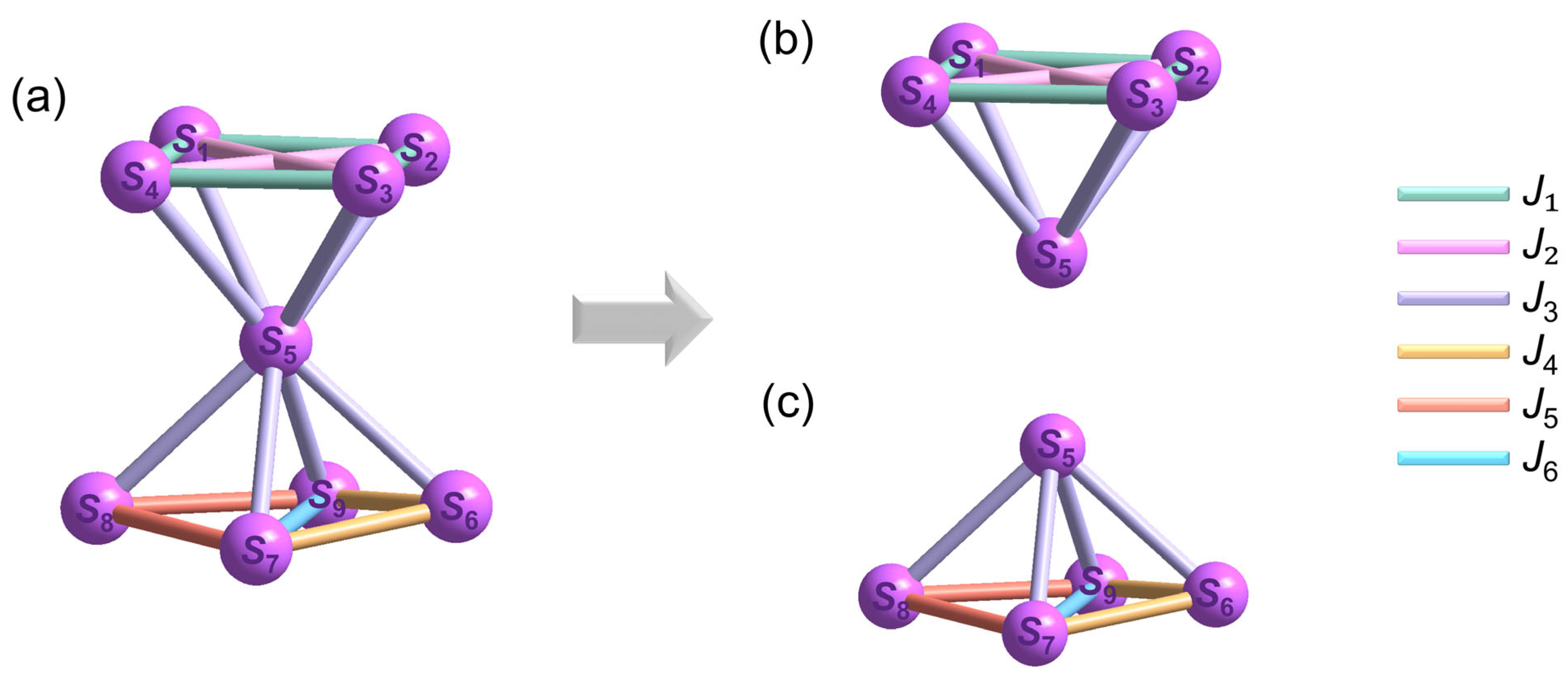
To further explore the magnetic interactions between Gd3+ ions in 1, we classify the exchange pathways into six distinct types (J1J6) based on their bridging motifs (Figure 5a). The magnetic interaction denoted as J1 stems from the coupling between Gd1 and Gd2, which are bridged by one μ4-OH, one μ3-OH, and a μ2-O ligand. Notably, ligand disorder along the Gd1–Gd4 edge, which leads to a mixed μ212 bridging mode, is not specifically considered in the magnetic model, similar to J1. J2 characterizes the diagonal magnetic interaction between Gd3+ ions in the upper Gd1–Gd4 quadrilateral base plane via a μ4-OH bridge. J3 describes the magnetic interaction between Gd5 and its adjacent Gd3+ ions through two μ3-OH groups. J4 accounts for the magnetic coupling between Gd3+ ions mediated by two μ3-OH and one μ2-O ligand. J5 quantifies the interaction between adjacent Gd3+ ions bridged by one μ3-OH and one μ2-O ligand. Finally, J6 defines the magnetic exchange interaction between Gd7 and Gd9, which are connected by a single μ3-OH bridge. Thus, the spin Hamiltonian of the system is derived by integrating these exchange interactions.
H ^ = 2 J 1 S 1 S 2 + S 2 S 3 + S 3 S 4 + S 1 S 4 2 J 2 S 1 S 3 + S 2 S 4 2 J 3 S 1 S 5 + S 2 S 5 + S 3 S 5 + S 4 S 5 + S 6 S 5 + S 7 S 5 + S 8 S 5 + S 9 S 5 2 J 4 S 6 S 7 + S 6 S 9 2 J 5 S 7 S 8 + S 9 S 8 2 J 6 S 7 S 9
In this model, S1S9 represent the spin of Gd1–Gd9, respectively, where positive values of J indicate ferromagnetic interactions, and negative values correspond to antiferromagnetic interactions.
To address computational challenges in modeling the magnetic properties of a nonanuclear cluster, we implement a reduced pentanuclear approximation. Given the prohibitively large exchange matrix for the full Gd9 system, we strategically retain the core magnetic unit comprising the central Gd5 ion and four vertex ions (Gd5: Gd1–Gd4 from the upper plane or Gd6–Gd9 from the lower plane) (Figure 5b,c). This simplification exploits the inherent S4 symmetry of the cluster, where Gd5 occupies the inversion center and the upper/lower Gd4 planes exhibit congruent coordination environments. The resultant Hamiltonian matrix enables computationally tractable fitting while preserving the dominant exchange pathways.
H ^ = 2 J 1 S 1 S 2 + S 2 S 3 + S 3 S 4 + S 1 S 4 2 J 2 S 1 S 3 + S 2 S 4 2 J 3 S 1 S 5 + S 2 S 5 + S 3 S 5 + S 4 S 5
H ^ = 2 J 3 S 6 S 5 + S 7 S 5 + S 8 S 5 + S 9 S 5 2 J 4 S 6 S 7 + S 6 S 9 2 J 5 S 7 S 8 + S 9 S 8 2 J 6 S 7 S 9
Considering the approximate treatments applied during the fitting, the χMT data for both Gd5 models were derived by converting the χMT values of 1 (MW = 3784.03 g mol−1): χMT (Gd5) = χMT (1)/9 × 5 (Figure S7). The optimal fitting of the experimental χMT vs. T data yields the magnetic parameters J1 = −0.055 cm−1, J2 = −0.114 cm−1, J3 = 0.019 cm−1, and g = 1.979 using Equation (2), and J3 = 0.015 cm−1, J4 = −0.097 cm−1, J5 = −0.082 cm−1, J6 = −0.072 cm−1, and g = 1.980 using Equation (3) [70]. These results are consistent with established magneto-structural relationships [42,71,72]. The ferromagnetic J3 coupling originates from μ3-OH bridges, aligning with empirical models where the Gd–O–Gd bond angles between 105 and 135° favor ferromagnetic interactions in Gd1–Gd5 (Table S4, bold font) [42,71,72]. However, in Gd5–Gd9, only 62.5% of the Gd–O–Gd bond angles fall within the 105–135° range. This may account for the smaller magnitude of the J3 in Equation (3) compared to that in Equation (2). In contrast, the antiferromagnetic J1, J2, J4, and J6 arise from μ4-OH/μ3-OH/μ2-O bridges, which is consistent with the correlation that excessively large or small bond angles result in antiferromagnetic coupling (Table S4, underlined font and wavy font, respectively). Compared with other hourglass-like Ln9 clusters [24,41,42,43,45,46,47,48,49,50,51,52,53,54,55,56,57,58,59,60,61,62,63,64,65,66,67,68,69], the structural differences in compound 1—beyond variations in ligands and symmetry—are mostly reflected in bond angles and Gd···Gd distances. For the upper base, the presence of μ4-OH bridging groups restricts the ability of sulfur-containing ligands to modify the distances between central metal ions during coordination. For the lower base, however, as only three Gd3+ ions are connected by one μ3-OH bridging group, the influence of sulfur-containing ligands becomes more prominent. They widen the Gd···Gd distances (Gd7···Gd8 = 3.96 Å, Gd8···Gd9 = 3.87 Å), make contributions to reducing magnetic exchange pathways [32], and also affect the distribution of Gd–O–Gd bond angles (Table S11). All these factors directly influence the magnetic coupling between metal ions.
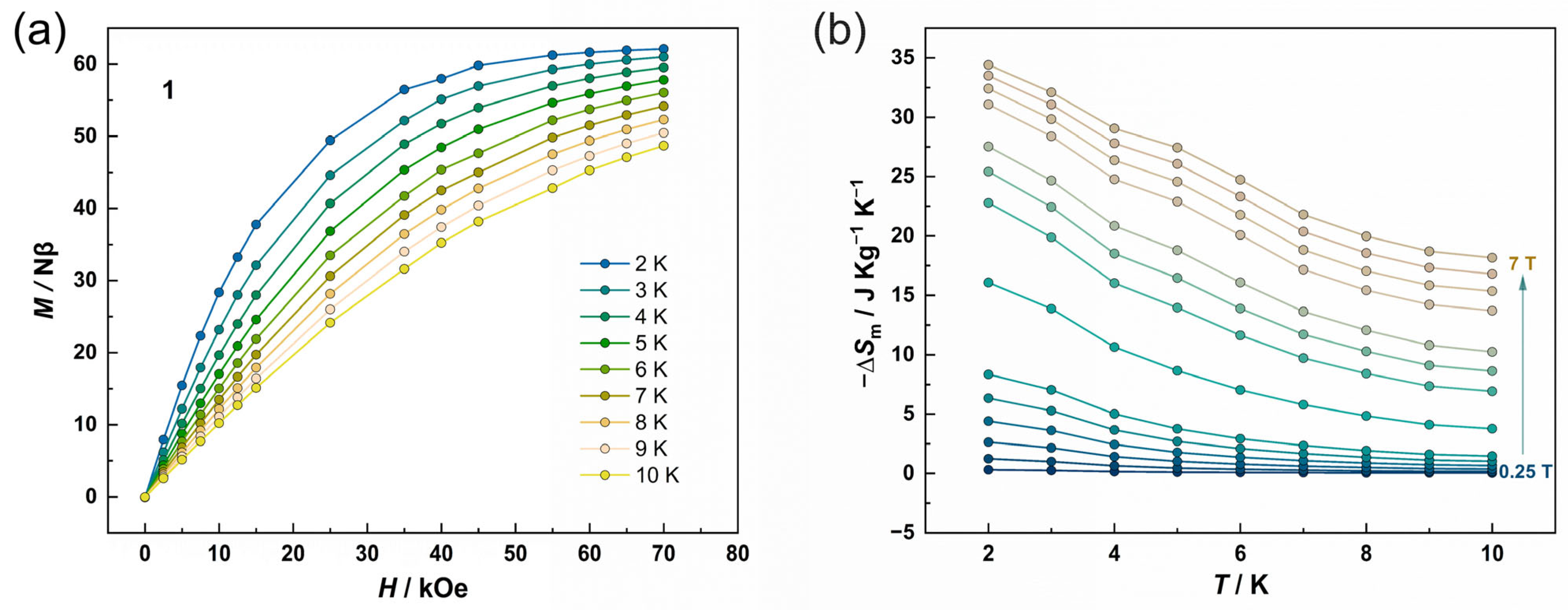
The field-dependent magnetization of complex 1 was measured at discrete temperatures (2–10 K) under applied magnetic fields up to 7 T (Figure 6a). The magnetization (M) versus field curves exhibit a nearly linear increase at low fields (< 2 T), followed by a gradual approach to saturation, reaching a value of 62.13 Nβ at 2 K and 7 T, which is in close agreement with the theoretical saturation magnetization of 63 Nβ for nine non-interacting Gd3+ ions. Given the weak magnetic interactions observed in 1 and the well-known MCE in Gd3+-based clusters, the −ΔSm values were calculated by the magnetic data using the Maxwell equation:
Δ S m ( T )   = [ M ( T ,   H ) / T ] H d H
where T is the testing temperature, H is the applied magnetic field, and M is magnetization.
As shown in Figure 6b, the −ΔSm values demonstrate a monotonic increase with decreasing temperature and increasing magnetic field change (ΔH), reaching a maximum value of −ΔSm = 34.41 J kg−1 K−1 at 2 K under ΔH = 7 T (Figure 6b). This value is approximately 84% of the theoretical limit of 41.2 J kg−1 K−1 calculated by −ΔSmmax = nRln(2S + 1)/MW, with the reduction primarily attributed to the presence of weak interactions between adjacent metal ions. Notably, while moderate among molecular magnetic coolers generally [4,8,9,10,11], this −ΔSm value ranks as the second highest reported for Gd9 clusters (Table S12). When the number of central metal ions is the same, magnetic density—a key parameter that determines entropy change—can be readily inferred from the molecular weight. For instance, 1 has the largest molar mass (MW = 3784.03 g mol−1), corresponding to the lowest value of NGd/MW among these Gd9 clusters [41,42]. In MCE materials research, a larger molar mass and lower magnetic density are generally considered unfavorable for achieving high −ΔSm values, as a lower concentration of magnetic ions per unit mass tends to limit the degree of magnetic ordering that contributes to entropy change under an external magnetic field [4]. Despite this, 1 exhibits a −ΔSm value comparable to that of Gd9(L1)4(μ4-OH)2(μ3-OH)8(μ2-OCH3)4(NO3)8(H2O)8}(OH)·2H2O (HL1 = 2,6-dimethoxyphenol, MW = 3015.28 g mol−1). This compound, although not the lightest, displays weak antiferromagnetic and ferromagnetic interactions and holds the highest −ΔSm value (40.60 J kg−1 K−1 at 2 K under ΔH = 7 T) reported for Gd9 clusters to date (Table S12) [41,42,43]. Meanwhile, 1 has a −ΔSm slightly larger than that of the third-ranked cluster, [Gd9(L2)4(NO3)12(μ3-OH)8(μ4-OH)2(CH3CH2OH)4]·4.5CH3CH2OH·3Et3NH (H2L2 = N-methyldiethanolamine, MW = 3288.90 g mol−1, −ΔSm = 33.65 J kg−1 K−1 at 3 K under ΔH = 7 T) [41], and much larger than that of the fourth-ranked cluster, [Gd9(L3)8(NO3)8(μ3-OH)8(μ4-OH)2]·NO3 (HL3 = 2-(2-aminoethoxy)ethanol; acac = acetylacetonate; MW = 2976.45 g mol−1, −ΔSm = 25.22 J kg−1 K−1 at 3 K under ΔH = 7 T) [41] with the highest magnetic density and lowest molecular weight in Gd9 clusters with MCE (Table S12).
Research findings reveal that both magnetic density and magnetic interactions affect the −ΔSm value. For complex 1, magnetic density seems to play a minor role in determining the large −ΔSm. We thus propose three potential factors contributing to its enhanced magnetocaloric performance. First, the incorporation of the heavy p-block S atoms from the HL ligands alters the cluster architecture, particularly affecting the Gd3+ ions at the vertices of the upper and lower basal planes, which are bridged by a μ4-OH and μ3-OH, respectively. Next, these sulfur-containing ligands in terminal coordination mode modulate key structural parameters—specifically Gd–μ2/μ3/μ4-O–Gd bond angles and Gd···Gd distances—rendering the structure distinct from previously reported Gd9 clusters ligated solely by N/O donors. Last, these geometric perturbations are expected to influence the nature and strength of magnetic exchange pathways, including both ferromagnetic and antiferromagnetic interactions. Collectively, these effects enhance the magnetic entropy change, despite the higher atomic mass of sulfur compared to conventional N/O-coordinating atoms.
Magnetization versus field measurements for complex 2 at 2 K reveal a gradual increase to a maximum moment of 46.31 Nβ at 7 T without a saturation plateau (Figure S8). Alternating-current (ac) magnetic susceptibility measurements under zero dc field with an oscillating field of 1.5 Oe show no detectable out-of-phase magnetic susceptibility (χ″) signal down to 2 K, suggesting the absence of magnetic relaxation (Figure S9). Consistently, hysteresis measurements at 2 K at a sweep rate of 200 Oe s−1 exhibit vanishingly small coercivity (Figure S10). This collective behavior precludes SMM behavior under these conditions.
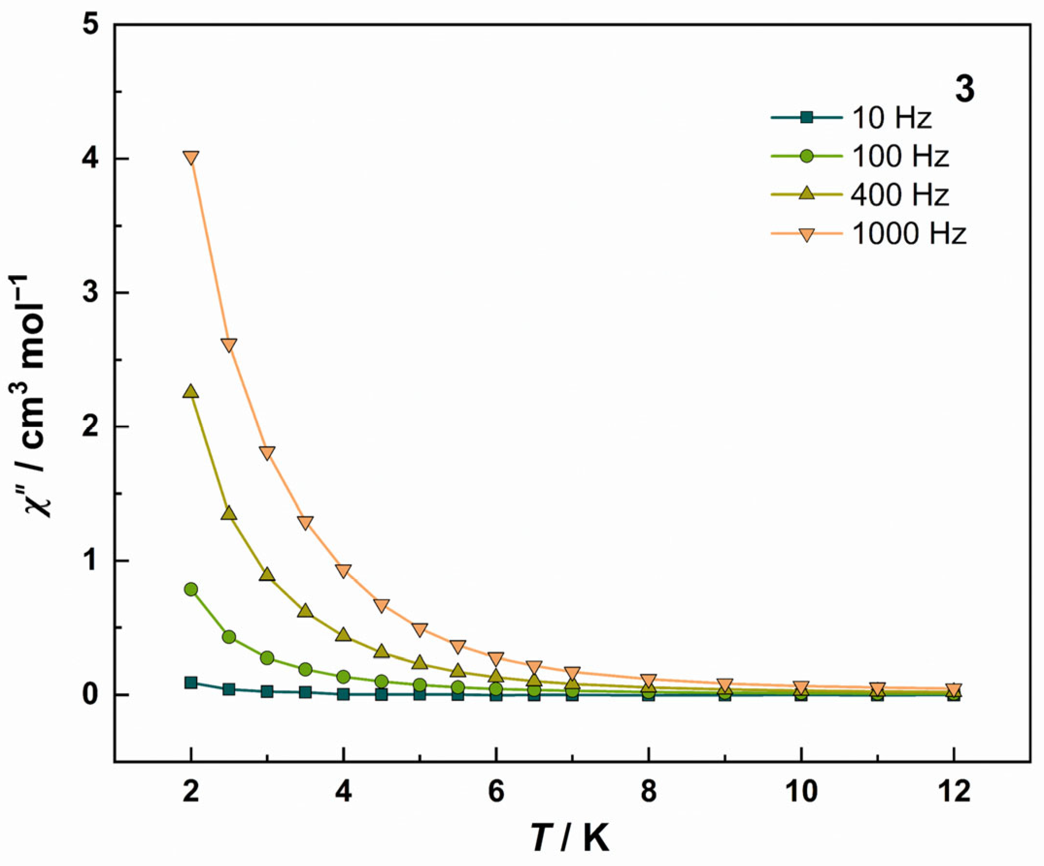
Magnetization versus field measurements for complex 3 at 2 K show a gradual approach to 50.91 Nβ at 7 T (Figure S11). The absence of distinct saturation and temperature-dependent non-superposition of M(H) curves possibly originates from strong intrinsic magnetic anisotropy. Frequency dependence of χ″ under 12 K confirms the slow magnetic relaxation characteristic of SMM behavior (Figure 7 and Figure S12). However, the absence of a discernible peak in the χ″ signal prevents the determination of relaxation time and thus the relaxation energy barrier. This behavior is similar to most reported nonanuclear Dy3+-based compounds [48,50,56,63]—except for one Dy9 compound with an energy barrier of Ueff = 9.24 K [47]. Zero-field-cooled and field-cooled (ZFC–FC) susceptibility measurements at H = 50 Oe show no divergence, ruling out long-range magnetic ordering or superparamagnetic transitions (Figure S13). Additionally, hysteresis measurements at 2 K at a sweep rate of 200 Oe s−1 exhibit minimal coercivity (Figure S14).

4. Conclusions

In summary, three nonanuclear lanthanide-based clusters [Ln9(L)17(μ3-OH)9(μ4-OH)]·nH2O (1: Ln = Gd, n = 3; 2: Ln = Tb, n = 3; 3: Ln = Dy, n = 1) were synthesized under hydrothermal conditions with sulfur-containing bidentate ligand 2-pyridinethiol 1-oxide. Remarkably, all three clusters exhibit a unique asymmetric architecture in which the upper and lower Ln9 cores are bridged by μ4-OH and μ3-OH groups, respectively. Of these three clusters, 1 exhibits an excellent cryogenic MCE with −ΔSm = 34.41 J kg−1 K−1 at 2 K under ΔH = 7 T, and 3 shows the SMM behavior. It is worth noting that, compared with the other four reported Gd9 clusters with MCE, 1 exhibits a magnetic entropy change value that is second only to the highest among Gd9 systems, despite having the largest molar mass and the lowest magnetic density, indicating the dominant role of magnetic coupling in determining the magnetocaloric performance. Specifically, the sulfur-containing bidentate ligands, coordinated in a terminal mode, influence key structural parameters of the nonanuclear cluster, including increasing the distances between Gd3+ ions, modifying the Gd–O–Gd bond angles, and reducing the number of bridging groups. This work not only expands research on the MCE of Gd9 clusters but also provides a new synthetic strategy for constructing high-performance molecular-based magnetic cooling materials using ligands containing heavy p-block elements.

Supplementary Materials

The following supporting information can be downloaded at https://www.mdpi.com/article/10.3390/magnetochemistry11090070/s1. Figure S1: The coordination environment diagram of compound 1. Color code: O (red), Gd (purple), S (yellow), and H (white). A disordered L with μ2-S (Mode III) coordination, free solvent molecules, and pyridine rings are omitted for clarity; Figure S2: The stacking pattern within a unit cell of 1. Color code: O (red), C (gray), N (blue), Gd (purple), and S (yellow). Disordered L with μ2-S, free solvent molecules, and H atoms are omitted for clarity; Figure S3: The part crystal structure diagram of compound 1 (a) and 3 (b) containing Ln6–Ln9. Color code: O (red), C (gray), N (blue), Gd/Dy (purple), S (yellow), and H (white). The extra carbon atoms on the pyridine rings with their hydrogen atoms and free solvent molecules are omitted for clarity; Figure S4: The coordination environment diagram of compound 3. Color code: O (red), C (gray), N (blue), Dy (purple), S (yellow), and H (white). Disordered L ligands, free solvent molecules, and pyridine rings are omitted for clarity; Figure S5: The stacking pattern within a unit cell of 3. Color code: O (red), C (gray), N (blue), Dy (purple), and S (yellow). Unordered L ligands, free solvent molecules, and H atoms are omitted for clarity; Figure S6: The PXRD curves of fresh samples of 13. The two calculated curves are simulated from the single crystal data of 1 and 3; Figure S7. Temperature dependence of χM and χMT for Gd5: Gd1–Gd5 (a) and Gd5–Gd9 (b). The experimental data of Gd5 were obtained by converting the data of 1, while the solid lines represent the best fits through the PHI program [70]; Figure S8: The plot of M vs. H at 2 K for 2; Figure S9: Temperature dependence of the in-phase (χ′) (a) and out-of-phase (χ′′) (b) ac susceptibility for 2 under zero dc field in the temperature range of 2–12 K; Figure S10: Hysteresis loop for 2 measured at 2 K with a sweep rate of 200 Oe/s; Figure S11: The plots of M vs. H at various temperatures for 3; Figure S12: Temperature dependence of the in-phase (χ′) (a) and out-of-phase (χ′′) (b) ac susceptibility for 3 under zero dc field in the temperature range of 2–12 K; Figure S13: FC and ZFC of 3 at an applied field of 50 Oe; Figure S14: Hysteresis loop for 3 measured at 2 K with a sweep rate of 200 Oe/s. Illustration: an enlarged view of the area near the zero field. Table S1: Crystallographic data and structure refinement for 13; Table S2: Continuous Shape Measures (CShMs) calculation for 1; Table S3: Selected bond lengths (Å) for 1; Table S4: Selected angles (deg) for 1; Table S5: Hydrogen bonding parameters in clusters 13; Table S6: π···π interaction parameters (Å, deg) for 1 and 3; Table S7: C–H···π interaction parameters (Å, deg) for clusters 1 and 3; Table S8: Continuous Shape Measures (CShMs) calculation for 3; Table S9: Selected bond lengths (Å) for 3; Table S10: Selected angles (deg) for 3; Table S11: The selected Gd–O–Gd angles of the reported Gd9 clusters with MCE [41,42,43]; Table S12: The MCE properties of the previously reported Gd9 clusters [41,42,43].

Author Contributions

Conceptualization, T.H.; methodology, Y.-W.G. and T.G.; investigation, Y.-W.G. and X.-Q.W.; data curation, T.G. and Y.-W.G.; writing—original draft preparation, Y.-W.G.; writing—review and editing, T.H.; supervision, T.H.; funding acquisition, T.H. Of the authors, Y.-W.G. and T.G. contributed equally to this work. All authors have read and agreed to the published version of the manuscript.

Funding

This research was funded by the National Natural Science Foundation of China (funding number: 22271228) and the Fundamental Research Funds for the Central Universities (funding number: xzy012023031).

Institutional Review Board Statement

Not applicable.

Informed Consent Statement

Not applicable.

Data Availability Statement

The original contributions presented in this study are included in the article and the Supplementary Materials.

Acknowledgments

We thank Chang Huang and Dan He at the Instrument Analysis Center of Xi’an Jiaotong University for assistance with single-crystal X-ray diffraction measurement and magnetic measurement, respectively.

Conflicts of Interest

The authors declare no conflicts of interest.

References

  1. Lucia, U.; Grisolia, G. Magnetocaloric refrigeration in the context of sustainability: A review of thermodynamic bases, the state of the art, and future prospects. Energies 2024, 17, 3585. [Google Scholar] [CrossRef]
  2. Zhao, S.; Wang, P.; Chen, W.; Wang, L.; Wang, Q.C.; Chen, W.Q. Supply and demand conflicts of critical heavy rare earth element: Lessons from gadolinium. Resour. Conserv. Recycl. 2023, 199, 1–9. [Google Scholar] [CrossRef]
  3. Nehan, P.Z.Z.; Vitayaya, O.; Munazat, D.R.; Manawan, M.T.E.; Darminto, D.; Kurniawan, B. The magnetocaloric effect properties for potential applications of magnetic refrigerator technology: A review. Phys. Chem. Chem. Phys. 2024, 26, 14476–14504. [Google Scholar] [CrossRef]
  4. Zheng, Y.-Z.; Zhou, G.-J.; Zheng, Z.; Winpenny, R.E.P. Molecule-based magnetic coolers. Chem. Soc. Rev. 2014, 43, 1462–1475. [Google Scholar] [CrossRef] [PubMed]
  5. Pecharsky, V.K.; Gschneidner, K.A. Magnetocaloric effect and magnetic refrigeration. J. Magn. Magn. Mater. 1999, 200, 44–56. [Google Scholar] [CrossRef]
  6. De Haas, W.J. Extremely low temperatures. Nature 1933, 132, 372–373. [Google Scholar] [CrossRef]
  7. Phan, M.H.; Yu, S.C. Review of the magnetocaloric effect in manganite materials. J. Magn. Magn. Mater. 2007, 308, 325–340. [Google Scholar] [CrossRef]
  8. Hu, J.J.; Peng, Y.; Liu, S.J.; Wen, H.R. Recent advances in lanthanide coordination polymers and clusters with magnetocaloric effect or single-molecule magnet behavior. Dalton Trans. 2021, 50, 15473–15487. [Google Scholar] [CrossRef]
  9. Konieczny, P.; Sas, W.; Czernia, D.; Pacanowska, A.; Fitta, M. Magnetic cooling: A molecular perspective. Dalton Trans. 2022, 51, 12762–12780. [Google Scholar] [CrossRef] [PubMed]
  10. Zhong, X.; Hu, J.-J.; Yao, S.-L.; Zhang, R.-J.; Wang, J.-J.; Cai, D.-G.; Luo, T.-K.; Peng, Y.; Liu, S.-J.; Wen, H.-R. Gd(III)-based inorganic polymers, metal-organic frameworks and coordination polymers for magnetic refrigeration. CrystEngComm 2022, 24, 2370–2382. [Google Scholar] [CrossRef]
  11. Fan, C.H.; Bai, K.P.; Luo, Q.C.; Han, T.; Huang, C.; Zheng, Y.Z.; Chen, W.P. A new family of SO42−-templated 3d-4f high-nuclearity clusters: Syntheses, structures, and magnetic properties. Chin. J. Chem. 2024, 42, 2438–2444. [Google Scholar] [CrossRef]
  12. Liu, S.J.; Han, S.D.; Zhao, J.P.; Xu, J.L.; Bu, X.H. In-situ synthesis of molecular magnetorefrigerant materials. Coord. Chem. Rev. 2019, 394, 39–52. [Google Scholar] [CrossRef]
  13. Liu, J.L.; Chen, Y.C.; Tong, M.L. Molecular design for cryogenic magnetic coolants. Chem. Rec. 2016, 16, 825–834. [Google Scholar] [CrossRef]
  14. Han, S.D.; Miao, X.H.; Liu, S.J.; Bu, X.H. Magnetocaloric effect and slow magnetic relaxation in two dense (3,12)-connected lanthanide complexes. Inorg. Chem. Front. 2014, 1, 549–552. [Google Scholar] [CrossRef]
  15. Zheng, X.Y.; Jiang, Y.H.; Zhuang, G.L.; Liu, D.P.; Liao, H.G.; Kong, X.J.; Long, L.S.; Zheng, L.S. A gigantic molecular wheel of {Gd140}: A new member of the molecular wheel family. J. Am. Chem. Soc. 2017, 139, 18178–18181. [Google Scholar] [CrossRef]
  16. Luo, X.M.; Hu, Z.B.; Lin, Q.F.; Cheng, W.W.; Cao, J.P.; Cui, C.H.; Mei, H.; Song, Y.; Xu, Y. Exploring the performance improvement of magnetocaloric effect based Gd-exclusive cluster Gd60. J. Am. Chem. Soc. 2018, 140, 11219–11222. [Google Scholar] [CrossRef] [PubMed]
  17. Zhitomirsky, M.E. Enhanced magnetocaloric effect in frustrated magnets. Phys. Rev. B 2003, 67, 1–7. [Google Scholar] [CrossRef]
  18. Chang, L.X.; Xiong, G.; Wang, L.; Cheng, P.; Zhao, B. A 24-Gd nanocapsule with a large magnetocaloric effect. Chem. Commun. 2013, 49, 1055–1057. [Google Scholar] [CrossRef]
  19. Ho, T.L. Hard soft acids bases (HSAB) principle and organic-chemistry. Chem. Rev. 1975, 75, 1–20. [Google Scholar] [CrossRef]
  20. Zheng, X.Y.; Kong, X.J.; Zheng, Z.P.; Long, L.S.; Zheng, L.S. High-nuclearity lanthanide-containing clusters as potential molecular magnetic coolers. Acc. Chem. Res. 2018, 51, 517–525. [Google Scholar] [CrossRef]
  21. Zheng, X.Y.; Xie, J.; Kong, X.J.; Long, L.S.; Zheng, L.S. Recent advances in the assembly of high-nuclearity lanthanide clusters. Coord. Chem. Rev. 2019, 378, 222–236. [Google Scholar] [CrossRef]
  22. Iki, N.; Tanaka, T.; Hiro-oka, S.; Shinoda, K. Self-assembly of a trilanthanide(III) core sandwiched between two thiacalix[4] arene ligands. Eur. J. Inorg. Chem. 2016, 2016, 5020–5027. [Google Scholar] [CrossRef]
  23. Tian, H.W.; Hong, J.Q.; Wang, K.; del Rosal, I.; Maron, L.; Zhou, X.G.; Zhang, L.X. Unprecedented reaction mode of phosphorus in phosphinidene rare-earth complexes: A joint experimental-theoretical study. J. Am. Chem. Soc. 2018, 140, 102–105. [Google Scholar] [CrossRef] [PubMed]
  24. Li, Z.P.; Wang, D.; Zhou, Z.H.; Zhao, G.Y.; Li, Q.; Bi, Y.F.; Zheng, Z.P. Thiacalix[4]arene-sandwiched sandglass-like Ln9 Clusters (Ln = Tb and Eu): Insights into the selective luminescence quenching properties by p-nitrobenzene derivatives. Inorg. Chem. 2022, 61, 20814–20823. [Google Scholar] [CrossRef] [PubMed]
  25. Ying, J.; Gao, N.; Mou, H.C.; Tian, A.X. Rare earth mono-substituted Keggin dimmers: Structures, electrochemical, supercapacitor and magnetic properties. J. Solid State Chem. 2022, 314, 1–11. [Google Scholar] [CrossRef]
  26. Ohama, K.; Komiya, M.; Yamatoya, T.; Sawamura, R.; Karashimada, R.; Gao, S.; Ozawa, Y.; Osada, K.; Aoki, I.; Nagasaki, T.; et al. Albumin nanoparticle electrostatically loaded with highly anionic Gd-thiacalixarene complex for MRI-guided neutron capture therapy. Colloids Surf., A 2024, 699, 1–13. [Google Scholar] [CrossRef]
  27. Marin, R.; Gálico, D.A.; Gayfullina, R.; Moilanen, J.O.; Carlos, L.D.; Jaque, D.; Murugesu, M. A zero-field single-molecule magnet with luminescence thermometry capabilities containing soft donors. J. Mater. Chem. C 2022, 10, 13946–13953. [Google Scholar] [CrossRef]
  28. Chen, S.M.; Xiong, J.; Zhang, Y.Q.; Yuan, Q.; Wang, B.W.; Gao, S. A soft phosphorus atom to "harden" an erbium(III) single-ion magnet. Chem. Sci. 2018, 9, 7540–7545. [Google Scholar] [CrossRef]
  29. Liu, S.S.; Lang, K.; Zhang, Y.Q.; Yang, Q.; Wang, B.W.; Gao, S. A distinct magnetic anisotropy enhancement in mononuclear dysprosium-sulfur complexes by controlling the Dy-ligand bond length. Dalton Trans. 2016, 45, 8149–8153. [Google Scholar] [CrossRef]
  30. Canaj, A.B.; Dey, S.; Céspedes, O.; Wilson, C.; Rajaraman, G.; Murrie, M. There is nothing wrong with being soft: Using sulfur ligands to increase axiality in a Dy(III) single-ion magnet. Chem. Commun. 2020, 56, 1533–1536. [Google Scholar] [CrossRef] [PubMed]
  31. Tziotzi, T.G.; Gracia, D.; Dalgarno, S.J.; Schnack, J.; Evangelisti, M.; Brechin, E.K.; Milios, C.J. A {Gd12Na6} molecular quadruple-wheel with a record magnetocaloric effect at low magnetic fields and temperatures. J. Am. Chem. Soc. 2023, 145, 7743–7747. [Google Scholar] [CrossRef]
  32. Braun, J.; Seufert, D.; Anson, C.E.; Tang, J.K.; Powell, A.K. An effectively uncoupled Gd8 cluster formed through fixation of atmospheric CO2 showing excellent magnetocaloric properties. Int. J. Mol. Sci 2024, 25, 264. [Google Scholar] [CrossRef]
  33. Wang, Y.F.; Wang, Y.X.; Yang, Q.Q.; Yin, B. Auxiliary rather than dominant. the role of direct Dy-S coordination in single-molecule magnet unveiled via ab initio study. J. Phys. Chem. A 2024, 128, 5285–5297. [Google Scholar] [CrossRef]
  34. Kumari, N.; Jassal, M.; Agrawal, A.K. Effect of metal ion on UV protective and antimicrobial properties of in-situ synthesized pyrithione complexes on cellulosic textiles. Mater. Today Commun. 2023, 34, 1–13. [Google Scholar] [CrossRef]
  35. Han, T.; Wang, X.Q.; Guo, T.; Cao, H.S.; Chen, W.P.; He, L. Modulation of the magnetic dynamics in two air-stable sulfur-ligated dysprosium complexes via polymerization. Dalton Trans. 2023, 52, 7551–7557. [Google Scholar] [CrossRef]
  36. Yuan, F.; Li, T.; Zhang, M.; Qian, H. Synthesis and crystal structure of tri(pyrithione) cerium complex and its catalytic property for oxidation of benzoin to benzil. Synth. React. Inorg. Met.-Org. Nano-Met. Chem. 2013, 43, 1510–1513. [Google Scholar] [CrossRef]
  37. Niu, D.Z.; Mu, L.L.; Lu, Z.S.; Song, B.L.; Sun, B.W. Synthesis and crystal structure of Pr3(DMSO)2 (Hmpo=2-mercaptopyridine N-oxide). J. Chem. Crystallogr. 2004, 34, 195–198. [Google Scholar] [CrossRef]
  38. Xiong, R.G.; Zuo, J.L.; You, X.Z.; Huang, X.Y. Synthesis and X-ray structure of the first lanthanide 2-mercaptopyridine N-oxide complex, tris-(1-hydroxy-2(1H)-pyridinethionato-O,S) bis sulfinylbis(methane) samarium(III). Polyhedron 1996, 15, 3321–3324. [Google Scholar] [CrossRef]
  39. Hu, Y.H.; Weng, L.H.; Huang, L.G.; Chen, X.T.; Wu, D.X.; Kang, B.S. Structure of tris(2-mercaptopyridine 1-oxido)cobalt(III) acetonitrile solvate. Acta Crystallogr. Sect. C Cryst. Struct. Commun. 1991, 47, 2655–2656. [Google Scholar] [CrossRef]
  40. West, D.X.; Frank, C.A. 2 series of rare-earth complexes of the 2-thiopyridine N-oxide anion. J. Inorg. Nucl. Chem. 1979, 41, 49–53. [Google Scholar] [CrossRef]
  41. Wang, T.; Wang, H.; Sun, W. Sandglass-like Ln9 nanoclusters with magnetocaloric effect and lanthanide-centered luminescence. Cryst. Growth Des. 2024, 24, 8076–8084. [Google Scholar] [CrossRef]
  42. Pandey, P.; Chauhan, D.; Walawalkar, M.G.; Gupta, S.K.; Meyer, F.; Rajaraman, G.; Murugavel, R. Hourglass-shaped homo- and heteronuclear nonanuclear lanthanide clusters: Structures, magnetism, photoluminescence, and theoretical analysis. Inorg. Chem. 2024, 63, 11963–11976. [Google Scholar] [CrossRef]
  43. Hu, P.; Li, S.H.; Cao, L.H.; Liu, A.G.; Zhuang, G.L.; Ji, L.D.; Li, B. Construction of a high nuclear gadolinium cluster with enhanced magnetocaloric effect through structural transition. Acs Omega 2022, 7, 38782–38788. [Google Scholar] [CrossRef]
  44. Casanova, D.; Cirera, J.; Llunell, M.; Alemany, P.; Avnir, D.; Alvarez, S. Minimal distortion pathways in polyhedral rearrangements. J. Am. Chem. Soc. 2004, 126, 1755–1763. [Google Scholar] [CrossRef]
  45. Panguluri, S.P.K.; Jourdain, E.; Chakraborty, P.; Klyatskaya, S.; Kappes, M.M.; Nonat, A.M.; Charbonnière, L.J.; Ruben, M. Yb-to-Eu cooperative sensitization upconversion in a multifunctional molecular nonanuclear lanthanide cluster in solution. J. Am. Chem. Soc. 2024, 146, 13083–13092. [Google Scholar] [CrossRef] [PubMed]
  46. Knighton, R.C.; Soro, L.K.; Lecointre, A.; Pilet, G.; Fateeva, A.; Pontille, L.; Francés-Soriano, L.; Hildebrandt, N.; Charbonnière, L.J. Upconversion in molecular hetero-nonanuclear lanthanide complexes in solution. Chem. Commun. 2021, 57, 53–56. [Google Scholar] [CrossRef] [PubMed]
  47. Hu, P.; Cao, L.H.; Liu, A.G.; Zhang, Y.Q.; Zhang, T.L.; Li, B. Modulating the relaxation dynamics via structural transition from a dinuclear dysprosium cluster to a nonanuclear cluster. Dalton Trans. 2021, 50, 12814–12820. [Google Scholar] [CrossRef] [PubMed]
  48. Huang, Y.; Qin, Y.R.; Ge, Y.; Cui, Y.F.; Zhang, X.M.; Li, Y.H.; Yao, J.L. Rationally assembled nonanuclear lanthanide clusters: Dy9 displays slow relaxation of magnetization and Tb9 serves as luminescent sensor for Fe3+, CrO42− and Cr2O72−. New J. Chem. 2019, 43, 19344–19354. [Google Scholar] [CrossRef]
  49. Griffiths, K.; Kühne, I.A.; Tizzard, G.J.; Coles, S.J.; Kostakis, G.E.; Powell, A.K. Twists to the spin structure of the Ln9-diabolo motif exemplified in two {Zn2Ln2}Ln9{Zn2} coordination clusters. Inorg. Chem. 2019, 58, 2483–2490. [Google Scholar] [CrossRef]
  50. Wang, H.S.; Long, Q.Q.; Yin, C.L.; Xu, Z.W.; Pan, Z.Q. Syntheses, crystal structures and magnetic properties of sandglass Dy9III and irregular tetrahedron Dy4III complexes. Polyhedron 2018, 141, 69–76. [Google Scholar] [CrossRef]
  51. Chen, H.F.; Yang, X.P.; Jiang, D.M.; Shi, D.L.; Zhang, L.J. Construction of NIR luminescent polynuclear lanthanide-based nanoclusters with sensing properties towards metal ions. Dalton Trans. 2018, 47, 13880–13886. [Google Scholar] [CrossRef]
  52. Zheng, X.Y.; Peng, J.B.; Livera, M.; Luo, Y.; Wang, Y.Y.; Kong, X.J.; Long, L.S.; Zheng, Z.P.; Zheng, L.S. Selective formation of chromogen I from N-acetyl-D-glucosamine upon lanthanide coordination. Inorg. Chem. 2017, 56, 110–113. [Google Scholar] [CrossRef]
  53. Mylonas-Margaritis, I.; Kitos, A.A.; Panteli, C.C.; Skordi, K.; Tasiopoulos, A.J.; Bekiari, V.; Escuer, A.; Perlepes, S.P. 2-hydroxybenzophenone-controlled self-assembly of enneanuclear lanthanide(III) hydroxo coordination clusters with an “hourglass”-like topology. Inorg. Chem. Commun. 2017, 83, 118–122. [Google Scholar] [CrossRef]
  54. Omagari, S.; Nakanishi, T.; Kitagawa, Y.; Seki, T.; Fushimi, K.; Ito, H.; Meijerink, A.; Hasegawa, Y. Critical role of energy transfer between terbium ions for suppression of back energy transfer in nonanuclear terbium clusters. Sci. Rep. 2016, 6, 1–11. [Google Scholar] [CrossRef] [PubMed]
  55. Greisch, J.F.; Chmela, J.; Harding, M.E.; Klopper, W.; Kappes, M.M.; Schooss, D. Gas-phase photoluminescence characterization of stoichiometrically pure nonanuclear lanthanoid hydroxo complexes comprising europium or gadolinium. Inorg. Chem. 2016, 55, 3316–3323. [Google Scholar] [CrossRef] [PubMed]
  56. Zou, H.H.; Sheng, L.B.; Chen, Z.L.; Liang, F.P. Lanthanide nonanuclear clusters with sandglass-like topology and the SMM behavior of dysprosium analogue. Polyhedron 2015, 88, 110–115. [Google Scholar] [CrossRef]
  57. Omagari, S.; Nakanishi, T.; Seki, T.; Kitagawa, Y.; Takahata, Y.; Fushimi, K.; Ito, H.; Hasegawa, Y. Effective photosensitized energy transfer of nonanuclear terbium clusters using methyl salicylate derivatives. J. Phys. Chem. A 2015, 119, 1943–1947. [Google Scholar] [CrossRef]
  58. Nakanishi, T.; Suzuki, Y.; Doi, Y.; Seki, T.; Koizumi, H.; Fushimi, K.; Fujita, K.; Hinatsu, Y.; Ito, H.; Tanaka, K.; et al. Enhancement of optical faraday effect of nonanuclear Tb(III) complexes. Inorg. Chem. 2014, 53, 7635–7641. [Google Scholar] [CrossRef]
  59. Greisch, J.F.; Harding, M.E.; Schäfer, B.; Ruben, M.; Klopper, W.; Kappes, M.M.; Schooss, D. Characterization of nonanuclear europium and gadolinium complexes by gas-phase luminescence spectroscopy. J. Phys. Chem. Lett. 2014, 5, 1727–1731. [Google Scholar] [CrossRef]
  60. Zhang, B.; Xiao, T.; Liu, C.M.; Li, Q.; Zhu, Y.Y.; Tang, M.S.; Du, C.X.; Song, M.P. Systematic study of the luminescent europium-based nonanuclear clusters with modified 2-hydroxybenzophenone ligands. Inorg. Chem. 2013, 52, 13332–13340. [Google Scholar] [CrossRef]
  61. Singh-Wilmot, M.A.; Sinclair, R.A.; Andrews, M.; Rowland, C.; Cahill, C.L.; Murugesu, M. Nonanuclear lanthanide(III) nanoclusters: Structure, luminescence and magnetic properties. Polyhedron 2013, 53, 187–192. [Google Scholar] [CrossRef]
  62. Xu, X.B.; Zhao, L.; Xu, G.F.; Guo, Y.N.; Tang, J.K.; Liu, Z.L. A diabolo-shaped Dy9 cluster: Synthesis, crystal structure and magnetic properties. Dalton Trans. 2011, 40, 6440–6444. [Google Scholar] [CrossRef]
  63. Alexandropoulos, D.I.; Mukherjee, S.; Papatriantafyllopoulou, C.; Raptopoulou, C.P.; Psycharis, V.; Bekiari, V.; Christou, G.; Stamatatos, T.C. A new family of nonanuclear lanthanide clusters displaying magnetic and optical properties. Inorg. Chem. 2011, 50, 11276–11278. [Google Scholar] [CrossRef] [PubMed]
  64. Baril-Robert, F.; Petit, S.; Pilet, G.; Chastanet, G.; Reber, C.; Luneau, D. Site-selective lanthanide doping in a nonanuclear yttrium(III) cluster revealed by crystal structures and luminescence spectra. Inorg. Chem. 2010, 49, 10970–10976. [Google Scholar] [CrossRef]
  65. Petit, S.; Baril-Robert, F.; Pilet, G.; Reber, C.; Luneau, D. Luminescence spectroscopy of europium(III) and terbium(III) penta-, octa- and nonanuclear clusters with β-diketonate ligands. Dalton Trans. 2009, 6809–6815. [Google Scholar] [CrossRef] [PubMed]
  66. Volpe, M.; Bombieri, G.; Clemente, D.A.; Foresti, E.; Grillone, M.D. Synthesis and physico-chemical characterization of a new series of hydroxide ion acetylacetonate lanthanide(III)-ditungsten decacarbonyl hydride complexes. J. Alloys Compd. 2004, 374, 382–386. [Google Scholar] [CrossRef]
  67. Addamo, M.; Bombieri, G.; Foresti, E.; Grillone, M.D.; Volpe, M. Assembling process of charged nonanuclear cationic lanthanide(III) clusters assisted by dichromium decacarbonyl hydride. Inorg. Chem. 2004, 43, 1603–1605. [Google Scholar] [CrossRef]
  68. Xu, G.; Wang, Z.M.; He, Z.; Lu, Z.; Liao, C.S.; Yan, C.H. Synthesis and structural characterization of nonanuclear lanthanide complexes. Inorg. Chem. 2002, 41, 6802–6807. [Google Scholar] [CrossRef]
  69. Hubert-Pfalzgraf, L.G.; Miele-Pajot, N.; Papiernik, R.; Vaissermann, J. A novel example of self-assembly in lanthanide chemistry: Synthesis and molecular structure of Na(EtOH)6Y94-O)23-OH)8{μ-η2-MeC(O)CHC(O)OEt}82-MeC(O)CHC(O)OEt}8. J. Chem. Soc. Dalton Trans. 1999, 4127–4130. [Google Scholar] [CrossRef]
  70. Chilton, N.F.; Anderson, R.P.; Turner, L.D.; Soncini, A.; Murray, K.S. PHI: A powerful new program for the analysis of anisotropic monomeric and exchange-coupled polynuclear d- and f-block complexes. J. Comput. Chem. 2013, 34, 1164–1175. [Google Scholar] [CrossRef]
  71. Singh, M.K.; Rajeshkumar, T.; Kumar, R.; Singh, S.K.; Rajaraman, G. Role of (1,3) {Cu-Cu} interaction on the magneto-caloric effect of trinuclear {CuII-GdIII-CuII} complexes: Combined DFT and experimental studies. Inorg. Chem. 2018, 57, 1846–1858. [Google Scholar] [CrossRef] [PubMed]
  72. Rajeshkumar, T.; Singh, S.K.; Rajaraman, G. A computational perspective on magnetic coupling, magneto-structural correlations and magneto-caloric effect of a ferromagnetically coupled {GdIII-GdIII} pair. Polyhedron 2013, 52, 1299–1305. [Google Scholar] [CrossRef]
Figure 1. Ligand coordination modes: Modes I (a), II (b), and III (c).
Magnetochemistry 11 00070 g001
Figure 2. The crystal structure diagram of 1 (a) and 3 (b). Color code: O (red), C (gray), N (blue), Gd/Dy (purple), S (yellow), and H (white). A disordered L with μ2-S (Mode III) coordination in 1, disordered L ligands in 3, and free solvent molecules and hydrogen atoms of pyridine rings in 1 and 3 are omitted for clarity.
Magnetochemistry 11 00070 g002
Figure 3. Representation of the core structure of 1 (a) and 3 (b). Color code: O (red), Gd/Dy (purple), and S (yellow). The gray dotted line represents the disordered components within the structure.
Magnetochemistry 11 00070 g003
Figure 4. (a) Temperature-dependent susceptibilities for 13 under a dc magnetic field of 1000 Oe; (b) the χM−1 vs. T plot of 1. The solid black line represents the best fits with the Curie–Weiss law.
Magnetochemistry 11 00070 g004
Figure 5. Magnetic coupling schematic diagram of 1 (a), Gd5 including Gd1–Gd5 (b), and Gd5 including Gd5–Gd9 (c) systems in 1. Color code: Gd (purple), J1 (cyan), J2 (pink), J3 (light purple), J4 (yellow), J5 (orange), and J6 (blue).
Magnetochemistry 11 00070 g005
Figure 6. (a) Plots of M vs. H at various temperatures for 1. (b) Plots of experimental magnetic entropy change versus T for 1 at 0.25–7 T from 2 to 10 K.
Magnetochemistry 11 00070 g006
Figure 7. Temperature dependence of the χ″ ac susceptibility for 3 under zero dc field in the temperature range of 2–12 K.
Magnetochemistry 11 00070 g007
Disclaimer/Publisher’s Note: The statements, opinions and data contained in all publications are solely those of the individual author(s) and contributor(s) and not of MDPI and/or the editor(s). MDPI and/or the editor(s) disclaim responsibility for any injury to people or property resulting from any ideas, methods, instructions or products referred to in the content.