Article

Vernonia amygdalina Leaf Extract Loaded Electrosprayed Particles for Inhibiting Phytophthora spp. Causing Citrus Root Rot

by
Pratchaya Tipduangta
1,
Sunee Chansakaow
1,
Sirinthicha Thakad
1,
Pawitrabhorn Samutrtai
1,
Aekkhaluck Intharuksa
1,
Ratchadawan Cheewangkoon
2,
Anuruddha Karunarathna
2,
Tipprapa Promthep
2 and
Busaban Sirithunyalug
1,*
1
Department of Pharmaceutical Sciences, Faculty of Pharmacy, Chiang Mai University, Chiang Mai 50200, Thailand
2
Department of Entomology and Plant Pathology, Faculty of Agriculture, Chiang Mai University, Chiang Mai 50200, Thailand
*
Author to whom correspondence should be addressed.
Horticulturae 2023, 9(9), 969; https://doi.org/10.3390/horticulturae9090969
Submission received: 24 July 2023 / Revised: 23 August 2023 / Accepted: 24 August 2023 / Published: 25 August 2023

Abstract

:
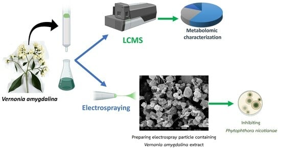
Citrus is an important economic plant in Thailand. The infection of citrus roots by Phytophthora nicotianae leads to root rot, reduced growth, and branch death. Although fosetyl aluminum and metalaxyl are commonly employed to address citrus root rot, they possess limitations in terms of their ability to diffuse to the root of citrus. Vernonia amygdalina leaf ethyl acetate extract (VLE) has been demonstrated to effectively inhibit Pythium deliense, a fungus closely related to Phytophthora nicotianae. This study aimed to investigate the anti-fungus activity of fractions obtained from the ethyl acetate extract of Vernonia amygdalina leaf against Phytophthora nicotianae, identify the most effective fraction, and formulate it into polymeric micro/nanoparticles using the electrospray process. The findings revealed that the VLE fraction eluted with ethanol:chloroform 1:1 had a high alkaloid content from metabolomic study and exhibited the potential to inhibit Phytophthora nicotianae at a concentration of 200 µg/mL. Consequently, this fraction was selected for incorporation into polymer blends of Poly Vinyl Alcohol/cellulose acetate to generate electrosprayed particles with a diameter of 0.97 ± 0.55 microns. These particles effectively suppressed in vitro Phytophthora nicotianae, thereby suggesting that VLE-containing electrosprayed particles have the potential to be applied and their in vivo performance in the treatment of citrus root rot evaluated in future experiments.

Graphical Abstract

1. Introduction

Thailand, one of the leading citrus-producing countries in Southeast Asia, had an overall citrus production of 1.1 million tons in 2021 [1]. The citrus industry in Thailand has experienced significant growth over the past years, and the country is renowned for its diverse range of citrus fruits. Oranges, tangerines, pomelos, and lemons are among the most commonly cultivated citrus fruits in Thailand [2].
Root rot disease is a widespread issue in citrus trees worldwide, including Thailand. It is typically caused by fungus pathogens that thrive in poorly drained or waterlogged soils. The most prevalent fungus species associated with root rot in citrus trees are Phytophthora spp., particularly Phytophthora nicotianae and Phytophthora citrophthora [3,4]. Symptoms of root rot in citrus trees include wilting, yellowing or browning of leaves, stunted growth, a decline in overall tree health, and dieback. Infected trees may also exhibit a general decline in fruit quality and yield. If left untreated, root rot can lead to the death of the tree through dieback [5,6]. Various approaches exist for managing root rot caused by Phytophthora nicotianae, such as pruning to avoid waterlogged conditions [7], biological control using Chaetomium spp. [8], and antifungus chemical control [9]. Fosetyl aluminum and metalaxyl have been used to control Phytophthora spp., although there are limitations in their ability to diffuse to the roots and easily degraded in soil [9,10].
Bitter leaf (Vernonia amygdalina Delile) has been found to possess anti-fungus properties. It has been reported that the ethyl acetate extract of bitter leaf demonstrated efficacy against Pythium deliense, a major cause of root rot disease in Catharanthus roseus (L.) D.Gon [11]. Pythium and Phytophthora belong to the class Oomycota and share a similar ancestor [12,13]. Thus, our study hypothesizes that the ethyl acetate leaf extract from V. amygdalina will have potency against P. nicotianae.
Next, the extract will be formulated into polymeric particles for easier storage and better compound stability. In this study, cellulose acetate and a blend of cellulose acetate/polyvinyl alcohol have been selected as a polymer matrix for the V. amygdalina leaf extract. Electrospray, a technique that employs electrostatic force to create particles from a polymeric solution, will be used to prepare micro- to nanoparticles [14]. This process can produce smaller particles without involving heat, unlike the common spray drying technique [15]. Hence, it is a suitable technique for preparing V. amygdalina leaf extract polymeric particles. Therefore, our study aims to explore an alternative approach to inhibiting P. nicotianae using V. amygdalina leaf extract and develop them as electrosprayed microparticles.

2. Materials and Methods

2.1. Materials

Organic solvents including ethyl acetate, hexane, absolute ethanol, chloroform, acetone and dimethylacetamide were analytical grade and purchased from Fisher Co. Ltd. and distributed by Union Sci Chiang Mai. Silica gel was purchased from Merck KGaA (Damstadt, Germany). Polyvinyl alcohol (PVA) Mw 13,000–23,000, 87–89% hydrolyzed and cellulose acetate (CA) number average molecular weight (Mn) 30,000 were purchased from Sigma-Aldrich Chemie GmbH (Taufkirchen, Germany). The mycology and bio-assay processes: Potato Dextrose Agar (PDA) from HiMedia Laboratories Pvt. Ltd. Dimethyl sulfoxide (DMSO) from RCI laboratories Ltd. Distributed by V. S. Chem house, Bangkok, Thailand.

2.2. Vernonia Amygdalina Identification

To assure the accuracy of botanical origin, Vernonia amygdalina Del. (Nan Chao Wei) samples were collected from the medicinal plant garden of the Faculty of Pharmacy, Chiang Mai University, Chiang Mai, Thailand in the dry season of November 2021. The aerial parts of both vegetative and reproductive structures of at least three V. amygdalina samples were carefully examined for morphological identification. The living specimen of Vernonia amygdalina was then identified following the plant description of the World Flora Online database [16]. The morphological characters of flowers and fruits were carried out under a stereo microscope in triplicate. The integral features of plant structures that are helpful to identify were investigated.

2.3. Vernonia Amygdalina Leaf Extraction and Fraction Separation

Vernonia amygdalina leaves were washed thoroughly with tap water and dried at room temperature. Dried leaves were placed in a tray and oven-dried at 60 °C for 16 h. The dry leaves were powdered. The coarse powder was macerated in hexane and periodically stirred every 8 h for 24 h. Then, this extraction process was repeated in triplicate. After that hexane was removed by filtering through filter papers (Whatman No.4). The hexane extraction was performed to remove non-polar compounds from the Vernonia amygdalina leaves extract and it was excluded from this study. The Vernonia amygdalina leaf coarse powder from the above step was then macerated in ethyl acetate (EtOAc) and stirred periodically every 8 h for 24 h. After that, the extraction procedure was conducted three times. The EtOAc extract was combined and filtered using filter papers (Whatman No.4). The solvent in the filtrate was removed using the rotary evaporator model Rotavapor® R-300 (Buchi, Flawil, Switzerland). The resultant dark green crude was stored in an amber glass bottle at 4 °C until use.
The crude extract of V. amygdalina leaf extraction (3 g) was isolated by column chromatography (CC) using silica gel 60 (particle size 0.063–0.200 nm) as a stationary phase. A gradient of chloroform and methanol was used in the mobile phase to obtain 7 fractions. The fraction was determined by thin-layer chromatography (TLC) with 70% hexane in ethyl acetate, and determined under 254 and 365 nm UV light. Each fraction was concentrated by a rotary evaporator model Rotavapor® R-300 (Buchi, Flawil, Switzerland).

2.4. Metabolomic Profiles Analysis Using LC-MS/MS

Untargeted metabolomics analysis of each V. amygdalineacha fraction was conducted using a Q-exactive Quadrupole Orbitrap Mass Spectrometer (MS) coupled with an UltiMate 3000 LC system. To retrieve the full separation between each metabolite in the LC system, a total of 5 µL sample injections were used at a flow rate of 0.3 mL/min. The Hypersil GOLD™ column and auto-sampler temperatures were maintained at 60 °C and 6 °C, respectively. The mobile phase was composed of 50%/50% methanol/water with 0.1% formic acid (MP: A), and acetonitrile with 0.1% formic acid (MP: B) (LC-MS grade, Sigma). Gradient starting conditions were 99% MP: A and 5% MP: B. Starting conditions were held for 1 min before rising to 95% B over 23 min. The column was flushed with 100% B for 5 min before returning to the starting conditions for 12 min. The total time of each analysis was 40 min. A blank sample (0.1% formic acid/methanol) was administered after every injection. All metabolites in each sample were elucidated by the MS system in a positive mode. A spray voltage of 3.8 kV in both positive mode, sheath gas, and auxiliary gas flow rates were set at 48 and 11 arbitrary units (AU), respectively. The capillary temperature was 350 °C. The MS analysis alternated between MS full scans and data-dependent MS/MS scans with dynamic exclusion. LC-MS for full MS: scan range, 75–700 m/z; resolution 120,000; AGC target 3 × 10⁶; max IT 30 ms and LC-MS for full MS/MS; resolution 15,000; AGC target 1 × 10⁵; max IT 50 ms. Up to five ions (Top5) with the most intense signal were fragmented. All LC-MS runs were acquired and the metabolites were identified against the database using the Xcalibur 3.1 software (Thermo Scientific, Waltham, MA, USA) [17]. All metabolites were categorized by their chemical structures and then were compared between fractions.

2.5. Selection and Identification of the Fungus Strain

The citrus root rot causing Phytophthora nicotianae (YY002) was obtained from the Department of Entomology and Plant Pathology culture collection, Faculty of Agriculture, Chiang Mai University, Chiang Mai, Thailand. Chomchid et al. (2020) have confirmed the nomenclature and the pathogenicity of P. nicotianae (YY002) [18].

2.6. Phytophthora Nicotianae Inhibition Assay against V. amygdalina Leaf Extract Fractions and Its Crude by Using Poison Media

The P. nicotianae (YY002) obtained from the Department of Entomology and Plant Pathology was transferred to a new Potato Dextrose Agar (PDA) plate and incubated for five days. Mycelial plug disks (5.5 mm) were prepared from the growing end of the colony. The crude extract and the EtOAc fractions were dissolved in 0.2% DMSO. The resultant mixtures were filtered using a Whatman™ Puradisc™ Nylon Syringe filter and added to PDA (40 °C) to make the concentration gradient: 0, 200, 500, and 1000 µg/mL. The 0 µg/mL was considered as the negative control. The PDA mixtures were then poured into Petri dishes (15 mL/plate). Previously prepared mycelial plugs were transferred to poison PDA and incubated at room temperature. The results were recorded daily until the control reached full maturity by measuring the colony diameter to calculate the percent inhibition of radial growth (PIRG) and effective dose (ED50). PIRG can be calculated by using Equation (1) where the R1 and R2 method is illustrated in Figure 1.
PIRG = [R1 − R2)/R1] × 100

2.7. Electrosprayed Particle Preparation

The V. amygdalina leaf extraction fraction that exhibited the highest effectiveness against P. nicotianae in experiment 2.6, identified as fraction 7, was dissolved in a mixture of acetone and dimethylacetamide (in a ratio of 2:1). Following this, polymers (PVA and/or cellulose acetate) were added and the solution volume was adjusted to 15 mL. The composition of each formulation is shown in Table 1. The electrosprayed solution was loaded in a 10 mL plastic syringe and equipped with an 18 G blunt metal needle, then it was installed in a syringe pump model NE-300 (New Era Pump System Inc., Farmingdale, NY, USA). The solution feeding rate was set at 0.8 mL.h−1. The distance between the needle tip and the foil-covered aluminum plate collector was 15 cm. The ES-30 high voltage generator (Gamma High Voltage Research, Ormond Beach, FL, USA) supplied voltage at 12 kV throughout the electrospraying. The electrospraying process was performed at ambient conditions (30 ± 2 °C/55 ± 5% RH) for 3 h. The electrosprayed particles were carefully harvested by using a thin blade scratched on the aluminum foil sheet on the plate collector. The electrosprayed particles in each formulation were stored at room temperature in a small tightly closed container before further use.

2.8. Electrosprayed Particle Morphology Study and Diameter Measurement

A few milligrams of electrosprayed particles from each formula were carefully placed on adhesive carbon tape affixed to a metal stub, which was subsequently coated with a layer of gold. All samples were then subjected to examination using a Scanning Electron Microscope, specifically the model JSM-IT300LV (JOEL, Tokyo, Japan). The examination was performed at magnifications of 5000 and 10,000 times to achieve detailed analysis. To determine the electrosprayed particle diameter, Martin’s diameter measurement method was employed [19]. This measurement was carried out on 90 electrosprayed particles from each formulation. ImageJ software (National Institute of Health, Bethesda, MD, USA) was used to accurately assess the particle sizes and ensure precise data collection.

2.9. Fourier Transform Infrared Spectrometry

A few milligrams of PVA, CA, fraction 7 of V. amygdalina leaf extraction (7-VLE) and electrosrayed particle formulae containing fraction 7 of V. amygdalina leaf extraction were examined by a Fourier transform infrared spectrometer model Frontier L128-0010 S/N: 98779 (PerkinElmer Instruments, Walthan, MA, USA). Samples were placed on an attenuated total reflectance (ATR) stage and spectrum was acquired with resolution 2 cm−1 and 16 scans. The FTIR spectra were plotted using Origin 8.0 software (OriginLab Corporation, Northampton, MA, USA).

2.10. P. nicotianae Inhibition Assay against V. Amygdalina Extract Loaded Electrosprayed Particles

Previously isolated P. nicotianae isolate was transferred to a new PDA plate and incubated for five days. Mycelial plug disks (5.5 mm) were prepared from the growing end of the cultures. Electrosprayed particles from Formula 2 and Formula 5 were suspended in PDA to prepare concentration gradients 0, 600, 1500, and 3000 µg/mL. The PDA mixtures were then poured into Petri dishes (15 mL/plate). Previously prepared mycelial plugs were transferred to poison PDA and incubated at room temperature. The results were recorded daily until the control reached full maturity by measuring the colony diameter to calculate the percent inhibition of radial growth (PIRG) and effective dose (ED50).

2.11. Statistical Analysis

All the statistical analysis was analyzed with SPSS version 26 (IBM Corp., Armonk, NY, USA) and p < 0.05 is considered as the minimum level of significance in all cases. The results are expressed as mean with standard deviation (SD). All data showed normal distribution with skewness and kurtosis values less than 1 (or more than −1). The parametric data were evaluated using one-way ANOVA with post hoc Turkey’s test.

3. Results

3.1. V. amygdalina Leaf Extract Fractions Inhibition Efficacy against P. nicotianae

Each fraction of the V. amygdalina leaf extract exhibited different levels of inhibition (Figure 2). Fraction 5 and the crude extract (F0) showed no effectiveness against P. nicotianae. The other fractions PIRG range, in descending order, was F7 > F3 > F4 > F6 > F1 > F2. Based on the statistical analysis of the PIRG data, the fractions can be categorized into three groups: F7 and F3 with significantly higher PIRG values, F1 and F2 with the lowest PIRG values, and F4 and F6 with intermediate PIRG values. F0 exhibited no inhibition due to its nature as a crude extract composed of a variety of compounds, only containing a limited quantity of bioactive components. In contrast, fractions represent concentrated blends of bioactive compounds present in larger proportions. As a result, fractions generally demonstrate elevated levels of activity compared to the crude extract. Fraction trends to have higher activity than crude extract. Hence, the F7 fraction was selected for further studies. Fraction F7 showed an inhibitory effect on P. nicotianae growth at all tested concentrations (Figure 3, Table 2). However, 200 μg/mL did not demonstrate a significant mean difference in suppressing P. nicotianae growth. Significant inhibition was observed at 500 and 1000 μg/mL (Table 2).

3.2. Metabolomic Profile of Each V. amygdalina Fraction

The LC-MS/MS analysis of each fraction of V. amygdalina established the difference in both the metabolomic profile and the quantity of those metabolites. We identified 112, 243, 167, 217, 123, 94, and 134 chemicals in fractions 1, 2, 3, 4, 5, 6, and 7, respectively. The metabolites were categorized by types of structure into hydrocarbons, carboxylic acids, peptides, fatty acids and derivatives, alkaloids, terpenoids, glycosides, flavonoids, steroids, chlorophylls and derivatives, and other phenolic compounds. The metabolomic profile of each V. amygdalina fraction is illustrated in Figure 4. The most common metabolites found in all fractions were in the phenolic compounds group whereas the least common type was peptides. In addition, each fraction of V. amygdalina extract had different metabolite profiles. For example, chlorophylls and derivatives were not identified in fractions 1, 3, and 4 and peptides were not found in fractions 3 and 6. Fraction 6 did not contain the glycosides.

3.3. Characterization of Electrosprayed Particles Containing V. amygdalina Leaf Extract

Our preliminary findings indicate that the maximum loading of V. amygdalina leaf extract is less than 10% w/w. Beyond this loading percentage, particles adhere to the aluminum foil on the plate collector. Consequently, we examined five formulations with different types of polymers and concentrations of V. amygdalina leaf extract (5–9% w/w). Figure 5 illustrates the morphology of the electrosprayed particles. Generally, the particles displayed an irregular shape (Figure 5A,B). However, when the CA content in Formula 3 increased, an irregularly shaped particle transformed into a beaded-on-string particle, as depicted in Figure 5C. The diameter of the electrosprayed particles was measured as 0.98 ± 0.53 μm, 0.95 ± 0.48 μm, and 0.75 ± 0.33 μm for Formulas 1–3, respectively. The bead-on-string structure in Formula 3 contributed to a slightly reduced diameter size. This phenomenon occurred due to the concentration of CA in Formula 3, which caused polymer chains to entangle but failed to produce a fibrous structure. Consequently, the structure collapsed and appeared as a bead-on-string formation, resulting in a decrease in particle diameter. Based on these observations, it is not advisable to produce electrospray with a CA component equal to or higher than that found in Formula 3. Subsequently, we conducted an experiment involving the blending of CA and PVA to create a polymer blend electrosprayed formula. The SEM image of Formulas 4 and 5 revealed particles with irregular shapes, similar to Formulas 1 and 2 (Figure 5D,E). Formulas 4 and 5 exhibited particle diameters of 0.82 ± 0.54 μm and 0.97 ± 0.55 μm, respectively. According to the statistical analysis, the mean size of the electrosprayed particles did not significantly differ across the various formulas. Consequently, blending PVA with CA at a ratio of 1:1 had no discernible impact on particle shape or diameter size compared to CA at the same weight percentage. In summary, Formulas 2 and 5 were chosen to continue the study on FTIR and Phytophthora nicotianae inhibition efficacy.
Figure 6 displays the FTIR spectra of electrosprayed particle Formulas 2 and 5, along with the spectra of the raw materials (CA, PVA, and F7). The CA spectra exhibit a carbonyl stretching peak at 1744 cm−1, a C-O stretching peak at 1367 cm−1, and a C-H bending peak at 1219 and 1033 cm−1. The PVA spectrum demonstrates a hydroxyl stretching peak at 3272 cm−1, the asymmetric stretching peaks of CH2 at 2950 and 2907 cm−1, the C=O stretching peak at 1715 cm−1, the C-O stretching peak of primary alcohol at 1241 cm−1 and 1086 cm−1, and the C-H bending peak at 840 cm−1. These characteristic IR peaks of CA and PVA are consistent with the literature [20,21]. The major IR peaks in F7 include the OH stretching peak at 3362 cm−1, the CH2 asymmetric stretching peak at 2924 cm−1, and strong peaks at 1701 and 1024 cm−1. Although F7 is a mixed compound, its FTIR peak may not clearly indicate its functional group. However, it can still be utilized for identification and extraction quality control purposes. The primary peaks of Formula 2 closely resemble the reference CA IR spectra at 1745, 1370, 1232, and 1050 cm−1. Similarly, Formula 5 exhibits prominent peaks at 1734, 1367, 1228, and 1033 cm−1, which are located similarly to the characteristic peaks of CA and PVA. The absence of F7 characteristics in Formulas 2 and 5 may contribute to the low loading amount of F7.

3.4. Phytophthora Nicotianae Inhibition Efficacy of Electrosprayed Particle Formulae

The PIRGs of two different electrosprayed particle formulations (Formula 2 and 5) are shown in Table 3. Phytophthora nicotianae growth assay in F7 loaded microspheres poison media is demonstrated in Figure 7. Formula 5 greatly outperforms P. nicotianae in terms of PIRG at concentrations of 1500 and 600 µg/mL. Even after being produced as an electrosprayed particle, F7 remained an effective suppressor of P. nicotianae growth.

4. Discussion

Phytophthora nicotianae was previously known as P. parasitica. However, in current nomenclature, the P. parasitica has been synonymized into P. nicotianae [22]. The P. nicotianae is well known for causing citrus root rot disease. However, P. nicotianae shows a wider host range, which includes many tropical fruit crops. Phytophthora diseases are soil-borne diseases and cause many issues in the agriculture management systems [23]. Currently, a broad range of commercial fungicides is used to control Phytophthora spp., viz., etridiazole, fosetyl-Al, phosphonic, and metalaxyl [24,25]. However, Phytophthora spp. shows the resistivity of many of the commercially available fungicides. Kongtragoul et al. revealed the metalaxyl resistivity of Phytophthora spp. found in Thailand [26]. Further, the Sustainable Development Goals (SDG) and Bio-circular-green Economy (BCG) concepts in Thailand lead to pesticide and fungicide-free agriculture in Thailand [27]. Hence, it is important to research the alternatives for the fungicides available in the market.
The P. nicotianae strain used in the current study is reported to cause citrus root rot [18]. The PIRG values of different fractions of the VLE showed different PIRG on P. nicotianae. The comparison of metabolomic profiles revealed that the alkaloids present in the extracts may affect the mycelial growth of P. nicotianae. These differences in metabolite profiles may explain the distinction in the antifungal efficacy of V. amygdalina, since fractions 3 and 7 were among the most effective against Phytophthora spp. and the alkaloids were more abundant in these fractions. Therefore, alkaloids might be one of the important substances in V. amygdalina that influenced the inhibition of P. nicotianae.
Alkaloids are heterocyclic aromatic compounds containing N-atom. The bioactivities of alkaloids were reported from physiological and pharmacological to toxicological activities with the latter emphasizing the alkaloids’ application against pathogens [28]. Many studies described the utilization of particular alkaloids for their antifungal activities. Alhilal et al. (2021) stated that the diterpenoid alkaloids, delcarpum, hydrodavisine, and peregrine from Delphinium peregrinum L. var. eriocarpum Boiss were competent for the inhibition of fungi and bacteria [29]. Yang et al. (2020) indicated promising applications in pesticides and insecticides of luotonin A analogs, the alkaloid derivatives [30]. Chen and colleagues (2021) reported that alkaloids quinine and quinoline derivatives were effective against phytopathogenic and agriculturally important fungi, Botrytis cinerea, Fusarium graminearum, Rhizoctonia solani, and Sclerotinia sclerotiorum [31]. The reported antifungal activities of naturally derived alkaloids supported our study to develop the alkaloid-rich extract from V. amygdalina into a pesticide.
Electrospray is a technique that generates nanoparticles by elongating liquid droplets, resulting in solvent evaporation, increased charge density, and dispersion of smaller droplets from the microscale to the nanoscale [32]. In our study, we utilized this method to produce VLE in the form of micro- to nanosize particles. These electrosprayed particles demonstrate remarkable stability and controlled release properties, making them promising candidates for diverse applications, including drug delivery systems and nanocomposites [33,34,35]. Several factors influence the shape and size of electrosprayed particles, with the type of polymer, its molecular weight, loading ingredient quantity, and polymer concentration being the most significant factors [36,37]. In our investigation, the electrosprayed particle matrices were composed of CA or a 1:1 mixture of CA and PVA, with a polymer concentration ranging from 2% to 6% w/v. The increased polymer concentration resulted in the formation of rod-shaped particles or beaded-on-string structures, while further elevating the polymer concentration led to the formation of electrospun fibers [37,38]. The addition of various loading substances, particularly herbal extracts containing diverse chemical compounds, can interfere with the structure of the matrices, plasticize the entire structure, and hinder particle formation. To address this, we limited the loading of F7 to 9% or less of the total weight. Through careful selection of polymer concentration and F7 loading amount, we successfully prepared VLE electrosprayed particles resulting in mixed diameters ranging from the nano- to micron-scale, with an average particle size below 1 micron. The compatibility between F7 and the polymers was confirmed through observed interactions in the FTIR analysis. The efficacy of P. nicotianae inhibition is significantly influenced by the type of electrospray matrix. Formula 5, which is a blend of PVA and CA matrices, exhibited superior P. nicotianae inhibition compared to Formula 2, which is CA-based. In Formula 5, PVA rapidly dissolved upon contact with the medium, facilitating the release of F7. In contrast, in Formula 2, F7 diffused from the undissolved CA matrices, resulting in lower inhibition due to a smaller content of released F7. The ability to adjust the polymer ratio enables further customization of release kinetics to meet specific requirements in various fields [39].
Nevertheless, the utilization of VLE electrosprayed particles in practical citrus farming remains at an early developmental stage, posing challenges for effectively bringing this product to market. Substantial further research is required to enrich the current findings, with specific emphasis on identifying a biomarker substance for monitoring in vitro release, assessing the stability of VLE electrospray particles, and evaluating the in vivo performance of these particles within the context of citrus farming. Another noteworthy consideration pertains to the potential phytotoxicity of nanoparticles, which demands thorough investigation. Lin and Xing (2007) demonstrated that ZnO nanoparticles hinder seed germination in ryegrass and corn, while also impeding root growth in radish, rape, ryegrass, lettuce, corn, and cucumber [39]. The ZnO nanoparticles’ IC50 values, ranging approximately from 20 to 50 mg/L, exhibited variations depending on the species tested [40]. Further exploration of the phytotoxicity of VLE electrosprayed particles is essential to ensure their efficacy and safety in citrus applications.
In the current study, our electrospray apparatus is custom-designed for laboratory-scale operations. Notably, industrial-scale electrospray equipment is commercially available (e.g., Yflow® Ltd., Málaga, Spain), facilitating the potential upscaling of the electrospray process [41]. The utilization of VLE electrosprayed particles as a potential mixed fertilizer strategically applied around the citrus root system is a speculative proposition. The validation of this concept necessitates conducting in vivo experiments to comprehensively assess the practical efficacy and impact of employing these particles in such a manner. Moreover, our research lays a foundational cornerstone for the investigation of pesticide-free alternatives derived from plant extracts, aimed at supporting sustainable agricultural practices in Thailand.

5. Conclusions

The fractions of V. amygdalina leaf that were first extracted with hexane and then extracted with ethyl acetate revealed variations in their ability to inhibit P. nicotianae. Among them, F7 exhibited the highest potency against P. nicotianae. Chemical components analysis of F7 revealed a rich concentration of alkaloids, which are compounds known to inhibit P. nicotianae. By using CA and CA/PVA as polymer matrices, F7 was effectively incorporated into the electrosprayed particles. Formula 5 (CA/PVA) demonstrated superior potency in inhibiting P. nicotianae. compared to Formula 2 (CA). Hence, the F7 electrosprayed particles exhibit promising potential for advancing into in vivo assessments as a treatment approach for citrus root rot induced by P. nicotianae. This marks the initial step towards establishing a platform of pesticide-free alternatives derived from plant extracts, further supporting the foundation of sustainable agricultural practices.

Author Contributions

Conceptualization, B.S., P.T., S.C., P.S. and R.C.; methodology, S.C., S.T., P.S., A.I. and T.P.; investigate, S.T., P.S., A.I. and T.P. validation, R.C., P.T. and B.S.; resources, R.C. and P.T.; writing—original draft preparation, B.S., A.K., P.T., P.S. and R.C.; writing—review and editing, B.S., A.K., P.T., S.C., A.I., P.S. and R.C.; supervision, R.C., B.S. and P.T.; project administration, B.S. and P.T.; funding acquisition, B.S. and P.T. All authors have read and agreed to the published version of the manuscript.

Funding

This research project was supported by Fundamental Fund 2022, Chiang Mai University. Funding No. FRB650031/0162.

Data Availability Statement

No data was used for the research described in this article.

Acknowledgments

All authors would like to thank Fundamental Fund 2022, Chiang Mai University for funding support. The authors gratefully acknowledge the Faculty of Pharmacy, Chiang Mai University for providing facilities and the Department of Entomology and Plant Pathology, Faculty of Agriculture, Chiang Mai University, Chiang Mai, Thailand. This research is partially supported by the Post-doctoral Fellowship 2022 for Reinventing Chiang Mai University.

Conflicts of Interest

The authors declare no conflict of interest or personal relationships that could have appeared to influence the work reported in this paper.

References

  1. Food and Agriculture Oranization of the United Nations. Available online: https://www.fao.org/faostat/en/#compare (accessed on 21 August 2023).
  2. Jungbluth, F. Economic Analysis of Crop Protection in Citrus Production in Central Thailand. Ph.D. Thesis, University of Hannover, Hanover, Germany, 2000. [Google Scholar]
  3. Ippolito, A.; Schena, L.; Nigro, F.; Soleti Ligorio, V.; Yaseen, T. Real-time detection of Phytophthora nicotianae and P. citrophthorain citrus roots and soil. Eur. J. Plant Pathol. 2004, 110, 833–843. [Google Scholar] [CrossRef]
  4. Watanarojanaporn, N.; Boonkerd, N.; Wongkaew, S.; Prommanop, P.; Teaumroong, N. Selection of arbuscular mycorrhizal fungi for citrus growth promotion and Phytophthora suppression. Sci. Hortic. 2011, 128, 423–433. [Google Scholar] [CrossRef]
  5. Graham, J.; Timmer, L. Phytophthora diseases of citrus. Plant Dis. Int. Importance 1992, 3, 250–269. [Google Scholar]
  6. Ezrari, S.; Radouane, N.; Tahiri, A.; El Housni, Z.; Mokrini, F.; Özer, G.; Lazraq, A.; Belabess, Z.; Amiri, S.; Lahlali, R. Dry root rot disease, an emerging threat to citrus industry worldwide under climate change: A review. Physiol. Mol. Plant Pathol. 2022, 117, 101753. [Google Scholar] [CrossRef]
  7. Graham, J.; Feichtenberger, E. Citrus Phytophthora diseases: Management challenges and successes. J. Citr. Path. 2015, 2. [Google Scholar] [CrossRef]
  8. Hung, P.M.; Wattanachai, P.; Kasem, S.; Poeaim, S. Efficacy of Chaetomium Species as Biological Control Agents against Phytophthora nicotianae Root Rot in Citrus. Mycobiology 2015, 43, 288–296. [Google Scholar] [CrossRef]
  9. Afek, U.; Sztejnberg, A. Effects of fosetyl-Al and phosphorous acid on scoparone, a phytoalexin associated with resistance of citrus to Phytophthora citrophthora. Phytopathology 1989, 79, 736–739. [Google Scholar] [CrossRef]
  10. Cohen, Y.; Coffey, M.D. Systemic fungicides and the control of oomycetes. Annu. Rev. Phytopathol. 1986, 24, 311–338. [Google Scholar] [CrossRef]
  11. Intaparn, P. Controlling Pythium Root Rot by Nano-Particle Extracts from Cordyceps and Herbs. Ph.D. Thesis, Graduate School, Chiang Mai University, Chiang Mai, Thailand, 2020. [Google Scholar]
  12. Villa, N.O.; Kageyama, K.; Asano, T.; Suga, H. Phylogenetic relationships of Pythium and Phytophthora species based on ITS rDNA, cytochrome oxidase II and β-tubulin gene sequences. Mycologia 2006, 98, 410–422. [Google Scholar] [CrossRef]
  13. Ho, H.H. The taxonomy and biology of Phytophthora and Pythium. J. Bacteriol. Mycol. Open Access 2018, 6, 40–45. [Google Scholar] [CrossRef]
  14. Sridhar, R.; Ramakrishna, S. Electrosprayed nanoparticles for drug delivery and pharmaceutical applications. Biomatter 2013, 3, e24281. [Google Scholar] [CrossRef]
  15. Peltonen, L.; Valo, H.; Kolakovic, R.; Laaksonen, T.; Hirvonen, J. Electrospraying, spray drying and related techniques for production and formulation of drug nanoparticles. Expert Opin. Drug Delivery 2010, 7, 705–719. [Google Scholar] [CrossRef]
  16. World Flora Online Database Gymnanthemum Amygdalinum (Delile) Sch.Bip. Available online: http://www.worldfloraonline.org/taxon/wfo-0000096111. (accessed on 5 December 2021).
  17. Du, X.; Smirnov, A.; Pluskal, T.; Jia, W.; Sumner, S. Metabolomics Data Preprocessing Using ADAP and MZmine 2. In Computational Methods and Data Analysis for Metabolomics; Li, S., Ed.; Springer: New York, NY, USA, 2020; pp. 25–48. [Google Scholar]
  18. Chomchid, S.; McGovern, R.J.; Cheewankoon, R.; To-anun, C. Efficiency of Chaetomium in controlling citrus root rot. Khon Kaen Agric. J. 2020. [Google Scholar] [CrossRef]
  19. USP-NF Online. <776> Optical Microscopy; United States Pharmacopeia: Rockville, MD, USA, 2023. [Google Scholar]
  20. Hou, J.; Wang, Y.; Xue, H.; Dou, Y. Biomimetic Growth of Hydroxyapatite on Electrospun CA/PVP Core–Shell Nanofiber Membranes. Polymers 2018, 10, 1032. [Google Scholar] [CrossRef]
  21. Franca, T.; Goncalves, D.; Cena, C. ATR-FTIR spectroscopy combined with machine learning for classification of PVA/PVP blends in low concentration. Vib. Spectrosc. 2022, 120, 103378. [Google Scholar] [CrossRef]
  22. Index Fungorum. Available online: http://www.indexfungorum.org/ (accessed on 28 June 2023).
  23. Panabieres, F.; Ali, G.S.; Allagui, M.B.; Dalio, R.J.D.; Gudmestad, N.C.; Kuhn, M.-L.; Roy, S.G.; Schena, L.; Zampounis, A. Phytophthora nicotianae diseases worldwide: New knowledge of a long-recognised pathogen. Phytopathol. Mediterr. 2016, 55, 20–40. [Google Scholar]
  24. Ferrin, D.; Kabashima, J. In vitro insensitivity to metalaxyl of isolates of Phytophthora citricola and P. parasitica from ornamental hosts in southern California. Plant Dis. 1991, 75, 1041–1044. [Google Scholar] [CrossRef]
  25. Pongpisutta, R. Fruit Rot of Durian cv. Mon Thong Caused by Phytophthora palmivora (Butl.) Butl. and Its Control. Master’s Thesis, Kasetsart University, Bangkok, Thailand, 1992. [Google Scholar]
  26. Kongtragoul, P.; Ishikawa, K.; Ishii, H. Metalaxyl Resistance of Phytophthora palmivora Causing Durian Diseases in Thailand. Horticulturae 2021, 7, 375. [Google Scholar] [CrossRef]
  27. National Science and Technology Development Agency. BCG Concept Background. Available online: https://www.bcg.in.th/eng/background/ (accessed on 7 July 2023).
  28. Adamski, Z.; Blythe, L.L.; Milella, L.; Bufo, S.A. Biological Activities of Alkaloids: From Toxicology to Pharmacology. Toxins 2020, 12, 210. [Google Scholar] [CrossRef]
  29. Alhilal, M.; Sulaiman, Y.A.M.; Alhilal, S.; Gomha, S.M.; Ouf, S.A. Antifungal Activity of New Diterpenoid Alkaloids Isolated by Different Chromatographic Methods from Delphinium peregrinum L. var. eriocarpum Boiss. Molecules 2021, 26, 1375. [Google Scholar] [CrossRef]
  30. Yang, G.-Z.; Zhang, J.; Peng, J.-W.; Zhang, Z.-J.; Zhao, W.-B.; Wang, R.-X.; Ma, K.-Y.; Li, J.-C.; Liu, Y.-Q.; Zhao, Z.-M.; et al. Discovery of luotonin A analogues as potent fungicides and insecticides: Design, synthesis and biological evaluation inspired by natural alkaloid. Eur. J. Med. Chem. 2020, 194, 112253. [Google Scholar] [CrossRef] [PubMed]
  31. Chen, Y.-J.; Ma, K.-Y.; Du, S.-S.; Zhang, Z.-J.; Wu, T.-L.; Sun, Y.; Liu, Y.-Q.; Yin, X.-D.; Zhou, R.; Yan, Y.-F.; et al. Antifungal Exploration of Quinoline Derivatives against Phytopathogenic Fungi Inspired by Quinine Alkaloids. J. Agric. Food Chem. 2021, 69, 12156–12170. [Google Scholar] [CrossRef] [PubMed]
  32. Jaworek, A.; Sobczyk, A.T. Electrospraying route to nanotechnology: An overview. J. Electrost. 2008, 66, 197–219. [Google Scholar] [CrossRef]
  33. Zhao, S.; Huang, C.; Yue, X.; Li, X.; Zhou, P.; Wu, A.; Chen, C.; Qu, Y.; Zhang, C. Application advance of electrosprayed micro/nanoparticles based on natural or synthetic polymers for drug delivery system. Mater. Des. 2022, 220, 110850. [Google Scholar] [CrossRef]
  34. Bock, N.; Woodruff, M.A.; Hutmacher, D.W.; Dargaville, T.R. Electrospraying, a Reproducible Method for Production of Polymeric Microspheres for Biomedical Applications. Polymers 2011, 3, 131–149. [Google Scholar] [CrossRef]
  35. Nguyen, D.N.; Clasen, C.; Van den Mooter, G. Pharmaceutical Applications of Electrospraying. J. Pharm. Sci. 2016, 105, 2601–2620. [Google Scholar] [CrossRef]
  36. Almería, B.; Deng, W.; Fahmy, T.M.; Gomez, A. Controlling the morphology of electrospray-generated PLGA microparticles for drug delivery. J. Colloid Interface Sci. 2010, 343, 125–133. [Google Scholar] [CrossRef]
  37. Adibkia, K.; Selselehjonban, S.; Emami, S.; Osouli-Bostanabad, K.; Barzegar-Jalali, M. Electrosprayed polymeric nanobeads and nanofibers of modafinil: Preparation, characterization, and drug release studies. Bioimpacts 2019, 9, 179–188. [Google Scholar] [CrossRef]
  38. Morais, A.Í.S.; Vieira, E.G.; Afewerki, S.; Sousa, R.B.; Honorio, L.M.C.; Cambrussi, A.N.C.O.; Santos, J.A.; Bezerra, R.D.S.; Furtini, J.A.O.; Silva-Filho, E.C.; et al. Fabrication of Polymeric Microparticles by Electrospray: The Impact of Experimental Parameters. J. Funct. Biomater. 2020, 11, 4. [Google Scholar] [CrossRef]
  39. Tipduangta, P.; Belton, P.; Fábián, L.; Wang, L.Y.; Tang, H.; Eddleston, M.; Qi, S. Electrospun Polymer Blend Nanofibers for Tunable Drug Delivery: The Role of Transformative Phase Separation on Controlling the Release Rate. Mol. Pharm. 2016, 13, 25–39. [Google Scholar] [CrossRef]
  40. Lin, D.; Xing, B. Phytotoxicity of nanoparticles: Inhibition of seed germination and root growth. Environ. Pollut. 2007, 150, 243–250. [Google Scholar] [CrossRef] [PubMed]
  41. Anu Bhushani, J.; Anandharamakrishnan, C. Electrospinning and electrospraying techniques: Potential food based applications. Trends Food Sci. Technol. 2014, 38, 21–33. [Google Scholar] [CrossRef]
Figure 1. Percent inhibition of radial growth (PIRG).
Horticulturae 09 00969 g001
Figure 2. Bar chart shows the mean inhibition zone (mm) against P. nicotianae growth: F0 refers to crude and F1–F7 refer to fractions 1–7 of V. amygdalina leaf extract and the control was distilled water. Values are means (+/− sd), n = 5. Different letters indicate a significant difference in the means.
Horticulturae 09 00969 g002
Figure 3. Phytophthora nicotianae growth assay with F7 poison media: (a) control, (b) 200, (c) 500, (d) 1000 μg/mL.
Horticulturae 09 00969 g003
Figure 4. The metabolic profiles of V. amygdalina leaf extracts (F1–7) categorized by metabolite structural types. The data are presented as percent composition; fraction 1 (gray), fraction 2 (diagonal stripe), fraction 3 (dotted), fraction 4 (horizontal stripe), fraction 5 (black), fraction 6 (white), and fraction 7 (vertical stripe).
Horticulturae 09 00969 g004
Figure 5. SEM images and particle size distribution histogram of electrosprayed particles: (A) Formula 1, (B) Formula 2, (C) Formula 3, (D) Formula 4, (E) Formula 5, and diameter size of polymeric electrosprayed particle containing F7.
Horticulturae 09 00969 g005
Figure 6. Fourier transform infrared spectroscopy spectra of raw materials and electrosprayed particle formulae.
Horticulturae 09 00969 g006
Figure 7. Phytophthora nicotianae growth assay in F7 loaded microspheres poison media: (a,e) control; (b,f) 600; (c,g) 1500; (d,h) 3000 μg/mL.
Horticulturae 09 00969 g007
Table 1. Components of each electrosprayed particle formula in grams and % w/w cellulose acetate (CA) polyvinyl alcohol (PVA) Fraction 7 (F7).
Formula
No.
F7
(g)
CA
(g)
PVA
(g)
F7
%w/w
CA
%w/w
PVA
%w/w
10.030.30-991-
20.030.45-6.2593.75-
30.030.60-595-
40.030.300.30547.547.5
50.030.2250.2256.2546.87546.875
Table 2. The percentage (%) inhibition of radial growth (PIRG) of Phytophthora nicotianae using different concentrations of F7 fraction (n = 5). Different letters indicate a significant difference in the means.
F7 Concentration (µg/mL)PIRG
(%)
Control 0.00 C
200 3.88 C
50010.7 B
1000 34.82 A
LSD5.87
CV15.34
Table 3. Inhibition percentage of radial growth (IPRG) caused by electrospray particles containing F7 Formula 2, or Formula 5, included in the growth medium of P. nicotianae (n = 5). Different letters indicate a significant difference in the means.
Concentration
(µg/mL)
PIRG
of Formula 2 (%)
PIRG
of Formula 5 (%)
Control 0.00 A0.00 A
600 25.77 B67.82 B
150090.35 C100.00 C
3000 100.00 D100.00 C
LSD3.750.68
CV5.180.75
Disclaimer/Publisher’s Note: The statements, opinions and data contained in all publications are solely those of the individual author(s) and contributor(s) and not of MDPI and/or the editor(s). MDPI and/or the editor(s) disclaim responsibility for any injury to people or property resulting from any ideas, methods, instructions or products referred to in the content.