Article

Saffron Stigmas Apocarotenoid Contents from Saffron Latent Virus (SaLV)-Infected Plants with Different Origins and Dehydration Temperatures

by
Cándida Lorenzo
1,†,
Golnaz Shadmani
2,†,
Hajar Valouzi
2,
Natalia Moratalla-López
1,
Habibullah Bahlolzada
3,
Rosario Sánchez-Gómez
1,
Akbar Dizadji
2,* and
Gonzalo L. Alonso
1,*
1
Cátedra de Química Agrícola, ETS de Ingeniería Agronómica y de Montes y Biotecnología, Universidad de Castilla-La Mancha, Campus Universitario, 02071 Albacete, Spain
2
Department of Plant Protection, College of Agriculture and Natural Resources, University of Tehran, Karaj 31587-77871, Iran
3
Agronomy Department, Faculty of Agriculture, Bamyan University, Bamyan 1601, Afghanistan
*
Authors to whom correspondence should be addressed.
These authors contributed equally to this work.
Horticulturae 2023, 9(8), 933; https://doi.org/10.3390/horticulturae9080933
Submission received: 24 July 2023 / Revised: 13 August 2023 / Accepted: 15 August 2023 / Published: 17 August 2023

Abstract

:
Saffron is a spice that is obtained by dehydrating the stigmas of the Crocus sativus flower. Iran is the country that produces the largest amount of saffron, exceeding 90% of world production. Currently, there is a growing medicinal use which implies that there is more demand than supply worldwide, in turn, a large amount of labor is required to obtain it; for these two reasons, it reaches a high price in the international market. This demand is due to the high concentration of apocarotenoid metabolites that it biosynthesizes. In this work, the content of these metabolites of saffron from six production areas of Iran and neighbouring countries infected with saffron latent virus (SaLV) and dehydrated at two temperatures is compared. The corms of the six provenances were planted in a homogeneous plot and the stigmas analyzed were those of the second year after planting. The analysis showed that corms do not completely retain the memory of their original origin. In general, the ratio of the sum of mmol/kg of HTCC derivatives to the sum of the mmol of crocins is greater than two. This implies that the biosynthesis of saffron apocarotenoids due to the degradation of β-carotene towards HTCC is more important than that of zeaxanthin formation, which later gives HTCC and crocetin dialdehyde.

1. Introduction

Crocus sativus L. (C.s.) is a plant belonging to the family Iridaceae. It is sterile [1], and its reproduction occurs through the budding of its underground, thickened stems called corms. This asexual reproduction results in no genetic differences among cultivated plants, and there are no crosses or different varieties found worldwide. It is the same variety that has adapted to various soil, climate, and cultural conditions since its origin, which is undeniably represented in the pictorial reproductions of Minoan culture at the palace of Knossos in Crete or the frescoes of the archaeological site of Akrotiri on the Greek island of Santorini, which are dated before the eruption of the last active volcano in the Aegean Sea in 1615 BC [2]. From then until the present day, for over thirty-six centuries, this plant has been cultivated similarly to obtain the dried stigmas of its flowers, which constitute the spice saffron.
Iran, India, Greece, Afghanistan, Morocco, Spain, Italy, China, and Azerbaijan are the major saffron-producing countries worldwide [3]. Saffron production is an old traditional agriculture in Iran [4]. Currently, Iran is the country with the highest saffron production, accounting for over 88.8% of global output, with an average yield of 3.3 kg per hectare. About 60% of the saffron cultivation fields are located in the two neighbouring provinces of southern Khorasan and Razavi Khorasan provinces in Iran [5], although saffron cultivation in Iran is expanding to other provinces. Saffron cultivation started in Afghanistan in 1998, and then expanded to different provinces, e.g., Herat, Mazar-i Sharif, Baghlan, Kabul, Wardak, Bamyan, and Logar. Since 2003, saffron is expected to be an alternative cultivation to poppy [6,7]. In recent years, its applications as a medicinal plant and in the pharmaceutical industry have experienced significant growth [8], competing with traditional culinary uses. This has caused an increase in spice demand while production remains limited. This is due to the fact that the cultivation of saffron is constrained by the reproduction of corms as seeds.
The vegetative cycle of C.s. lasts for two years, as depicted in Figure 1 [9]. It can be observed that the flowers of the first year emerge using the reserves from the previous year. This occurs because the roots start to sprout from the corm at the same time the flowers grow, which emerge from the bud that will become the new corm. The flowers of the second year emerge from the corm formed in the previous year, and therefore, their characteristics are influenced by how well-nourished the developing corm was during the first year.
In autumn, when the ambient temperature drops below 16 °C, the corm blooms [10]. During the harvest, the flower is collected, and the stigma, composed of three filaments, is separated. By subjecting the filaments to a dehydration process, where approximately 80% of their mass is lost as water, saffron spice is obtained. This process significantly influences the concentration of the spice’s metabolites [11,12]. The main metabolites responsible for the key characteristics that make this spice commercially valuable are apocarotenoids. Among these, crocetin esters are the primary compounds responsible for the colour [13,14], picrocrocin for the bitter taste [15], and safranal contributes to the aroma [16]. It is also important to mention hydroxy-β-cyclocitral (4-hydroxy-2,6,6-trimethyl-1-cyclohexene-1-carboxaldehyde, known as HTCC), an intermediate compound involved in the transformation from picrocrocin to safranal [17]. In plants, geranylgeranyl-diphosphate (GGPP) is the precursor in the biosynthesis of C40 carotenoids, which are mostly lipophilic. Lycopene is the first branch point for the rest of them, which is biosynthesized in plastids from the methylerythritol phosphate (MEP) pathway. Lycopene produces β-carotene through the action of a lycopene-β-cyclase (β-LYC), which also produces α-carotene through the action of other enzymes [18] (Figure 2). These apocarotenoids are biosynthesized in the stigma from β-carotene. Through the action of CCD4a/b, β-carotene can be converted into HTCC and β-ionone [19]. Alternatively, it can follow a pathway to produce zeaxanthin, which, with the help of CCD2, yields two molecules of HTCC and one molecule of crocetin dialdehyde. HTCC, catalyzed by UGT790G1, can produce picrocrocin, which can further convert to safranal or undergo a process to form safranal directly by dehydration. On the other hand, crocetin dialdehyde can be transformed into crocetin by ALDH. Crocetin, through esterification with sugars via UGT4AD1 and UGT91P3, gives rise to the family of crocetin esters, with trans-4-GG- and trans-3-Gg-crocetin esters being the predominant ones [20]. When the flower is harvested, these enzymatic processes continue to evolve until the catalyzing enzymes become inactive. This can occur during the dehydration process, especially when conducted at high temperatures, as is the case in saffron-producing regions like Italy and Spain. As mentioned earlier, the dehydration temperature is crucial for determining the composition of saffron’s apocarotenoids. So far, several studies were carried out concerning the effects of the dehydration process on secondary metabolites of saffron [21,22,23,24].
Recently, a novel potyvirus, namely, saffron latent virus (SaLV), has been described from C. sativus in Iran [25] as the most prevalent saffron-infecting virus (>70% infection rate) in the saffron fields in Iran [26]. Then, SaLV was detected in Crocus almehensis, which is cultivated as an ornamental plant/flower in Iran. While SaLV is able to transmit with aphids (i.e., Aphis gossypii, Aphis fabae, and Myzus persicae), its high prevalence in Iran can be associated with vegetative propagation of C. sativus by corm, which facilitates the distribution of the virus through the corm exchanging among different provinces of Iran [27]. Previous studies [11,28] have demonstrated that the concentration of spice’s metabolites in saffron is affected by its origin, the dehydration process, and the presence of saffron latent virus (SaLV) contamination. Moreover, other works classify the origin traceability of saffron by different methods [29,30,31]. However, it remained unclear whether corms of different origins, planted in the same plot with identical soil and climatic conditions and cultural practices and infected with the same virus, would retain the memory of their original corm procedence.
Therefore, the aim of this work was to study the concentration of the main saffron metabolites derived from corms planted under the same soil and climatic conditions and cultural practices and infected with the same virus in order to determine whether the origin of the first planted corm significantly influences the metabolic composition of the spice obtained from them. Additionally, a secondary objective was to examine whether there is a difference between dehydrating saffron at 40 °C or 50 °C, temperatures at which protein denaturation typically occurs.

2. Materials and Methods

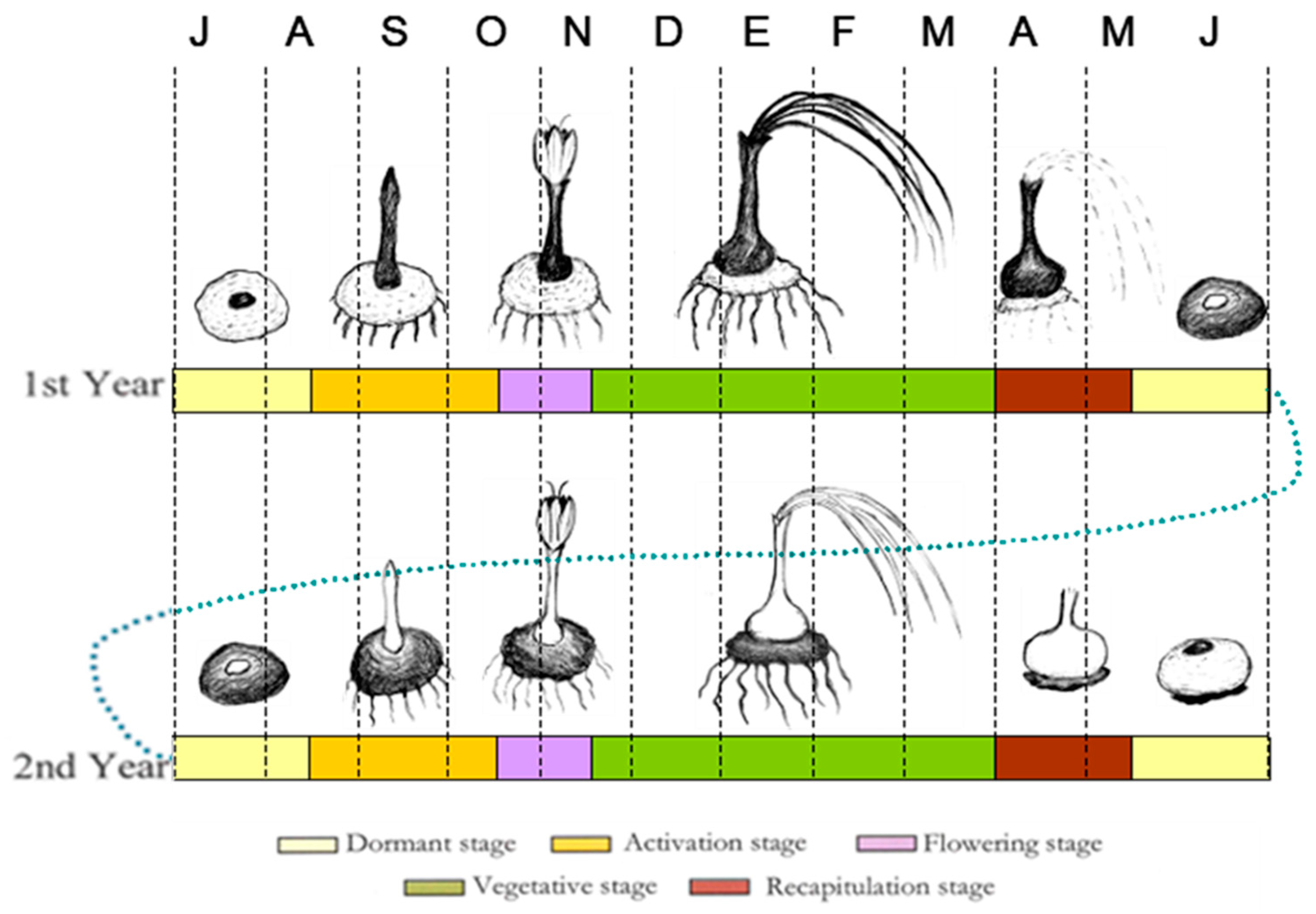
2.1. Plant Materials

In 2015, corms of C.s. were acquired, directly from the farmers, from the Bamyan province of Afghanistan and the five provinces with the highest saffron production in Iran. They were planted in six plots within a homogeneous plot at the experimental field (with clay loam soil) located at the University of Tehran, Karaj, Iran. In each area, corms from the same farmer were planted in individual subplots, as indicated in Table 1. After two years of planting, flowers were collected from each zone. Once their stigmas were separated, they were dehydrated at two temperatures until reaching constant mass: 40 °C for 120 min and 55 °C for 45 min. The codes for the different origins of the corms were as follows: Afghanistan (Bamyan province)—Af (1); Iran including five provinces of Fars (Shiraz)—Sh (2), Kerman province (Koohbanan)—Ko (3), Tehran—Th (4), Isfahan—Es (5), and Razavi Khorasan province—Kh (6).

2.2. Extraction

Saffron aqueous extracts were prepared following the standard procedures outlined by the International Organization for Standardization (ISO:3632-2, [32] with a minor modification). Briefly, instead of using 500 mg of tissues, 50 mg was taken for HPLC-DAD analysis, as described in a previous study [33]. The resulting homogenate was filtered through a polytetrafluoroethylene (PTFE) syringe filter with a pore size of 0.45 µm (Millipore, Bedford, MA, USA) and subsequently transferred to a vial for HPLC-DAD analysis.

2.3. HPLC-DAD Analysis

The analysis of crocetin esters, picrocrocin, safranal, and HTCC was conducted using an Agilent 1200 HPLC chromatograph (Palo Alto, CA, USA) equipped with a 150 mm × 4.6 mm inner diameter and 5 μm Luna C18 column (Tecknokroma, Sant Cugat del Vallès, Spain) at a temperature of 30 °C. The detection wavelengths were set at 440 nm for crocetin esters, 330 nm for safranal, and 250 nm for picrocrocin and HTCC using the DAD detector (Hewlett Packard, Waldbronn, Germany). HPLC-grade acetonitrile was sourced from Panreac® (Barcelona, Spain), and ultrahigh-purity water was produced using a Milli-Q system (Millipore, Burlington, MA, USA), as it is described by García-Rodríguez et al. [33]. Each sample was analyzed in duplicate, and two measurements were taken for each replicate.

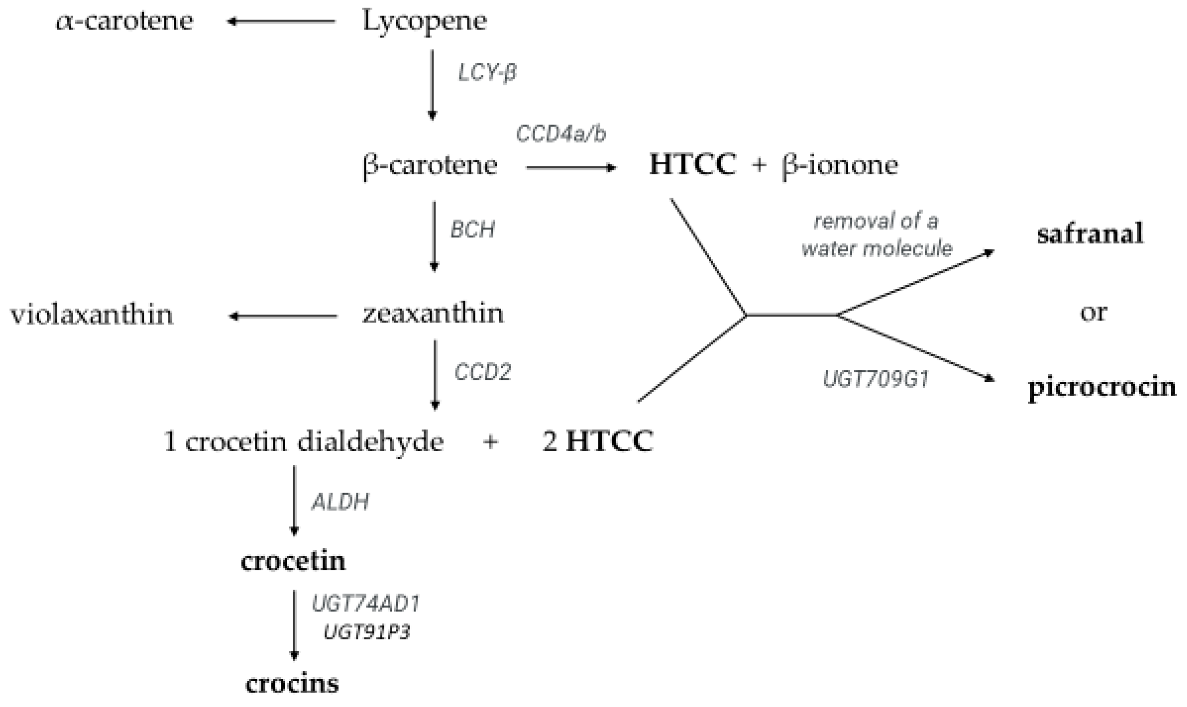
2.4. Identification and Quantification of Crocetin Esters, Picrocrocin, Safranal, Kaempferol Glycosides, and HTCC

Identification of crocetin esters, picrocrocin, HTCC, and safranal was carried out as previously reported [13]. Quantification was based on the following calibration curves [34]: Ci = (0.00746 ± 0.00004)Ai − (0.00571 ± 0.12863), correlation coefficient (R2) = 0.9999 for trans-5-tG, trans-5-nG, and trans-4-GG; Ci = (0.00713 ± 0.00003)Ai − (0.00472 ± 0.05608), R2 = 0.9999 for trans-3-Gg, and trans-2-gg; Ci = (0.00531 ± 0.0004)Ai − (0.00571 ± 0.12863), R2 = 0.9999 for cis-4-GG; Ci = (0.00500 ± 0.00003)Ai − (0.00331 ± 0.05608), R2 = 0.9999 for cis-3-Gg, and cis-2-gg; Ci = (0.02900 ± 0.00002)Ai + (0.51940 ± 0.02631), R2 = 0.9999 for picrocrocin and HTCC, and Ci = (0.03227 ± 0.00063)Ai + (0.05101 ± 0.03103), R2 = 0.9989 for safranal. Limits of detection (LOD) and quantification (LOQ) as described by García-Rodríguez et al. [33] were taken into consideration. Safranal (purity ≥ 88%) and crocetin esters (trans-4-GG and trans-3-Gg, purity ≥ 99%) were obtained from Sigma-Aldrich (Madrid, Spain) and Phytolab GmbH & Co. KG (Vestenbergs-greuth, Bravaria, Germany), respectively, and picrocrocin was isolated, as described by Sánchez et al. [34].

2.5. Nomenclature for Crocetin Esters

Abbreviations for crocetin esters (also known as crocins) were adopted from [13] as follows: trans-5-tG, trans-crocetin (β-D-triglucosyl)-(β-D-gentiobiosyl) ester; trans-5-nG, trans-crocetin (β-D-neapolitanosyl)-(β-D-gentiobiosyl) ester; trans-4-GG, trans-crocetin di-(β-D-gentiobiosyl) ester; trans-3-Gg, trans-crocetin (β-D-glucosyl) (β-D-gentiobiosyl) ester; trans-2-gg, trans-crocetin di-(β-D-glucosyl) ester; cis-4-GG, cis-crocetin di-(β-D-gentiobiosyl) ester; cis-3-Gg, cis-crocetin (β-D-glucosyl)-(β-D-gentiobiosyl) ester, and cis-2-gg, cis-crocetin di-(β-D-glucosyl) ester.

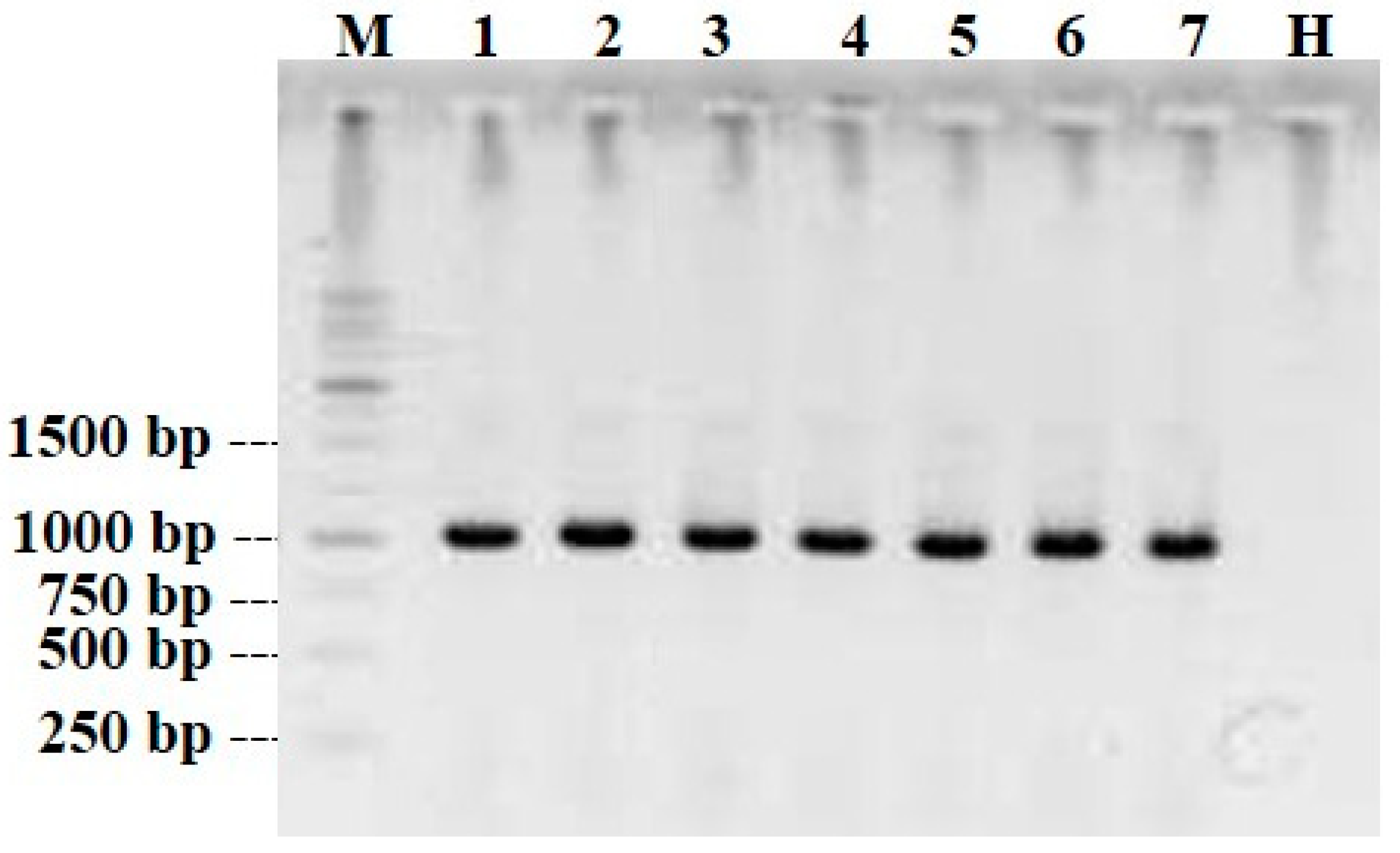
2.6. Serological and Molecular Assays

Following the sprouting of the saffron corms in the subplots, leaf tissue samples were collected and checked for virus infection. Since there is no species-specific antibody provided against recently described SaLV, the presence of potyviruses in leaf tissue samples of C. sativus plants was checked by Antigen-Coated-Plate ELISA (ACP-ELISA) kit (RT-0573/1, DSMZ, Braunschweig, Germany) following the protocol described before (Richter et al., 1995). The samples with positive reaction in ELISA assay were subjected to a reverse transcription-polymerase chain reaction (RT-PCR) to check the SaLV infection status using SaLV-P1-F (GTTCTCTTTGAACTTTTCGCACC)/P1-R (CCTATCGAAAACTTGTTTCCAGCC) specific primers. Overall, total RNA extraction, cDNA synthesis, RT-PCR, and sequencing were performed according to Movi et al. [27]. Briefly, total RNA extraction was performed using RNX-Plus Kit (Sinaclone, Tehran, Iran) according to the manufacturer’s protocol from saffron plants’ leaf tissues. Following cDNA synthesis by SaLV-P1-R primer (at 42 °C for 1 h), all PCRs were done using a Thermal Cycler (Eppendorf-MasterCycler®personal, Hamburg, Germany) and Taq DNA Polymerase Master Mix RED (Ampliqon, Odense, Denmark), with the thermal profile comprising an initial denaturing step as 94 °C for 3 min, followed by 35 cycles of 94 °C for 1 min, 57 °C for 30 s, and 72 °C for the 70 s, and a final extension time step at 72 °C for 5 min.

2.7. Statistical Analysis

A one-way analysis of variance (ANOVA) was conducted to compare the mean values, and Duncan’s test was employed at a significance level of p < 0.05. Additionally, discriminant function analysis was carried out using the statistical software package SPSS 24.0 (SPSS Inc., Chicago, IL, USA). Multivariate analysis of variance (MANOVA) was conducted to study the effect of the principal factors (dehydration temperatures and the different provenances of the first corms that were planted) on the different saffron metabolites. Additionally, a principal component analysis (PCA) was performed with the purpose of obtaining an overall view of the influence of the main saffron metabolites in the separation of corms from different provinces studied. Both statistical analyses were carried out using the Statgraphics Centurion statistical program (version 19.4.02; StatPoint, Inc., The Plains, VA, USA).

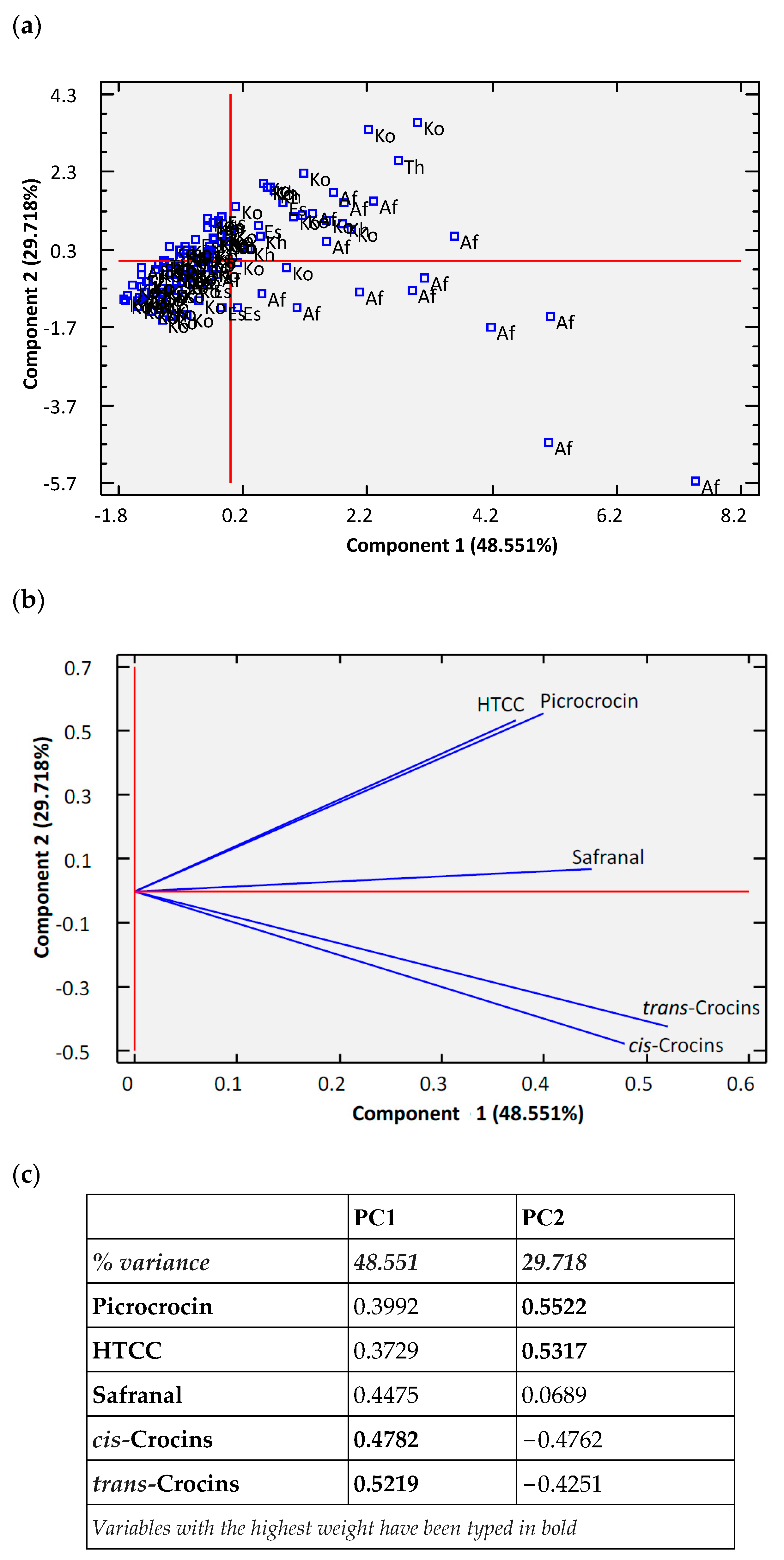
3. Results and Discussion

The potyvirus infection was detected in all 146 saffron plant samples, originating from Iran and Afghanistan, by ACP-ELISA using potyvirus genus-specific antibodies, demonstrating 100% potyvirus infection. Extracted total RNA from all samples was subsequently subjected to RT-PCR using the SaLV-specific primer pair SaLV-P1-F/R. This specific PCR primer produced an amplicon with an expected size of 1016 bp (Figure 3) in all samples and confirmed the presence of SaLV in all saffron plants (100%). This result is in accordance with our recent studies showing a high prevalence of SaLV, rated 70–94%, in saffron plants collected from different locations in Iran [25,26,27]. Interestingly, all 26 saffron plants originating from the Bamyan province of Afghanistan were SaLV infected; to our knowledge, this is the first report of SaLV infection in Afghanistan saffron plants. This is to be expected because Iran and Afghanistan have historical and cultural relations, with a more than 500-mile frontier.
A comparison of different secondary metabolites of freeze-dried saffron stigmas between SaLV-infected and -uninfected samples of the same provinces of Iran showed that the secondary metabolite content was higher in SaLV-uninfected than SaLV-infected samples. It seems that the content of several secondary metabolites of saffron could be modified by the SaLV presence in saffron samples from the same province. Indeed, some environmental characteristics (altitude, temperature, and precipitation/rainfall) of different geographical origins could affect the content of several compounds of C. sativus stigmas and the presence of SaLV could modify these effects [28].
To study the effect of “temperature x time” until reaching constant mass (40 °C for 120 min and 55 °C for 45 min), stigmas were taken from 25 subplots in four different zones (Af: 8; Sh: 2; Kh: 4; Ko: 11) as indicated in Table 1. The results of the average concentration of the major metabolites of the spice obtained in each zone are presented in Table 2. The saffron that shows the highest concentration of HTCC derivatives are those of Af origin at both temperatures, while those of Sh origin exhibit the lowest concentration.
In general, the sum of all the crocins is higher when the saffron is dehydrated at 40 °C than when it is dehydrated at 55 °C, except in the case of Sh. In Afghanistan, this difference is very large, being six times greater than the value of the sum of crocins when dehydrated at 40 °C. The high value is especially due to trans-4-GG, trans-3-Gg, trans-2-G, and trans-1-g. Previous records showed that the sum of crocetin esters’ content in dark-dried saffron samples was higher than in the freeze-dried ones, while freeze-dried saffron samples obtained a greater concentration of picrocrocin and safranal than the dark-dried ones. In other words, dark-drying can nullify the adverse effect of SaLV on crocin content [12].
Table 3 shows the multivariate analysis of variance (MANOVA) of different saffron metabolites depending on the zone and dehydration temperature. In this analysis, we can observe that the effect of the two dehydration temperatures on the composition of metabolites is less significant than the effect of the origin of the corms used for the initial planting. We could say that the corms retain a memory of their origin.
Table 4 shows the mean of the values of the different secondary metabolites of saffron and the sum of all the crocins. Significant differences are observed in different metabolites depending on the zone to which the samples belong. There were especially significant differences between Afghanistan and the rest of the areas, that is, Afghanistan was characterized by having more safranal and more crocins, although crocin trans-5-nG was more abundant in Es and picocrocin more abundant in Tehran. However, HTCC did not show significant differences in any area from this study.
Figure 4a depicts the principal component analysis (PCA) of saffron samples, which shows that there are two components explaining 78.269% of the variance. The first principal component (PC1) explains 48.55% of the variance and the second one (PC2) explains 29.72%. The parameters that defined the PC1 were crocins (cis and trans), whereas the variables that most contributed to the differentiation in PC2 were picrocrocin and HTCC (Figure 4b,c). According to the projection of the samples, both components did not separate the samples according to the provinces significantly, being that only the samples from Afghanistan showed a slight separation from the rest, and they are oriented towards where the trans and cis crocins are heading. However, some samples of Ko are positioned towards the directions of the metabolites derived from HTCC. In Figure 4b, it can be observed that PC2 separates the samples in opposite directions based on their metabolite content, with samples from the same metabolic pathway clustering together. This highlights that the majority of the concentration of HTCC derivatives comes from a different pathway than that of the crocins. In other words, the β-carotene degradation pathway that, by CCD4a/b action, is more important in the biosynthesis of HTCC and picrocrocin than the one that produces zeaxanthin, and, subsequently, by CCD2 action, gives two molecules of HTCC and one molecule of crocetin dialdehyde. Later, safranal is formed from HTCC through a chemical process involving the elimination of water. This explains why the orientation of safranal in the PCA diagram is intermediate.
To confirm that there is no differentiation based on the origin of the corms, we performed a discriminant analysis by creating six groups corresponding to different regions. The results verified that a possible differentiation between zones could exist; this dissipates after the corms have been in the experimentation field of the University for two years. Only the corms from Afghanistan have a trend to differentiate from the rest. This differentiation is practically due to function 1, which explains 76.4% of the variance, and it depends mainly on trans-3-Gg and cis-3-Gg.

4. Conclusions

The analysis of the concentration of the main apocarotenoids of saffron from corms of different geographical origins reveals that after two years of remaining in the same edaphoclimatic conditions, being subjected to the same cultural practices, in the same edaphoclimatic conditions, and infected with the virus itself they do not fully retain the memory of their geographic origin. This leads us to think that if we leave the corms in these conditions for longer, their saffron will exhibit the same quality as if they originated from the same area. On the other hand, the fact that the ratio between the “sum of HTCC derivatives” and the “sum of crocins” is much higher than two indicates that the route of degradation of β-carotene to HTCC is preferred to its transformation to give crocins and HTCC.

Author Contributions

Conceptualization, A.D. and G.L.A.; methodology, A.D., C.L., G.S. and H.V.; software, R.S.-G. and H.V.; validation, N.M.-L. and H.B.; formal analysis, C.L. and G.S.; investigation, N.M.-L., R.S.-G. and H.B.; resources, A.D., H.B. and N.M.-L.; data curation, R.S.-G. and H.B.; writing—original draft preparation, C.L. and G.S.; writing—review and editing, C.L., G.S., A.D. and G.L.A.; visualization, N.M.-L. and H.B.; supervision, A.D. and G.L.A.; project administration, A.D. and G.L.A.; funding acquisition, A.D. and G.L.A. All authors have read and agreed to the published version of the manuscript.

Funding

This research was financially supported by the Iran National Science Foundation (INSF, Project No. 4000914), the University of Tehran (Grant No. 73148924/6/19), and the AZUVOL II Project (Ref.: SBPLY/21/180501/000014) financed by the Government of Castilla–La Mancha (Spain) in collaboration with FEDER.

Data Availability Statement

New data was not created.

Conflicts of Interest

The authors declare no conflict of interest.

References

  1. Grilli Caiola, M.; Canini, A. Ultrastructure of Chromoplasts and Other Plastids in Crocus sativus L. (Iridaceae). Plant Biosyst. Int. J. Deal. All Asp. Plant Biol. 2004, 138, 43–52. [Google Scholar] [CrossRef]
  2. Dewan, R. Bronze Age Flower Power: The Minoan Use and Social Significance of Saffron and Crocus Flowers; Institute for European and Mediterranean Archaeology: New York, NY, USA, 2015; Volume 5, pp. 42–55. [Google Scholar]
  3. Shahnoushi, N.; Abolhassani, L.; Kavakebi, V.; Reed, M.; Saghaian, S. Economic Analysis of Saffron Production. In Saffron; Elsevier: Amsterdam, The Netherlands, 2020; pp. 337–356. ISBN 978-0-12-818638-1. [Google Scholar]
  4. Bazrafshan, O.; Ramezani Etedali, H.; Gerkani Nezhad Moshizi, Z.; Shamili, M. Virtual Water Trade and Water Footprint Accounting of Saffron Production in Iran. Agric. Water Manag. 2019, 213, 368–374. [Google Scholar] [CrossRef]
  5. Ramezani, M.; Dourandish, A.; Jamali Jaghdani, T.; Aminizadeh, M. The Influence of Dense Planting System on the Technical Efficiency of Saffron Production and Land Use Sustainability: Empirical Evidence from Gonabad County, Iran. Agriculture 2022, 12, 92. [Google Scholar] [CrossRef]
  6. Wyeth, P.; Malik, N. A Strategy for Promoting Afghan Saffron Exports; ICARDA and Washington State University: Pullman, WA, USA, 2008. [Google Scholar]
  7. Mollafilabi, A.; Aslami, M.H.; Shoorideh, H. Replacement of Saffron (Crocus sativus L.) with Poppy (Papaver somniferum L.) and Its Socio-Economic Results. Acta Hortic. 2010, 850, 299–302. [Google Scholar] [CrossRef]
  8. Bagur, M.J.; Alonso Salinas, G.; Jiménez-Monreal, A.; Chaouqi, S.; Llorens, S.; Martínez-Tomé, M.; Alonso, G. Saffron: An Old Medicinal Plant and a Potential Novel Functional Food. Molecules 2018, 23, 30. [Google Scholar] [CrossRef] [PubMed]
  9. Carmona, M.; Zalacain, A.; Alonso, G.L. The Chemical Composition of Saffron: Color, Taste and Aroma; Bomarzo SL: Albacete, Spain, 2006. [Google Scholar]
  10. Molina, R.V.; García-Luis, A.; Coll, V.; Ferrer, C.; Valero, M.; Navarro, Y.; Guardiola, J.L. Flower Formation in the Saffron Crous (Crocus sativus L.) the Role of Temperature. Acta Hortic. 2004, 39–47. [Google Scholar] [CrossRef]
  11. Carmona, M.; Zalacain, A.; Pardo, J.E.; López, E.; Alvarruiz, A.; Alonso, G.L. Influence of Different Drying and Aging Conditions on Saffron Constituents. J. Agric. Food Chem. 2005, 53, 3974–3979. [Google Scholar] [CrossRef]
  12. Moratalla-López, N.; Parizad, S.; Habibi, M.K.; Winter, S.; Kalantari, S.; Bera, S.; Lorenzo, C.; García-Rodríguez, M.V.; Dizadji, A.; Alonso, G.L. Impact of Two Different Dehydration Methods on Saffron Quality, Concerning the Prevalence of Saffron Latent Virus (SaLV) in Iran. Food Chem. 2021, 337, 127786. [Google Scholar] [CrossRef]
  13. Carmona, M.; Zalacain, A.; Sánchez, A.M.; Novella, J.L.; Alonso, G.L. Crocetin Esters, Picrocrocin and Its Related Compounds Present in Crocus sativus Stigmas and Gardenia Jasminoides Fruits. Tentative Identification of Seven New Compounds by LC-ESI-MS. J. Agric. Food Chem. 2006, 54, 973–979. [Google Scholar] [CrossRef]
  14. D’Archivio, A.; Di Donato, F.; Foschi, M.; Maggi, M.; Ruggieri, F. UHPLC Analysis of Saffron (Crocus sativus L.): Optimization of Separation Using Chemometrics and Detection of Minor Crocetin Esters. Molecules 2018, 23, 1851. [Google Scholar] [CrossRef]
  15. del Campo, C.P.; Carmona, M.; Maggi, L.; Kanakis, C.D.; Anastasaki, E.G.; Tarantilis, P.A.; Polissiou, M.G.; Alonso, G.L. Picrocrocin Content and Quality Categories in Different (345) Worldwide Samples of Saffron (Crocus sativus L.). J. Agric. Food Chem. 2010, 58, 1305–1312. [Google Scholar] [CrossRef]
  16. García-Rodríguez, M.V.; López-Córcoles, H.; Alonso, G.L.; Pappas, C.S.; Polissiou, M.G.; Tarantilis, P.A. Comparative Evaluation of an ISO 3632 Method and an HPLC-DAD Method for Safranal Quantity Determination in Saffron. Food Chem. 2017, 221, 838–843. [Google Scholar] [CrossRef] [PubMed]
  17. Himeno, H.; Sano, K. Synthesis of Crocin, Picrocrocin and Safranal by Saffron Stigma-like Structures Proliferated in vitro. Agric. Biol. Chem. 1987, 51, 2395–2400. [Google Scholar] [CrossRef]
  18. Diretto, G.; Ahrazem, O.; Rubio-Moraga, Á.; Fiore, A.; Sevi, F.; Argandoña, J.; Gómez-Gómez, L. UGT709G1: A Novel Uridine Diphosphate Glycosyltransferase Involved in the Biosynthesis of Picrocrocin, the Precursor of Safranal in Saffron (Crocus sativus). New Phytol. 2019, 224, 725–740. [Google Scholar] [CrossRef] [PubMed]
  19. Ahrazem, O.; Rubio-Moraga, A.; Berman, J.; Capell, T.; Christou, P.; Zhu, C.; Gómez-Gómez, L. The Carotenoid Cleavage Dioxygenase CCD 2 Catalysing the Synthesis of Crocetin in Spring Crocuses and Saffron Is a Plastidial Enzyme. New Phytol. 2016, 209, 650–663. [Google Scholar] [CrossRef]
  20. Sharma, M.; Kaul, S.; Dhar, M.K. Transcript Profiling of Carotenoid/Apocarotenoid Biosynthesis Genes during Corm Development of Saffron (Crocus sativus L.). Protoplasma 2019, 256, 249–260. [Google Scholar] [CrossRef]
  21. Acar, B.; Sadikoglu, H.; Ozkaymak, M. Freeze Drying of Saffron (Crocus sativus L.). Dry. Technol. 2011, 29, 1622–1627. [Google Scholar] [CrossRef]
  22. Chaouqi, S.; Moratalla-López, N.; Lage, M.; Lorenzo, C.; Alonso, G.L.; Guedira, T. Effect of Drying and Storage Process on Moroccan Saffron Quality. Food Biosci. 2018, 22, 146–153. [Google Scholar] [CrossRef]
  23. del Campo, C.P.; Carmona, M.; Maggi, L.; Kanakis, C.D.; Anastasaki, E.G.; Tarantilis, P.A.; Polissiou, M.G.; Alonso, G.L. Effects of Mild Temperature Conditions during Dehydration Procedures on Saffron Quality Parameters. J. Sci. Food Agric. 2010, 90, 719–725. [Google Scholar] [CrossRef]
  24. Tong, Y.; Zhu, X.; Yan, Y.; Liu, R.; Gong, F.; Zhang, L.; Hu, J.; Fang, L.; Wang, R.; Wang, P. The Influence of Different Drying Methods on Constituents and Antioxidant Activity of Saffron from China. Int. J. Anal. Chem. 2015, 2015, 953164. [Google Scholar] [CrossRef]
  25. Parizad, S.; Dizadji, A.; Koohi Habibi, M.; Winter, S.; Kalantari, S.; Movi, S.; García-Arenal, F.; Ayllón, M.A. Description and Genetic Variation of a Distinct Species of Potyvirus Infecting Saffron (Crocus sativus L.) Plants in Major Production Regions in Iran. Ann. Appl. Biol. 2018, 173, 233–242. [Google Scholar] [CrossRef]
  26. Parizad, S.; Dizadji, A.; Habibi, M.K.; Winter, S.; Kalantari, S.; Garcıa, F. Prevalence of Saffron Latent Virus (SaLV), a New Potyvirus Species, in Saffron Fields of Iran. J. Plant Pathol. 2017, 99, 802. [Google Scholar]
  27. Movi, S.; Dizadji, A.; Parizad, S.; Zarghani, S.N. Biological Characteristics and Genetic Variation Analyses of Saffron Latent Virus (SaLV) Based on Genomic P1-Pro and P3 Regions. Eur. J. Plant Pathol. 2022, 164, 299–312. [Google Scholar] [CrossRef]
  28. Parizad, S.; Dizadji, A.; Habibi, M.K.; Winter, S.; Kalantari, S.; Movi, S.; Lorenzo Tendero, C.; Alonso, G.L.; Moratalla-Lopez, N. The Effects of Geographical Origin and Virus Infection on the Saffron (Crocus sativus L.). Qual. Food Chem. 2019, 295, 387–394. [Google Scholar] [CrossRef]
  29. Zhi, L.; Xianmei, G.; Jian, Y.; Duoyong, Z.; Bin, L.; Zihong, Z.; Piao, C.; Dongguang, W. Quality Evaluation and Origin Traceability of the Imported and Domestic Saffron Spice (Crocus sativus L.) Products in China Market Using Chemical Composition and Stable Isotope Analysis. J. Food Compos. Anal. 2023, 118, 105202. [Google Scholar] [CrossRef]
  30. El Hani, O.; García-Guzmán, J.J.; Palacios-Santander, J.M.; Digua, K.; Amine, A.; Gharby, S.; Cubillana-Aguilera, L. Geographical Classification of Saffron (Crocus sativus L.) Using Total and Synchronous Fluorescence Combined with Chemometric Approaches. Foods 2023, 12, 1747. [Google Scholar] [CrossRef]
  31. Nie, J.; Yang, J.; Liu, C.; Li, C.; Shao, S.; Yao, C.; Chen, B.; Tao, Y.; Wang, F.; Zhang, Y.; et al. Stable Isotope and Elemental Profiles Determine Geographical Origin of Saffron from China and Iran. Food Chem. 2023, 405, 134733. [Google Scholar] [CrossRef]
  32. ISO 3632; Saffron (Crocus sativus L.). Part 1 (Specification) and Part 2 (Test Methods). International Organization for Standardization (ISO): Geneva, Switzerland, 2011.
  33. García-Rodríguez, M.V.; Serrano-Díaz, J.; Tarantilis, P.A.; López-Córcoles, H.; Carmona, M.; Alonso, G.L. Determination of Saffron Quality by High-Performance Liquid Chromatography. J. Agric. Food Chem. 2014, 62, 8068–8074. [Google Scholar] [CrossRef]
  34. Sánchez, A.M.; Carmona, M.; del Campo, C.P.; Alonso, G.L. Solid-Phase Extraction for Picrocrocin Determination in the Quality Control of Saffron Spice (Crocus sativus L.). Food Chem. 2009, 116, 792–798. [Google Scholar] [CrossRef]
Figure 1. Vegetative cycle of Crocus sativus L. Capital letters at the top of the figure refer to the months of the year (adapted from Carmona et al. [9]).
Horticulturae 09 00933 g001
Figure 2. Diagram of the biosynthesis of saffron apocarotenoids from lycopene [18].
Horticulturae 09 00933 g002
Figure 3. Amplification of SaLV-P1 with specific primers SaLV-P1-F/R. Lane 1, 1 kb DNA-ladder (Fermentas); lanes 1–7, SaLV isolates originated from different locations; lane H, negative control (healthy plant).
Horticulturae 09 00933 g003
Figure 4. Principal component analysis (PCA) of saffron samples. (a) Projection of saffron samples in the plane formed by the two main components. (b) Projection of the saffron metabolites studied in the first two principal components. (c) Weights of the variables most influential in the first two principal components.
Horticulturae 09 00933 g004
Table 1. The number of subplots where the used stigmas came from.
Number of SubplotsAfShKhThEsKo
corms from the same farmer in that area26101531478
stigmas were dehydrated at 40 °C for 120 min1881131467
stigmas were dehydrated at 55 °C for 45 min8240011
Table 2. The content of the main compounds with different dehydration temperatures in the different zones by HPLC-DAD analysis.
Zones Compound (mmol/kg Saffron) *
AfShKhKo
Dehydration Temperature (°C)4055405540554055
Picrocrocin194.41 a216.20 a169.07 a94.72 a183.23 a174.40 a111.08 a108.63 a
Safranal13.09 b5.03 a,b005.75 a,b005.09 a,b
HTCC278.42 a263.08 a159.92 a120.91 a240.78 a224.15 a216.69 a258.09 a
∑ HTCC derv.485.92 a484.31 a328.99 a215.63 a429.76 a398.55 a327.77 a371.81 a
trans-5-tG1.33 b0.23 a0.30 a0.36 a0.29 a0.67 a0.21 a0.19 a
trans-5-nG3.50 a1.27 a0.80 a2.41 a8.56 a9.33 a5.15 a1.10 a
trans-4-GG71.45 b15.50 a15.32 a18.26 a11.02 a2.43 a6.04 a10.01 a
trans-4-ng4.04 a0.38 a0.34 a0.83 a2.89 a12.15 b4.31 a0.94 a
trans-3-Gg73.53 b17.59 a12.24 a16.87 a14.82 a3.63 a15.03 a11.96 a
trans-2-gg19.96 b6.21 a3.32 a6.91 a5.90 a0.39 a8.74 a4.37 a
cis-4-GG5.84 b1.16 a1.27 a1.75 a1.50 a1.02 a1.12 a0.96 a
trans-2-G34.80 b0.71 a0.83 a1.43 a1.43 a1.19 a0.66 a0.56 a
cis-3-Gg4.94 b1.41 a1.01 a2.61 a,b2.64 a,b1.50 a3.22 a,b1.64 a
trans-1-g16.58 b0.25 a0.38 a0.45 a0.36 a0.59 a0.43 a0.46
cis-2-gg3.35 b0.49 a0.30 a1.39 a1.02 a0.49 a0.83 a0.31 a
∑ Crocins239.32 b38.82 a32.60 a45.04 a44.53 a29.24 a35.78 a27.30 a
* Values are the mean of 8 in Af, 2 in Sh, 4 in Khand, and 11 in Ko. One-way analysis of variance (ANOVA) for each row is included. Different letters (a, b) represent statistically significant differences (p < 0.05) in saffron samples for each metabolite’s concentration.
Table 3. F-Values from the multivariate analysis of variance (MANOVA) of saffron compounds attending to zone and dehydration temperature.
ZoneDehydration Temperature (°C)
Picrocrocin4.82 ***0.11
HTCC1.621.08
Safranal8.63 ***1.29
trans-5-tG9.25 ***4.22 *
trans-5-nG9.56 ***0.30
trans-4-GG14.50 ***5.48 *
trans-4-ng4.73 ***2.41
trans-3-Gg17.72 ***10.56 **
trans-2-gg9.18 ***10.72 *
cis-4-GG5.60 ***3.83
trans-2-G14.66 ***11.16 **
cis-3-Gg3.88 **8.18 **
trans-1-g15.79 ***13.19 ***
cis-2-gg2.158.20 **
Significant differences according to Fisher’s LSD test are indicated as * p < 0.05, ** p < 0.01, or *** p < 0.001.
Table 4. The content of the main compounds of saffron samples by HPLC-DAD analysis.
Compound
(mmol/kg Saffron) *
Zones
AfShKhTeEsKo
Picrocrocin190.18 a,b125.18 a145.03 a282.84 b117.11 a105.73 a
Safranal12.23 b1.38 a3.82 a6.65 a1.83 a1.67 a
HTCC255.21 a130.03 a219.35 a190.75 a193.96 a189.13 a
∑ derv. HTCC457.62 a256.59 a368.20 a480.24 a312.90 a296.53 a
trans-5-tG1.57 b0.23 a0.32 a1.03 a,b0.58 a0.22 a
trans-5-nG3.86 a0.98 a12.22 a5.12 a30.14 b4.07 a
trans-4-GG85.42 b13.12 a5.51 a5.28 a0.48 a4.94 a
trans-3-Gg76.15 b13.26 a10.09 a3.60 a7.41 a12.56 a
trans-2-gg19.46 b4.95 a4.34 a0.41 a0.36 a10.28 a,b
cis-4-GG10.33 b1.52 a0.89 a1.13 a0.84 a1.04 a
trans-2-G28.76 b0.94 a1.52 a0.63 a1.37 a0.65 a
cis-3-Gg6.39 b1.65 a2.18 a1.93 a2.47 a2.52 a
cis-2-gg3.46 b0.36 a,b1.11 a,b0.47 a1.80 a,b1.41 a,b
∑ Crocins191.03 b31.50 a38.21 a26.12 a55.29 a32.42 a
* One-way analysis of variance (ANOVA) for each row is included. Different letters (a, b) represent statistically significant differences (p < 0.05) in saffron samples for each metabolite’s concentration.
Disclaimer/Publisher’s Note: The statements, opinions and data contained in all publications are solely those of the individual author(s) and contributor(s) and not of MDPI and/or the editor(s). MDPI and/or the editor(s) disclaim responsibility for any injury to people or property resulting from any ideas, methods, instructions or products referred to in the content.

