Article

Effect of Heat Stress on Root Architecture, Photosynthesis, and Antioxidant Profile of Water Spinach (Ipomoea aquatica Forsk) Seedlings

1
Key Laboratory for Quality Regulation of Tropical Horticultural Crops of Hainan Province, Sanya Nanfan Research Institute, Hainan University, Sanya 572025, China
2
Key Laboratory for Quality Regulation of Tropical Horticultural Crops of Hainan Province, School of Horticulture, Hainan University, Haikou 570228, China
3
Hainan Yazhou Bay Seed Laboratory, Sanya 572025, China
*
Authors to whom correspondence should be addressed.
Horticulturae 2023, 9(8), 923; https://doi.org/10.3390/horticulturae9080923
Submission received: 7 July 2023 / Revised: 31 July 2023 / Accepted: 10 August 2023 / Published: 13 August 2023

Abstract

:
Crop productivity around the world is being seriously affected by adverse environmental conditions. High temperature (HT) stress has severely hampered plant growth, yield, and quality. Water spinach is a significant heat-resilient green leafy vegetable that can mitigate prolonged HT stress. However, the morphological, physiological, and biochemical alterations that occur in its response to heat stress remain unknown. In this study, the physiological response to HT stress in water spinach plants with different temperature (25-control, 30, 35, 40, 45 °C) tolerances was investigated. When plants were subjected to HT over a long period of time, their growth was stunted. The results showed that no significant difference was seen between the control (25 °C) and 30 °C for some traits (root shoot fresh weight, root morphological traits, and leaf gas exchanges parameters). Further, HT (35, 40, and 45 °C) stress significantly reduced the growth status, the gas exchange parameters, the pigment content, the photosystem function, and the root architecture system of water spinach. Conversely, HT stress considerably enhanced secondary metabolites in terms of total phenolics, flavonoids, soluble sugars, and anthocyanin content. Furthermore, heat stress remarkably increased the accumulation of reactive oxygen species (ROS) and caused cellular membrane damage. HT stress effectively altered the antioxidant defense system and caused oxidative damage. Generally, HT has an adverse effect on the enzyme activity of water spinach, leading to cell death. However, the current study found that temperatures ≥35 °C had an adverse effect on the growth of water spinach. Further research will be needed to examine the mechanism and the gene expression involved in the cell death that is caused by temperature stress in water spinach plants.

1. Introduction

The global average temperature has risen considerably since the turn of the century, and current climate change indicates that it will continue to do so [1,2]. Climate change has resulted from global warming, particularly HT stress, and these changes are considered a serious challenge to global agricultural productivity [3]. Several environmental problems, including increasing temperatures, are now being raised throughout the world [4,5]. Globally, temperature changes limit crop productivity [6]. In the current century, experts predict that seasonal temperature increases in tropical and subtropical regions will significantly exceed those of the earlier century. As a result, a 1 °C increase in yearly temperature will result in agricultural production decreases of 2.5–16% [7]. This increasing temperature has significantly hindered crop growth and development in various horticultural species, notably vegetables, resulting in considerably lower production. Furthermore, by the end of the 21st century, an elevation in global temperature of 3–5 °C is predicted, which will cause severe damage to future horticultural produce [8]. Heat stress generally occurs when plants are subjected to temperatures exceeding their ideal growing range for a long period of time, and it causes considerable plant damage.
The primary root system is the plant’s initial line of defense against the damaging effects of abiotic stress [9]. A healthy root system has been identified as a crucial attribute that increases a plant’s ability to absorb water and nutrients as well as to tolerate high temperatures [10]. Heat stress may indirectly influence roots by decreasing root water relations due to increased shoot water demand, which affects root development and nutrient absorption [11]. Plants are able to recover from environmental stresses, such as HT stress, due to this adaptability. Understanding the root system’s response mechanism to HT stress is thus crucial for increasing crop tolerance.
Photosynthesis is the most essential metabolic mechanism in plants, and plants are therefore very sensitive to extreme temperatures [12]. Heat stress induces photosynthetic acclimatization, directly modifies physiological functions, and indirectly modifies developmental processes [13]. All phases, stages, and functions of photosynthesis are susceptible to rising temperatures [14]. The photosynthetic systems of most plants are especially sensitive to HT, and they are hampered when temperatures exceed 38 °C [13]. Jahan et al. [15] reported that plants subjected to severe temperatures have lower photosynthetic efficiency and lower levels of photosynthetic pigments in their leaves, and the example used was the tomato plant. Heat stress rapidly eliminates the water from leaves and causes cellular membrane integrity [7]. Rubisco is a heat-sensitive photosynthetic enzyme and plays a direct role in CO2 fixation. Temperature fluctuation affects both the rates of RuBP production and the carboxylation sites of Rubisco [16,17]. Ahammed et al. [18] noticed that heat stress dramatically reduced photosynthetic assimilation rate, chlorophyll fluorescence traits, and photosynthetic pigment levels in tomato seedlings. HT (42 °C) stress considerably reduced pigments and carotenoid concentration in water spinach. In addition, heat stress effectively decreased Fv/Fm and photosystem II, resulting in PSII reaction center damage under HT stress [1]. Heat stress-induced chloroplast damage inactivates heat-sensitive proteins, including RCA activity, and downregulates chloroplast elements, resulting in reduced photosynthetic capacity, an imbalance of redox homeostasis, and cell death [19]. Chloroplasts function as metabolic centers, and they contribute significantly to heat stress adaptation mechanisms. Heat stress damages the photosynthetic machinery in chloroplasts, which activates cellular heat-stress signaling [20,21].
Changes in enzyme activity and temperature fluctuations may disrupt metabolic functions. During heat stress, reactive oxygen species (ROS) were generated and accumulated in many species, resulting in oxidative damage in plants [22]. HT stress caused the over-production of malonaldehyde (MDA) and thus decreased the level of cellular membrane integrity [23]. Plants, being sessile organisms, establish an effective antioxidative defense mechanism to detoxify excess ROS in an extreme environment, which assists in preventing cellular membrane damage [24]. This antioxidant defense system is composed of a wide range of enzyme-based antioxidants, such as superoxide dismutase (SOD), catalase (CAT), peroxidase (POD), ascorbate peroxidases (APX), glutathione reductase (GR), monodehydroascorbate reductase (MDHAR), and dehydroascorbate reductase (DHAR), as well as non-enzymatic antioxidants, including ascorbate (AsA), glutathione (GSH), phenolics, flavonoids, soluble sugars, and anthocyanin content [25].
In China and other Asian countries, water spinach (Ipomoea aquatica Forsk) is a green leafy vegetable that is extensively grown and consumed [26]. In terms of nutritional and economic value, it is a popular green vegetable. It contains several nutrients that have phenolic content, and it contains folic acid, carotenoids, and vitamins [27]. Water spinach is a heat-resistant species, and it may thus prove valuable for research into the molecular mechanism of heat stress. Few pieces of research have investigated the morph-physiological response of the water spinach plant to HT stress, and more research is thus recommended. The response of water spinach to HT stress must be studied, primarily to find out at what temperature level the water spinach plants show stunted growth. The goal of the current research is (1) to examine the effects of different temperature levels on water spinach and find out at which temperature water spinach growth begins to be inhibited, (2) to examine the impact of HT on water spinach root morphology, photosynthetic apparatus, and secondary metabolite production, and (3) to evaluate the effect of HT on oxidative stress biomarkers and the antioxidant enzyme system of water spinach plants.

2. Materials and Methods

2.1. Experimental Setup

The water spinach (HNUWS003) cultivar was propagated using the nodal cutting method. Emerging shoots with two or three leaves (8 ± 0.5 cm in shoot length) were transplanted into a plastic tub containing nutrient-rich peat soil. After one week of the adaptation period, equal size plants were selected for HT stress treatment. For heat stress treatment, plants were shifted into separate controlled growth chambers with varying temperatures, and the treatments were as follows: (1) 25 °C (CK); (2) 30 °C; (3) 35 °C; (4) 40 °C; and (5) 45 °C. Plant samples were collected after 96 h of stress treatment and stored at −80 °C for subsequent analysis. There were four replications of each treatment, each containing ten plants. The trial was conducted under a controlled growth chamber (800–1000 μmol m−2 s−1 photosynthetic photon flux density with a photoperiod of 12/12 h dark/light period, and 85% relative humidity).

2.2. Growth Attributes and Root Morphology

Shoots and roots were separated after 96 h of stress treatment, and they were weighed individually to measure fresh biomass. The samples were then placed in an oven set at 80 °C for 3 days to determine dry weights. Three identical plants were selected for root harvesting. Running tap water was used to clean the roots. Root scanning was measured using the protocol described by Nawaz et al. [28].

2.3. Photosynthesis Related Parameters

Arnon’s [29] technique was used in order to determine the chlorophyll and carotenoid concentrations in the leaves. To quantify the content of chlorophyll a, chlorophyll b, and carotenoids, we crushed 0.5 g of upper fresh leaves in 10 mL of 80% acetone, centrifuged the homogenate at 5000× g for 10 min, and we then read the absorbance at 662 nm, 645 nm, and 470 nm in a spectrophotometer (160 UV–VIS; Thermo Scientific, Austin, TX, USA). Images of autofluorescence were emitted as previously reported by Mumtaz et al. [30] in their study. The leaf gas exchange characteristics were measured using a portable photosynthesis system (CIRAS-3, Hansatech Co., Amesbury, MA, USA). Fully developed leaves (upper 3rd leaf) were used to calculate the gas exchange parameters [31]. All measurements were performed under a photosynthetic photon flux density of 800 μmol/m2/s, a relative humidity of 65 ± 5%, a leaf temperature of 25 ± 2 °C, and external CO2 concentration of 360 ± 10 μmol/mol. Fully developed leaves (from the top) were used to detect chlorophyll fluorescence. After 30 min in the dark, the maximum efficiency of PSII photochemistry (Fv/Fm), the quantum yield of PSII photochemistry [Y(II)], photochemical quenching (qP), and non-photochemical quenching (NPQ) were calculated with an IMAGING-PAM Chlorophyll fluorescence detector (Heinz Walz, Effeltrich, Germany) [15].

2.4. SEM, TEM, and Leaf Paraffin Analysis

Fresh water spinach leaves were cut into 1 cm2 squares and then immediately placed in the electron microscope fixing solution for 2 h. The samples were washed three times for 15 min with 0.1 M PB (pH 7.4). At room temperature, the tissue pieces were kept in 0.1 M PB (pH 7.4) containing 1% OsO4 for 1–2 h. The pieces of tissue were then rinsed three times with 0.1 M PB (pH 7.4) for 15 min. Dehydration was carried out by a 15-min series of alcohol-isoamyl acetate concentrations, and the samples were dried in the critical point drier (Quorum K850, Laughton, UK) and gold-sputter-coated for 30 s using the Hitachi MC1000, Tokyo, Japan. Finally, SEM (Hitachi, SU8100, Tokyo, Japan) was used to examine the pictures [32].
Fresh leaves were cut into approximately 1 cm2 squares, and the preparation of sections was completed after fixation, dehydration at room temperature, resin penetration, embedding, polymerization, ultrathin sectioning, and staining, and then photographs were taken by using TEM (Hitachi, HT7800, Tokyo, Japan) [33].
Leaves of the water spinach plants were prepared as paraffin sections. Leaf paraffin sectioning was measured using the protocol described by Hewitson et al. [34] in their study. Image J152 software was used to measure the traits.

2.5. Secondary Metabolites

To measure the secondary metabolites, leaf samples (0.1 g) were crushed in cold methanol (70% v/v), formic acid (2% v/v), and ethanol (28% v/v). We first digested the homogenized leaf tissue (30 min), and we then stirred them at 250 rpm for 2 h at 30 °C. The samples were centrifuged at 10,000× g for 10 min at 4 °C. Soluble sugar concentration was determined according to the procedure described by [35]. Flavonoid, anthocyanins, and phenolic content were estimated as per the protocol given in detail by Jahan et al. [36] in their study.

2.6. Oxidative Stress Biomarkers and Antioxidant Enzymes Analysis

To determine the stress biomarkers and antioxidant enzyme activity, 0.5 g frozen tissue (leaf) samples were homogenized using liquid nitrogen. The ground tissue (leaf and root) samples were homogenized in 900 µL of 100 mM phosphate buffer (pH 7.4), as prescribed in the kit. Each homogenized sample was centrifuged at 12,000× g for 15 min at 4 ◦C. After that, the supernatant was added to a new falcon tube for further analysis. The activities of APX (A123–1), CAT (A007–1), DAHR (BC0660), GR (A062–1), GST (A004), MDHAR (BC0650), POD (A084-3-1), and SOD (A001–1), and the levels of H2O2 (A064), MDA (A003-3), and O2•− (A052) were calculated using the instructions included in the kits. The absorbance values were measured on a spectrophotometer (160 UV–VIS; Thermo Scientific) at 290, 405, 412, 340, 412, 340, 420, 550, 405, 530, and 550 nm, respectively.

2.7. Leaf H2O2, O2•−, and MDA Staining

NBT and DAB staining were used for histochemical staining of H2O2 and O2•− as previously quoted by Altaf et al. [37] in their study. According to Pompella et al. [38], freshly obtained leaves were stained in Schiff’s reagent for 60 min until a red color appeared, and then extra staining was removed by washing in potassium sulfite solution (0.5%, w/v, K2S2O5 in 0.05 M HCl).

2.8. Statistical Analysis

The statistical analysis was performed using SPSS software. Results were analyzed via one-way ANOVA, and then Fisher’s least significant difference (LSD) test was used to evaluate the significant difference at p ≤ 0.05.

3. Results

3.1. Plant Growth and Root Morphology

After 96 h of HT stress treatment, we noticed that temperature levels (25 (CK) and 30 °C) had shown no significant difference; conversely, HT levels (35, 40, and 45 °C) hindered water spinach growth and biomass production (Figure 1). In addition, we found that increasing temperature levels decreased the growth characteristics. The results revealed that growth traits (fresh shoot weight, FSW; fresh root weight, FRW; dry shoot weight, DSW; dry root weight, DRW) after 96 h of temperature (30 °C) treatment were very similar to CK (25 °C) (Table 1). In contrast, HT treatments dramatically decreased these growth traits (Table 1). The FSW was reduced by 13.76, 33.11, and 50.38%; the FRW was decreased by 24.32, 44.59, and 60.13%; the DSW declined by 21.38, 39.88, and 57.80% (Figure 2C); and DRW was reduced by 17.64, 35.29, and 54.29% at temperatures of 35, 40, and 45 °C, respectively (Table 1). Additionally, plant biomass production decline was more noticeable at 40 and 45 °C temperatures when compared with 35 °C temperature treatment (Table 1).
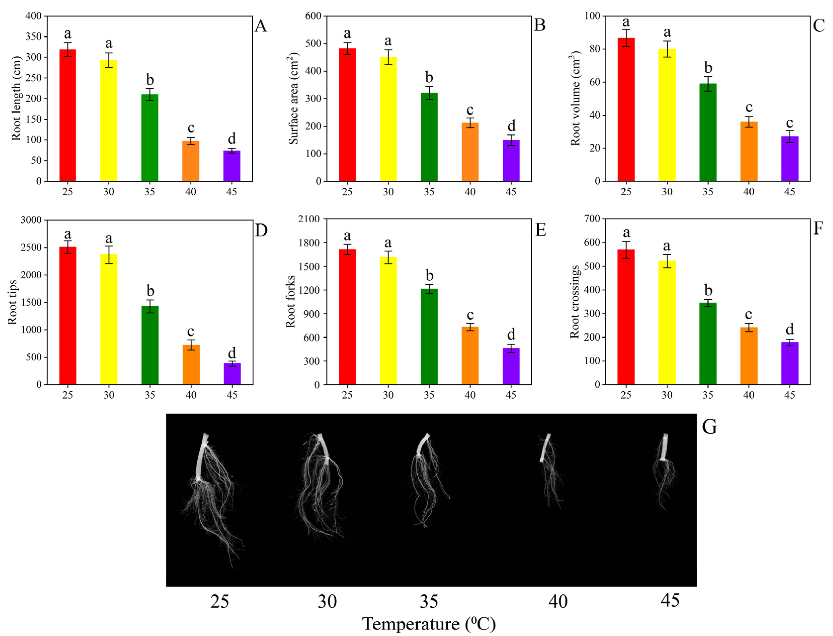
The root growth of water spinach plants was considerably decreased by HT treatments, as revealed by a decrease in root growth characteristics (Figure 2A–G). Additionally, when the temperature levels rose, the root growth attributes decreased, but the most damaging effect was observed at 35, 40, and 45 °C temperatures compared with the CK group (Figure 2). The root length was reduced by 34.16, 69.59, and 76.81% (Figure 2A); the surface area decreased by 33.40, 55.79, and 69.08% (Figure 2B); and the root volume declined by 31.39, 58.13, and 68.60% (Figure 2C) at temperatures of 35, 40, and 45 °C, respectively. Considerable alterations were observed at temperatures of 35–45 °C, and they showed decreased root tips from 43.07 to 84.71% (Figure 2D), root forks from 29.12 to 72.98% (Figure 2E), and root crossings from 39.36 to 68.54% (Figure 2F). The 30 °C temperature treatment maintained the same root characteristics as the 25 °C CK treatment (Figure 2).

3.2. Photosynthesis Related Parameters

The results showed that water spinach’s photosynthetic characteristics were significantly affected by different temperatures (35, 40, and 45 °C) (Figure 3). When compared to CK (25 °C), the Pn was decreased by 29.41, 46.38, and 61.58%, and the Gs rate was reduced by 36.05, 58.41, and 63.84%, respectively, when the water spinach plants were exposed to temperatures of 35, 40, and 45 °C, respectively (Figure 3A,B). The Ci and Tr were greatly reduced by 39.95, 52.31, and 71.04%, and 31.25, 52.85, and 64.80%, respectively, following treatment with temperatures of 35, 40, and 45 °C (Figure 3C,D). The leaf photosynthetic parameters of water spinach plants grown at a 30 °C temperature were found to be statistically comparable to those of water spinach plants grown at 25 °C temperature (Figure 3).
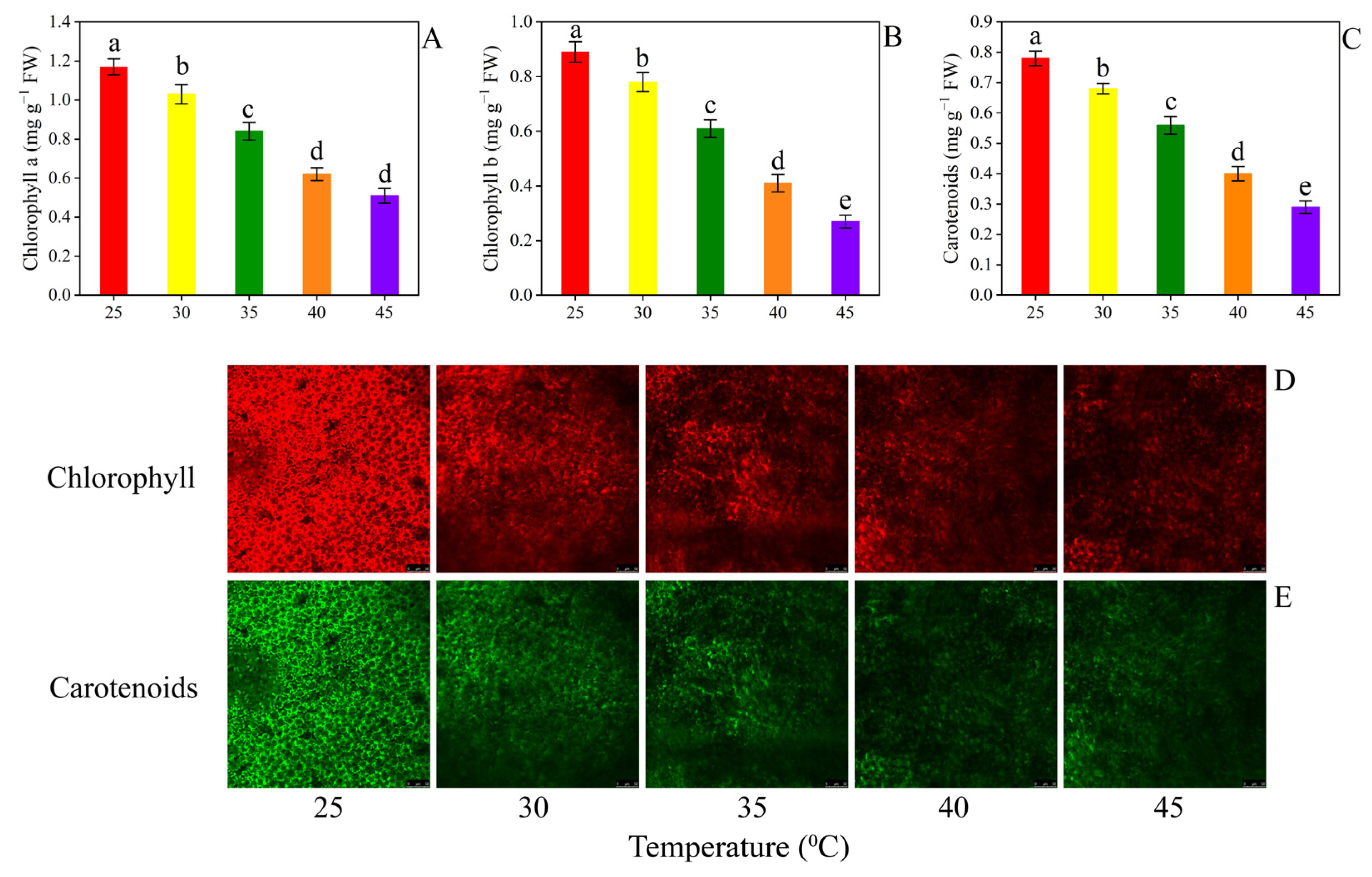
The results revealed that HT treatment from 30 to 45 °C noticeably lowered the level of leaf pigments compared to CK (25 °C) plants (Figure 4). The chlorophyll a and chlorophyll b content decreased up to 11.96, 28.20, 47.01, and 56.41%, and 12.35, 31.46, 53.93, and 69.66% when plants were treated with temperatures of 30, 35, 40, and 45 °C, respectively (Figure 4A,B). Carotenoids’ concentration in water spinach leaves decreased by 12.82, 28.21, 48.71, and 62.82%, respectively, following treatment with temperatures of 30, 35, 40, and 45 °C compared to CK plants (Figure 4C).
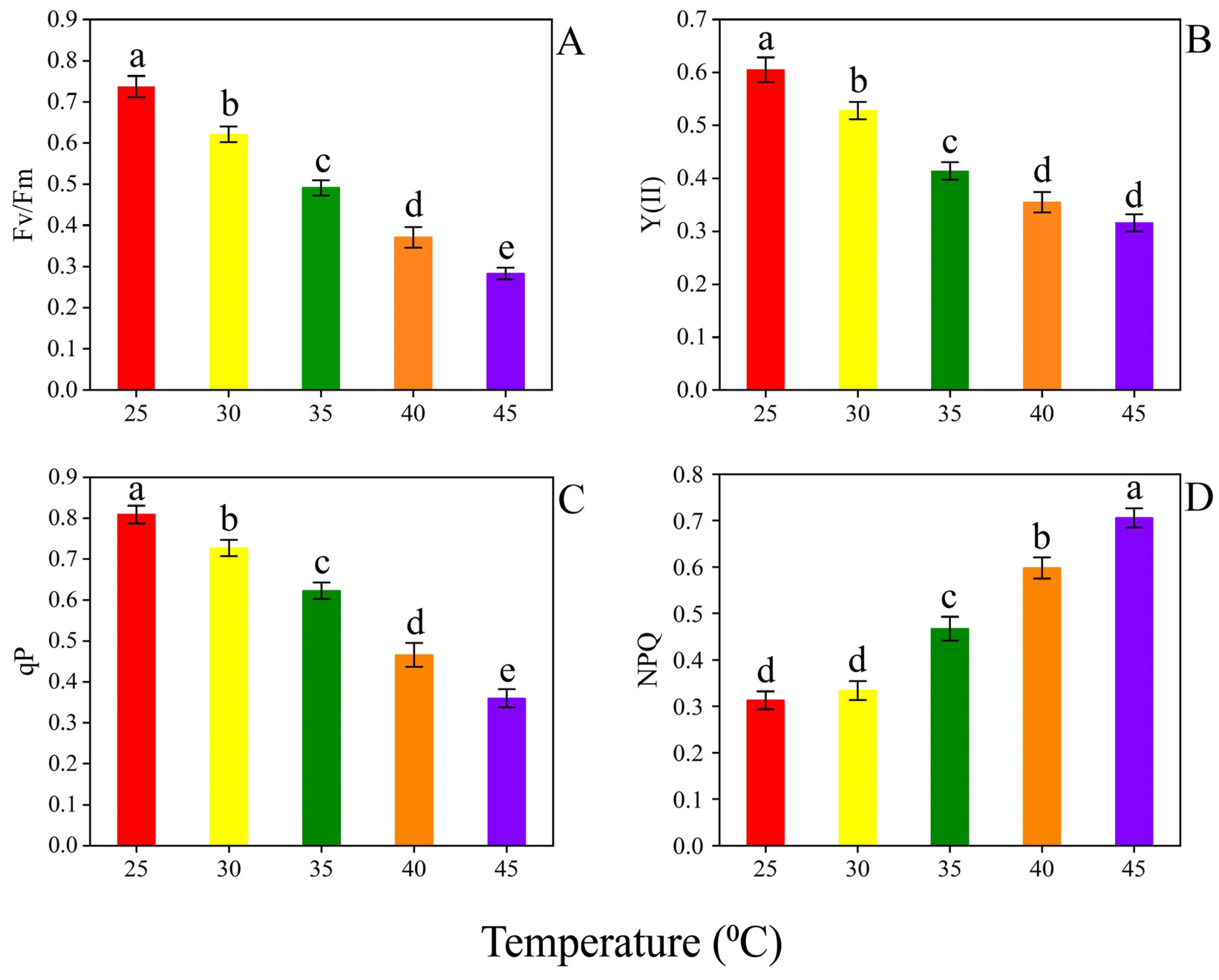
Temperature treatments caused considerable alterations in the water spinach plant’s chlorophyll fluorescence traits (Figure 5). To characterize the performance of photosystem II at varying temperatures, the maximum quantum yield (Fv/Fm), effective quantum efficiencies (Y(II)), non-photochemical quenching (NPQ), and photochemical quenching coefficient (qP) of water spinach leaves were all measured (Figure 5). When temperature levels increased, the Fv/Fm, Y(II), and qP decreased, but the value of NPQ nonetheless improved (Figure 5). Furthermore, when compared with CK, the Fv/Fm was decreased by 15.37, 33.36, 49.66, and 61.60%, Y(II) was reduced by 12.72, 31.57, 41.32, and 47.76%, and qP was reduced by 10.13, 22.99, 42.39, and 55.37% when plants were treated with temperatures of 30, 35, 40, and 45 °C, respectively (Figure 5A–C). In addition, the values of NPQ were considerably enhanced 0.07-fold, 0.49-fold, 0.91-fold, and 1.25-fold following treatment with temperatures of 30, 35, 40, and 45 °C, respectively (Figure 5D).

3.3. Effects of Temperature on the Ultrastructure and Paraffin Analysis of Leaves

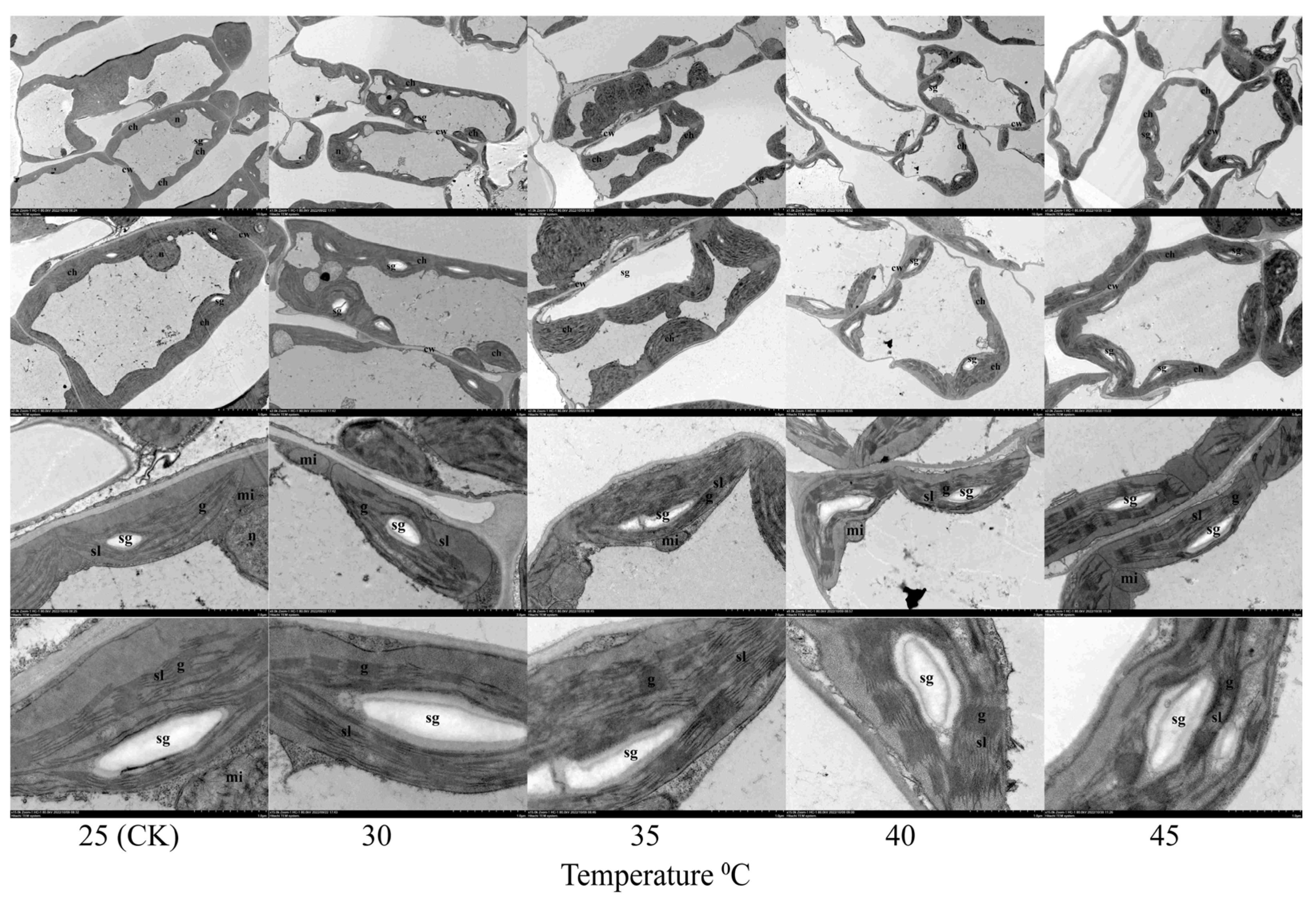
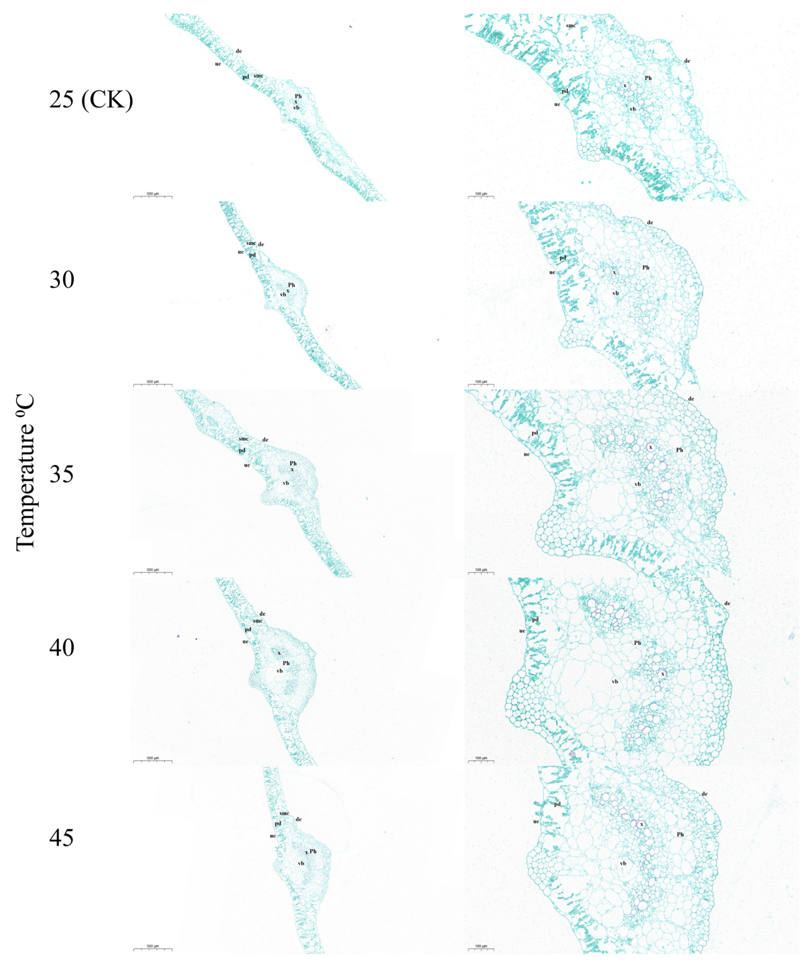
Plant cells need structural integrity to develop normally. In this investigation, TEM was used to examine how temperature stress affected the ultrastructural alterations in the water spinach leaves (Figure 6). After stress treatment, the chloroplast number declined and organelle sections clumped together, chloroplasts broke down, and less starch grains were produced. The outer protective membrane of the starch granules was distorted and inclined to blur, while the thylakoids were loose and damaged. Further, mitochondria deformed inward and decreased. In addition, under the influence of temperature stress, the chloroplast’s structure was altered. The present findings revealed that the ultrastructure of water spinach leaves was altered by HT stress (Figure 6). The anatomy indexes of water spinach leaf under different temperature levels are shown in Figure 7. High temperature stress causes slight damage to the palisade tissue through histological observation (Figure 7).

3.4. Secondary Metabolites

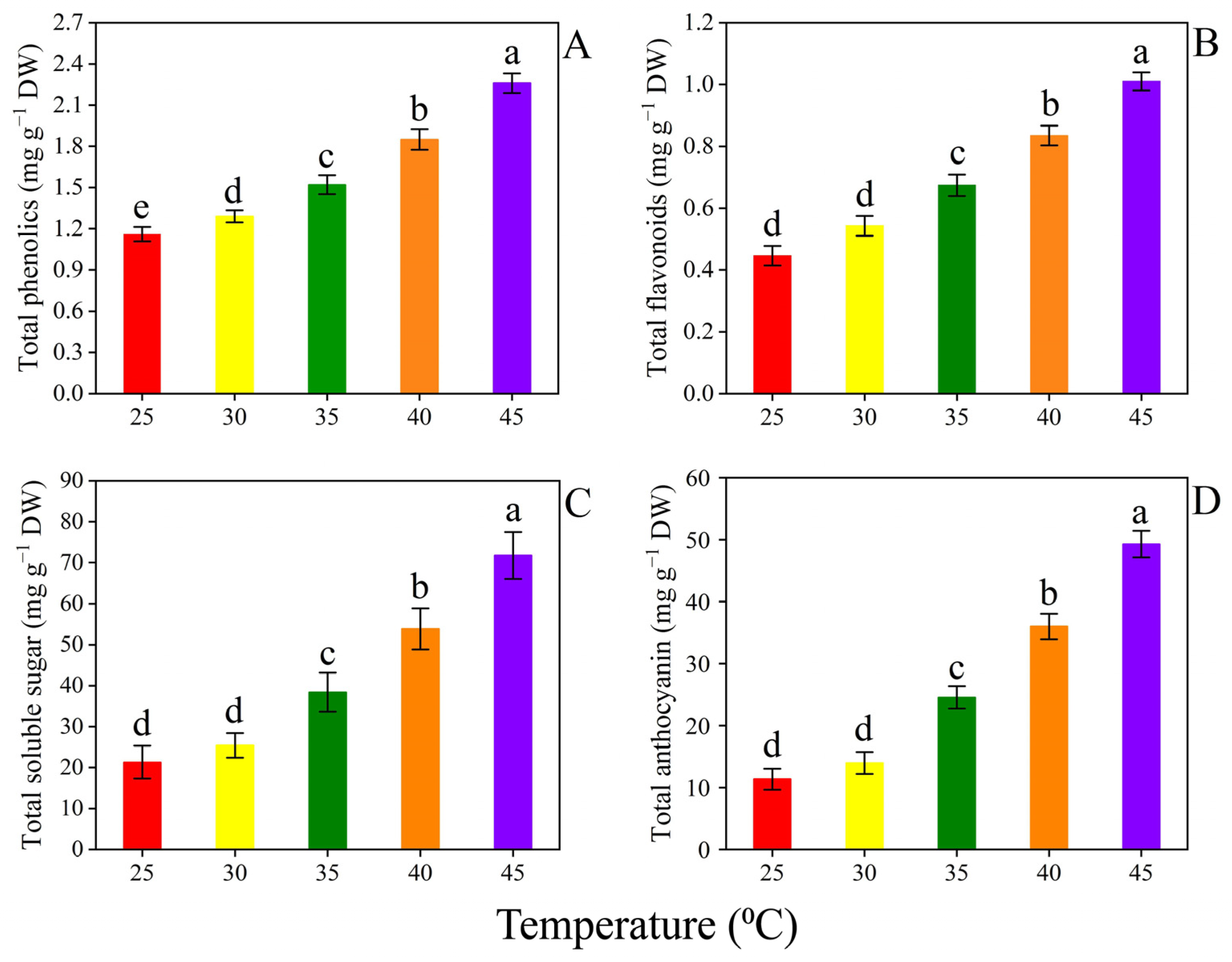
There was a significant increase in the contents of phenolics and flavonoids of the leaves when various temperature treatments were applied (Figure 8A,B). The contents of phenolics and flavonoids were efficiently improved up to 0.11-fold, 0.31-fold, 0.59-fold, and 0.98-fold and 0.21-fold, 0.51-fold, 0.87-fold, and 1.26-fold, respectively, following treatment with temperatures of 30, 35, 40, and 45 °C, respectively, when compared to CK plants (Figure 8A,B). Similarly, when water spinach plants were treated with 30, 35, 40, and 45 °C temperatures, the total soluble sugar and total anthocyanin content were increased by 0.19-fold, 0.80-fold, 1.52-fold, and 2.36-fold and 0.22-fold, 1.15-fold, 2.16-fold, and 3.33-fold, respectively, when compared to CK plants (Figure 8C,D).

3.5. Oxidative Stress Biomarkers, Antioxidant Enzymes, and Leaf Staining Analysis

Compared to CK treatment, the water spinach plants were subjected to various temperature treatments, and the H2O2 and O2•−, and MDA concentrations of water spinach leaves were increased (Figure 9). By increasing temperature levels, the oxidative stress biomarkers significantly improved (Figure 9). Furthermore, a considerable improvement was seen after 35–45 °C temperatures, showing enhancement in all of the above traits. During leaf staining analysis, brown spots dots indicated H2O2, blue dot spots represented O2•−, and red dot spots represented MDA accumulation in the water spinach leaves, as shown in Figure 9.
Plants exposed to different temperature treatments noticed substantial changes in antioxidant enzyme activities (Figure 10). The current findings indicated that SOD, CAT, POD, APX, GR, GST, DHAR, and MDHAR activity remained similar at 25 (CK) and 30 °C temperatures (Figure 10). The activity of antioxidant enzymes (SOD, CAT, POD, APX, GR, GST, DHAR, and MDHAR) in leaves was enhanced progressively as the temperature level improved from 35 to 45 °C (Figure 10).

4. Discussion

The current climate as well as other variables influence plant growth and development all over the world. Increasing temperatures are the most prominent sign of future global climate change [13]. Heat stress has become a serious threat to sustainable agricultural productivity in many regions of the world. Indeed, the effects of heat stress on cells, tissues, and organs are readily apparent [1]. This study has revealed that heat stress can cause significant changes to significant biological functions in water spinach plants, as measured by cellular membrane integrity level, photosynthetic mechanism, root architecture system, stress biomarkers, and antioxidant enzyme activity.

4.1. Plant Growth and Root Morphology

Heat stress has a negative impact on the plant (Figure 1), which is shown in the phenotypic character of the plant. Heat stress remarkably hindered the growth characteristics of tomato seedlings [3]. The current findings have shown that HT levels considerably reduced the growth attributes of water spinach (Table 1). The current findings have shown a negative correlation between temperature levels and plant growth parameters. Heat stress has a significant impact on plant growth and productivity [39]. Various factors cause losses in plant yield upon exposure to HT. Heat stress reduced the growth of pepper seedlings [40], and the growth status of pepper, okra, tomato, potato, and melon was inhibited by HT levels [41,42,43,44]. HT impaired normal plant growth by distributing ion homeostasis.
Root size and root architecture are crucial factors affecting the efficacy of nutrient assimilation in plants [28]. The current findings have shown that increasing temperature levels effectively reduced root growth characteristics (Figure 2). HT stress reduced the growth of plants [45]. HT may cause a nutritional interruption in roots by altering the root architecture system. The root architecture system was adversely damaged by abiotic stresses in tomato, pepper, and watermelon [28,31,36]. Furthermore, the root growth, the biomass yield, and the metabolism of eggplant were all influenced by abiotic stress [11,46]. These results are comparable with those reported earlier by Kadir et al. [46] on strawberry, by Arai-Sanoh et al. [47] on rice, and by Gladish and Rost [48] on pea under HT stress.

4.2. Photosynthesis Related Parameters

Photosynthesis is a vital component of a plant’s physiology that modulates plant growth and survival. The major source of energy for photosynthesis is chlorophyll [49]. The photosynthetic apparatus is inhibited by HT stress. HT stress commonly inhibits photosynthesis before other cellular processes are hampered [49,50]. Current results revealed that HT level remarkably reduced gas exchange parameters (Figure 3). Jahan et al. [15] observed that heat stress considerably reduced the photosynthetic traits of tomato seedlings. Furthermore, Wang et al. [51] revealed that HT stress reduced leaf photosynthesis and the relative chlorophyll content of grapevine. The findings of this study are consistent with other investigations that assessed photosynthesis in response to HT stress, such as pea [52], tomato [53], cucumbers [54], and tomato [55]. Plants create a diverse array of pigment substances. Chlorophyll a and chlorophyll b, as well as carotenoids, are the primary pigment molecules involved in photosynthesis. HT levels markedly decreased pigment molecules in water spinach leaves (Figure 4). Camejo et al. [42] revealed that HT levels markedly reduced leaf pigment concentration in tomato. Heat stress also remarkably impaired pigment concentration in pea, wheat, and maize [56,57,58]. Under HT stress, photosynthesis pigment levels decreased, which may be related to constituent deterioration and changes in membrane permeability caused by oxidative damage [15]. The production of reactive oxygen species (ROS) is a significant contributor to the decline in leaf chlorophyll content [59]. The same findings were observed with Citrullus lanatus [60], Daucus carota [61], lettuce [62], and potato [63] under HT stress. In general, increasing chlorophyll concentration leads to enhanced photosynthesis and, as a result, better plant performance. This is similar to the results of Naz et al. [64] in potato under HT stress. Chlorophyll fluorescence has evolved into a powerful technique for evaluating plant photosynthetic properties under severe environmental conditions [42]. The disintegration of the photosynthetic apparatus and a reduction in the optimal Fv/Fm caused by stress-induced photosynthesis inhibition is considered to be the effective controls of photosystem II for photo-oxidative production [55]. The PSII response center encounters photoinhibition caused by HT stress [15]. In agreement with these findings, we found that HT stress considerably decreased Fv/Fm, PSII, and qP (Figure 5). The photosynthetic apparatus of water spinach plants was severely harmed by HT stress, as shown by the reduction in Fv/Fm, PSII, and qP as well as the rise in NPQ. These results were in line with earlier research on tomato, wheat, and cucumbers [53,65,66,67]. HT stress significantly damaged the ultrastructure of the leaves of water spinach. The HT level damages the structural integrity of the cell (Figure 6). Tang et al. [68] and Bukhari et al. [69] described the same results.

4.3. Secondary Metabolites

Soluble sugars are the most significant metabolites produced in plant tissues under stressful conditions. In water spinach plants, soluble sugar production showed lowered leaf water content, and it caused cellular dehydration and osmotic stress (Figure 8). Alhaithloul et al. [70] agreed with these results, indicating that under HT stress, soluble sugar content increased in Catharanthus roseus. Stress was found to increase the concentration of soluble sugars [71]. In addition, soluble sugar concentration was increased in tomato [72], pepper [73], and strawberry [74] under heat stress. Secondary metabolites, such as anthocyanin, phenols, and flavonoids, provide a second line of defense against free radical scavenging. Water spinach was subjected to various temperature levels, and the leaf’s concentration of secondary metabolites was enhanced (Figure 8). These findings confirmed earlier studies on eggplant and potato under HT stress [75,76]. In addition, increasing temperature levels increased the flavonoid content in spinach [77].

4.4. Oxidative Stress Biomarkers and Antioxidant Enzymes Analysis

Several studies have been conducted to investigate the negative impact of abiotic stressors on membrane integrity. Under stressed conditions, plants often produce an excessive ROS accumulation, which causes oxidative damage and membrane lipid peroxidation [78]. When water spinach plants were treated with various temperature levels, they produced large amounts of H2O2 and O2•−. The current findings show that increasing temperature levels considerably improved the generation rate of H2O2 and O2•− in the leaves of water spinach plants (Figure 9). Similarly, Jahan et al. [7] noticed that heat stress remarkably improved the ROS concentration in tomato plants. Moreover, during heat stress, similar findings were validated by several studies, and the levels of H2O2 and O2•− were improved in sweet potato, radish, and pea [79,80,81]. MDA is a key marker of oxidative damage to cell membrane integrity. HT levels dramatically enhanced MDA accumulation in water spinach leaves (Figure 9). Jahan et al. [7] revealed that heat stress considerably increased MDA concentration in tomato plants. In addition, our findings showed that HT stress caused significant oxidative damage, which is supported by extensive literature on pea [82], tomato [83], carrot [84], and spinach [85] under heat stress.
The antioxidative defense mechanism and redox balance of plants are both crucial in preventing ROS generation and oxidative damage under abiotic stress conditions. Plants activate an antioxidant defense system under HT temperature in order to protect themselves from oxidative damage [83]. The current findings have noticed that antioxidant enzyme levels were increased by increased HT levels (Figure 10). Similarly, when mulberry plants were subjected to various HT treatments, the enzyme activity of the anitoxidants was improved [86]. HT stress considerably increased oxidative damage, and it altered the antioxidant defense system in tomato seedlings [3]. Furthermore, Balal et al. [66] revealed that HT stress increased the antioxidant enzyme activity in cucumber and also enhanced heat stress tolerance. The antioxidant enzyme level was enhanced in tomato during various temperature levels, too [87]. Similar findings were found in this research as in the work of Hu et al. [88] on pepper, Gulen and Eris [89] on strawberry, and Zhao et al. [90] on lettuce under heat stress.

5. Conclusions

In the present study, we showed that different temperature ranges resulted in considerable growth retardation, restrictions on the photosynthetic machinery, and the production of excessive ROS, all of which affected the water spinach’s capacity to withstand heat stress. In this investigation, increased temperature levels (25, 30, 35, 40, and 45 °C) inhibited water spinach growth by affecting the plant’s physiological and metabolic functions. The results showed that temperatures of 35, 40, and 45 °C caused a significant decline in pigment molecules and gas exchange attributes that led to an impairment in plant growth, whereas temperatures of 30 °C produced a less significant reduction in plant growth. The findings of this study also indicate that HT (35, 40, and 45 °C) may have detrimental effects on the plant cells of water spinach. The oxidative damage caused by HT stress led to a generation of ROS, which enhanced the antioxidant enzyme activity in water spinach plants. Further research will be needed to examine the mechanism and the gene expression involved in the cell death that is caused by temperature stress in water spinach plants.

Author Contributions

Conceptualization, X.W., M.A.A. and Z.W.; methodology, X.W. and M.A.A.; software, X.W. and M.A.A.; validation, X.W. and Y.H.; formal analysis, X.W. and M.A.A.; investigation, X.W.; resources, G.Z.; writing—original draft preparation, X.W. and M.A.A.; writing—review and editing, X.W.; visualization, X.W.; supervision, G.Z.; project administration, Z.W.; funding acquisition, Z.W. and G.Z. All authors have read and agreed to the published version of the manuscript.

Funding

This work was supported by the Natural Science Foundation of Hainan Province (320RC495), the National Natural Science Foundation of China (31960084), and the National Key Research and Development Program of China (2018YFD1000800).

Data Availability Statement

The original contributions presented in the study are included in the article.

Conflicts of Interest

The authors declare no conflict of interest.

References

  1. Guo, R.; Wang, X.; Han, X.; Chen, X.; Wang-Pruski, G. Physiological and transcriptomic responses of water spinach (Ipomoea aquatica) to prolonged heat stress. BMC Genom. 2020, 21, 533. [Google Scholar] [CrossRef] [PubMed]
  2. Altaf, M.A.; Sharma, N.; Singh, J.; Samota, M.K.; Sankhyan, P.; Singh, B.; Kumar, A.; Naz, S.; Lal, M.K.; Tiwari, R.K.; et al. Mechanistic insights on melatonin-mediated plant growth regulation and hormonal cross-talk process in solanaceous vegetables. Sci. Hortic. 2023, 308, 111570. [Google Scholar] [CrossRef]
  3. Jahan, M.S.; Shu, S.; Wang, Y.; Chen, Z.; He, M.; Tao, M. Melatonin alleviates heat-induced damage of tomato seedlings by balancing redox homeostasis and modulating polyamine and nitric oxide biosynthesis. BMC Plant Bio. 2019, 19, 414. [Google Scholar]
  4. Hasan, M.M.; Skalicky, M.; Jahan, M.S.; Hossain, M.N.; Anwar, Z.; Nie, Z.F. Spermine: Its emerging role in regulating drought stress responses in plants. Cells 2021, 10, 261. [Google Scholar] [CrossRef] [PubMed]
  5. Ahmar, S.; Zolkiewicz, K.; Gruszka, D. Analyses of genes encoding the Glycogen Synthase Kinases in rice and Arabidopsis reveal mechanisms which regulate their expression during development and responses to abiotic stresses. Plant Sci. 2023, 332, 111724. [Google Scholar] [CrossRef] [PubMed]
  6. Raza, A.; Razzaq, A.; Mehmood, S.S.; Zou, X.; Zhang, X.; Lv, Y.; Xu, J. Impact of climate change on crops adaptation and strategies to tackle its outcome: A review. Plants 2019, 8, 34. [Google Scholar] [CrossRef] [PubMed] [Green Version]
  7. Jahan, M.S.; Guo, S.; Sun, J.; Shu, S.; Wang, Y.; Abou El-Yazied, A. Melatonin-mediated photosynthetic performance of tomato seedlings under high-temperature stress. Plant Physiol. Biochem. 2021, 167, 309–320. [Google Scholar] [CrossRef]
  8. Aleem, S.; Sharif, I.; Amin, E.; Tahir, M.; Parveen, N.; Aslam, R. Heat tolerance in vegetables in the current genomic era: An overview. Plant Growth Regul. 2020, 92, 497–516. [Google Scholar] [CrossRef]
  9. Yang, X.; Zhu, X.; Wei, J.; Li, W.; Wang, H.; Xu, Y.; Yang, Z.; Xu, C.; Li, P. Primary root response to combined drought and heat stress is regulated via salicylic acid metabolism in maize. BMC Plant Bio. 2022, 22, 417. [Google Scholar]
  10. Wu, W.; Ma, B.L.; Whalen, J.K. Enhancing rapeseed tolerance to heat and drought stresses in a changing climate: Perspectives for stress adaptation from root system architecture. Adv. Agron. 2018, 151, 87–157. [Google Scholar]
  11. Heckathorn, S.A.; Giri, A.; Mishra, S.; Bista, D. Heat stress and roots. In Climate Change and Plant Abiotic Stress Tolerance; Wiley: Hoboken, NJ, USA, 2013; pp. 109–136. [Google Scholar]
  12. Tao, M.Q.; Jahan, M.S.; Hou, K.; Shu, S.; Wang, Y.; Sun, J. Bitter melon (Momordica charantia L.) rootstock improves the heat tolerance of cucumber by regulating photosynthetic and antioxidant defense pathways. Plants 2020, 9, 692. [Google Scholar] [CrossRef]
  13. Wahid, A.; Farooq, M.; Hussain, I.; Rasheed, R.; Galani, S. Responses and management of heat stress in plants. In Environmental Adaptations and Stress Tolerance of Plants in the Era of Climate Change; Springer: New York, NY, USA, 2012; pp. 135–157. [Google Scholar]
  14. Ahmad, P.; Prasad, M.N.V. Environmental Adaptations and Stress Tolerance of Plants in the Era of Climate Change; Springer Science & Business Media: Berlin/Heidelberg, Germany, 2011. [Google Scholar]
  15. Jahan, M.S.; Shu, S.; Wang, Y.; Hasan, M.; El-Yazied, A.A.; Alabdallah, N.M. Melatonin pretreatment confers heat tolerance and repression of heat-induced senescence in tomato through the modulation of ABA-and GA-mediated pathways. Front. Plant Sci. 2021, 12, 650955. [Google Scholar] [CrossRef]
  16. Kattge, J.; Knorr, W. Temperature acclimation in a biochemical model of photosynthesis: A reanalysis of data from 36 species. Plant Cell Environ. 2007, 30, 1176–1190. [Google Scholar] [CrossRef] [PubMed]
  17. Liu, X.; Huang, B. Photosynthetic acclimation to high temperatures associated with heat tolerance in creeping bentgrass. J. Plant Physiol. 2008, 165, 1947–1953. [Google Scholar] [CrossRef] [PubMed]
  18. Ahammed, G.J.; Xu, W.; Liu, A.; Chen, S. COMT1 silencing aggravates heat stress-induced reduction in photosynthesis by decreasing chlorophyll content, photosystem II activity, and electron transport efficiency in tomato. Front. Plant Sci. 2018, 9, 998. [Google Scholar] [CrossRef]
  19. Hu, S.; Ding, Y.; Zhu, C. Sensitivity and responses of chloroplasts to heat stress in plants. Front. Plant Sci. 2020, 11, 375. [Google Scholar] [CrossRef] [PubMed] [Green Version]
  20. Sun, A.Z.; Guo, F.Q. Chloroplast retrograde regulation of heat stress responses in plants. Front. Plant Sci. 2016, 7, 398. [Google Scholar] [CrossRef] [PubMed] [Green Version]
  21. Lal, M.K.; Tiwari, R.K.; Altaf, M.A.; Kumar, A.; Kumar, R. Abiotic and biotic stress in horticultural crops: Insight into recent advances in the underlying tolerance mechanism. Front. Plant Sci. 2023, 14, 1212982. [Google Scholar] [CrossRef]
  22. Iqbal, Z.; Sarkhosh, A.; Balal, R.M.; Rauf, S.; Khan, N.; Altaf, M.A.; Camara-Zapata, J.M.; Garcia-Sanchez, F.; Shahid, M.A. Silicon nanoparticles mitigate hypoxia-induced oxidative damage by improving antioxidants activities and concentration of osmolytes in southern highbush blueberry plants. Agronomy 2021, 11, 2143. [Google Scholar] [CrossRef]
  23. Narayanan, S.; Tamura, P.J.; Roth, M.R.; Prasad, P.V.; Welti, R. Wheat leaf lipids during heat stress: I. High day and night temperatures result in major lipid alterations. Plant Cell Environ. 2016, 39, 787–803. [Google Scholar] [CrossRef] [Green Version]
  24. Mittler, R.; Vanderauwera, S.; Suzuki, N.; Miller, G.A.D.; Tognetti, V.B.; Vandepoele, K. ROS signaling: The new wave? Trends Plant Sci. 2011, 16, 300–309. [Google Scholar] [CrossRef]
  25. Allakhverdiev, S.I.; Kreslavski, V.D.; Klimov, V.V.; Los, D.A.; Carpentier, R. Heat stress: An overview of molecular responses in photosynthesis. Photosynth Res. 2008, 98, 541–550. [Google Scholar] [CrossRef] [PubMed]
  26. Tang, T.; Liu, X.; Wang, L.; Zuh, A.A.; Qiao, W.; Huang, J. Uptake, translocation and toxicity of chlorinated polyfluoroalkyl ether potassium sulfonate (F53B) and chromium co-contamination in water spinach (Ipomoea aquatica Forsk). Environ. Poll. 2020, 266, 115385. [Google Scholar] [CrossRef]
  27. Fu, H.; Xie, B.; Ma, S.; Zhu, X.; Fan, G.; Pan, S. Evaluation of antioxidant activities of principal carotenoids available in water spinach (Ipomoea aquatica). J. Food Comp. Anal. 2011, 24, 288–297. [Google Scholar] [CrossRef]
  28. Nawaz, M.A.; Jiao, Y.; Chen, C.; Shireen, F.; Zheng, Z.; Imtiaz, M. Melatonin pretreatment improves vanadium stress tolerance of watermelon seedlings by reducing vanadium concentration in the leaves and regulating melatonin biosynthesis and antioxidant-related gene expression. J. Plant Physiol. 2018, 220, 115–127. [Google Scholar] [CrossRef]
  29. Arnon, D.I. Copper enzymes in isolated chloroplasts. Polyphenol oxidase in Beta vulgaris. Plant Physiol. 1949, 24, 1–15. [Google Scholar] [CrossRef] [PubMed] [Green Version]
  30. Mumtaz, M.A.; Hao, Y.; Mehmood, S.; Shu, H.; Zhou, H.; Jin, W. Physiological and Transcriptomic analysis provide molecular Insight into 24- epibrassinolide mediated Cr (VI)-Toxicity tolerance in pepper plants. Environ. Pollut. 2022, 306, 119375. [Google Scholar] [CrossRef]
  31. Altaf, M.A.; Shu, H.; Hao, Y.; Mumtaz, M.A.; Lu, X.; Wang, Z. Melatonin Affects the Photosynthetic Performance of Pepper (Capsicum annuum L.) Seedlings under Cold Stress. Antioxidants 2022, 11, 2414. [Google Scholar] [CrossRef]
  32. Pathan, A.K.; Bond, J.; Gaskin, R.E. Sample preparation for scanning electron microscopy of plant surfaces–horses for courses. Micron 2008, 39, 1049–1061. [Google Scholar] [CrossRef]
  33. Kuo, J. Processing plant tissues for ultrastructural study. Methods Mol. Biol. 2014, 1117, 39–55. [Google Scholar]
  34. Hewitson, T.D.; Wigg, B.; Becker, G.J. Tissue preparation for histochemistry: Fixation, embedding, and antigen retrieval for light microscopy. Methods Mol. Biol. 2010, 611, 3–18. [Google Scholar]
  35. Dubois, M.; Gilles, K.A.; Hamilton, J.K.; Rebers, P.T.; Smith, F. Colorimetric method for determination of sugars and related substances. Anal. Chem. 1956, 28, 350e356. [Google Scholar] [CrossRef]
  36. Jahan, M.S.; Guo, S.; Baloch, A.R.; Sun, J.; Shu, S.; Wang, Y. Melatonin alleviates nickel phytotoxicity by improving photosynthesis, secondary metabolism and oxidative stress tolerance in tomato seedlings. Ecotoxicol. Environ. Saf. 2020, 197, 110593. [Google Scholar] [CrossRef]
  37. Altaf, M.A.; Hao, Y.; He, C.; Mumtaz, M.A.; Shu, H.; Fu, H. Physiological and biochemical responses of pepper (Capsicum annuum L.) seedlings to nickel toxicity. Front. Plant Sci. 2022, 13, 950932. [Google Scholar] [CrossRef]
  38. Pompella, A.; Maellaro, E.; Casini, A.F.; Comporti, M. Histochemical detection of lipid peroxidation in the liver of bromobenzene–poisoned mice. Am. J. Pathol. 1987, 129, 295–301. [Google Scholar]
  39. Masouleh, S.S.S.; Sassine, Y.N. Molecular and biochemical responses of horticultural plants and crops to heat stress. Ornam. Hortic. 2020, 26, 148–158. [Google Scholar] [CrossRef]
  40. Utami, D.; Aryanti, E. Impact of heat stress on germination and seedling growth of chili pepper (Capsicum annuum L.). In IOP Conference Series: Earth and Environmental Science; IOP Publishing: Bristol, UK, 2021; Volume 637, p. 012032. [Google Scholar]
  41. Gulen, H.; Eris, A. Some physiological changes in strawberry (Fragaria× ananassa ‘Camarosa’) plants under heat stress. J. Hortic. Sci. Biotech. 2003, 78, 894–898. [Google Scholar] [CrossRef]
  42. Camejo, D.; Rodríguez, P.; Morales, M.A.; Dell’Amico, J.M.; Torrecillas, A.; Alarcón, J.J. High temperature effects on photosynthetic activity of two tomato cultivars with different heat susceptibility. J. Plant Physiol. 2005, 162, 281–289. [Google Scholar] [CrossRef] [PubMed]
  43. Pagamas, P.; Nawata, E. Sensitive stages of fruit and seed development of chili pepper (Capsicum annuum L. var. Shishito) exposed to high-temperature stress. Sci. Hortic. 2008, 117, 21–25. [Google Scholar] [CrossRef]
  44. Tang, R.; Niu, S.; Zhang, G.; Chen, G.; Haroon, M.; Yang, Q. Physiological and growth responses of potato cultivars to heat stress. Botany 2018, 96, 897–912. [Google Scholar] [CrossRef] [Green Version]
  45. Benlloch-Gonzalez, M.; Bochicchio, R.; Berger, J.; Bramley, H.; Palta, J.A. High temperature reduces the positive effect of elevated CO2 on wheat root system growth. Field Crops Res. 2014, 165, 71–79. [Google Scholar] [CrossRef]
  46. Kadir, S.; Sidhu, G.; Al-Khatib, K. Strawberry (Fragaria× ananassa Duch.) growth and productivity as affected by temperature. Hort. Sci. 2006, 41, 1423–1430. [Google Scholar] [CrossRef] [Green Version]
  47. Arai-Sanoh, Y.; Ishimaru, T.; Ohsumi, A.; Kondo, M. Effects of soil temperature on growth and root function in rice. Plant Prod. Sci. 2010, 13, 235–242. [Google Scholar] [CrossRef]
  48. Gladish, D.K.; Rost, T.L. The effects of temperature on primary root growth dynamics and lateral root distribution in garden pea (Pisum sativum L., cv.“Alaska”). Environ. Exp. Bot. 1993, 33, 243–258. [Google Scholar] [CrossRef]
  49. Mathur, S.; Agrawal, D.; Jajoo, A. Photosynthesis: Response to high temperature stress. J. Photochem. Photobiol. B 2014, 137, 116–126. [Google Scholar] [CrossRef]
  50. Berry, J.; Bjorkman, O. Photosynthetic response and adaptation to temperature in higher plants. Annual Rev. Plant Physiol. 1980, 31, 491–543. [Google Scholar] [CrossRef]
  51. Wang, L.J.; Fan, L.; Loescher, W.; Duan, W.; Liu, G.J.; Cheng, J.S. Salicylic acid alleviates decreases in photosynthesis under heat stress and accelerates recovery in grapevine leaves. BMC Plant Bio. 2010, 10, 34. [Google Scholar]
  52. Haldimann, P.; Feller, U.R.S. Growth at moderately elevated temperature alters the physiological response of the photosynthetic apparatus to heat stress in pea (Pisum sativum L.) leaves. Plant Cell Environ. 2005, 28, 302–317. [Google Scholar] [CrossRef]
  53. Camejo, D.; Jiménez, A.; Alarcón, J.J.; Torres, W.; Gómez, J.M.; Sevilla, F. Changes in photosynthetic parameters and antioxidant activities following heat-shock treatment in tomato plants. Funct. Plant Bio. 2006, 33, 177–187. [Google Scholar] [CrossRef] [PubMed]
  54. Ding, X.; Jiang, Y.; Hao, T.; Jin, H.; Zhang, H.; He, L. Effects of heat shock on photosynthetic properties, antioxidant enzyme activity, and downy mildew of cucumber (Cucumis sativus L.). PLoS ONE 2016, 11, e0152429. [Google Scholar] [CrossRef] [Green Version]
  55. Tan, W.; Meng, Q.W.; Brestic, M.; Olsovska, K.; Yang, X. Photosynthesis is improved by exogenous calcium in heat-stressed tobacco plants. J. Plant Physiol. 2011, 168, 2063–2071. [Google Scholar] [CrossRef]
  56. Georgieva, K.; Lichtenthaler, H.K. Photosynthetic response of different pea cultivars to low and high temperature treatments. Photosynthetica 2006, 44, 569–578. [Google Scholar] [CrossRef]
  57. Feng, B.; Liu, P.; Li, G.; Dong, S.T.; Wang, F.H.; Kong, L.A. Effect of heat stress on the photosynthetic characteristics in flag leaves at the grain-filling stage of different heat-resistant winter wheat varieties. J. Agro. Crop Sci. 2014, 200, 143–155. [Google Scholar] [CrossRef]
  58. Yüzbaşıoğlu, E.; Dalyan, E.; Akpınar, I. Changes in photosynthetic pigments, anthocyanin content and antioxidant enzyme activities of maize (Zea mays L.) seedlings under high temperature stress conditions. Trak. Univ. J. Nat. Sci. 2017, 18, 97–104. [Google Scholar]
  59. Chary, N.S.; Kamala, C.; Raj, D.S.S. Assessing risk of heavy metals from consuming food grown on sewage irrigated soils and food chain transfer. Ecotoxicol. Environ. Saf. 2008, 69, 513–524. [Google Scholar] [CrossRef] [PubMed]
  60. Khandaker, M.M.; Fazil, R.; Saifuddin, M.A.A.M.; Zakaria, A.J. Effects of temperature treatment on seed germination, root development and seedling growth of Citrullus lanatus (watermelon). Bulg. J. Agri. Sci. 2020, 26, 558–566. [Google Scholar]
  61. Ibrahim, M.A.; Nissinen, A.; Prozherina, N.; Oksanen, E.J.; Holopainen, J.K. The influence of exogenous monoterpene treatment and elevated temperature on growth, physiology, chemical content and headspace volatiles of two carrot cultivars (Daucus carota L.). Environ. Exp. Bot. 2016, 56, 95–107. [Google Scholar] [CrossRef]
  62. Yang, X.; Han, Y.; Hao, J.; Qin, X.; Liu, C.; Fan, S. Exogenous spermidine enhances the photosynthesis and ultrastructure of lettuce seedlings under high-temperature stress. Sci. Hortic. 2022, 291, 110570. [Google Scholar] [CrossRef]
  63. Aien, A.; Khetarpal, S.; Pal, M. Photosynthetic characteristics of potato cultivars grown under high temperature. Am. Eurasian J. Agric. Environ. Sci. 2011, 11, 633–639. [Google Scholar]
  64. Naz, N.; Durrani, F.; Shah, Z.; Khan, N.A.; Ullah, I. Influence of heat stress on growth and physiological activities of potato (Solanum tuberosum L.). Phyton 2018, 87, 225. [Google Scholar]
  65. Wang, P.; Sun, X.; Chang, C.; Feng, F.J.; Liang, D.; Cheng, L.L. Delay in leaf senescence of Malus hupehensis by long-term melatonin application is associated with its regulation of metabolic status and protein degradation. J. Pineal Res. 2013, 55, 424–434. [Google Scholar] [CrossRef]
  66. Balal, R.M.; Shahid, M.A.; Javaid, M.M.; Iqbal, Z.; Anjum, M.A.; Garcia-Sanchez, F. The role of selenium in amelioration of heat-induced oxidative damage in cucumber under high temperature stress. Acta Physiol. Plantar. 2016, 38, 158. [Google Scholar] [CrossRef]
  67. Bhusal, N.; Sharma, P.; Sareen, S.; Sarial, A.K. Mapping QTLs for chlorophyll content and chlorophyll fluorescence in wheat under heat stress. Bio. Plantar. 2018, 62, 721–731. [Google Scholar] [CrossRef]
  68. Tang, L.; Hamid, Y.; Zehra, A.; Sahito, Z.A.; He, Z.; Khan, M.B. Mechanisms of water regime effects on uptake of cadmium and nitrate by two ecotypes of water spinach (Ipomoea aquatica Forsk.) in contaminated soil. Chemosphere 2020, 246, 125798. [Google Scholar] [CrossRef]
  69. Bukhari, S.A.H.; Zheng, W.; Xie, L.; Zhang, G.; Shang, S.; Wu, F. Cr-induced changes in leaf protein profile, ultrastructure and photosynthetic traits in the two contrasting tobacco genotypes. Plant Growth Regul. 2015, 79, 147.e156. [Google Scholar] [CrossRef]
  70. Alhaithloul, H.A.; Soliman, M.H.; Ameta, K.L.; El-Esawi, M.A.; Elkelish, A. Changes in ecophysiology, osmolytes, and secondary metabolites of the medicinal plants of Mentha piperita and Catharanthus roseus subjected to drought and heat stress. Biomolecules 2019, 10, 43. [Google Scholar] [CrossRef] [PubMed] [Green Version]
  71. Shahid, M.; Saleem, M.F.; Anjum, S.A.; Shahid, M.; Afzal, I. Effect of terminal heat stress on proline, secondary metabolites and yield components of wheat (Triticum aestivum L.) genotypes. Philipp. Agric. Sci. 2017, 100, 278–286. [Google Scholar]
  72. Paupière, M.J.; Müller, F.; Li, H.; Rieu, I.; Tikunov, Y.M.; Visser, R.G.; Bovy, A.G. Untargeted metabolomic analysis of tomato pollen development and heat stress response. Plant Reprod. 2017, 30, 81–94. [Google Scholar] [CrossRef] [Green Version]
  73. Li, J.; Xie, J.; Yu, J.; Lyv, J.; Zhang, J.; Ding, D.; Li, N.; Zhang, J.; Bakpa, E.P.; Yang, Y.; et al. Melatonin enhanced low-temperature combined with low-light tolerance of pepper (Capsicum annuum L.) seedlings by regulating root growth, antioxidant defense system, and osmotic adjustment. Front. Plant Sci. 2022, 13, 998293. [Google Scholar] [CrossRef]
  74. Neocleous, D.; Vasilakakis, M. Antioxidant Response of Salt-Treated Strawberry Plants to Heat Stress. In Workshop on Berry Production in Changing Climate Conditions and Cultivation Systems. COST-Action 863: Euroberry Research: From Genomics to Sustainable Production, Quality and Health; ISHS: Leuven, Belgium, 2008; Volume 838, pp. 217–222. [Google Scholar]
  75. Wu, X.; Zhang, S.; Liu, X.; Shang, J.; Zhang, A.; Zhu, Z. Chalcone synthase (CHS) family members analysis from eggplant (Solanum melongena L.) in the flavonoid biosynthetic pathway and expression patterns in response to heat stress. PLoS ONE 2020, 15, e0226537. [Google Scholar] [CrossRef] [Green Version]
  76. Liu, B.; Kong, L.; Zhang, Y.; Liao, Y. Gene and metabolite integration analysis through transcriptome and metabolome brings new insight into heat stress tolerance in potato (Solanum tuberosum L.). Plants 2021, 10, 103. [Google Scholar] [CrossRef]
  77. Uz Zaman, Q.; Abbasi, A.; Tabassum, S.; Ashraf, K.; Ahmad, Z.; Siddiqui, M.H. Calcium induced growth, physio-biochemical, antioxidants, osmolytes adjustments and phytoconstituents status in spinach under heat stress. S. Afr. J. Bot. 2022, 149, 701–711. [Google Scholar] [CrossRef]
  78. Huang, B.; Chen, Y.E.; Zhao, Y.Q.; Ding, C.B.; Liao, J.Q.; Hu, C. Exogenous melatonin alleviates oxidative damages and protects photosystem II in maize seedlings under drought stress. Front. Plant Sci. 2019, 10, 677. [Google Scholar] [CrossRef] [PubMed] [Green Version]
  79. Sharkey, T.D. Effects of moderate heat stress on photosynthesis: Importance of thylakoid reactions, rubisco deactivation, reactive oxygen species, and thermotolerance provided by isoprene. Plant Cell Environ. 2015, 28, 269–277. [Google Scholar] [CrossRef]
  80. Fukuoka, N.; Hamada, T. Effects of heat stress on the biological maillard reaction, oxidative stress, and occurrence of internal browning in Japanese radish (Raphanus sativus L.). J. Plant Physiol. 2021, 256, 153326. [Google Scholar] [CrossRef]
  81. Xin, Q.; Liu, B.; Sun, J.; Fan, X.; Li, X.; Jiang, L. Heat shock treatment promoted callus formation on postharvest sweet potato by adjusting active oxygen and phenylpropanoid metabolism. Agriculture 2022, 12, 1351. [Google Scholar] [CrossRef]
  82. Sharma, V.; Singh, C.M.; Chugh, V.; Prajapati, P.K.; Mishra, A.; Kaushik, P. Morpho-Physiological and Biochemical Responses of Field Pea Genotypes under Terminal Heat Stress. Plants 2023, 12, 256. [Google Scholar] [CrossRef]
  83. Zhou, R.; Kong, L.; Yu, X.; Ottosen, C.O.; Zhao, T.; Jiang, F. Oxidative damage and antioxidant mechanism in tomatoes responding to drought and heat stress. Acta Physiol. Plantar. 2019, 41, 20. [Google Scholar] [CrossRef]
  84. Commisso, M.; Toffali, K.; Strazzer, P.; Stocchero, M.; Ceoldo, S.; Baldan, B. Impact of phenylpropanoid compounds on heat stress tolerance in carrot cell cultures. Front. Plant Sci. 2016, 7, 1439. [Google Scholar] [CrossRef] [Green Version]
  85. Anjos Neto, A.P.D.; Oliveira, G.R.F.; Mello, S.D.C.; Silva, M.S.D.; Gomes-Junior, F.G.; Novembre, A.D.D.L.C. Seed priming with seaweed extract mitigate heat stress in spinach: Effect on germination, seedling growth and antioxidant capacity. Bragantia 2020, 79, 502–511. [Google Scholar] [CrossRef]
  86. Chaitanya, K.V.; Sundar, D.; Masilamani, S.; Ramachandra Reddy, A. Variation in heat stress-induced antioxidant enzyme activities among three mulberry cultivars. Plant Growth Regul. 2002, 36, 175–180. [Google Scholar] [CrossRef]
  87. Mazorra, L.M.; Nunez, M.; Hechavarria, M.; Coll, F.; Sánchez-Blanco, M.J. Influence of brassinosteroids on antioxidant enzymes activity in tomato under different temperatures. Bio. Plantar. 2002, 45, 593–596. [Google Scholar] [CrossRef]
  88. Hu, W.H.; Xiao, Y.A.; Zeng, J.J.; Hu, X.H. Photosynthesis, respiration and antioxidant enzymes in pepper leaves under drought and heat stresses. Bio. Plantar. 2010, 54, 761–765. [Google Scholar] [CrossRef]
  89. Gulen, H.; Eris, A. Effect of heat stress on peroxidase activity and total protein content in strawberry plants. Plant Sci. 2004, 166, 739–744. [Google Scholar] [CrossRef]
  90. Zhao, X.; Sui, X.; Zhao, L.; Gao, X.; Wang, J.; Wen, X. Morphological and physiological response mechanism of lettuce (Lactuca sativa L.) to consecutive heat stress. Sci. Hortic. 2002, 301, 111112. [Google Scholar] [CrossRef]
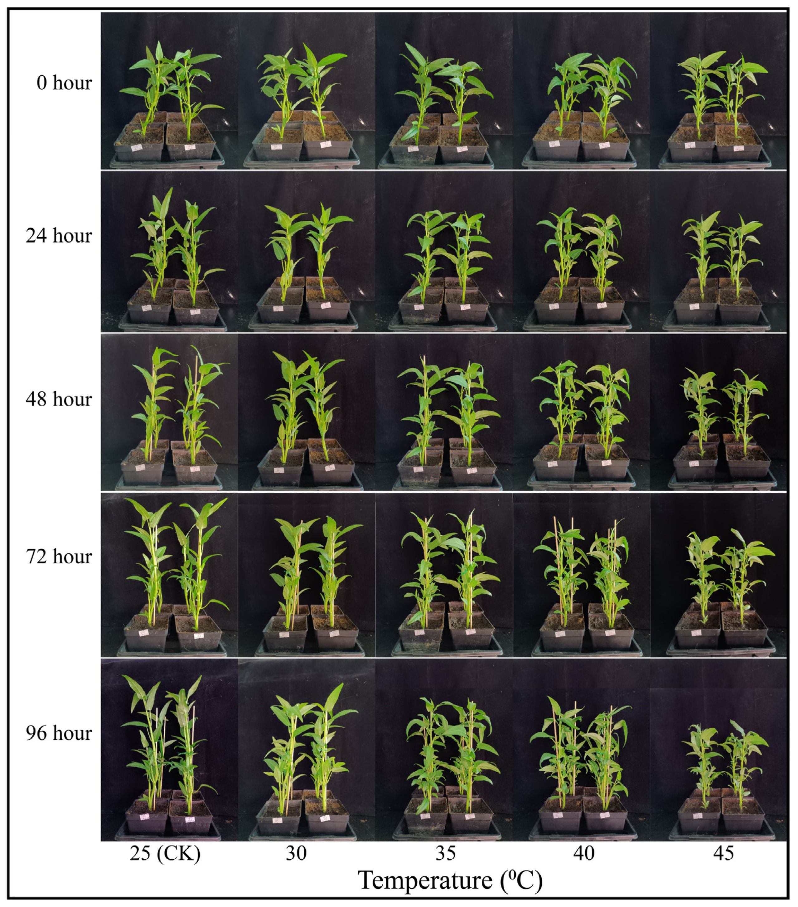
Figure 1. Visual illustration of water spinach plants under various temperature treatments.
Horticulturae 09 00923 g001
Figure 2. Effect of different temperature treatments on root length (A), surface area (B), root volume (C), root tips (D), root forks (E), root crossing (F), and root architecture system (G) in water spinach plants. Mean ± SE of four replicate. Lowercase letters exhibit significant differences (p < 0.05) according to the Fisher’s LSD test.
Horticulturae 09 00923 g002
Figure 3. Effect of different temperature treatments on photosynthesis [Pn (A); Gs (B), Ci (C), and Tr (D)], as well as stomatal properties (E) in water spinach. Mean ± SE of four replicate. Lowercase letters exhibit significant differences (p < 0.05) according to Fisher’s LSD test.
Horticulturae 09 00923 g003
Figure 4. Effect of different temperature treatments on chlorophyll a (A), chlorophyll b (B), carotenoids (C), and auto-fluorescence (D,E) microscopic observations in water spinach plants. Mean ± SE of four replicate. Lowercase letters exhibit significant differences (p < 0.05) according to Fisher’s LSD test.
Horticulturae 09 00923 g004
Figure 5. Effect of different temperature treatments on chlorophyll fluorescence [Fv/Fm (A), Y(II) (B); qP (C), and NPQ (D)] characteristics in water spinach plants. Mean ± SE of four replicate. Lowercase letters exhibit significant differences (p < 0.05) according to Fisher’s LSD test.
Horticulturae 09 00923 g005
Figure 6. Effect of different temperature treatments on the leaf ultrastructure of water spinach. SG: starch granule; n, nucleus; ch, chloroplast; cw-cell wall; sg, starch granule; sl, stromal lamellae; g, granum; mi, mitochondria.
Horticulturae 09 00923 g006
Figure 7. Effect of different temperature treatments on leaf tissue by paraffin section analysis. UE: upper epidermis; SMC: spongy mesophyll cells; Ph, phloem; vb, vascular bundle; de, down-epidermis; pd, palisade mesophyll cell; vb, vascular bundle; x, xylem.
Horticulturae 09 00923 g007
Figure 8. Effect of different temperature treatments on total phenolics (A), total flavonoids (B), total soluble sugar (C), and total anthocyanin (D) in water spinach. Mean ± SE of four replicate. Lowercase letters exhibit significant differences (p < 0.05) according to Fisher’s LSD test.
Horticulturae 09 00923 g008
Figure 9. Influence of various temperature levels on oxidative stress biomarkers [H2O2 (A); O2•− (B), and MDA (C)] in water spinach and leaf histochemical localization of H2O2, O2•−, and MDA accumulation. Mean ± SE of four replicate. Lowercase letters exhibit significant differences (p < 0.05) according to Fisher’s LSD test.
Horticulturae 09 00923 g009
Figure 10. Effect of different temperature treatments on antioxidant enzymes [SOD (A); CAT (B), POD (C), APX (D), GR (E), GST (F), DHAR (G), and MDHAR (H)] activity in water spinach plants. Mean ± SE of four replicate. Lowercase letters exhibit significant differences (p < 0.05) according to Fisher’s LSD test.
Horticulturae 09 00923 g010
Table 1. The influence of different temperature treatment on fresh and dry biomass of water spinach plants.
Temperature
(°C)
Biomass Weight/Plant (g)
FreshDry
ShootRootShootRoot
25 (CK)7.71 ± 0.27 a1.483 ± 0.301 a1.48 ± 0.05 b0.106 ± 0.001 b
307.61 ± 0.25 a1.432 ± 0.035 a1.72 ± 0.06 a0.114 ± 0.001 a
356.64 ± 0.20 b1.126 ± 0.021 b1.26 ± 0.05 c0.077 ± 0.003 c
405.15 ± 0.24 c0.823 ± 0.013 c0.84 ± 0.05 d0.051 ± 0.002 d
453.82 ± 0.15 d0.596 ± 0.018 d0.51 ± 0.04 e0.036 ± 0.001 e
Mean ± SE of four replicate. Lowercase letters exhibit significant differences (p < 0.05) according to Fisher’s LSD test.
Disclaimer/Publisher’s Note: The statements, opinions and data contained in all publications are solely those of the individual author(s) and contributor(s) and not of MDPI and/or the editor(s). MDPI and/or the editor(s) disclaim responsibility for any injury to people or property resulting from any ideas, methods, instructions or products referred to in the content.