Article

Exploring the Bioprotective Potential of Halophilic Bacteria against Major Postharvest Fungal Pathogens of Citrus Fruit Penicillium digitatum and Penicillium italicum

1
Phytopathology Unit, Department of Plant Protection, Ecole Nationale d’Agriculture de Meknès, Km 10, Rte Haj Kaddour, BP S/40, Meknès 50001, Morocco
2
African Genome Center, Mohammed VI Polytechnic University (UM6P), Lot 660, Hay Moulay Rachid, Ben Guerir 43150, Morocco
3
Laboratory of Functional Ecology and Environmental Engineering, Sidi Mohamed Ben Abdellah University, Route d’Imouzzer, Fez 30050, Morocco
4
Microbiology Unit, Laboratory of Bioresources, Biotechnology, Ethnopharmacology and Health, Faculty of Medicine and Pharmacy Oujda, University Mohammed Premier, Oujda 60000, Morocco
5
Plant Protection Laboratory, Regional Center of Agricultural Research of Meknès, National Institute of Agricultural Research, Km 13, Route Haj Kaddour, BP.578, Meknès 50001, Morocco
6
Biotechnology Unit, Regional Center of Agricultural Research, INRA-Morocco, Rabat 10080, Morocco
7
Unité de Recherche Résistance Induite et Bioprotection des Plantes, Faculty of Sciences, Université de Reims Champagne-Ardenne, USC INRAe 1488, 51687 Reims, France
8
Plant Pathology Laboratory, AgroBiosciences, College of Sustainable Agriculture and Environmental Sciences, Mohammed VI Polytechnic University (UM6P), Lot 660, Hay Moulay Rachid, Ben Guerir 43150, Morocco
*
Authors to whom correspondence should be addressed.
Horticulturae 2023, 9(8), 922; https://doi.org/10.3390/horticulturae9080922
Submission received: 6 May 2023 / Revised: 4 August 2023 / Accepted: 10 August 2023 / Published: 13 August 2023
(This article belongs to the Special Issue Plant Pathology in Horticultural Production)

Abstract

:
Citrus fruits are vulnerable to green mold (caused by Penicillium digitatum) and blue mold (caused by Penicillium italicum) during storage, posing significant challenges to the industry. Therefore, biological control utilizing antagonistic bacteria has emerged as a dependable strategy for managing postharvest diseases. In this study, halophilic bacterial isolates were carefully selected from diverse saline ecosystems, including the Dead Sea, the Agadir Sea, the Rabat Sea, saline soil, and water of the Amassine Oued in Taounate, based on rigorous in vitro and in vivo antagonism bioassays. Out of 21 bacteria from different saline environments, 10 were chosen for further characterization based on the 16S rDNA gene. Notably, the EAM1 isolate demonstrated exceptional inhibitory effects, reaching a 90% inhibition rate against P. digitatum, while the ER2 isolate closely followed with an 89% inhibition rate against P. italicum. Furthermore, in bacterial supernatant experiments, six bacterial isolates effectively curbed the growth of P. digitatum, and three demonstrated efficacy against P. italicum development. In an in vivo trial spanning ten days of incubation, three highly effective isolates against P. digitatum displayed zero severity, and two of these isolates also demonstrated zero severity against P. italicum. Interestingly, a comparison of bacterial filtrates revealed that all isolates exhibited a severity level of over 50% against the pathogen causing green rot (P. digitatum), while the severity was lower than 50% for the supernatants of the two isolates used against P. italicum. In conclusion, this study highlights the promising role of halophilic bacteria, specifically Bacillus amyloliquefaciens EAM1 and B. amyloliquefaciens ER2, in controlling postharvest fruit pathogens. The findings shed light on the potential of utilizing these bioprotective agents to address the challenges posed by green and blue citrus molds, providing valuable insights for the citrus industry.

1. Introduction

Citrus species (Citrus spp.) are renowned for their nutritional benefits, being rich sources of vitamin C and bioactive compounds with antioxidant properties, which fulfill essential dietary requirements [1]. The citrus industry plays a pivotal role in the global economy, as it not only provides valuable commodity production but also generates employment opportunities at various stages of its production cycle [2,3].
The citrus group, encompassing oranges, mandarins, tangerines, and other varieties, thrives in cultivation across more than 100 countries worldwide [4]. Brazil and the United States, both known for their favorable climate, stand as two of the world’s top orange-producing nations, contributing to a global orange yield increase of 4.2 million tons in 2018/2019, rising from 47.6 million tons in 2017. According to the latest data from the Food and Agriculture Organization, the overall citrus fruit production surpassed 150 million tons in 2018, with oranges comprising 50% of the total output [5]. Notably, Morocco holds a significant position as a leading citrus producer in the Mediterranean region, dedicating 126,600 hectares to citrus cultivation [6].
Despite concerted efforts, the challenge of preserving fresh produce quality and safety remains a pressing concern. Microorganisms play a crucial role in diminishing the quality of fruits, posing significant economic and health risks, particularly with fungal infections that may produce mycotoxins. Citrus and other fruit products can be exposed to pathogens during field growth and may encounter additional threats during postharvest handling processes [7]. Physical damage during transportation, like bruising, further increases susceptibility to fungal infections [6]. These postharvest diseases are identified as the primary cause of losses in fruits and vegetables after harvesting, resulting in substantial economic impact. In regions with high humidity levels, approximately 20–25% of harvested fruits and vegetables are susceptible to postharvest infections caused by phytopathogenic microorganisms during handling [8,9]. For citrus, different microbes, including fungi, bacteria, and viruses, induce diseases at different stages of production. Notably, postharvest citrus fruit diseases are often attributed to several pathogenic fungi, such as Aspergillus niger, Geotrichum candidum, Penicillium spp., and Rhizopus stolonifera [10,11].
The postharvest phase of citrus cultivation is highly susceptible to more than 20 different types of diseases, resulting in substantial economic losses [12]. Two major diseases that significantly impact citrus fruits during storage are green mold and blue mold. These diseases are primarily caused by the pathogens Penicillium digitatum and Penicillium italicum, respectively. Notably, P. digitatum is the primary pathogen, responsible for approximately 90% of the total losses [13,14,15].
Contamination arises from various sources, including wounds caused by insect and mechanical lesions, along with other environmental factors [16]. The precise dynamics of the interaction between the pathogens and the host remain incompletely understood. Nonetheless, certain factors are believed to influence this interaction, leading to an increase in fungal infections. Both fungi (P. digitatum and P. italicum) are known for secreting organic acids that create an optimal condition for infection [17]. In comparison to green mold, blue mold can present a more significant challenge due to its capacity to nest and initiate the infection process even in the absence of fruit injury [18]. P. italicum may produce hydrolytic enzymes that weaken the nearby fruit skin, establishing a favorable environment for infection [19].
In recent decades, research efforts have primarily focused on utilizing chemical fungicides to combat both fungi. Among the commonly employed options, thiabendazole (TBZ) and imazalil (IMZ) have become widely used fungicides [20]. However, the extensive use of these fungicides has led to the development of resistant strains, rendering these treatments less effective. Moreover, concerns regarding environmental pollution and the potential accumulation of harmful residues in food have emerged [21,22]. Finding suitable alternative control strategies has become a challenging task for scientists. In response, the employment of antagonistic microorganisms and natural antibacterial compounds has been proposed as a potential alternative to synthetic fungicides [23].
Biological control is emerging as a critical alternative for effectively managing postharvest diseases in fruits, highlighting the need for more efficient biocontrol strategies. Notably, the successful control of infections using antagonistic bacteria has been widely reported [23,24]. However, finding suitable alternative control systems remains a challenge for scientists. Consequently, advocacy for employing antagonistic microorganisms and natural antimicrobial compounds as alternatives to chemical fungicides is gaining momentum [6]. The primary objective of the present study is to explore alternative approaches for the biological management of postharvest rot in citrus fruits caused by P. digitatum and P. italicum. The research aims to evaluate the potential of halophilic bacteria isolates from diverse biotopes as biocontrol agents and to assess their antifungal activities against green and blue mold. Additionally, the study endeavors to characterize the most effective bacterial isolates, identify their traits, and investigate the production of antifungal compounds. By delving into these investigations, the study seeks to enhance our understanding of effective strategies for controlling postharvest rot in citrus fruits.

2. Materials and Methods

2.1. Pathogen Sources

The fungi utilized in this study were previously isolated from decaying orange and lemon fruits. These fungal strains were sourced from the phytopathology department of Morocco’s National School of Agriculture in Meknes. To maintain the strains, fungal pathogens were subcultured on potato dextrose agar (PDA), and antibiotics (50 g/mL chloramphenicol and streptomycin sulfate) were added. The cultures were subsequently incubated at 25 °C for seven days in the absence of light.

2.2. Fruit Preparation

In vivo experiments were carried out using oranges (Citrus reticulata Blanco) of the ‘Valencia-late’ cultivar. Fruits with uniform size and maturity stages were carefully selected and stored until required for the experiments. Before each trial, the oranges underwent a thorough cleaning process, followed by surface disinfection with 0.1% sodium hypochlorite for 5 min. After disinfection, the oranges were rinsed with sterile distilled water (SDW) and allowed to dry at room temperature.

2.3. Isolating Bacteria from Saline Water and Soil

Water samples were collected from four distinct regions, namely the Dead Sea, the Agadir Sea, the Rabat Sea, and the Amassine Oued region. Subsequently, serial dilutions were prepared by adding 1 mL of each water sample to 9 mL of SDW. In an Erlenmeyer flask, 10 g of soil from the Amassine Oued region was mixed with 100 mL of SDW. Dilutions of this mixture were spread onto PDA Petri plates and incubated for 72–96 h at 2 °C, with periodic shaking every 30 min. Colonies displaying various morphologies on lysogeny broth (LB) medium Petri dishes were subcultured until pure cultures were obtained. In total, 21 bacterial colonies were collected and preserved in the phytopathology laboratory in Meknes, Morocco, for further experimentation. To adjust the bacterial suspension, a 24-hour-old culture was utilized, and its concentration was set to 1 × 10⁸ CFU using a spectrophotometer at 620 nm. This standardized suspension is now ready for subsequent tests and analyses.

2.4. Screening for Antagonistic Activity by Direct Confrontation

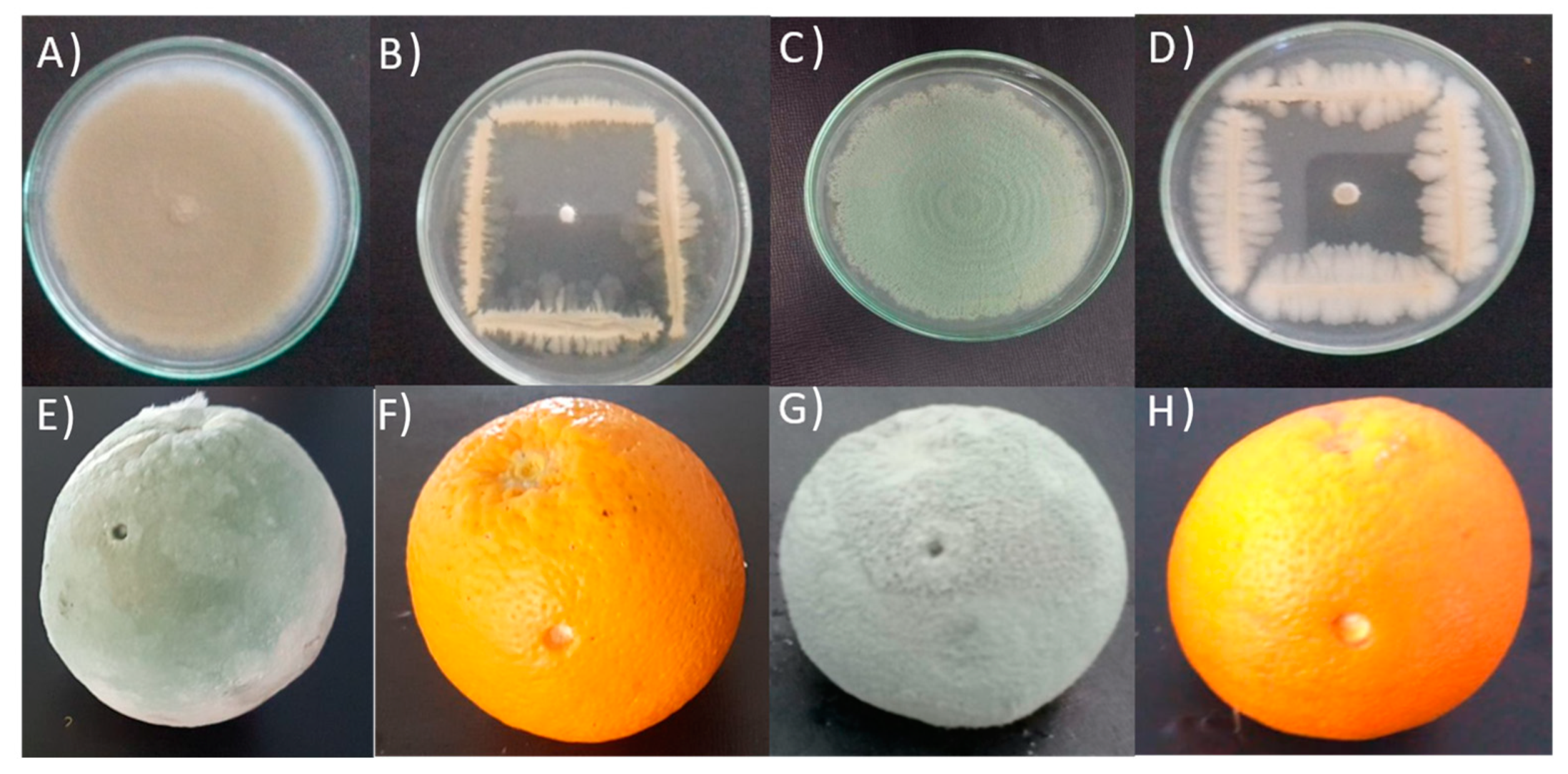
2.4.1. In Vitro Dual Culture

Antagonism tests were conducted by directly confronting bacterial isolates with fungal pathogens to evaluate their potential in halting the growth of both fungi [23,24]. For each bacterium, streaks were made along the media. Subsequently, 10 μL of the spore solution of each pathogenic fungus, adjusted to 1 × 10⁶ CFU, was injected into the central petri plate containing the PDA medium and the bacterial isolate. Controls were prepared separately, containing only the spore solution. To quantify the inhibition rate (IR), the formula reported by Ezrari et al. [23] was employed.

2.4.2. In Vivo Antagonism Experiment

To assess the efficacy of bacterial isolates in controlling postharvest decay in Valencia late fruits, two equatorial incisions were made using a sterile needle, each measuring 2 mm in diameter and 3 mm in depth. The obtained incisions were treated with 50 µL of bacterial suspension at a concentration of 108 CFU, while SDW was used as a control. After four hours, the pretreated wounds were inoculated with 20 µL of conidial suspension containing 104 spores/mL for both P. digitatum and P. italicum. The treated fruits were placed in plastic bags with Whatman paper soaked in SDW to maintain humidity and prevent moisture loss. The boxes were then incubated at 4 °C in a growth chamber with a photoperiod of 12 h. The experiment was conducted in two runs, each consisting of four fruits with eight wounds. The lesion diameter was measured on days 10 and 20 after inoculation, and the calculation method described by Ezrari et al. [23] was used for evaluation.

2.5. Bacterial Identification

The ten bacterial isolates, which had previously demonstrated significant antagonist activity against both pathogens, were subjected to identification. DNA extraction for each bacterial isolate was carried out following the protocol described by Llop et al. [25].
To identify the antagonist microorganisms, the polymerase chain reaction (PCR) technique was employed, amplifying the partial gene expressing 16sDNA using the universal primers Fd1: 5’-AGA GTT TGA TCC TGG CTC AG-3’ and RP2: 5’-ACG GCT ACC TTG TTA CGA CTT-3’. The PCR reaction was conducted in a total volume of 25 µL as per the manufacturer’s instructions, using EnzimaGoTaq DNA polymerase (Promega, Madison, USA ). The amplifications were carried out in an Eppendorf thermal cycler, following the settings specified by Ezrari et al. [23]. Subsequently, the PCR products obtained were subjected to sequencing using the Sanger dideoxy method [26]. The sequences were then aligned, corrected, and analyzed for similarity in the NCBI-BLAST collection (National Center for Biotechnology Information, http://blast.ncbi.nlm.nih.gov/Blast.cgi accessed on 2 February 2023) and through the DNAMAN tool (version 6.0, Lynnon Biosoft, San Ramon, CA, USA). The output sequences from the analysis were added to GenBank [23].

2.6. An In-Depth Characterization of Antagonistic Mechanisms

Multiple in vitro experiments were performed to investigate the biocontrol activity mechanisms demonstrated by the ten chosen bacterial isolates.

2.6.1. Indirect Antagonist Activity

Volatile Compounds (VOCs) Bioassay

To evaluate the potential generation of volatile organic compounds (VOCs) capable of inhibiting fungal development, each isolate was cultivated on an LB medium and then incubated at 28 °C. After 24 h, 10 µL of the spore solution of each pathogenic fungus, adjusted to 1 × 10⁶ CFU, was added to the PDA medium in the Petri dishes. The lids of the Petri dishes were removed, and the bottoms containing the medium were sealed together using translucent adhesive tape (Parafilm®, Gennevilliers, France). The control Petri dish had no bacteria [27]. Observations were made after incubation at 28 °C for 5 and 10 days. The inhibition rate was calculated following the method reported by Trivedi et al. [28].

In Vitro Bacterial Cell-Free Filtrates Effect

In vitro effect
The test aimed to investigate the impact of cell-free filtrates from the ten selected bacteria on both fungi. The protocol was conducted following the method adopted by Balouiri et al. [29]. A 100 µL bacterial suspension containing 1 × 10⁸ CFU was introduced to liquid LB medium and incubated at 28 °C on a rotatory shaker set to 130 revolutions per minute for 4 days. Subsequently, the mixture was centrifuged at 5500 rpm for 25 min, and the resulting supernatant was filtered through a 0.22-diameter membrane filter. A 10% (v/v) portion of the filtered supernatant was then added to the PDA medium, heated to 45–50 °C. To evaluate antifungal activity, 10 μL aliquots of the pathogen’s spore solution (1 × 10⁶ CFU) were placed in the center of Petri dishes. The control group received a 10% liquid LB medium instead of the bacterial supernatant. Antifungal activity (%) was calculated using the formula IR = ((DC − DT)/DC) × 100 after 6 days of incubation at 28 °C. DC represents the growth diameter in the control, while DT corresponds to the growth diameter with the bacterial supernatant.
In Vivo Effect
After creating wounds in the fruit, 50 μL of the filtrate supernatant from antagonistic bacteria was applied to each wound. After 24 h, the wounded fruits were treated with 20 μL of a 1 × 10⁴ spores/mL conidial suspension of the respective fungal pathogen. For the control group, 20 μL of liquid LB medium was administered before pathogen injection. Subsequently, the fruits were placed in plastic boxes and incubated at 4 °C. The development of decay was assessed 10–20 days post-inoculation and the lesion diameters were recorded using a digital caliper. Disease severity (DS) was determined according to the following formula [24,30]:
Disease severity (%): (DT/DC) × 100
where DT is the average lesion diameter (mm) of treated wounds (antagonist following by pathogen) and DC is the average diameter (mm) of the wounds in the control treatment.

2.6.2. Bacterial Isolates’ Biochemical Characteristics

Amylase Activity

The evaluation of amylase production was carried out using nutrient agar medium (NA) supplemented with 1% soluble starch. A Petri plate containing NA medium and a 48-h-grown isolate (1 × 10⁸ CFU) was placed in the center and incubated at 28 °C for 72 h. To assess starch hydrolysis, 3 mL of Lugol’s iodine solution was added to the medium and allowed to react for 3 min. The presence of a clear zone surrounding the bacterial colony indicated amylase activity, while starch-containing zones remained constant and opalescent [31]. The amylolytic index was determined using the following formula: transparent halo diameter divided by the colony diameter [32].

Protease Activity

The determination of protease activity by the selected bacterial antagonist was conducted using a solid medium containing skimmed milk, following the protocol described by Ezrari et al. [23]. Each 24-hour-old bacterium was spotted with 5 μL of a suspension at a concentration of 1 × 10⁸ CFU in the middle of the medium. Subsequently, the Petri dishes were incubated at 28 °C for 48 h. Effective protease action was indicated by the presence of a distinct halo forming around the bacterial colonies. To calculate the proteolytic index, the diameter of the translucent halo was divided by the colony diameter using the following formula: transparency of the halo divided by the colony diameter [32].

Cellulase Activity

Cellulase production was evaluated using carboxymethyl cellulose (CMC) as the substrate [33]. Each bacterial suspension (1 × 10⁸ CFU) was inoculated using 10 μL. The Petri plates were then incubated at 28 °C for four days to monitor cellulase production. Subsequently, the plates were incubated with a 0.1% Red Congo solution for 15 min, followed by three rinses with a 1 M NaCl solution. Healthy cellulase activity was indicated by the presence of a distinct halo forming around the bacterial colonies. To calculate the cellulase index, the diameter of the transparent halo was divided by the colony diameter using the following formula: transparent halo diameter divided by colony diameter [32].

Hydrocyanic Acid (HCN) Production

The assessment of hydrogen cyanide (HCN) synthesis by a subset of bacterial isolates was conducted using a technique outlined in a previous study, following the protocol described by Trivedi et al. [28]. To test HCN production, a 24-hour fresh bacterial culture was adjusted to 1 × 10⁸ CFU, and 100 μL of this culture was inoculated into Petri dishes containing solid LPGA medium (yeast peptone glucose agar) with 4.4 g/L glycine. A Whatman paper disc treated with a picrate solution (2.5% picric acid in 12.5% anhydrous sodium carbonate (Na2CO3) solution) was placed on the lid of each dish. Control plates using SDW instead of bacteria were also prepared. The dishes were then inverted and incubated for four days at 28 °C. The presence of HCN production was determined by observing the color change in the Whatman paper, which indicated the results of the test.

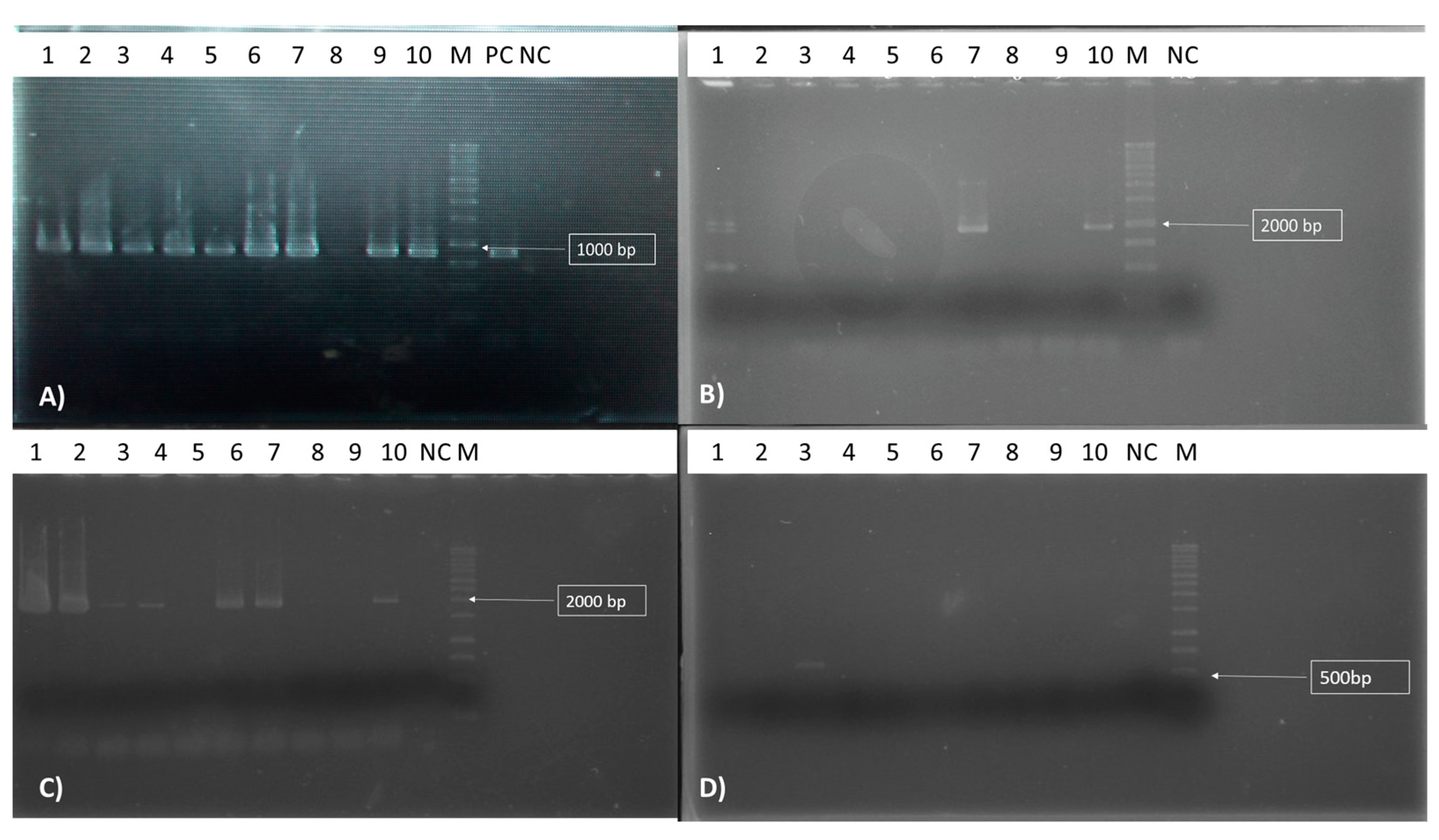
The Antibiotic Biosynthetic Gene Detection

The 10 selected bacteria underwent DNA extraction, and the obtained results were utilized to identify the presence of genes associated with bacillomycin, surfactin, iturin, and fengycin. The PCR experiment was conducted following the method described by Lahlali et al. [24] using specific primers: BACC1F/BACC1R for bacillomycin, FEND1F/FEND1R for fengycin, ITUP1F/ITUP2R for iturin, and P17/P18 for surfactin [34] (Table 1).

2.7. Semi-Practical Trials

The effectiveness of isolates (ER2, ER6, and EAM3) which exhibited the highest efficacy under in vivo conditions was evaluated in a large-scale, semi-commercial study conducted in packinghouses for the treatment of both blue and green citrus rot diseases. Citrus fruits were subjected to four evenly spaced wounds, followed by immersion in each bacterial solution (1 × 10⁸ CFU) for 2 min. As a control, citrus fruits were dipped in SDW. Subsequently, ten citrus fruits per bag were placed in plastic bags, and three replicates were prepared. The bags were then incubated in a growth chamber at 4 °C. After a 24-h incubation period, the citrus fruits were infected with each fungus (1 × 10⁵ spores/mL) and incubated at 4 °C in the growth chamber. Simultaneously, treatment with the fungicidal thiabendazole (50%) was carried out [24].

2.8. Statistical Analysis

The tests were conducted multiple times to ensure accuracy. The data were presented as mean ± SD (standard deviation), and a randomized methodology was employed. For statistical analysis, the SPSS program (IBM SPSS Statistics 20, New York, NY, USA) was used to perform an analysis of variance (ANOVA). In cases where the impact was significant at a p-value of 0.05, Duncan’s test was applied for means separation.

3. Results

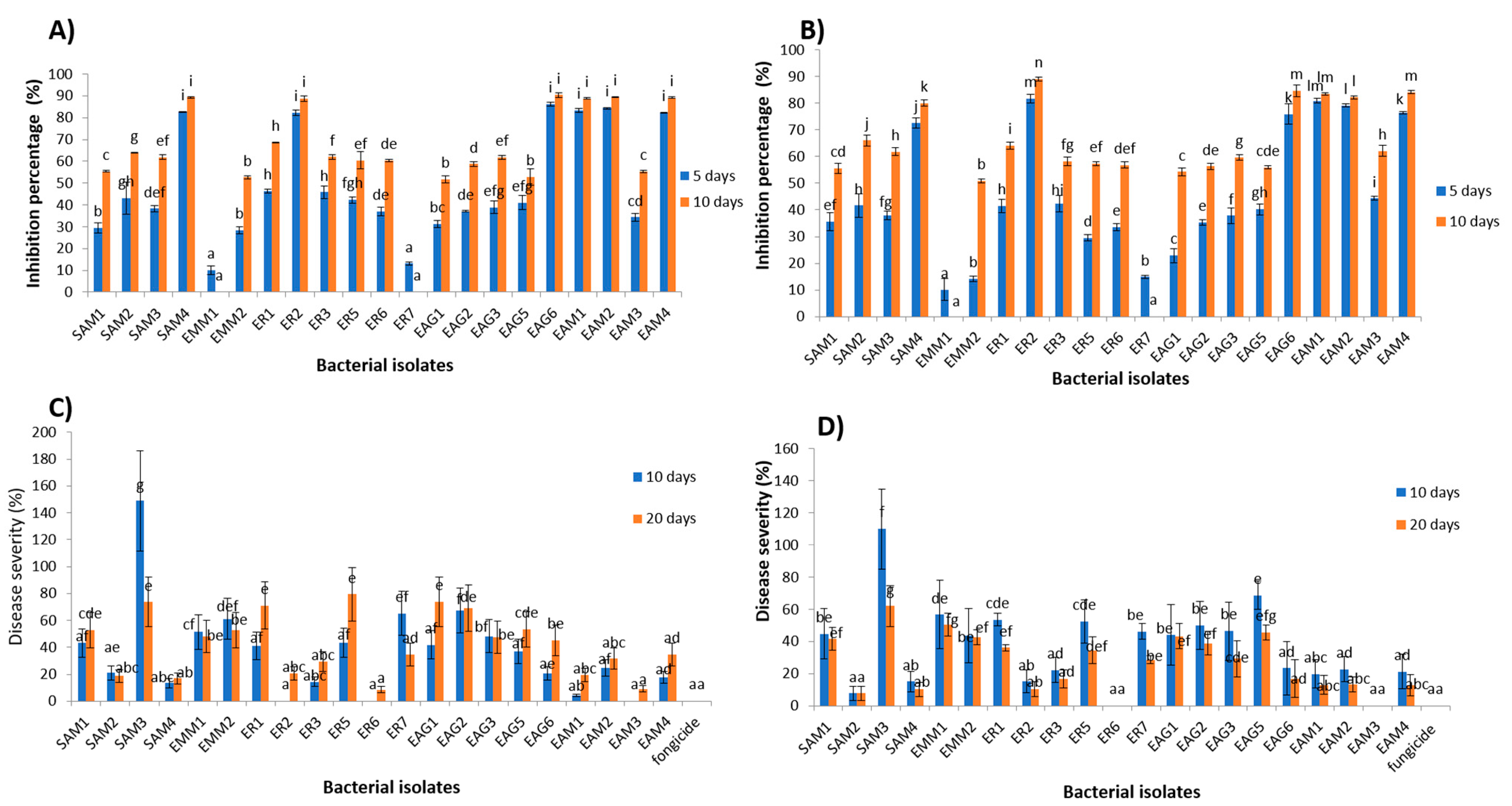
3.1. In Vitro Antagonistic Activity

Using a dual culture bioassay, the antibacterial activity of 21 morphologically distinct bacterial colonies obtained from various locations was assessed against the development of both fungi. The antifungal properties of the 21 isolates exhibited varying levels of effectiveness against the two fungal species. Statistical analysis revealed significant differences between the isolates (p < 0.05), indicating notable distinctions in their antifungal activities (Figure 1 and Figure 2). Among the tested isolates, only six (SAM4, ER2, EAG6, EAM1, EAM2, and EAM4) displayed a considerable ability to inhibit the mycelial development of both harmful fungi, with a rate exceeding 50% after 5 days of incubation. Furthermore, these isolates exhibited a remarkable antagonistic effect after 10 days, surpassing 80% inhibition against the fungi.

3.2. In Vivo Antagonism Experiment

The statistical analysis revealed a significant difference (p < 0.05) among the bacterial isolates (Figure 1 and Figure 2). Three halophilic bacteria (ER2, ER6, and EAM3) displayed sustained antifungal activity against citrus green rot, with 100% inhibition (severity = 0%) after 10 days of incubation. In the in vivo confrontation bioassay between P. italicum and the 21 halophilic bacteria, two isolates (EAM3 and ER6) demonstrated a 100% inhibition rate after 10 days and 20 days of incubation.

3.3. Bacterial Identification

The results obtained from both in vitro and in vivo antagonism experiments revealed that 10 bacteria, namely SAM2, SAM4, ER2, ER3, ER6, EAG6, EAM1, EAM2, EAM3, and EAM4, exhibited strong antagonistic effects against both pathogens. Through sequencing the partial region of the 16S rDNA, these 10 isolates were identified as species belonging to the genus Bacillus (9) (B. amyloliquefaciens (4), B. subtilis (3), B. velezensis (1), B. vallismortis (1)) and Ochrobactrum (1) (Ochrobactrum thiophenivorans). The obtained sequences have been deposited in the GenBank database, and their accession numbers are presented in Table 2.

3.4. In Vitro Antagonism via the Bacterial Supernatant

The impact of bacterial cell-free culture filtrate on antifungal activity displayed considerable variation across different bacterial isolates. Notably, the filtrates from six isolates, namely SAM4, ER2, EAG6, EAM1, EAM2, and EAM4, exhibited an inhibition rate surpassing 50% against P. digitatum. However, only three bacterial supernatants (EAG6, EAM1, and EAM4) achieved a similar growth reduction exceeding 50% against P. italicum.

3.5. In Vivo Antagonism via the Bacterial Supernatant

The in vivo confrontation trials between 10 culture filtrates obtained from bacterial isolates and P. digitatum revealed that the disease severity was notably higher compared to the confrontation of bacterium against fungus. All tested isolates showed a severity percentage exceeding 50%. The statistical analysis highlighted significant differences among the results after just 10 days of incubation. Conversely, only two out of the 10 culture filtrates obtained from bacterial and P. italicum isolates exhibited a severity lower than 50%. As incubation time increased, the severity of blue rot decreased for all isolates, as depicted in Figure 3. The statistical analysis demonstrated a highly significant difference in the effects of the various isolates during both incubation periods.

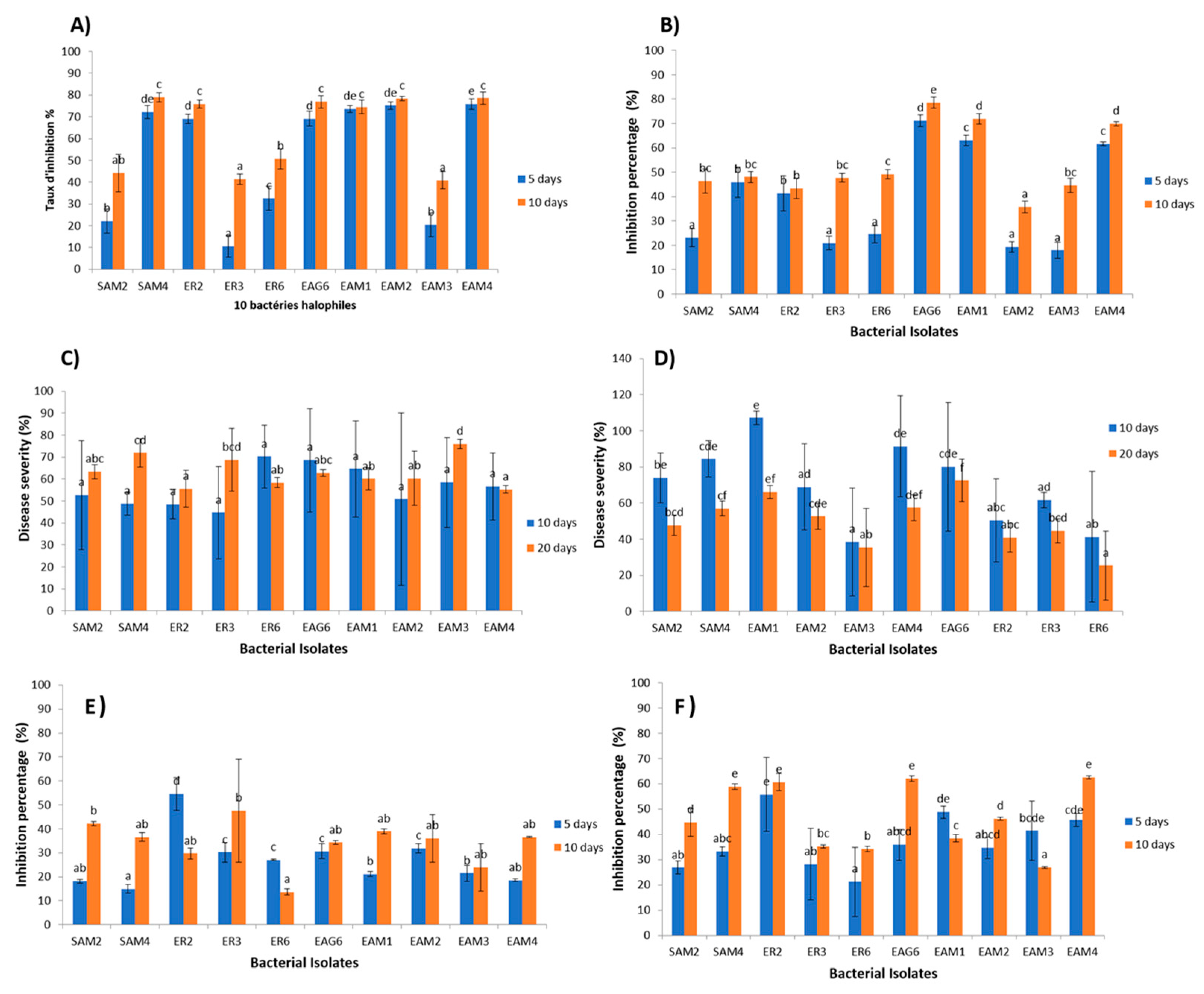
3.6. In Vitro Volatility-Mediated Antagonism

Among the bacterial strains tested, only ER2 exhibited significant antifungal activity (>50%) against P. digitatum through the release of VOCs, as depicted in Figure 3. Additionally, four out of the ten bacterial isolates (EAM4, SAM4, ER2, and EAG6) showed a notable antagonistic effect against P. italicum through VOC emission, with the effect increasing to 60% for these isolates after 10 days of incubation. The statistical analysis revealed a highly significant difference in antifungal activity among the various isolates for both tested pathogens after 5 and 10 days of incubation.

3.7. Biochemical Traits

The ten chosen bacterial isolates were assessed for their enzymatic activity, and the results are presented in Table 3.

3.7.1. Amylase Activity

The findings indicated that out of the tested isolates, eight demonstrated the ability to produce amylase. Notably, the ER2 isolate exhibited a significantly higher cellulolytic index of 2.2.

3.7.2. Proteolytic Activity

The results indicated that nine of the tested isolates could produce protease. These isolates demonstrated varying proteolytic capacities, as evidenced by the formation of distinct halos around the producing bacteria, with differing sizes. Notably, the bacterial isolate EAG6 displayed a particularly high proteolytic activity, with a proteolytic index of 4.

3.7.3. Cellulose Degradation

The results indicated that five bacterial isolates exhibited cellulase production. Notably, the isolate EAM3 demonstrated a notably high amylolytic index.

3.7.4. Hydrogen Cyanide Production

Upon examination of the Whatman paper, it was evident that only EAM1 and EAM3 bacterial isolates exhibited HCN production, as signified by a distinct color change.

3.8. Detection of the Antibiotic Biosynthetic Gene

PCR was employed to investigate the presence of biocontrol genes in a group of twenty antagonist bacteria. The results regarding the detection of genes related to the biosynthesis of lipopeptides, specifically bacillomycin, fengycin, iturin, and surfactin, are summarized in Figure 4. The evaluation of bacterial isolates’ ability to produce bacillomycin showed that only nine isolates (EAM1, EAM2, EAM3, EAM4, SAM2, SAM4, ER2, ER6, and EAG6) harbored the bamC gene responsible for bacillomycin synthesis (Figure 4A). Concerning fengycin secretion, only three isolates were found to possess this gene (EAM1, ER2, and EAG6) (Figure 4B). As for iturin production, the results indicated the presence of the expected PCR product in seven bacterial isolates (EAM1, EAM2, EAM3, EAM4, SAM4, ER2, and EAG6) (Figure 4C). However, for surfactin production, it was observed that only one bacterial isolate (EAM3) harbored this gene (Figure 4D).

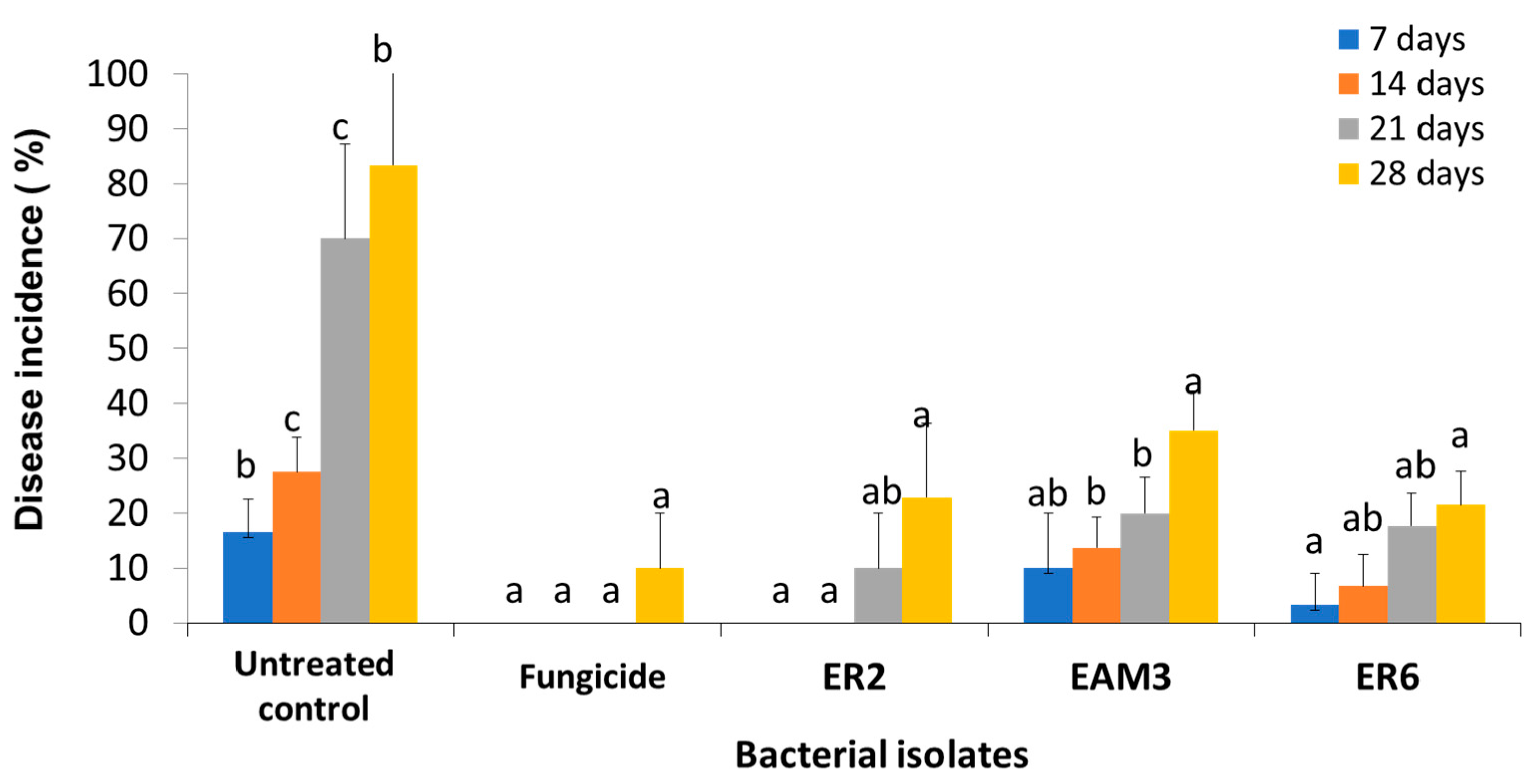
3.9. Semi-Practical Trials

The obtained results demonstrated the statistical significance of all treatments (p < 0.05). The disease incidence progressively increased over time (from 7 to 28 days) irrespective of the treatment. Notably, no disease symptoms were observed after two weeks of incubation, with a 0% incidence rate for ER2 and a 30% incidence rate for EAM3 (Figure 5).

4. Discussion

Our study’s findings emphasize the valuable potential of saline biotopes as a rich source of antagonistic bacteria, offering a promising eco-friendly strategy to effectively combat citrus postharvest diseases. The halophilic bacterial strains’ ability to inhibit pathogens and thrive in extreme environments makes them highly attractive as Biological Control Agents (BCAs). Previous research has also demonstrated the antimicrobial capabilities of halophilic bacteria against fungal pathogens [37]. Pérez-Inocencio et al. [38] highlighted the successful colonization of halophilic bacteria in salt-affected soil, showcasing their potential for disease suppression. Additionally, halophilic bacteria have been effectively employed in the bioremediation of organic pollutants in saline environments [39]. These findings underscore the significant potential of halophilic bacterial strains as efficient biocontrol agents for both agricultural and environmental applications. We isolated 21 bacteria from five distinct saline biotopes and evaluated their antifungal efficacy against both fungi, both in vitro (dual culture bioassay) and in vivo. The isolated bacteria exhibited substantial antifungal efficacy against both infections, as revealed by the results.
To date, limited research has explored the potential antifungal effects of halophilic bacteria in controlling postharvest and fungal plant diseases. This highlights the need for further investigation in this area [40,41,42]. In our study, we investigated the ability of halophilic bacteria to inhibit the mycelial growth of both fungal pathogens on citrus fruits, and the selected isolates demonstrated promising activity against citrus green and blue mold. Based on the 16S rDNA sequence, the identified halophilic bacteria belonged to the species B. amyloliquefaciens, B. subtilis, B. velezensis, B. vallismortis, and O. thiophenivorans. Previous studies have also reported the potential of various bacteria as biocontrol agents against postharvest diseases [43,44,45,46,47]. Bacillus bacteria, in particular, are recognized for their capacity to produce a range of antibacterial compounds, making them promising candidates for biocontrol [45,48,49].
The work carried out by Tian et al. [50] highlighted the significant antagonistic activities of bacteria of the Bacillus genus against P. digitatum. Their study also revealed that the cell-free supernatant from these bacteria caused cell vacuolation in P. digitatum [50]. Similarly, Li et al. (2022) [51] observed that a strain of B. subtilis L1–21 produced essential antifungal compounds, leading to substantial suppression of P. digitatum development during fruit storage [51]. These findings further emphasize the potential of Bacillus strains as effective biocontrol agents against postharvest fungal pathogens.
Bacillus amyloliquefaciens has been previously reported as an effective control agent against Penicillium spp. [52]. In vitro tests conducted by Calvo et al. [53] demonstrated that the B. amyloliquefaciens BUZ-14 strain reduced the growth of P. italicum fungus and inhibited the development of both fungi in preventive treatments on orange fruits [53]. Similarly, Bacillus velezensis (SAM4) exhibited antifungal activity against both fungi. Several studies have reported the capacity of this bacterium as a BCA against different pathogenic fungi [54], with Calvo et al. [55] demonstrating that it significantly inhibited the development of P. italicum by 80%. Moreover, B. vallismortis showed promise as a BCA against Corn stalk rot [56], leading to an 80.3% decrease in the growth of Monilinia fructicola, and fumigation with the culture solution resulted in a significant reduction of brown rot in peach by 77.1% [57]. Park et al. [58] highlighted the effectiveness of B. vallismortis in reducing the infection of Colletotrichum acutatum on mature fruits [58].
Furthermore, Duan et al. [59], in their study on B. vallismortis, reported its important antagonist activity against Fusarium spp., which is known to cause apple replant disease, resulting in reduced severity and incidence of the disease. Additionally, both B. vallismortis and B. amyloliquefaciens were recently found to possess antagonistic activities against Phytophthora capsica [59].
Few studies have explored the potential of species belonging to the Ochrobactrum genus as biological control agents [60,61]. Surprisingly, our present study revealed that Ochrobactrum thiophenivorans completely inhibited citrus green rot. Previously, this species has been noted for its sustainable bioremediation of pesticides in the soil environment [62].
Antagonistic bacteria inhibit fungal growth and suppress diseases by secreting hydrolytic enzymes, volatile substances, and secondary metabolites. Our study indicates that the chosen antagonist bacteria, which exhibit potent antifungal activity, possess similar biochemical activities, and employ various biocontrol mechanisms. These bacteria produce extracellular lytic enzymes that can destroy the fungal cell wall, interrupting the process of infection [63]. From our results, we found that eight of the antagonist bacteria showed amylase activity. Although amylase is reported to play a minor role in the control of some fungi, several studies have suggested its potential use [57]. Moreover, proteolytic activity was observed in nine out of the ten isolates tested. Our findings align with previous studies that have highlighted the production of proteases by halophilic bacteria isolated from saline biotopes [64,65]. These enzymes were reported to play a role in the control of Penicillium expansum, Botrytis cinerea, and C. acutatum. Similarly, the research conducted by Dunne et al. [66] emphasized that proteases secreted by Tenotrophomonas maltophilia W81 can control the growth of the fungal pathogen Pythium ultimum [66]. Regarding cellulose degradation, 50% of bacterial isolates showed positive results. The role of this enzyme has been demonstrated in previous studies as well [67]. Bibi et al. (2018) [68] tested the potential of halophilic bacteria to synthesize cellulase and confirmed its role in the control of Phytophthora capsici and P. ultimum [68]. Two of the tested isolates (20%) exhibited the production of hydrogen cyanide (HCN), a volatile metabolite known for its biocontrol activity against fungal diseases. This finding is consistent with Bano and Musarrat (2003) [69], who demonstrated that Pseudomonas aeruginosa can inhibit the growth of Fusarium oxysporum, Trichoderma herizum, and Alternaria alternata through the production of HCN and other metabolites [69].
Furthermore, we investigated the biosynthesis of genes encoding lipopeptide production in the selected bacteria. These bacteria displayed genes responsible for the synthesis of lipopeptides, which have been reported to inhibit the growth of several pathogens [70]. Lipopeptides are considered major contributors to the antifungal activity of different Bacillus spp. Several studies have reported the potential of bacillomycin, fengycin, surfactin, and iturin in controlling fungi. Moreover, the presence of these genes was reported in most Bacillus isolates [23,71]. Dimkić et al. [34] and Arrebola et al. [72] revealed that iturin is the most common antibiotic produced by the Bacillus genus, which is particularly effective against postharvest diseases [34,73]. Bacillomycin, surfactin, and fengycin have also been reported to exhibit antifungal activity [73]. Arroyave-Toro et al. [48] demonstrated that iturin and fengycin, produced by B. subtilis, have significant biocontrol capacity against B. cineria and C. acutatum [48]. Similarly, Rodríguez-Chávez et al. [74] reported that iturins and fengycins produced by B. subtilis impact conidia germination and reduce P. expansum lesions on apple fruits [74].
The in vivo trials’ findings demonstrated that halophilic bacteria have a significant impact on the development of citrus rot. B. amyloliquefaciens and B. vallismortis effectively controlled P. digitatum-caused green rot, as previously reported by Leelasuphakul et al. [75], showing an 86.7% decrease in disease incidence and a delay of up to 6 days in disease symptoms, with decay symptoms reduced up to day 9 using the biocontrol agent B. subtilis [75].
Moreover, the difference in a strain’s ability to combat fungi in vitro and in vivo can be attributed to various factors. In vitro assays provide controlled conditions, while in vivo settings involve complex interactions with the host plant, other microorganisms, and environmental factors. These factors, such as competition, varying environmental conditions, and host defense mechanisms, can affect the strain’s effectiveness in inhibiting fungi. Conducting field trials under realistic conditions can bridge the gap between in vitro and in vivo results, providing a more comprehensive understanding of the strain’s efficacy.
In summary, our work demonstrates the potential of halophilic bacteria derived from saline environments as natural and eco-friendly biological control agents for postharvest diseases. The experimental trials demonstrated their efficacy in controlling various plant pathogens. These findings suggest that harnessing the capabilities of these bacteria could prove valuable in postharvest disease management strategies. However, further research is necessary to develop a commercial product based on these bacteria.

Author Contributions

Conceptualization, N.R., H.A., S.E. and R.L.; methodology, R.L., S.E. and N.R.; software, R.L., H.A. and S.E.; validation, R.L., S.E., Z.B. and E.A.B.; formal analysis, N.R., S.E. and J.K.; investigation, R.L., F.M. and E.A.B.; resources, R.L.; data curation, H.A., N.R. and R.L.; writing—original draft preparation, N.R., S.E. and J.K.; writing—review and editing, R.L., Z.B., F.M. and E.A.B.; supervision, R.L.; project administration, R.L. All authors have read and agreed to the published version of the manuscript.

Funding

This research was financially supported by the Phytopathology Unit of the Department of Plant Protection and Environment, Ecole Nationale d’Agriculture de Meknes.

Institutional Review Board Statement

Not applicable.

Informed Consent Statement

Not applicable.

Data Availability Statement

Data are available under reasonable demand.

Conflicts of Interest

The authors declare no conflict of interest.

References

  1. Mditshwa, A.; Magwaza, L.S.; Tesfay, S.Z.; Opara, U.L. Postharvest factors affecting vitamin C content of citrus fruits: A review. Sci. Hortic. 2017, 218, 95–104. [Google Scholar] [CrossRef]
  2. Chinnici, G.; Zarbà, C.; Hamam, M.; Pecorino, B.; D’Amico, M. A model of circular economy of citrus industry. Int. Multidiscip. Sci. GeoConf. SGEM 2019, 19, 19–26. [Google Scholar]
  3. Babcock, B.A. Economic Impact of California’s Citrus Industry in 2020. J. Citrus Pathol. 2022, 9, 36–39. [Google Scholar] [CrossRef]
  4. Ismail, M.; Zhang, J. Post-harvest citrus diseases and their control. Outlooks Pest Manag. 2004, 15, 29. [Google Scholar] [CrossRef]
  5. Chen, J.; Shen, Y.; Chen, C.; Wan, C. Inhibition of key citrus postharvest fungal strains by plant extracts in vitro and in vivo: A review. Plants 2019, 8, 26. [Google Scholar] [CrossRef] [Green Version]
  6. Ezrari, S.; Radouane, N.; Tahiri, A.; El Housni, Z.; Mokrini, F.; Özer, G.; Lazraq, A.; Belabess, Z.; Amiri, S.; Lahlali, R. Dry root rot disease, an emerging threat to citrus industry worldwide under climate change: A review. Physiol. Mol. Plant Pathol. 2022, 117, 101753. [Google Scholar] [CrossRef]
  7. Dwiastuti, M.E.; Soesanto, L.; Aji, T.G.; Devy, N.F. Hardiyanto Biological control strategy for postharvest diseases of citrus, apples, grapes and strawberries fruits and application in Indonesia. Egypt. J. Biol. Pest Control 2021, 31, 141. [Google Scholar] [CrossRef]
  8. Gomes, A.A.M.; Queiroz, M.V.; Pereira, O.L. Mycofumigation for the biological control of post-harvest diseases in fruits and vegetables: A review. Austin J. Biotechnol. Bioeng. 2015, 2, 1–8. [Google Scholar]
  9. Bradford, K.J.; Dahal, P.; Van Asbrouck, J.; Kunusoth, K.; Bello, P.; Thompson, J.; Wu, F. The dry chain: Reducing postharvest losses and improving food safety in humid climates. In Food Industry Wastes; Elsevier: Amsterdam, The Netherlands, 2020; pp. 375–389. [Google Scholar]
  10. Sommer, N.F. Postharvest Handling Practices. Plant Dis. 1982, 66, 357. [Google Scholar] [CrossRef]
  11. Holmes, G.J.; Eckert, J.W. Sensitivity of Penicillium digitatum and P. italicum to postharvest citrus fungicides in California. Phytopathology 1999, 89, 716–721. [Google Scholar] [CrossRef] [Green Version]
  12. Zacarias, L.; Cronje, P.J.R.; Palou, L. Postharvest technology of citrus fruits. In The Genus Citrus; Elsevier: Amsterdam, The Netherlands, 2020; pp. 421–446. [Google Scholar]
  13. Macarisin, D.; Cohen, L.; Eick, A.; Rafael, G.; Belausov, E.; Wisniewski, M.; Droby, S. Penicillium digitatum suppresses production of hydrogen peroxide in host tissue during infection of citrus fruit. Phytopathology 2007, 97, 1491–1500. [Google Scholar] [CrossRef] [Green Version]
  14. Moss, M.O. Fungi, quality and safety issues in fresh fruits and vegetables. J. Appl. Microbiol. 2008, 104, 1239–1243. [Google Scholar] [CrossRef] [PubMed]
  15. Zamani, M.; Tehrani, A.S.; Ahmadzadeh, M.; Hosseininaveh, V.; Mostofy, Y. Control of Penicillium digitatum on orange fruit combining Pantoea agglomerans with hot sodium bicarbonate dipping. J. Plant Pathol. 2009, 91, 437–442. [Google Scholar]
  16. Perez, M.F.; Ibarreche, J.P.; Isas, A.S.; Sepulveda, M.; Ramallo, J.; Dib, J.R. Antagonistic yeasts for the biological control of Penicillium digitatum on lemons stored under export conditions. Biol. Control 2017, 115, 135–140. [Google Scholar] [CrossRef]
  17. Prusky, D.; McEvoy, J.L.; Saftner, R.; Conway, W.S.; Jones, R. Relationship between host acidification and virulence of Penicillium spp. on apple and citrus fruit. Phytopathology 2004, 94, 44–51. [Google Scholar] [CrossRef] [Green Version]
  18. Mercier, J.; Smilanick, J.L. Control of green mold and sour rot of stored lemon by biofumigation with Muscodor albus. Biol. Control 2005, 32, 401–407. [Google Scholar] [CrossRef]
  19. Gong, L.; Liu, Y.; Xiong, Y.; Li, T.; Yin, C.; Zhao, J.; Yu, J.; Yin, Q.; Gupta, V.K.; Jiang, Y. New insights into the evolution of host specificity of three Penicillium species and the pathogenicity of P. Italicum involving the infection of Valencia orange (Citrus sinensis). Virulence 2020, 11, 748–768. [Google Scholar] [CrossRef]
  20. Moussa, H.; El Omari, B.; Chefchaou, H.; Tanghort, M.; Mzabi, A.; Chami, N.; Remmal, A. Action of thymol, carvacrol and eugenol on Penicillium and Geotrichum isolates resistant to commercial fungicides and causing postharvest citrus decay. Can. J. Plant Pathol. 2020, 43, 26–34. [Google Scholar] [CrossRef]
  21. Chen, D.; Forster, H.; Adaskaveg, J.E. Baseline Sensitivities of Major Citrus, Pome, and Stone Fruits Postharvest Pathogens to Natamycin and Estimation of the Resistance Potential in Penicillium digitatum. Plant Dis. 2021, 105, 2114–2121. [Google Scholar] [CrossRef]
  22. Tanaka, T.; Suzuki, J.; Inomata, A.; Moriyasu, T. Combined effects of maternal exposure to fungicides on behavioral development in F1-generation mice: 3. Fixed-dose study of imazalil. Birth Defects Res. 2021, 113, 1390–1406. [Google Scholar] [CrossRef]
  23. Ezrari, S.; Mhidra, O.; Radouane, N.; Tahiri, A.; Polizzi, G.; Lazraq, A.; Lahlali, R. Potential role of rhizobacteria isolated from citrus rhizosphere for biological control of citrus dry root rot. Plants 2021, 10, 872. [Google Scholar] [CrossRef]
  24. Lahlali, R.; Mchachti, O.; Radouane, N.; Ezrari, S.; Belabess, Z.; Khayi, S.; Mentag, R.; Tahiri, A.; Barka, E.A. The Potential of Novel Bacterial Isolates from Natural Soil for the Control of Brown Rot Disease (Monilinia fructigena) on Apple Fruits. Agronomy 2020, 10, 1814. [Google Scholar] [CrossRef]
  25. Llop, P.; Bonaterra, A.; Peñalver, J.; López, M.M. Development of a Highly Sensitive Nested-PCR Procedure Using a Single Closed Tube for Detection of Erwinia amylovora in Asymptomatic Plant Material. Appl. Environ. Microbiol. 2000, 66, 2071–2078. [Google Scholar] [CrossRef] [PubMed] [Green Version]
  26. Walker, S.E.; Lorsch, J. Sanger Dideoxy Sequencing of DNA. In Methods in Enzymology; Academic Press: Cambridge, MA, USA, 2013; pp. 171–184. [Google Scholar]
  27. Lamia, L.; Souad, A.-G. Action de Paenibacillus polymyxa SGK2 sur quelques champignons de la fusariose du blé dur (Triticum durum) en Algérie. Alger. J. Nat. Prod. 2014, 2, 35–42. [Google Scholar]
  28. Trivedi, P.; Pandey, A.; Palni, L.M.S. In vitro evaluation of antagonistic properties of Pseudomonas corrugata. Microbiol. Res. 2008, 163, 329–336. [Google Scholar] [CrossRef]
  29. Balouiri, M.; Sadiki, M.; Ibnsouda, S.K. Methods for in vitro evaluating antimicrobial activity: A review. J. Pharm. Anal. 2016, 6, 71–79. [Google Scholar] [CrossRef] [Green Version]
  30. Lahlali, R.; Aksissou, W.; Lyousfi, N.; Ezrari, S.; Blenzar, A.; Tahiri, A.; Ennahli, S.; Hrustić, J.; MacLean, D.; Amiri, S. Biocontrol activity and putative mechanism of Bacillus amyloliquefaciens (SF14 and SP10), Alcaligenes faecalis ACBC1, and Pantoea agglomerans ACBP1 against brown rot disease of fruit. Microb. Pathog. 2020, 139, 103914. [Google Scholar] [CrossRef]
  31. Sodhi, H.K.; Sharma, K.; Gupta, J.K.; Soni, S.K. Production of a thermostable α-amylase from Bacillus sp. PS-7 by solid state fermentation and its synergistic use in the hydrolysis of malt starch for alcohol production. Process Biochem. 2005, 40, 525–534. [Google Scholar] [CrossRef]
  32. Nisa, R.M.; Irni, M.; Amaryllis, A.; Sugeng, S.; Iman, R. Chitinolytic bacteria isolated from chili rhizosphere: Chitinase characterization and its application as biocontrol for whitefly (Bemisia tabaci Genn.). Am. J. Agric. Biol. Sci. 2010, 5, 430–435. [Google Scholar]
  33. Cattelan, A.J.; Hartel, P.G.; Fuhrmann, J.J. Screening for plant growth–promoting rhizobacteria to promote early soybean growth. Soil Sci. Soc. Am. J. 1999, 63, 1670–1680. [Google Scholar] [CrossRef]
  34. Dimkić, I.; Živković, S.; Berić, T.; Ivanović, Ž.; Gavrilović, V.; Stanković, S.; Fira, D. Characterization and evaluation of two Bacillus strains, SS-12.6 and SS-13.1, as potential agents for the control of phytopathogenic bacteria and fungi. Biol. Control 2013, 65, 312–321. [Google Scholar] [CrossRef]
  35. Ramarathnam, R.; Bo, S.; Chen, Y.; Fernando, W.G.D.; Xuewen, G.; De Kievit, T. Molecular and biochemical detection of fengycin-and bacillomycin D-producing Bacillus spp., antagonistic to fungal pathogens of canola and wheat. Can. J. Microbiol. 2007, 53, 901–911. [Google Scholar] [CrossRef] [Green Version]
  36. Hsieh, F.C.; Li, M.C.; Lin, T.C.; Kao, S.S. Rapid detection and characterization of surfactin-producing Bacillus subtilis and closely related species based on PCR. Curr. Microbiol. 2004, 49, 186–191. [Google Scholar] [CrossRef] [PubMed]
  37. Etesami, H.; Glick, B.R. Halotolerant plant growth–promoting bacteria: Prospects for alleviating salinity stress in plants. Environ. Exp. Bot. 2020, 178, 104124. [Google Scholar] [CrossRef]
  38. Pérez-Inocencio, J.; Iturriaga, G.; Aguirre-Mancilla, C.L.; Ramírez-Pimentel, J.G.; Vásquez-Murrieta, M.S.; Álvarez-Bernal, D. Identification of Halophilic and Halotolerant Bacteria from the Root Soil of the Halophyte Sesuvium verrucosum Raf. Plants 2022, 11, 3355. [Google Scholar] [CrossRef]
  39. Diba, H.; Cohan, R.A.; Salimian, M.; Mirjani, R.; Soleimani, M.; Khodabakhsh, F. Isolation and characterization of halophilic bacteria with the ability of heavy metal bioremediation and nanoparticle synthesis from Khara salt lake in Iran. Arch. Microbiol. 2021, 203, 3893–3903. [Google Scholar] [CrossRef]
  40. Essghaier, B.; Fardeau, M.-L.; Cayol, J.-L.; Hajlaoui, M.R.; Boudabous, A.; Jijakli, H.; Sadfi-Zouaoui, N. Biological control of grey mould in strawberry fruits by halophilic bacteria. J. Appl. Microbiol. 2009, 106, 833–846. [Google Scholar] [CrossRef] [PubMed] [Green Version]
  41. Sadfi-Zouaoui, N.; Hannachi, I.; Andurand, D.; Essghaier, B.; Boudabous, A.; Nicot, P. Biological control of Botrytis cinerea on stem wounds with moderately halophilic bacteria. World J. Microbiol. Biotechnol. 2008, 24, 2871–2877. [Google Scholar] [CrossRef]
  42. Tabassum, B.; Samuel, A.O.; Bhatti, M.U.; Fatima, N.; Shahid, N.; Nasir, I.A. Bottlenecks in Commercialization and Future Prospects of Beneficial Halotolerant Microorganisms for Saline Soils. In Saline Soil-Based Agriculture by Halotolerant Microorganisms; Springer: Berlin/Heidelberg, Germany, 2019; pp. 187–208. [Google Scholar]
  43. Waewthongrak, W.; Pisuchpen, S.; Leelasuphakul, W. Effect of Bacillus subtilis and chitosan applications on green mold (Penicilium digitatum Sacc.) decay in citrus fruit. Postharvest Biol. Technol. 2015, 99, 44–49. [Google Scholar] [CrossRef]
  44. Hammami, R.; Oueslati, M.; Smiri, M.; Nefzi, S.; Ruissi, M.; Comitini, F.; Romanazzi, G.; Cacciola, S.O.; Sadfi Zouaoui, N. Epiphytic yeasts and bacteria as candidate biocontrol agents of green and blue molds of citrus fruits. J. Fungi 2022, 8, 818. [Google Scholar] [CrossRef]
  45. Chen, K.; Tian, Z.; He, H.; Long, C.; Jiang, F. Bacillus species as potential biocontrol agents against citrus diseases. Biol. Control 2020, 151, 104419. [Google Scholar] [CrossRef]
  46. Wang, Z.; Sui, Y.; Li, J.; Tian, X.; Wang, Q. Biological control of postharvest fungal decays in citrus: A review. Crit. Rev. Food Sci. Nutr. 2020, 62, 861–870. [Google Scholar] [CrossRef] [PubMed]
  47. Lastochkina, O.; Seifikalhor, M.; Aliniaeifard, S.; Baymiev, A.; Pusenkova, L.; Garipova, S.; Kulabuhova, D.; Maksimov, I. Bacillus spp.: Efficient biotic strategy to control postharvest diseases of fruits and vegetables. Plants 2019, 8, 97. [Google Scholar] [CrossRef] [PubMed] [Green Version]
  48. Arroyave-Toro, J.J.; Mosquera, S.; Villegas-Escobar, V. Biocontrol activity of Bacillus subtilis EA-CB0015 cells and lipopeptides against postharvest fungal pathogens. Biol. Control 2017, 114, 195–200. [Google Scholar] [CrossRef]
  49. Chen, K.; Tian, Z.; Luo, Y.; Cheng, Y.; Long, C. Antagonistic activity and the mechanism of Bacillus amyloliquefaciens DH-4 against citrus green mold. Phytopathology 2018, 108, 1253–1262. [Google Scholar] [CrossRef] [Green Version]
  50. Tian, Z.; Chen, C.; Chen, K.; Liu, P.; Fan, Q.; Zhao, J.; Long, C. Biocontrol and the mechanisms of Bacillus sp. w176 against postharvest green mold in citrus. Postharvest Biol. Technol. 2020, 159, 111022. [Google Scholar] [CrossRef]
  51. Li, Y.; Xia, M.; He, P.; Yang, Q.; Wu, Y.; He, P.; Ahmed, A.; Li, X.; Wang, Y.; Munir, S. Developing Penicillium digitatum Management Strategies on Post-Harvest Citrus Fruits with Metabolic Components and Colonization of Bacillus subtilis L1-21. J. Fungi 2022, 8, 80. [Google Scholar] [CrossRef]
  52. Hao, W.; Li, H.; Hu, M.; Yang, L.; Rizwan-ul-Haq, M. Integrated control of citrus green and blue mold and sour rot by Bacillus amyloliquefaciens in combination with tea saponin. Postharvest Biol. Technol. 2011, 59, 316–323. [Google Scholar] [CrossRef]
  53. Calvo, H.; Marco, P.; Blanco, D.; Oria, R.; Venturini, M.E. Potential of a New Strain of Bacillus Amyloliquefaciens BUZ-14 as a Biocontrol Agent of Postharvest Fruit Diseases; Elsevier: Amsterdam, The Netherlands, 2017; Volume 63, ISBN 3497676268. [Google Scholar]
  54. Jiang, C.H.; Liao, M.J.; Wang, H.K.; Zheng, M.Z.; Xu, J.J.; Guo, J.H. Bacillus velezensis, a potential and efficient biocontrol agent in control of pepper gray mold caused by Botrytis cinerea. Biol. Control 2018, 126, 147–157. [Google Scholar] [CrossRef]
  55. Calvo, H.; Mendiara, I.; Arias, E.; Gracia, A.P.; Blanco, D.; Venturini, M.E. Antifungal activity of the volatile organic compounds produced by Bacillus velezensis strains against postharvest fungal pathogens. Postharvest Biol. Technol. 2020, 166, 111208. [Google Scholar] [CrossRef]
  56. Li, Y.; Wang, R.; Liu, J.; Xu, L.; Ji, P.; Sun, L.; Pan, H.; Jiang, B.; Li, L. Identification of a biocontrol agent Bacillus vallismortis BV23 and assessment of effects of its metabolites on Fusarium graminearum causing corn stalk rot. Biocontrol Sci. Technol. 2019, 29, 263–275. [Google Scholar] [CrossRef]
  57. Liu, C.; Yin, X.; Wang, Q.; Peng, Y.; Ma, Y.; Liu, P.; Shi, J. Antagonistic activities of volatiles produced by two Bacillus strains against Monilinia fructicola in peach fruit. J. Sci. Food Agric. 2018, 98, 5756–5763. [Google Scholar] [CrossRef] [PubMed]
  58. Park, J.-W.; Balaraju, K.; Kim, J.-W.; Lee, S.-W.; Park, K. Systemic resistance and growth promotion of chili pepper induced by an antibiotic producing Bacillus vallismortis strain BS07. Biol. Control 2013, 65, 246–257. [Google Scholar] [CrossRef]
  59. Duan, Y.; Chen, R.; Zhang, R.; Jiang, W.; Chen, X.; Yin, C.; Mao, Z. Isolation and identification of Bacillus vallismortis HSB-2 and its biocontrol potential against apple replant disease. Biol. Control 2022, 170, 104921. [Google Scholar] [CrossRef]
  60. Zang, C.; Lin, Q.; Xie, J.; Lin, Y.; Zhao, K.; Liang, C. The biological control of the grapevine downy mildew disease using Ochrobactrum sp. Plant Prot. Sci. 2020, 56, 52–61. [Google Scholar] [CrossRef] [Green Version]
  61. Shoaib, A.; Ali, H.; Javaid, A.; Awan, Z.A. Contending charcoal rot disease of mungbean by employing biocontrol Ochrobactrum ciceri and zinc. Physiol. Mol. Biol. Plants 2020, 26, 1385–1397. [Google Scholar] [CrossRef]
  62. Erguven, G.O.; Demirci, U. Using Ochrobactrum thiophenivorans and Sphingomonas melonis for bioremediation of Imidacloprid. Environ. Technol. Innov. 2021, 21, 101236. [Google Scholar] [CrossRef]
  63. You, W.; Ge, C.; Jiang, Z.; Chen, M.; Li, W.; Shao, Y. Screening of a broad-spectrum antagonist-Bacillus siamensis, and its possible mechanisms to control postharvest disease in tropical fruits. Biol. Control 2021, 157, 104584. [Google Scholar] [CrossRef]
  64. Gao, L.; Ma, J.; Liu, Y.; Huang, Y.; Mohamad, O.A.A.; Jiang, H.; Egamberdieva, D.; Li, W.; Li, L. Diversity and biocontrol potential of cultivable endophytic bacteria associated with halophytes from the west Aral Sea Basin. Microorganisms 2021, 9, 1448. [Google Scholar] [CrossRef]
  65. Can-Herrera, L.A.; Gutierrez-Canul, C.D.; Dzul-Cervantes, M.A.A.; Pacheco-Salazar, O.F.; Chi-Cortez, J.D.; Carbonell, L.S. Identification by molecular techniques of halophilic bacteria producing important enzymes from pristine area in Campeche, Mexico. Braz. J. Biol. 2021, 83, e246038. [Google Scholar] [CrossRef]
  66. Dunne, C.; Crowley, J.J.; Moënne-Loccoz, Y.; Dowling, D.N.; O’Gara, F. Biological control of Pythium ultimum by Stenotrophomonas maltophilia W81 is mediated by an extracellular proteolytic activity. Microbiology 1997, 143, 3921–3931. [Google Scholar] [CrossRef]
  67. Schmoll, M.; Schuster, A. Biology and biotechnology of Trichoderma. Appl. Microbiol. Biotechnol. 2010, 87, 787–799. [Google Scholar]
  68. Bibi, F.; Strobel, G.A.; Naseer, M.I.; Yasir, M.; Khalaf Al-Ghamdi, A.A.; Azhar, E.I. Microbial Flora Associated with the Halophyte–Salsola imbricate and Its Biotechnical Potential. Front. Microbiol. 2018, 9, 65. [Google Scholar] [CrossRef] [PubMed]
  69. Bano, N.; Musarrat, J. Characterization of a New Pseudomonas aeruginosa Strain NJ-15 as a Potential Biocontrol Agent. Curr. Microbiol. 2003, 46, 324–328. [Google Scholar] [CrossRef]
  70. Fira, D.; Dimkić, I.; Berić, T.; Lozo, J.; Stanković, S. Biological control of plant pathogens by Bacillus species. J. Biotechnol. 2018, 285, 44–55. [Google Scholar] [CrossRef]
  71. Cao, Y.; Pi, H.; Chandrangsu, P.; Li, Y.; Wang, Y.; Zhou, H.; Xiong, H.; Helmann, J.D.; Cai, Y. Antagonism of Two Plant-Growth Promoting Bacillus velezensis Isolates Against Ralstonia solanacearum and Fusarium oxysporum. Sci. Rep. 2018, 8, 4360. [Google Scholar] [CrossRef] [Green Version]
  72. Arrebola, E.; Jacobs, R.; Korsten, L. Iturin A is the principal inhibitor in the biocontrol activity of Bacillus amyloliquefaciens PPCB004 against postharvest fungal pathogens. J. Appl. Microbiol. 2010, 108, 386–395. [Google Scholar] [CrossRef] [PubMed]
  73. Arrebola, E.; Sivakumar, D.; Korsten, L. Effect of volatile compounds produced by Bacillus strains on postharvest decay in citrus. Biol. Control 2010, 53, 122–128. [Google Scholar] [CrossRef]
  74. Rodríguez-Chávez, J.L.; Juárez-Campusano, Y.S.; Delgado, G.; Aguilar, J.R.P. Identification of lipopeptides from Bacillus strain Q11 with ability to inhibit the germination of Penicillium expansum, the etiological agent of postharvest blue mold disease. Postharvest Biol. Technol. 2019, 155, 72–79. [Google Scholar] [CrossRef]
  75. Leelasuphakul, W.; Hemmanee, P.; Chuenchitt, S. Growth inhibitory properties of Bacillus subtilis strains and their metabolites against the green mold pathogen (Penicillium digitatum Sacc.) of citrus fruit. Postharvest Biol. Technol. 2008, 48, 113–121. [Google Scholar] [CrossRef]
Figure 1. The effect of bacterial antagonism from saline ecosystems on the in vitro mycelial growth (mm) of Penicillium digitatum (A) and Penicillium italicum (B) at 25 °C. After incubation periods of 10 days and 20 days at 4 °C, the 21 bacterial isolates effectively mitigated the severity of green mold (C) and blue mold (D) infections on citrus fruits. For each incubation time, treatments having the same letters are not significantly different according to Duncan’s test at p ≤ 0.05.
Horticulturae 09 00922 g001
Figure 2. Impact of halophilic bacterial isolates on fungal growth: (A) Fungal growth and bacterial isolate (EAM2) against P. digitatum (B). Fungal pathogen (C) and bacterial isolate against P. italicum (EAM1) (D). Evaluation of green and blue rot on citrus fruits treated with halophilic bacterial isolates after incubation at 4 °C for 7 days. Citrus fruit inoculated with P. digitatum (E) and treated with ER2 (F). Citrus fruit inoculated with P. italicum (G) and treated with bacterial strain ER6 (H).
Horticulturae 09 00922 g002
Figure 3. Effects of 10% v/v bacterial cell-free culture filtrate on in vitro mycelial development of P. digitatum (A) and P. italicum (B) after 5 and 10 days of incubation at 25 °C, as well as on the severity of green rot (C) and blue rot (D) after 10 and 20 days of incubation at 4 °C. Additionally, the impact of VOCs on in vitro mycelial growth of P. digitatum (E) and P. italicum (F) after 5 and 10 days of incubation at 25 °C. For each incubation time, treatments with the same letters are not significantly different according to Duncan’s test at p ≤ 0.05.
Horticulturae 09 00922 g003
Figure 4. PCR was employed to detect antibiotic-producing genes in the antagonistic bacteria, specifically genes related to bacillomycin (A), fengycin (B), iturin (C), surfactin (D). The gel electrophoresis results are presented in lanes 1 to 10, representing the following bacterial isolates: EAM1, EAM2, EAM3, EAM4, SAM2, SAM4, ER2, ER3, ER6, and EAG6. Lanes NC, PC, and M correspond to the negative control, positive control, and DNA ladder, respectively.
Horticulturae 09 00922 g004
Figure 5. Detection of green rot disease caused by P. digitatum on citrus fruit treated with the selected effective bacteria after incubation at 4 °C. For each incubation time, treatments with the same letters are not significantly different according to Duncan’s test at p ≤ 0.05.
Horticulturae 09 00922 g005
Table 1. Oligonucleotide sequences of lipopeptides genes primers.
LipopeptidesPrimer PairPrimer SequenceProduct Length (bp)Annealing T°References
BacillomycinBACC1F
/BACC1R
GAAGGACACGGCAGAGAGTC/
CGCTGATGACTGTTCATGCT
875 bp60 °C[35]
FengycinFEND1F/
FEND1R
TTTGGCAGCAGGAGAAGTT/
GCTGTCCGTTCTGCTTTTTC
964 bp62 °C[35]
IturinITUP1F/
ITUP2R
AGCTTAGGGAACAATTGTCATCGGGGCTTC/
TCAGATAGGCCGCCATATCGGAATGATTCG
2 kb45 °C[34]
SurfactinP17/
P18
ATGAAGATTTACGGAATTTA/
TTATAAAAGCTCTTCGTACG
675 bp53 °C[36]
Table 2. Revealing the partial 16S rRNA locus of selected antagonistic bacterial strains against Penicillium species.
IsolatesSpeciesAccession Number
EAM1B. amyloliquefaciensON375996
EAM2B. subtilisMW646942
EAM3O. thiophenivoransMW644683
EAM4B. amyloliquefaciensMW644681
SAM2B. amyloliquefaciensMW644680
SAM4B. velezensisMW644682
ER2B. amyloliquefaciensON376334
ER3B. subtilisON376748
ER6B. vallismortisMW644685
EAG6B. subtilisMW644684
Table 3. The potential of bacterial isolates to synthesize lytic enzymes.
IsolatesAmylase ProductionProtease ProductionCellulase ProductionHCN Production
SAM20.00 ± 0.00 a (−)0.00 ± 0.00 a (−)0.00 ± 0.00 a (−)
SAM41.11 ± 0.04 b (+)1.62 ± 0.00 c (+)1.33 ± 0.03 b (+)
EAM11.81 ± 0.03 c (+)3.24 ± 0.25 e (+)1.40 ± 0.04 b (+)+
EAM22.28 ± 0.23 d (+)3.68 ± 0,32 f (+)1.23 ± 0.05 b (+)
EAM31.11+ 0.04 b (+)1.34 ± 0.06 b (+)0.00 ± 0.00 a (−)+
EAM41.89 ± 0.08 b (+)4.24 ± 0.06 g (+)0.00 ± 0.00 a (−)
ER21.11 b ± 0.03 (+)1.60 ± 0.02 bc (+)0.00 ± 0.00 a (−)
ER31.16 b ± 0.08 (+)2.28 ± 0.16 d (+)1.75 ± 0.22 c (+)
ER60.0 ± 0.00 a (−)2.20 ± 0.12 d (+)1.30 ± 0.19 b (+)
EAG61.18 ± 0.06 b (+)4.30 ± 0.01 g (+)0.00 ± 0.00 a (−)
Negative (−), Positive (+); In each column, treatments with the same letters are not significantly different according to Duncan’s test at p ≤ 0.05.
Disclaimer/Publisher’s Note: The statements, opinions and data contained in all publications are solely those of the individual author(s) and contributor(s) and not of MDPI and/or the editor(s). MDPI and/or the editor(s) disclaim responsibility for any injury to people or property resulting from any ideas, methods, instructions or products referred to in the content.

