Article

Effect of Red Visible Lighting on Postharvest Ripening of Bananas via the Regulation of Energy Metabolism

1
Academy of Agricultural Planning and Engineering, Ministry of Agriculture and Rural Affairs, Beijing 100125, China
2
Key Laboratory of Agro-Products Primary Processing, Ministry of Agriculture and Rural Affairs, Beijing 100121, China
3
College of Food Science and Pharmacy, Xinjiang Agricultural University, Urumqi 830000, China
4
Agricultural Information Institute, Chinese Academy of Agricultural Sciences, Beijing 100081, China
*
Author to whom correspondence should be addressed.
These authors contributed equally to this work.
Horticulturae 2023, 9(7), 840; https://doi.org/10.3390/horticulturae9070840
Submission received: 28 June 2023 / Revised: 12 July 2023 / Accepted: 21 July 2023 / Published: 23 July 2023

Abstract

:
The mechanism by which LED red light irradiation regulates postharvest banana ripening was evaluated in this study by the continuous irradiation of banana fruits at the mature-green stage. In this study, a self-developed LED banana fresh-keeping container lid was used to continuously irradiate the immature banana fruit. The light wavelength was 655.0 ± 1.0 nm, the light intensity was 800.0 ± 10.0 LX, and the height between the LED lamp and the fruit was 15.0 ± 0.5 cm. Bananas stored under dark conditions were used as the negative control group, and bananas stored under dark conditions after spraying with 500.0 mg/L ethephon diluent were used as the positive control group. Changes in physiological parameters related to postharvest banana ripening, such as the respiration rate, ethylene release, texture, color, carotenoid content, chlorophyll content, adenosine triphosphate content, and energy metabolism-related enzyme activities, were measured during 8 days of storage at 20.0 ± 0.1 °C to analyze the key factors determining postharvest banana ripening in response to red light. The red light-irradiated bananas had higher total color differences and higher rates of chlorophyll degradation and carotenoid synthesis than those of the ethephon-treated group during the storage period. Red light irradiation promoted banana fruit ripening and senescence mainly by promoting carotenoid synthesis, capturing absorbed light energy, accelerating energy metabolism, effectively enhancing the activities of the respiratory and energy metabolism-related enzymes H+ adenosine triphosphatase, Ca2+ adenosine triphosphatase, succinate dehydrogenase, cytochrome C oxidase, and malic enzyme, and promoting organic acid degradation. In conclusion, LED red light can be used as a new physical ripening technology for bananas, with a similar effect to that of traditional ethephon treatment.

1. Introduction

Banana (Musa nana Lour) is a widely consumed tropical fruit and is grown on all continents [1]. Bananas are a typical climacteric fruit, which, upon harvesting, undergo rapid respiration at normal temperatures and rapidly ripen at room temperature, resulting in a short storage life [2]. Therefore, low-temperature cold chain preservation methods are typically used for postharvest storage and transport to delay the ripening process. However, immature banana fruits, and ethephon spray must be applied to accelerate the ripening process before selling [3]. Cold chain storage and transportation combined with artificial ripening can minimize losses of tropical fruits, such as bananas and mangoes, and enhance marketability [4]. Therefore, the development of banana ripening technologies has important economic implications.
The exogenous ethylene and ethephon are commonly used in the mango, tomato, banana, and other fruit and vegetable industries [5]. Ethylene and ethephon are mainly treated in a uniform manner before fruit and vegetables are transported or after they arrive at the place of sale. The ripening agent represented by ethephon has the problem of non-standard use [6]. In addition, this chemical ripening method also has some limitations to expanding its application. For example, ethephon cannot be used in the home and in fresh fruit and vegetable store consumption environments. This is because with the increase in global consumer consumption, environmental awareness and attention to green food in recent years, consumers and fresh stores do not actively use chemical ripening agents to ripen fresh fruits and vegetables in the home and store environment. Therefore, a physical method as a means of ripening would make up for the blank application scenarios of ripening by chemical means [7]. Wang et al. [8] suggested that physical environmental factors, such as light, temperature, humidity, gas composition, and mechanical force, contribute to the regulation of the ripening of fruits and vegetables. Light is an important environmental factor affecting the development and postharvest ripening of fruits and vegetables, and different spectra have different effects [9]. Deng et al. [10] showed that LED blue light irradiation accelerates the color change, enhances chlorophyll degradation and carotenoid synthesis, increases the expression of genes related to ethylene-induced ripening, and increases the ethylene sensitivity of citrus fruits. However, the mechanism by which light promotes postharvest ripening and senescence in fruits has not been clearly established. Some studies have hypothesized that light can affect the postharvest ripening process by regulating the metabolism of ripening hormones, such as ethylene and abscisic acid, in fruits. In addition, energy metabolism is a very important factor affecting the postharvest ripening and senescence of fruits and vegetables. However, no studies have evaluated the effects of light treatment on patterns of postharvest ripening in fruits and vegetables from the perspective of energy metabolism.
Energy synthesis, dissipation, and translocation play important roles in the physiological quality of postharvest fruits and vegetables as well as in the onset of postharvest senescence. Some studies suggest that cellular energy in postharvest fruits and vegetables can regulate ripening and senescence processes [11]. Huang et al. [12] reported that N2 treatment reduces the transcript abundance of alternative oxidase and uncoupling protein genes in the mitochondrial inner membrane of kiwi fruits, maintaining a high energy charge and effectively delaying postharvest senescence; conversely, O2 treatment upregulated the transcript abundances of AOX and UCP in kiwi fruits, accelerating the decrease in energy charge levels and promoting postharvest senescence. Although there is no shortage of studies of exogenous light treatment and energy metabolism in postharvest fruits and vegetables, there is no clear evidence that exogenous LED illumination can modulate or influence the energy metabolism of postharvest fruits and vegetables and thus affect postharvest ripening and senescence. The aim of this study was to evaluate the correlation between the regulation of postharvest ripening and energy metabolism in banana fruits under red light irradiation, providing theoretical groundwork to guide research on the regulation of ripening by LED red light.

2. Materials and Methods

2.1. Materials and Reagents

Brazil bananas (Musa spp., AAA group, cv. ‘Brazil’) were purchased from Hainan at the mature-green stage and were not treated with any ripening agent before the experiment. To ensure the randomness of the test materials, the principle of non-selective and non-differential sampling was adopted, and only banana fingers that were diseased, damaged, rotten, or with large differences in maturity as indicated by the color were excluded. The banana fruits were wiped with a 0.1% sodium hypochlorite solution and dried naturally before use. The banana fruits were randomly divided into three experimental groups, the red light irradiation treatment group, ethephon treatment group, and control group, with at least 60 banana fingers per group.

2.2. Methods

2.2.1. Experimental Grouping and Conditions

In the control group (CK), bananas were placed in a self-developed LED banana preservation box [13], (China Invention Patent No. ZL 2021 2 0461369.8). No additional light was applied to the box during this period (Banana preservation box is shown in Figure 1).
In the exogenous ethephon treatment group (ET), a commercial ethephon solvent was diluted to 500.0 mg/L ethephon ripening solution. Banana fruits were soaked in the solution for 2.0 min, dried naturally and placed in banana preservation boxes at an ambient humidity of 75.0 ± 5.0% and a temperature of 20.0 ± 0.1 °C for storage. No additional light was applied to the box during this period, and exogenous light pollution was avoided by using a shading cloth.
In the red light irradiation treatment group (RL), bananas were placed flat in a banana preservation box with self-developed LEDs, with a light wavelength and intensity of 655.0 ± 1.0 nm and 800.0 ± 10.0 LX, respectively, and 15.0 ± 0.5 cm between the LEDs and the fruit. The box was continuously illuminated during the test period.
RL, ET and CK treatment groups were placed in banana preservation boxes, three layers of frames were stacked for each treatment group, and washed banana plantain fingers were placed in a single flat layer within a single frame. All treatment groups were at an ambient humidity of 75.0 ± 5.0% and a temperature of 20.0 ± 0.1 °C for storage. No additional light was applied to the box during this period, and exogenous light pollution was avoided by using a shading cloth and fruit physiological indexes were measured by regular sampling at time points of 0, 2, 4, 6, and 8 days. Ten pieces of fruit were sampled at each time point. After sampling, the pulp was ground into powder and mixed evenly. Each group of tests was repeated in three replicates and stored in a DW-86L388J ultra-low temperature storage box (Qingdao Haier Medical Co., Ltd., Qingdao, China, −80.0 °C) to determine other indicators.

2.2.2. Determination of the Ripening Parameters

The physiological indicators of banana ripeness include hardness, soluble solids content (SSC), color, respiration intensity, and ethylene release. The methods for measuring hardness, and SSC were referenced from Liu et al. [14]. Hardness was measured using a GY-3 fruit hardness tester (Hangzhou Top Instrument Co., Ltd., Hangzhou, China), with a measurement head diameter of 8.0 mm and a depth of 10.0 mm, and the hardness unit was expressed in N. SSC was measured using a PAL-1 digital refractometer (Zhejiang Naide Scientific Instrument Co., Ltd., Hangzhou, China), the results are measured as fruit sugar content, and the SSC unit was expressed as %. Color parameters were measured using an SC-10 precision colorimeter (Shanghai Gaozhi Precision Instruments Co., Ltd., Shanghai, China) to measure the L*, a*, and b* values of the banana skin. Where L* indicates the depth of black and white, a larger L* indicates whiter (lighter), a smaller L* indicates blacker (darker); the positive and negative values of a*, respectively, represent the red and green values, the positive values represent the red, and the negative values represent the green; the positive and negative values of b*, respectively, represent the yellow and blue values, the positive values represent the yellow, and the negative values represent the blue values. L 0 * , a 0 * , b 0 * represent the value of 0 days, and ΔE represents the total color difference. The calculation formula is as follows:
Δ E = ( L * L 0 * ) + ( a * a 0 * ) + ( b * b 0 * )
Ethylene release per unit time of bananas was detected with a FX950 ethylene analyzer (Beijing Sunshine billion Star Technology Co., Ltd., Beijing, China), and the experiment was repeated three times for each group. Each group of 10 bananas was placed in a gas tank and sealed for 30 min. When the ethylene production of the fruit reached a certain concentration (concentration within the detection limit), the ethylene analyzer was used to detect the ethylene release of the fruit, and the ethylene production of fruit was recorded and calculated per unit time. The unit is expressed in mg·kg−1·h−1.
Ethylene   release ( mg · kg 1 · h 1 ) = Ethylene   production   × ( Gas   tank   volume Fruit   volume ) Fruit   quality   × Airtight   time
A F-950 portable ethylene analyzer (Beijing Sunshine billion Star Technology Co., Ltd., Beijng, China) was used for the determination. Ten pieces of fruit were taken each time and placed in a gas tube, closed, the air pump started, the switch turned on after preheating for 30 min, and the CO2 generation per unit time measured. That is, the higher the CO2 production per unit time, the stronger the fruit respiration intensity, the unit is mg·kg−1·h−1.
respiratory   intensity ( mg · kg 1 · h 1 ) = Carbon   Dioxide   Production × ( Gas   tank   volume Fruit   volume ) Fruit   quality   × Airtight   time

2.2.3. Determination of the Energy Metabolism Index

The adenosine triphosphate (ATP) content was determined using the ATP Content Kit (Beijing Solarbio Science & Technology Co., Ltd., Beijing, China), 10.0 g of banana pulp was harvested for mitochondrial extraction and tested using 0.5 mL of crude mitochondrial extract for the determination of CCO, SDH, H+-ATPase and Ca2+-ATPase. Results (expressed in nmol/g) were obtained by a spectrophotometric method (UV-2100P UV–Visible spectrophotometer; Shanghai Huake Labware Co., Ltd., Shanghai, China), according to the manufacturer’s instructions. The energy metabolism related enzymes included H+ adenosine triphosphatase (H+-ATPase), Ca2+ adenosine triphosphatase (Ca2+-ATPase), cytochrome C oxidase (CCO), and succinate dehydrogenase (SDH). CCO and SDH were determined using an ELISA kit (Beijing Solarbio Science & Technology Co., Ltd., Beijing, China) and detected by a microplate reader (DNW-9602; Perlong Medical Equipment Co., Ltd., Nanjing, China); results are expressed in U/mg. H+-ATPase and Ca2+-ATPase activities were determined using the Nanjing Jiancheng Bioengineering Institute Inorganic Phosphorus Kit by spectrophotometry according to the manufacturer’s instructions. Results are expressed in µmol/(min·g).

2.2.4. Determination of Pigment Content

The determination of chlorophyll content was referenced from Zhang et al. [15] with slight modifications. A total of 1.5 g of frozen banana fruit was ground in liquid nitrogen and added to a centrifuge tube. Then, 10.0 mL of acetone–ethanol (2:1) solution was added, and the sample was shaken for 5.0 min. After centrifugation, the supernatant was collected and the absorbance was measured at 645 nm and 663 nm using a spectrophotometer. Chlorophyll content was expressed in μg/g. The determination of carotenoid content in the fruit was referenced from Liu et al. [14], and the units were expressed in μg/g.

2.2.5. Detection of Organic Acid Contents

Organic acids were extracted with reference to the method of Liu et al. [14] using an ion chromatograph (ICS-1100; Thermo Fisher Scientific, Waltham, MA, USA) for analytical detection, with slight modifications. A 3.0 g banana sample was ground in liquid nitrogen, supplemented with 20.0 mL of deionized water, ground to a homogenate, and shaken at room temperature (25 ± 1 °C) for 1 h. The supernatant was collected after centrifugation, passed through a 0.2 μm membrane filter, and then analyzed by ion chromatography. The organic acid components were identified and quantified by comparing the relative retention times and peak areas of the samples and reference standards. The organic acid content is expressed in mg/g.

2.2.6. Determination of the NADP-Malic Enzyme Activity and Malic Enzyme Protein Content of Banana

Step 1. Extraction of banana protein: To a total of 8 g of banana pulp, 1 mL of 1 mM Tris-HCI buffer was added and 300 mg of PVPP, before being ground completely and centrifuged at 4 °C and 10,000× g for 30 min. After the supernatant was collected, it was dialyzed at room temperature for 5 h in 5 mM Tris-HCI buffer (pH = 7.5). The dialyzed samples were freeze-dried for 24 h, then collected with 1500 µL deionized water and centrifuged for 30 min. The supernatant was collected, which was the banana protein extract.
Step 2. Separation of protein from banana pulp by SDS-PAGE: For the preparation of protein gel electrophoresis storage solution and glue refer to the method of Leammli [16]. The protein sample and the reductive sample buffer solution were mixed in equal volumes and shaken well. Next, the sample was heated in a boiling water bath for 5 min, cooled, and centrifuged for 30 s. The denaturing electrophoresis process was maintained at constant voltage, starting with 75 V and changing to 150 V after about half an hour until completion. After the electrophoresis was finished, the gel was stained by the Coomassie bright blue staining method.
Step 3. Detection of malidase in banana pulp by native PAGE: Preparation of the protein active gel electrophoresis storage solution and gel was performed by the method of Wang et al. [17]. The protein sample and the reducing sample buffer solution were mixed in equal volumes and shaken well, centrifuged for 30 s, and then the sample was loaded in 10 µL into each loading hole. Active electrophoresis and SDS-PAGE method. NADP-ME: The dye solution was 50 mM Tris, 10 mM MgCI2·6H20, 0.5 mM NADP-Na2, 20 mM L-malic acid, 20% (w/v) NBT, 2% (w/v) PMS. After active electrophoresis, the washed membrane was shaken with 10 mM PH7.5 Tris-HCI buffer 3 times for 5 min each. After washing, the solution was removed, the membrane placed into the dye solution and incubated in the dark at 37 °C for 40 min. After color development the membrane was fixed with 25% ethanol solution.
Step 4. Detection of malase activity in banana flesh by the Western-blot method: For the preparation of the malase polyclonal antibody, refer to Mehrabani and Hassanpouraghdam [18], with some modifications. Pure New Zealand white rabbits were selected as immune animals in the experiment. Strips containing the target protein were enriched to 300 µg with Freund complete adjuvant according to a 1:1 ratio of the fully mixed emulsification. Multi-point injection was performed on the soles of the feet, muscles of the back, and ear veins. After 14 days, three immunization enhancements were started, with 300 µg of Freund incomplete adjuvant emulsified antigen injected each time, with an immunization interval of 14 days. Rabbit serum was collected 14 days after the last immunization. The immune serum was stored overnight in a refrigerator at 4 °C, centrifuged at 5000× g for 10 min, and 1 mL of the supernatant was divided into 1.5 mL centrifuge tubes and stored at −80 °C for later use.
Step 5. Western blotting was using Mishra’s [19] method, SDS-PAGE electrophoresis was performed without staining, and protein electro-transfer was carried out. After electrophoresis, the gel was removed from the electrophoresis tank, the concentrated glue was cut off, the separation glue was placed in the electro-transfer buffer (the transfer buffer was 39 mM glycine, 48 mM Tris and 20% methanol). Next, the electro-transfer was assembled with a sponge, 4-layers of filter paper, NC membrane, separation glue, 4-layers of filter paper and another sponge to remove bubbles and placed in the electro-transfer device. Transfer buffer was poured into the electro-transfer tank and the membrane was transferred for 1.5 h at a current of 200 mA. After the transfer, the NC membrane was removed from the electrode tank, marked on both sides, and rinsed with TBST solution (TBST solution, 20 mMTris, 0.8% NaCl, and 0.1% Tween20). After rinsing, the membrane was placed in a sealing solution and sealed for 2 h by oscillating at room temperature (the sealing solution was prepared by dissolving 5% skim milk powder in TBST solution). After closure, 10 mL of the first antibody diluent was added to each membrane and incubated at room temperature with slight oscillation for 2 h. The membrane was washed with TBST solution 4 times after incubation. Then, a second antibody labeled with horseradish peroxidase was added and incubated at room temperature with slight oscillation for 2 h. After incubation, the membrane was washed 4 times with TBST. Then, the NC membrane was transferred to a dark environment and the target protein detected using the HRP-ECL luminescence method.
Animal experiments were approved and performed in accordance with the China Agricultural University Laboratory Animal Welfare and Animal Experimental Ethical Committee (Beijing, China) and conducted in compliance with regulatory guidelines (Approval No: CAU20190819-4). All efforts were made to reduce animal suffering.

2.3. Statistical Analysis

All indicator detection experiments were repeated three times. The data were statistically analyzed using Excel 2012 software to calculate the mean, standard deviation, and plot graphs. The experimental data were analyzed using variance analysis and multiple significant differences analysis with SPSS 26.0 software (p < 0.05).

3. Results and Analysis

3.1. Effect of LED Red Light Irradiation on the Physiological Quality of the Banana Fruit

As shown in Figure 2A,B, the epidermis of mature-green banana fruits gradually changed from green to yellow with the occurrence of anthrax spots to varying degrees as the storage time increased. In the first 4 days of storage, the total color difference among the three groups was not significant (p > 0.05); however, anthrax spots began to appear at day 4 in bananas in the ET group. On days 6 and 8, color differences were significantly higher in the ET and RL groups than in the CK group, with significant color differences between the ET and RL groups. Of note, bananas in the RL and ET groups did not show obvious and dense anthrax spots on day 8. As shown in Figure 2D, the SSC content showed a significant peak on day 6 during the natural postharvest ripening of bananas, and ET and RL treatment shifted this peak to day 4. Furthermore, RL also accelerated the decline in hardness, with similar values to those in the ET group. Bananas in the RL and ET groups showed more rapid softening from day 2 onwards. On day 4, the hardness values of RL and ET bananas were 52.6% and 46.0% of that in the CK group; however, the difference in hardness between the treatment groups and CK was reduced on day 8. During 8 days of storage, the chlorophyll content in banana fruit decreased gradually due to its degradation, and the carotenoid content increased. The chlorophyll content was lower in RL fruit than in CK (Figure 2E) and the carotenoid content was consistently higher than that of CK (Figure 2F). The rate of chlorophyll decline was significantly higher in RL fruits than in the CK group in the first 4 days of storage (p < 0.05); although rates in both groups showed a more rapid decline than that in ET, the differences were not significant (p > 0.05), indicating that RL had the same effect in promoting chlorophyll degradation as that of ethephon. Red light exerted a greater effect on banana fruit carotenoids than on chlorophyll contents (p < 0.05). On the eighth day of storage, the carotenoid content of the ET group was 19.54 μg/g, and that of the RL group was 23.04 μg/g. This may be explained by the effects of the light intensity, light quality, and photoperiod on light-absorbing pigments as well as the energy level of pigments during light absorption.
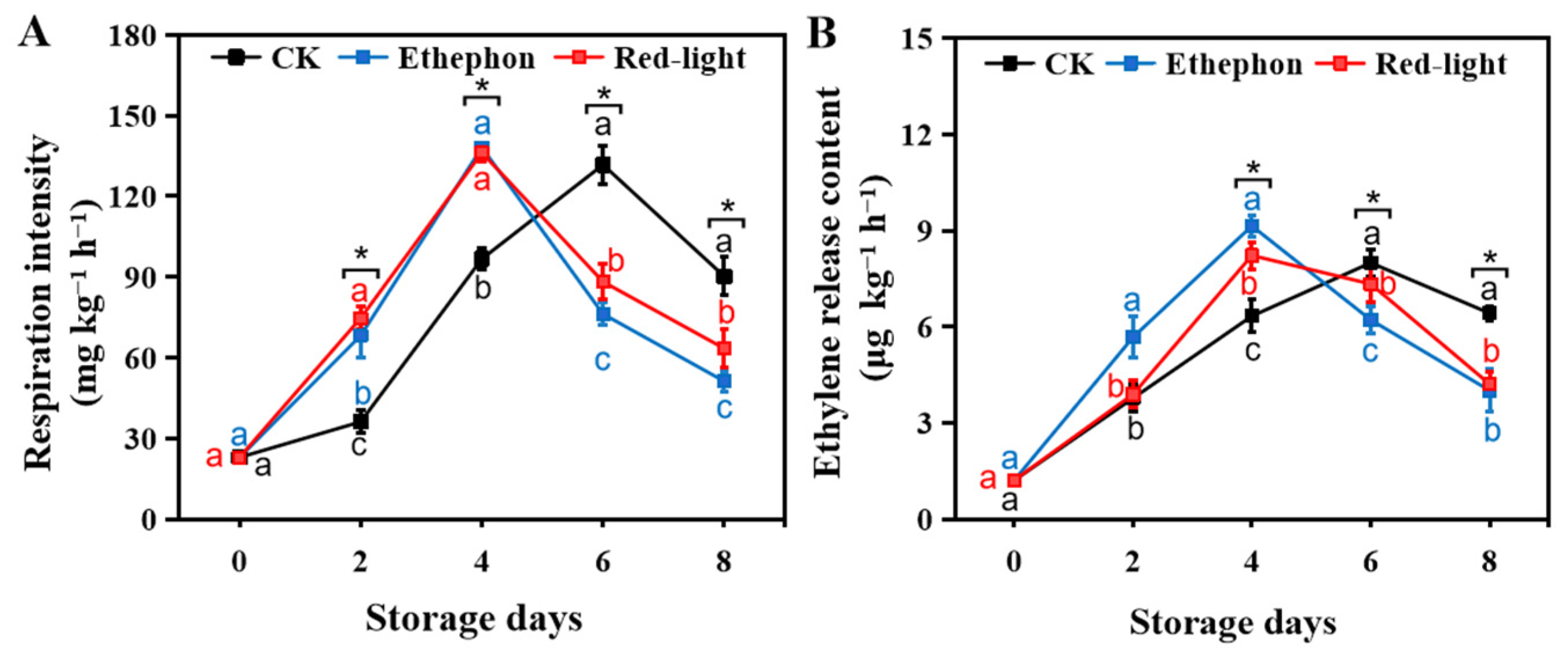
As shown in Figure 3, the peak respiration intensity and peak ethylene release of CK bananas in the case of natural ripening occurred on day 6. Similar to the results for SSC, both RL and ET resulted in advances in the respiration and ethylene peaks to day 4. However, there was no significant difference (p > 0.05) in the peak respiratory intensity of bananas among the CK, ET, and RL groups. There was no significant difference in peak ethylene release between the CK and RL groups (p > 0.05); however, the ethylene peak in the ET group at day 4 was significantly higher than those of the CK and RL groups (p < 0.05). It was evident from the banana physiological indexes that RL could exert a similar ripening effect to that of ET on bananas, accelerating softening, yellowing, and SSC accumulation. Although the effect of RL on ethylene, a key hormone for fruit ripening, was weaker than that of ET, RL could significantly increase the respiratory intensity of bananas (p < 0.05), similar to the effect of ethephon, and this might be related to the metabolic activity level of bananas.

3.2. Effect of LED Red Light Irradiation on the Energy Metabolism of Banana Fruits

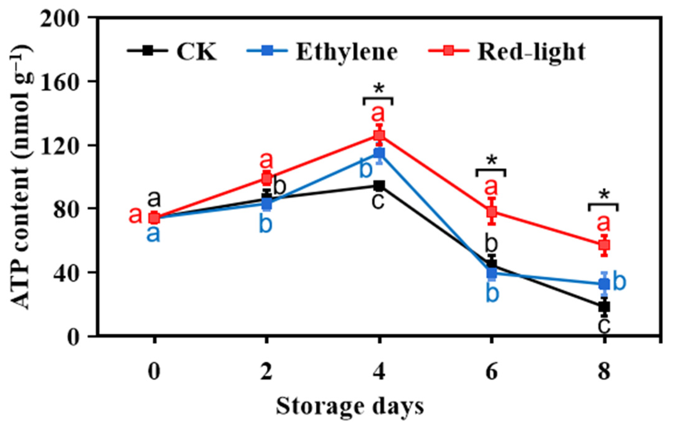
As shown in Figure 4, all treatment groups showed ATP peaks on day 4; however, RL had a significantly higher peak than those of ET and CK. Although the ATP content of all treatment groups decreased after day 4, the rate of decrease was lower in the RL group than in the other two groups, and the ATP content in the RL group was still significantly higher than those of the other two groups on day 6 (p < 0.05), while the difference between ET and CK was not significant. Combined with the effect of LED red light on the intensity of fruit respiration, the changes in ATP content corresponded to the peak respiration intensity on day 4, further suggesting that red light regulates the ATP content by regulating the intensity of fruit respiration, which in turn regulates fruit postharvest ripening. However, this may also be related to the activity of enzymes involved in ATP synthesis.
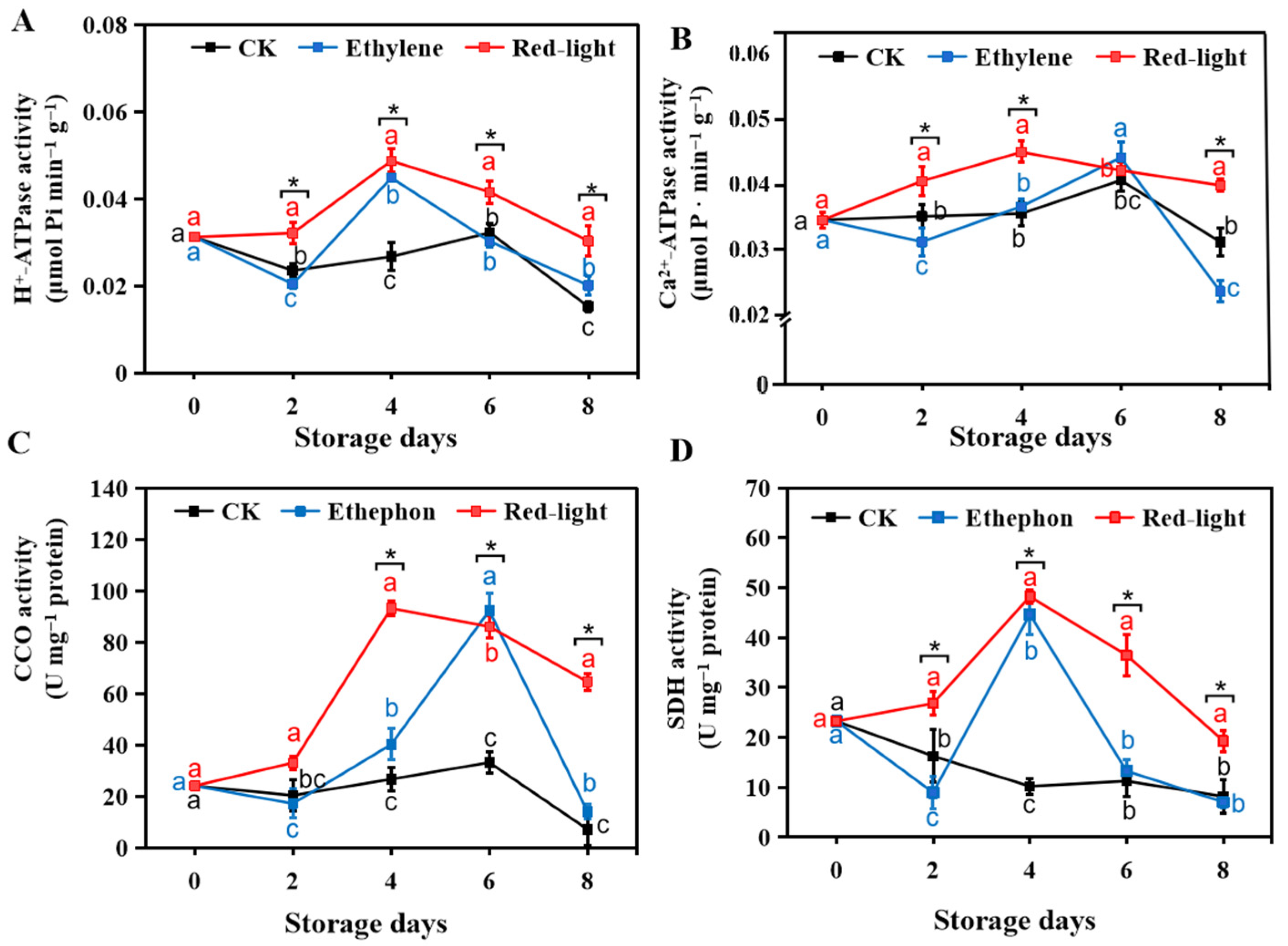
Figure 5 shows the effects of RL, ET, and CK on banana H+-ATPase, Ca2+-ATPase, CCO, and SDH during storage. H+-ATPase and Ca2+-ATPase are classified as P-type ATP synthases; they produce ATP by cyclical ion-transporting steps. The activity of CCO, a terminal enzyme in the respiratory chain, reflects the level of metabolic activity in fruit respiration; SDH (Figure 5D) catalyzes the final product, ATP, from substrates in the tricarboxylic acid cycle. Both RL and ET resulted in greater activity levels of these four enzymes than those in untreated bananas during the 8-day storage period. RL effectively induced and increased the activity of H+-ATPase, Ca2+-ATPase, CCO, and SDH, from day 2 and keeps it higher overall. Both RL and ET advanced H+-ATPase activity in bananas relative to that in the CK group up to day 4. In addition, RL elevated H+-ATPase activity to a higher level than that at day 0 by day 2. As shown in Figure 4C and Figure 5B, RL efficiently induced Ca2+-ATPase and CCO activity, advancing the peak enzyme activity to day 4. Furthermore, SDH activity levels in RL bananas were significantly higher than those in ET on days 2, 4, 6, and 8 (p < 0.05). Based on analyses of the key enzymes in energy metabolism and ATP content, RL increased ATP and maintained high levels mainly by elevating the activity of key enzymes in energy metabolism. In particular, Ca2+-ATPase and CCO may be critical enzymes for banana energy metabolism in response to RL.

3.3. Effect of LED Red Light Irradiation on the Metabolism of Malic Acid in Bananas

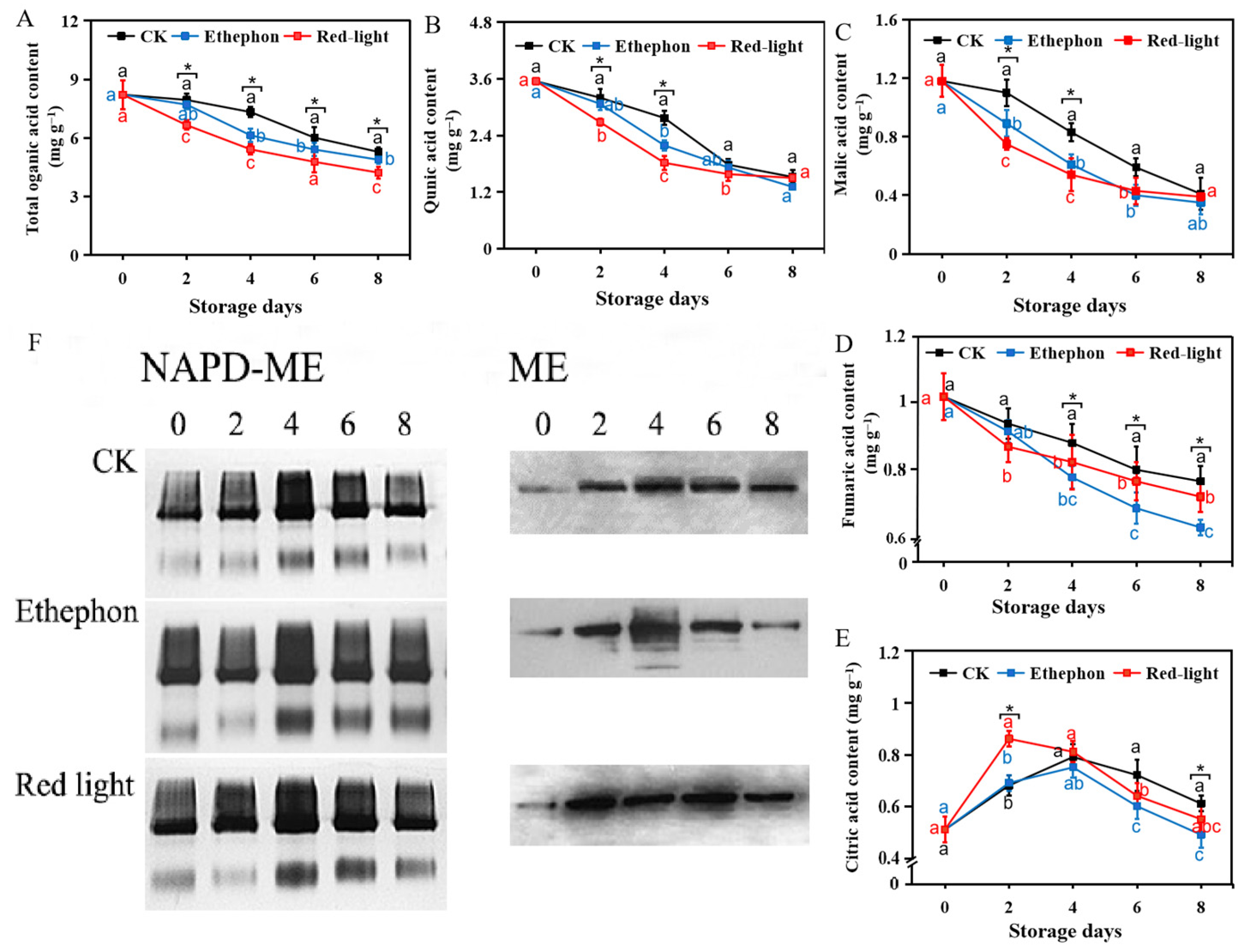
Changes in the energy metabolism of fruit were largely related to changes in their organic acid content. As shown in Figure 6, four major organic acids (i.e., quinic acid, malic acid, fumaric acid, and citric acid) were detected in banana fruits in this study, and quinic acid was the most abundant. Except for citric acid (Figure 6E), the contents of the total organic acids and the remaining three organic acids decreased as storage time increased. The total organic acid content of bananas decreased more substantially after ET and RL than in the CK group (Figure 6A). On day 2 of storage, the total organic acid content of RL bananas was already significantly lower than that of CK (p < 0.05). On day 8 of storage, the total organic acid content of RL bananas was 79.8% and 86.5% of those of the CK group and ET, respectively. The trends in quinic acid (Figure 6B) and malic acid (Figure 6C), the two most abundant organic acids in banana fruits, were similar. On day 2 of storage, the contents of quinic acid and malic acid in RL bananas were significantly lower than those in the CK and ET groups; however, the rate of decrease gradually slowed thereafter. There was no significant difference in the quinic and malic acid contents of RL bananas and ET bananas on day 6 (p > 0.05). The trend in fumaric acid differed from that of quinic acid (Figure 6D). The fumaric acid content of CK bananas was significantly lower than those of the other two treatment groups after 8 days of storage. As shown in Figure 6E, citric acid tended to accumulate and then decline during storage for 8 days. RL could advance the peak of citric acid synthesis to day 2; however, ET did not advance the peak of citric acid synthesis in bananas.
Figure 6F shows the electropherograms of ME activity and ME protein activity in bananas. As shown in Figure 6F, bananas in the RL group showed higher enzyme activity on day 2 and maintained higher band intensity levels on days 4, 6, and 8. In contrast, ET bananas showed higher activity on day 4 and showed a decline in ME activity on day 6. The results for NADP-ME were similar to those for ME, and the NADP-ME bands for RL bananas maintained a darker color on days 2, 4, and 6, indicating that RL bananas had higher NADP-ME protein activity. In contrast, the peak of the NADP-ME protein content in the ET and CK groups appeared only on day 4. These results indicate that RL can effectively stimulate malic enzyme activity, thus accelerating the degradation of organic acids, such as quinic acid, malic acid, and citric acid, and providing additional substrate for energy metabolism in bananas.

4. Discussion

Changes in fruit hardness, total color difference, and soluble solids are highly intuitive physiological indexes of fruit ripening after harvest. Red light irradiation treatment significantly promoted a change in the color of the fruit skin from green to red and increased the total color difference (p < 0.05), similar to the effects of ethephon (Figure 2A,B). Red light treatment shifted the peaks in fruit respiration and ethylene production to earlier time points and caused a rapid decrease in the hardness of bananas (Figure 2C), demonstrating that red light treatment could promote banana fruit ripening. Although there are few studies on the effect of LED light on fruit postharvest ripening and senescence, some studies have pointed out that suitable light conditions can promote or inhibit these processes [13]. For example, Xie et al. [20] clearly indicated that purple LED light significantly inhibits broccoli yellowing and senescence by down-regulating the expression of the chlorophyll degradation-related genes BoSGR, BoPAO, BoNYC1, and BoRCCR. Zhang et al. [15] showed that red light irradiation promotes the expression of phytochrome genes in tomato fruits and accelerates the expression of the ripening gene SlRIN, which in turn induced the expression of other ripening-related genes and promoted tomato fruit ripening. In this study, a comparison of various physiological indexes related to banana postharvest ripening revealed that red light treatment did not differentially affect the production of ethylene (p > 0.05), a key hormone for postharvest ripening, compared with ethephon treatment but effectively enhanced the respiration intensity in bananas (Figure 3). Moreover, the ATP content in the light irradiation group was higher than the non-irradiation group, which also promoted fruit ripening. In addition, the higher energy status in the light irradiation group was associated with the activities of enzymes related to the enhanced energy metabolism, and light-irradiation treatment maintained the higher levels of respiratory intensity. We propose that red light regulates respiration levels by increasing energy supply and may be a mechanism by which red light regulates banana fruit ripening and senescence.
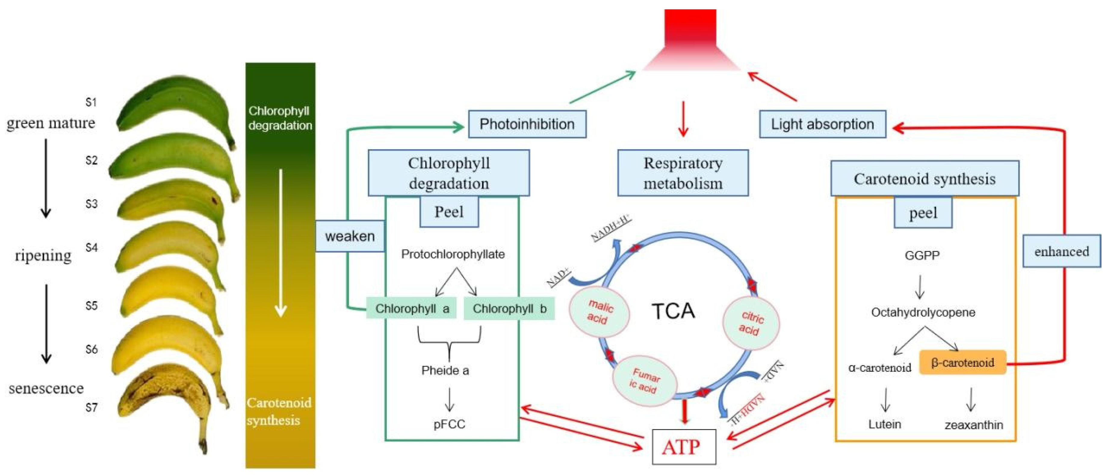
The coloration of banana fruits is mainly determined by a combination of carotenoid and chlorophyll levels. Most light-absorbing pigments (chlorophylls, carotenoids, etc.) in plants bind to proteins in a single peptide chain by non-covalent bonds to form pigment complexes, while a small fraction form reaction centers (RCs). The plant is able to convert the captured and absorbed light energy into chemical energy. RCs use the absorbed light energy to drive electron transfer reactions, producing ATP and reducing NADP to supply energy to the plant [21]. Usually, light absorption and capture by the plant are mediated by intracellular pigments. The ability of light-sensitive pigments, such as chlorophylls, carotenoids, and anthocyanins, to capture and absorb light is influenced by various factors, such as the light intensity, light quality, and light duration [22]. When intense light exposure exceeds the light energy absorbed and utilized by chlorophyll, the plant is in a state of excess light energy, which initiates a non-radiative energy dissipation pathway to consume the excess excitation energy. When the excess energy cannot be completely consumed, the plant produces reactive oxygen species and singlet chlorophyll, which leads to a decrease in light absorption, light-capture ability, and plant resistance, thereby promoting plant senescence. Similarly, carotenoids in plants can also play a role in light capture. In addition, they prevent photodamage that occurs under excessive light [23]. Carotenoids usually act as photoprotective agents in plants to prevent damage from excessive light exposure. In the present study, we used 800.0 ± 10.0 Lx red light to irradiate bananas and found that the outer skin of bananas showed a rapid change from green to yellow, which may also be a self-protection mechanism against strong light exposure. When the RL treatment time increased and the banana skin received a certain dose of light, chlorophyll was highly degraded and carotenoids were highly synthesized, thus avoiding photodamage. In addition, the degradation and anabolism of these two pigments may also accelerate the postharvest ripening of bananas. As shown in Figure 7, we propose a model by which various processes, including energy metabolism, carotenoid synthesis, and chlorophyll degradation, interact in banana fruits in response to RL signaling.
ATP is the most direct source of energy in living organisms and is mainly used for substance synthesis, nutrient transport, and the transmission of genetic information. In this study, we found that red light significantly elevated ATP levels in bananas (p < 0.05), enhanced the activity of ATP production-related enzymes in energy metabolism (Figure 4 and Figure 5), and accelerated fruit ripening. The study of Li et al. [24] showed that light could increase the ATP and ADP content in shiitake edodes and increase the activity of H+ -ATP and Ca2+-ATP enzymes, contrary to this study, which may also be related to species differences. Since the main sites of respiration and ATP production in postharvest fruits and vegetables are mitochondria, tri-H+-ATPase, Ca2+-ATPase, CCO, and SDH are important mitochondrial metabolic enzymes in fruits [25]. A study by Zhang et al. [15] indicated that external physical and chemical stimuli at a certain intensity could enhance energy metabolism in fruits and vegetables in response to stress. Jin et al. [26] performed mechanical shaking and found that moderate oscillatory stimulation increased energy levels and promoted ripening in kiwi fruits. In this experiment, continuous red light irradiation at 800.0 ± 10.0 Lx and 655.0 ± 1.0 nm promoted the increases in H+-ATPase, Ca2+-ATPase, CCO, and SDH activities in bananas and advanced the peak of Ca2+-ATPase and CCO activities by 2 days, accelerating respiration and energy metabolism in bananas and thus promoting postharvest ripening.
As fruits ripen during postharvest processing, respiratory metabolism gradually increases, and the organic acid content of the fruit begins to decrease [27]. Some studies have reported that organic acids are the first substrate for energy provision in postharvest fruits and vegetables and are consumed by respiratory metabolism in glycolysis and the tricarboxylic acid cycle, and these respiration-induced changes in organic acid metabolism usually accelerate fruit ripening [28,29]. The results of this study showed that red light could cause a rapid decrease in the organic acid content in bananas within 4 days, with a greater effect than that of ethephon treatment. In addition, since banana is abundant in malic acid, its malic and quinic acid contents both decreased rapidly during the first 4 days of storage, and the ATP content increased rapidly in the first 4 days. These findings indicate that fruit development and ripening require a large amount of energy supplied by organic acid consumption [30]. Malic enzymes are oxidative decarboxylases widely present in animals, plants, and microorganisms and contribute to the regulation of malic acid metabolism [31]. Malic enzymes are involved in the degradation of organic acids during fruit ripening, which is closely related to fruit senescence [32]. Onik et al. [33] proved that UV-C treatment upregulated the expression of NADP-ME, reduced the content of citric acid and malic acid, promoted the generation of fruit aroma, and then promoted the post-ripening aging of fruit. In this study, we found that red light irradiation not only effectively activated banana malic enzymes on day 2 but also increased the malic enzyme protein content (Figure 6F). This is consistent with the trend in banana organic acid contents. It is hypothesized that red light LED irradiation catalyzed the oxidative decarboxylation of organic acids, such as malic acid and quinic acid, to generate pyruvate, which produced NADPH, provided reducing power for cellular respiration, enhanced banana fruit respiration, and eventually accelerated fruit ripening.

5. Conclusions

In this study, red LED light was used to ripen banana fruits with ethephon treatment as a positive control and dark treatment as a negative control. Comparisons of energy metabolism-related enzyme activity, organic acid contents, and malic enzyme activity between red light-treated and ethephon-treated fruits revealed that continuous red LED light irradiation could induce more rapid carotenoid synthesis and chlorophyll degradation and regulate energy metabolism by capturing light energy through carotenoid absorption to maintain a higher ATP content, thus promoting ripening and senescence in banana fruits.

Author Contributions

B.L., X.Z (Xinqun Zhou) and J.S. designed the experimental trials. J.C., X.W. and B.L. performed the experiments and collected the samples. B.L., X.Z. (Xinqun Zhou), J.S. and J.C. wrote the article. B.L., X.Z. (Xuan Zhu), S.G. and X.G. and Q.C., revised and edited the article. X.Z. (Xinqun Zhou) and J.S. gave founding support to this paper. All authors have read and agreed to the published version of the manuscript.

Funding

This work was supported by the independent research and development program of the Academy of Agricultural Planning and Engineering, Ministry of Agriculture and Rural Affairs (QD202115) and scientific and technological innovation team project of primary processing of agricultural products (CHXTY-2021-08). All experiments in this work were supported by the Key Laboratory of Agro-Products Primary Processing.

Data Availability Statement

The data used to support the findings of this study can be made available by the corresponding author upon request.

Acknowledgments

We declare that we do not have any commercial or associative interest that represents a conflict of interest in connection with the work submitted.

Conflicts of Interest

The authors declare no conflict of interest.

References

  1. Mohandas, S.; Ravishankar, K.V. Banana: Genomics and Transgenic Approaches for Genetic Improvement; Springer: Singapore, 2016; pp. 112–113. [Google Scholar] [CrossRef]
  2. Bantayehu, M.; Alemayehu, M. Efficacy of post-harvest technologies on ripening behavior and quality of banana varieties grown in Ethiopia. Int. J. Fruit Sci. 2020, 20, 59–75. [Google Scholar] [CrossRef]
  3. Gamrasni, D.; Feldmesser, E.; Ben-Arie, R.; Raz, A.; Tabatznik, A.A.; Glikman, M.; Goldway, M. Gene expression in 1-Methylcyclopropene (1-MCP) treated tomatoes during pre-climacteric ripening suggests shared regulation of methionine biosynthesis, ethylene production and respiration. Agronomy 2020, 10, 1669. [Google Scholar] [CrossRef]
  4. Parijadi, A.; Yamamoto, K.; Ikram, M.; Dwivany, F.; Wikantika, K.; Putri, S.; Fukusaki, E. Metabolome analysis of banana (Musa acuminata) treated with chitosan coating and low temperature reveals different mechanisms modulating delayed ripening. Front. Sustain. Food Syst. 2022, 6, 835978. [Google Scholar] [CrossRef]
  5. Lu, W.; Mao, L.; Chen, J.; Han, X.; Ren, X.; Ying, T.; Luo, Z. Interaction of abscisic acid and auxin on gene expression involved in banana ripening. Acta Physiol. Plant. 2018, 40, 46. [Google Scholar] [CrossRef]
  6. Ishra, R.; Khananm, R.; Soar, J. Influence of food safety concerns on safe food purchasing at rural and urban consumers in Bangladesh. Appetite 2022, 179, 106306. [Google Scholar] [CrossRef]
  7. Hossain, R. Safe Food in Bangladesh: Perception and Influences on Safe Food Purchasing; Griffith University: Brisbane, Australia, 2019. [Google Scholar] [CrossRef]
  8. Wang, S.Y.; Chen, C.T.; Wang, C.Y. The influence of light and maturity on fruit quality and flavonoid content of red raspberries. Food Chem. 2009, 112, 676–684. [Google Scholar] [CrossRef]
  9. Loi, M.; Villani, A.; Paciolla, F.; Mulè, G.; Paciolla, C. Challenges and opportunities of light-emitting diode (Led) as key to modulate antioxidant compounds in plants. a review. Antioxidants 2020, 10, 42. [Google Scholar] [CrossRef]
  10. Deng, L.; Yuan, Z.; Xie, J.; Yao, S.; Zeng, K. Sensitivity to ethephon degreening treatment is altered by blue LED light irradiation in mandarin fruit. J. Agric. Food Chem. 2017, 65, 6158–6168. [Google Scholar] [CrossRef]
  11. Shu, C.; Cao, J.; Jiang, W. Post-harvest vibration-induced apple quality deterioration is associated with the energy dissipation system. Food Chem. 2022, 386, 132767. [Google Scholar] [CrossRef]
  12. Huang, Z.; Guo, L.; Wang, H.; Qu, H.; Ma, S.; Liu, Y.; Jiang, Y. Energy status of kiwifruit stored under different temperatures or exposed to long-term anaerobic conditions or pure oxygen. Post-Harvest Biol. Technol. 2014, 98, 56–64. [Google Scholar] [CrossRef]
  13. Liu, B.; Zhang, Y.; Ke, Z.; Sun, J.; Zhou, X.; Sun, J. Effects of LED light on the ripening regulation of green mature banana during storage and transportation. Trans. Chin. Soc. Agric. Eng. 2021, 37, 295–302. (in Chinese). [Google Scholar] [CrossRef]
  14. Liu, B.; Jiao, W.; Wang, B.; Shen, J.; Zhao, H.; Jiang, W. Near freezing point storage compared with conventional low temperature storage on apricot fruit flavor quality (volatile, sugar, organic acid) promotion during storage and related shelf life. Sci. Hortic. 2019, 249, 100–109. [Google Scholar] [CrossRef]
  15. Zhang, P.; Wei, Y.; Xu, F.; Wang, H.; Chen, M.; Shao, X. Changes in the chlorophyll absorbance index (I AD) are related to peach fruit maturity. N. Z. J. Crop Hortic. Sci. 2020, 48, 34–46. [Google Scholar] [CrossRef]
  16. Laemmli, U.K. Cleavage of structural proteins during the assembly of the head of bacteriophage T4. Nature 1970, 227, 680–685. [Google Scholar] [CrossRef]
  17. Wang, L.; Wang, M.; Li, Q.; Cai, T.; Jiang, W. Partial properties of an aspartic protease in bitter gourd (Momordica charantia L.) fruit and its activation by heating. Food Chem. 2008, 108, 496–502. [Google Scholar] [CrossRef]
  18. Mehrabani, L.V.; Hassanpouraghdam, M.B. Developmental variation of phenolic compounds in fruit tissue of two apple cultivars. Acta scientiarum polonorum. Technol. Aliment. 2012, 11, 259–264. Available online: http://www.food.actapol.net/volume11/issue3/5_3_2012 (accessed on 5 March 2023).
  19. Mishra, M.; Tiwari, S.; Gomes, A.V. Protein purification and analysis: Next generation Western blotting techniques. Expert Rev. Proteom. 2017, 14, 1037–1053. [Google Scholar] [CrossRef]
  20. Xie, C.; Tang, J.; Xiao, J.; Geng, X.; Guo, L. Purple light-emitting diode (LED) lights controls chlorophyll degradation and enhances nutraceutical quality of post-harvest broccoli florets. Sci. Hortic. 2022, 294, 110768. [Google Scholar] [CrossRef]
  21. Chen, L.S.; Li, P.; Cheng, L. Comparison of thermotolerance of sun-exposed peel and shaded peel of ‘Fuji’apple. Environ. Exp. Bot. 2009, 66, 110–116. [Google Scholar] [CrossRef]
  22. Moosavi-Nezhad, M.; Boshra, A.; Ahmad, E.; Nazim, S.; Sasan, A. Growth, Biomass Partitioning, and Photosynthetic Performance of Chrysanthemum Cuttings in Response to Different Light Spectra. Plants 2022, 11, 3337. [Google Scholar] [CrossRef]
  23. Muzzopappa, F.; Kirilovsky, D. Changing color for photoprotection: The orange carotenoid protein. Trends Plant Sci. 2020, 25, 92–104. [Google Scholar] [CrossRef] [PubMed]
  24. Li, Y.; Ding, S.; Xiang, T.; Kitazawa, H.; Sun, H.; Guo, Y. Effects of light irradiation on the textural properties and energy metabolism of postharvest shiitake mushrooms (Lentinula edodes). J. Food Process. Preserv. 2021, 45, e16066. [Google Scholar] [CrossRef]
  25. Aghdam, M.; Jannatizadeh, A.; Luo, Z.; Paliyath, G. Ensuring sufficient intracellular ATP supplying and friendly extracellular ATP signaling attenuates stresses, delays senescence and maintains quality in horticultural crops during postharvest life. Trends Food Sci. Technol. 2018, 76, 67–81. [Google Scholar] [CrossRef]
  26. Jin, P.; Zhu, H.; Wang, J.; Chen, J.; Wang, X.; Zheng, Y. Effect of methyl jasmonate on energy metabolism in peach fruit during chilling stress. J. Sci. Food Agric. 2013, 93, 1827–1832. [Google Scholar] [CrossRef]
  27. Osorio, S.; Scossa, F.; Fernie, A. Molecular regulation of fruit ripening. Front. Plant Sci. 2013, 4, 198. [Google Scholar] [CrossRef] [PubMed] [Green Version]
  28. González-Agüero, M.; Tejerina Pardo, L.; Zamudio, M.S.; Contreras, C.; Undurraga, P.; Defilippi, B.G. The unusual acid-accumulating behavior during ripening of cherimoya (Annona cherimola Mill.) is linked to changes in transcription and enzyme activity related to citric and malic acid metabolism. Molecules 2016, 21, 398. [Google Scholar] [CrossRef] [Green Version]
  29. Silva, G.; Silva, W.; Medeiros, D.; Salvador, A.; Cordeiro, M.; Silva, N.; Mizobutsi, G. The chitosan affects severely the carbon metabolism in mango (Mangifera indica L. cv. Palmer) fruit during storage. Food Chem. 2017, 237, 372–378. [Google Scholar] [CrossRef]
  30. Batista-Silva, W.; Nascimento, V.; Medeiros, D.; Nunes-Nesi, A.; Ribeiro, D.M.; Zsögön, A.; Araújo, W.L. Modifications in organic acid profiles during fruit development and ripening: Correlation or causation? Front. Plant Sci. 2018, 9, 1689. [Google Scholar] [CrossRef]
  31. Chen, Q.; Wang, B.; Ding, H.; Zhang, J.; Li, S. The role of NADP-malic enzyme in plants under stress. Plant Sci. 2019, 281, 206–212. [Google Scholar] [CrossRef]
  32. Jiang, C.; Fang, Z.; Zhou, D.; Pan, S.; Ye, X. Changes in secondary metabolites, organic acids and soluble sugars during the development of plum fruit cv. ‘Furongli’ (Prunus salicina Lindl). J. Sci. Food Agric. 2019, 99, 1010–1019. [Google Scholar] [CrossRef]
  33. Onik, J.C.; Xie, Y.; Duan, Y.; Hu, X.; Wang, Z.; Lin, Q. UV-C treatment promotes quality of early ripening apple fruit by regulating malate metabolizing genes during postharvest storage. PLoS ONE 2019, 14, e0215472. [Google Scholar] [CrossRef] [Green Version]
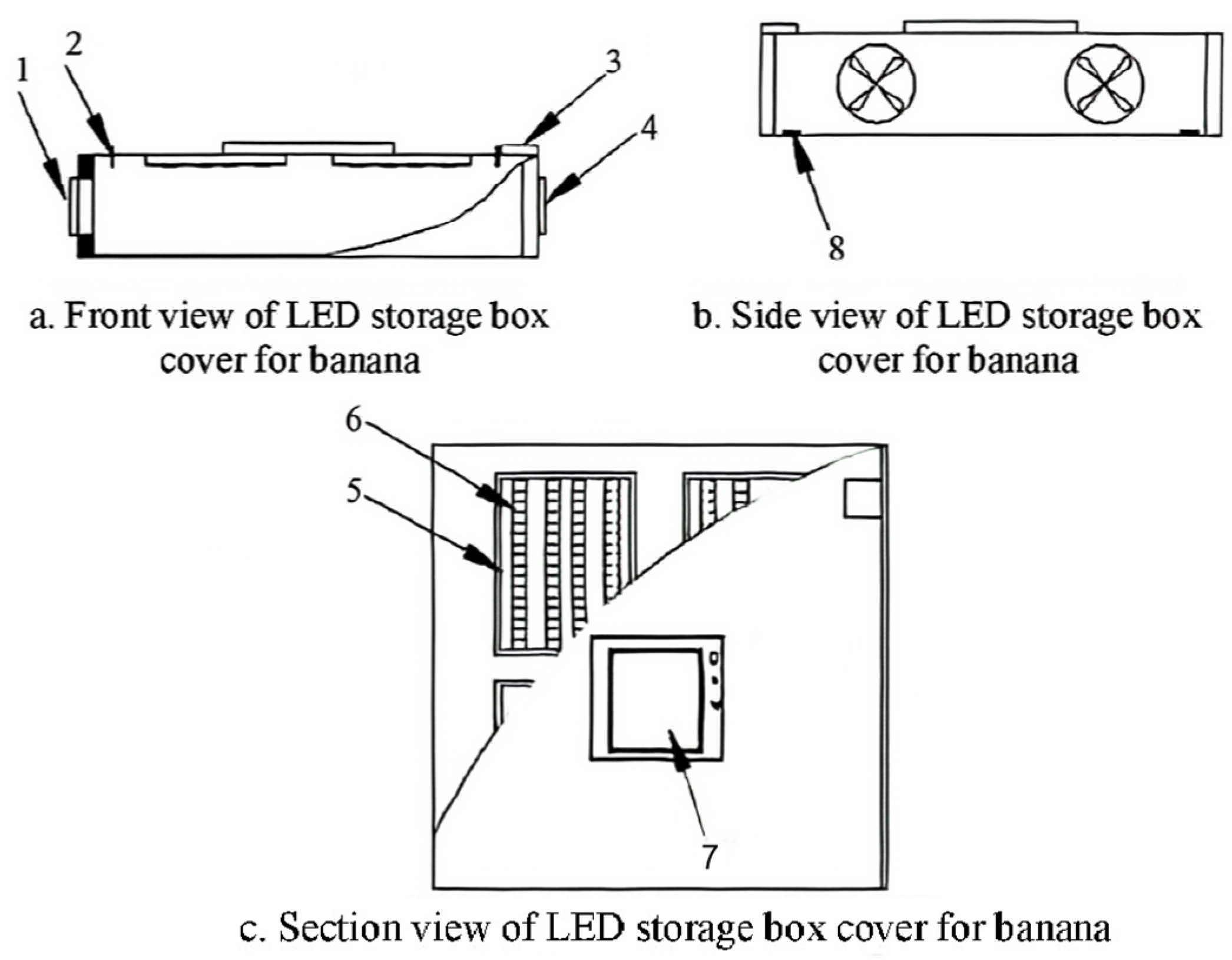
Figure 1. Banana preservation box from three perspectives. (Note: 1. Axial suction fan; 2. Temperature humidity sensor; 3. Powerswitch; 4. Axial flow blower; 5. LED light board; 6. LED lamp band; 7. Touch panel; 8. Buckle).
Horticulturae 09 00840 g001
Figure 2. Effect of LED red light treatment, ethephon treatment and control group on banana fruits (A), total color difference (B), firmness (C), soluble solid content (D), chlorophyll content (E), total carotenoid content (F) during storage. Vertical bars represent the standard deviation of the mean (n = 3). Different letters indicate statistically significant differences (p < 0.05).
Horticulturae 09 00840 g002
Figure 3. Effect of LED red light treatment, ethephon treatment and control group on bananas respiration intensity (A) ethylene release (B) content during storage. Vertical bars represent the standard deviation of the mean (n = 3). Different letters indicate statistically significant differences (p < 0.05).
Horticulturae 09 00840 g003
Figure 4. Effect of LED red light treatment, ethephon treatment and control group on bananas ATP content during storage. Vertical bars represent the standard deviation of the mean (n = 3). Different letters indicate statistically significant differences (p < 0.05).
Horticulturae 09 00840 g004
Figure 5. Effect of LED red light treatment, ethephon treatment and control group on bananas H+-ATPase (A), Ca2+-ATPase (B), CCO (C), and SDH (D) during storage. Vertical bars represent the standard deviation of the mean (n = 3). Different letters indicate statistically significant differences (p < 0.05).
Horticulturae 09 00840 g005
Figure 6. Effect of LED red light treatment, ethephon treatment and control group on bananas’ total organic acid (A), qunic acid content (B), malic acid content (C), fumaric acid content(D), citric acid content (E), malic enzyme activity and protein content (F) during storage. Vertical bars represent the standard deviation of the mean (n = 3). Different letters indicate statistically significant differences (p < 0.05).
Horticulturae 09 00840 g006
Figure 7. A model of LED red light signal-induced energy metabolism, carotenoid and chlorophyll metabolism in banana fruits.
Horticulturae 09 00840 g007
Disclaimer/Publisher’s Note: The statements, opinions and data contained in all publications are solely those of the individual author(s) and contributor(s) and not of MDPI and/or the editor(s). MDPI and/or the editor(s) disclaim responsibility for any injury to people or property resulting from any ideas, methods, instructions or products referred to in the content.