Article

Two Bacterial Bioagents Boost Onion Response to Stromatinia cepivora and Promote Growth and Yield via Enhancing the Antioxidant Defense System and Auxin Production

by
Hanan E. M. Osman
1,†,
Yasser Nehela
2,*,†,
Abdelnaser A. Elzaawely
2,
Mohamed H. El-Morsy
3,4 and
Asmaa El-Nagar
2
1
Biology Department, Faculty of Applied Science, Umm-Al-Qura University, Makkah 21955, Saudi Arabia
2
Department of Agricultural Botany, Faculty of Agriculture, Tanta University, Tanta 31527, Egypt
3
Deanship of Scientific Research, Umm Al-Qura University, Makkah 24243, Saudi Arabia
4
Plant Ecology and Range Management Department, Desert Research Center, Cairo 11753, Egypt
*
Author to whom correspondence should be addressed.
These authors contributed equally to this work.
Horticulturae 2023, 9(7), 780; https://doi.org/10.3390/horticulturae9070780
Submission received: 6 June 2023 / Revised: 29 June 2023 / Accepted: 6 July 2023 / Published: 8 July 2023

Abstract

:
White rot, caused by Stromatinia cepivora (Anamorph: Sclerotium cepivorum Berk), is a serious soil-borne disease of the onion that restricts its cultivation and production worldwide. Herein, we isolated and characterized a plant growth-promoting rhizobacterium Stenotrophomonas maltophilia from healthy onion roots and an endophytic bacterium Serratia liquefaciens from healthy bean leaves. Both isolates showed strong fungistatic activity against S. cepivora using the dual culture and culture filtrate methods. This effect might be due to the presence of several volatile compounds, especially menthol in both culture filtrates as shown with a GC-MS analysis. Additionally, the root drench application of cell-free culture filtrates of S. maltophilia and S. liquefaciens significantly reduced the incidence and severity of white rot disease on treated onion plants, which was associated with the activation of both enzymatic (POX and PPO) and non-enzymatic (phenolics and flavonoids) antioxidant defense machineries of S. cepivora-infected onion plants. Moreover, the culture filtrates of both bacterial bioagents remarkably enhanced the growth (as expressed by root length, plant height, and number of leaves) and yield parameters (as indicated by bulb circumference, fresh weight of the bulb, and bulb yield per plot) of treated onion plants under field conditions during two successive seasons (2020/2021 and 2021/2022). This might be because of a reduced disease severity and/or the accumulation of the main auxin, indole-3-acetic acid (IAA), and its precursor, the amino acid tryptophan. Our findings suggest that both bioagents might be utilized as eco-friendly alternative control measures to reduce the utilization of chemical fungicides entirely or partially for the safer production of onion in S. cepivora-infested soils.

1. Introduction

An onion (Allium cepa L.) is one of the oldest vegetable crops known to humans, as its cultivation dates back more than 5000 years [1], and, nowadays, it is one of the most cultivated vegetables all over the world. It is an important crop grown in Egypt and worldwide, whether for local consumption or exportation. Onions are rich in chemical compounds that have medical importance, such as flavonoids [2]. The global production of onions reached 106.59 million tons and the harvested area was 5.78 million hectares in 2021 [3]. Additionally, Egypt cultivated 94,457 hectares of onions, yielding 35.0684 tons per hectare, with a total production of 3.31 million tons [3]. An onion is attacked by several phytopathogens such as viruses, bacteria, fungi, and nematodes, which cause serious diseases and economic losses [4]. However, white rot disease caused by Sclerotium cepivorum Berk (teleomorph: Stromatinia cepivora) is the most serious disease and a limiting factor for onion production worldwide [5]. S. cepivora is a soil-borne fungus that can attack onion roots from seedlings to the harvest stage, resulting in plant death during a season [6].
The ascomycetous S. cepivora is a necrotrophic fungus that can infect numerous susceptible Allium species, particularly onions, leeks, and garlic, causing white rot disease. Although S. cepivora does not produce any known asexual spores, it survives and overwinters as sclerotia (the survival stage) [7]. Sclerotia are small, black, globular, solid structures that are produced by the pathogen at the end of its life cycle and can resist unfavorable environmental conditions and remain dormant in the soil for years even without a host [7]. It was reported previously that an unpredictable quantity of sclerotia produced naturally on infected hosts might decay shortly after the formation for unknown reasons; however, sclerotia that survive beyond the decay period are probable to persist viably for 20 years in the soil under field conditions in the absence of host plants [8]. Although sclerotia can spread from field to field through unsuccessful sanitation practices such as soil movement and/or contaminated water, only Allium root exudates stimulate sclerotia germination, which remains as the host range limited to Allium species [9].
Management of onion white rot is incredibly challenging and requires multi-pronged strategies due to the above-listed characteristics of sclerotia. These strategies include but are not limited to cultural controls such as crop rotation with nonhost plants, sanitation such as soil solarization [10], biological control using sclerotia germination stimulants, composted onion waste [11], or fungal and bacterial bioagents [12,13]. Although an individual control method does not give the desired level of disease management, chemical fungicides are considered the most effective means used to control this disease [14]. Despite the effectiveness of synthetic fungicides, the extensive and repeated use of these fungicides results in several environmental problems and has undesirable effects on humans, animals, and non-targeted microorganisms. Moreover, fungicides might disrupt natural biological systems via the development of fungicide-resistant fungal strains [15,16]. Consequently, it is necessary to prompt an intensive search for cheap eco-friendly alternatives that are safer for humans, animals, and the environment.
Biological control of soil-borne phytopathogens is a potential alternative to reduce the use of hazardous chemical fungicides entirely or partially. Biological control is one of the most sustainable alternative methods in controlling plant diseases due to its low cost and environmental friendliness [17,18]. Direct mechanisms/mode-of-actions of biological control against soil-borne phytopathogenic fungi involve, but are not limited to, antibiosis, cross-protection, hyperparasitism, predation, soil amendments, induced systemic resistance (ISR), as well as competition for a site and nutrient [17,18]. Moreover, plant growth-promoting rhizobacteria (PGPR) are considered one of the most important biological control agents for plant diseases that colonize the roots of many plants [19]. Stenotrophomonas maltophilia is a widespread PGPR that is isolated from the rhizosphere of many plants [20] such as cruciferous, corn, and beets [21]. Additionally, Garbeva et al. found that S. maltophilia colonizes and persists inside the tissues of potato plants [22]. S. maltophilia was effective in inhibiting the mycelial growth of Rhizoctonia solani, Verticillium dahliae [23], Sclerotium rolfsii [24], and Pyricularia oryzae (sexual morph Magnaporthe oryzae) [25]. Moreover, it had an antagonistic effect against Ralstonia solanacearum in vitro and on potato plants under greenhouse conditions [20].
On the other hand, endophytic bacteria could be isolated from different parts of a plant including roots, stems, leaves, flowers, fruits, and seeds, and be used in biological control for plant diseases [26]. Endophytic bacteria colonize the internal tissues of plants without causing any negative effects or disease symptoms [27]. In addition, they were identified as effective biological control agents for a variety of plant diseases [26]. Serratia liquefaciens is an endophytic bacterium isolated from Pinellia ternata for the first time by Liu et al. [28]. Additionally, S. liquefaciens and S. proteamaculans were investigated for their antifungal activity against several phytopathogenic fungi including Sclerotinia sclerotiorum Fusarium oxysporum, Rhizoctonia solani, Botrytis cinerea, and Alternaria alternata by Michail et al. [29].
In the current study, we isolated and characterized S. maltophilia and S. liquefaciens, and investigated their potential antifungal activity against S. cepivora, the causal agent of onion white rot disease, in vitro and in vivo under field conditions. We hypothesized that both bioagents may affect the growth and yield components of S. cepivora-infected onion plants. Moreover, we suggest that the plant growth-promoting properties of both bioagents are correlated with the activation of enzymatic and non-enzymatic antioxidant defense machinery and the accumulation of phytohormones such as auxins. Gas chromatography–mass spectrometry (GC-MS) was used to identify the chemical composition of culture filtrates of S. maltophilia and S. liquefaciens to better understand the function of these bacterial secretions, as well as their involvement in suppressing the growth of S. cepivora and reducing the disease severity of onion white rot.

2. Materials and Methods

2.1. Isolation of the Causal Agent of Onion White Rot Disease

The causal agent S. cepivora was isolated from onion bulbs and roots that exhibited typical symptoms of white rot disease. Samples were collected from different sites at EL-Gharbia governorate Egypt. Isolates of S. cepivora were obtained by scraping the mycelium or sclerotia from onion bulbs and roots then placed on a Potato Dextrose Agar (PDA) medium and incubated at 20 ± 2 °C for 7 days. Isolates were initially identified based on their cultural, morphological, and macroscopic characteristics and then tested for their pathogenicity.

2.1.1. Pathogenicity Test

The pathogenicity of seven isolates of S. cepivora was tested on 50-day-old healthy onion seedlings of the susceptible cultivar Giza 20. To prepare the fungal inoculum, 500 mL glass bottles containing 100 g of barley grains and 50 mL of water were autoclaved; five discs of each isolate were inoculated into the barley medium and incubated at 20 ± 2 °C for 30 days. Plastic pots (30 cm) were filled with sterilized sand–clay soil 1:1 (v/v) and infected with fungal inoculum 14 days before transplanting at a rate of 15 g·kg−1. Plants were watered as needed and other agricultural practices were conducted as recommended. Onions were uprooted at 150 days post transplanting (dpt) to evaluate the disease incidence and severity. The disease incidence was assessed by counting onion plants that had a visible white rot mycelium and/or sclerotia on the roots and scored as white rot-infected. The disease incidence (%) was calculated using Equation (1):
Disease   incidence   ( % ) = N u m b e r   o f   i n f e c t e d   p l a n t s N u m b e r   o f   t o t a l   p l a n t s × 100
Whereas the severity of onion white rot disease was evaluated according to a 5-degree symptom scale according to Tian and Bertolini [30] with slight modifications as follows: 0: Healthy; 1: Bulb covered with mycelium but not rotted; 2: Bulb covered with mycelium, and 1–25% of the bulb rotted; 3: Bulb covered with mycelium, 25–50% of the bulb rotted; 4: Bulb covered with mycelium, 50–75% of the bulb rotted; and 5: Bulb covered with mycelium, 75–100% of the bulb rotted.
Disease severity scores were converted into percentages according to Equation (2) as described by Zewide et al. [31] as follows:
Disease   severity   ( % ) = T o t a l   o f   a l l   r a t i n g s   T o t a l   n u m b e r   o f   p l a n t s × m a x i m u m   s c o r e × 100
Based on the pathogenicity test, isolate #4 was the most aggressive isolate and showed the highest pathogenicity among the tested isolates, so it was selected for molecular identification based on the sequence of its internal transcribed spacer (ITS) region. Moreover, it was selected for all subsequent experiments.

2.1.2. Molecular Identification of S. cepivora

The most aggressive isolate of S. cepivora (isolate #4) was subjected to molecular identification [32,33]. Briefly, this isolate was grown on a sterilized potato dextrose broth (PDB) and incubated at 20 ± 2 °C for 10 days. Subsequently, the mycelium and sclerotia were collected and filtered using cheesecloth, washed twice with sterilized deionized water, and dried using filter paper. Approximately 0.1 g of the mycelium and sclerotia were ground to a fine powder using liquid nitrogen. The total DNA of the pathogenic fungus was extracted using a Quick-DNA™ Fungal/Bacterial Miniprep Kit according to the manufacturer’s instructions and then purified, and the targeted sequences of the ITS region (ITS-5.8S rDNA) were amplified using PCR. The purified PCR products were sent for sequencing (Aoke Dingsheng Biotechnology Co., Beijing, China). Sanger sequencing was used to perform the two-directional sequencing of the ITS-5.8S rDNA sequences. DNABASER software (Heracle BioSoft S.R.L., Arges, Romania) was used to process and assemble consensus sequences. Subsequently, a Nucleotide-Nucleotide Basic Local Alignment Search Tool (BLASTn) was used to compare the assembled sequence with the most recent available data in GenBank and the national center for biotechnology information website (NCBI, http://www.ncbi.nlm.nih.gov/gene/; accessed on 7 February 2023).

2.1.3. Phylogenetic Analysis

An evolutionary analysis and phylogenetic trees were constructed using the assembled sequence of the ITS-5.8S rDNA based on the Maximum Likelihood method and Tamura-Nei model [34] using Molecular Evolutionary Genetics Analysis–Version 11 (MEGA 11) software using 500 bootstrap replications [35]. In addition to the query sequence, about 20 reference strains/isolates (Tables S1–S3 in Supplementary Material) were selected and used for multiple sequence alignment using ClustalW multiple sequence alignment algorithms.

2.2. Isolation of Plant Growth-Promoting Rhizobacterium and Endophytic Bacterium

A plant growth-promoting rhizobacterium (PGPR) was isolated from the rhizosphere soil of healthy onion plants growing next to diseased plants using the plate of soil dilution method as described by [36]. The soil attached to healthy plants’ roots was removed, collected, air dried, and mixed well. In total, 10 g of homogenized soil was placed in conical flasks, 100 mL of sterilized distilled water (SDW) was added—well shaken for 10 min (150 rpm at 30 °C)—and serial dilutions were prepared. Subsequently, 100 μL of the last dilution was spread on surfaces of Petri dishes containing a nutrient agar (NA) medium with a sterilized Drigalski glass triangle; the dishes were incubated at 28 ± 2 °C for 48 h. Growth in incubated dishes was examined and purified using a single colony.
Furthermore, an endophytic bacterium was isolated from fresh healthy leaves of common bean plants. Firstly, leaves were washed with tap water, air dried, and surface sterilized in 70% ethanol for 3 min, then in 2% sodium hypochlorite for 2 min, and finally rinsed with SDW 3 times. The samples were crushed in a sterilized mortar with 5 mL of an aqueous saline solution of 0.9% NaCl. In total, 100 µL of a suspension was spread on the surface of the Petri dishes containing the nutrient agar (NA) medium according to [37]; the dishes were incubated at 30 °C for 48 h.

Molecular Identification of S. maltophilia and S. liqufaciens

The tested isolates of bacteria were subjected to molecular identification through 16S rRNA sequencing. Total genomic DNA was extracted from a 2-day-old culture, using a Quick-DNA™ Fungal/Bacterial Miniprep Kit according to the manufacturer’s instructions. The PCR amplification and sequencing of the DNA extracts were performed. The PCR products were examined using agarose gel electrophoresis, and products that had shape bands were sent to a sequencing company (Aoke Dingsheng Biotechnology Co., Beijing, China) for sequencing. BLASTn was used to compare the raw fast sequence data with the NCBI nucleotide sequence database after being processed as described above.

2.3. In Vitro Antifungal Activity of S. maltophilia and S. liqufaciens

2.3.1. Dual Culture Assay

The dual culture assay was used to investigate the in vitro antifungal activity of S. maltophilia and S. liqufaciens isolates against S. cepivora. Briefly, bacterial isolates were separately streaked at 1 cm on one side of the outer edge of the Petri dishes containing a PDA medium and incubated at 28 ± 2 °C for 48 h. Then, a 6 mm mycelial disc of S. cepivora obtained from a freshly growing culture was placed on the other side of the plates. Plates inoculated with S. cepivora discs from one side and streaked with a line of sterile deionized water on the other side were used as a negative control. Likewise, Folicure 25% EC (Tebuconazole 25%), a triazole fungicide, was used as a positive control by dipping a 0.4 × 3.5 cm bar of filter papers in a solution of the recommended dose (25 mL per liter) and placed on the surface of the PDA on one side and a 6 mm mycelial disc of S. cepivora on the other side. All inoculated plates were incubated at 20 ± 2 °C for 10 days until the mycelial growth of the pathogenic fungus covered the entire control plates. The inhibition zone was calculated according to [38] as the distance between the bioagent bacteria and the edge of the pathogenic fungal mycelium.

2.3.2. Culture Filtrate Assay

A cell-free cultural filtrate of S. maltophilia or S. liqufaciens isolates was prepared in 250 mL flasks containing 100 mL of a sterilized nutrient broth medium (agar-free nutrient medium); flasks were inoculated with a loop of each bacteria, which was taken from a 2-day-old culture. Inoculated flasks were incubated on a rotary shaker at 200 rpm for 5 days at 28 ± 2 °C. The culture filtrates of bacteria were obtained through centrifugation for 10 min at 10,000 rpm, then the supernatant was collected and filtrated with a 0.45 µm pore-size syringe filter. Subsequently, the effect of the culture filtrate of S. maltophilia and S. liqufaciens on the radial growth of S. cepivora was tested using PDA plates amended with culture filtrates of the test bacteria. An appropriate volume of the S. maltophilia and S. liqufaciens culture filtrate was mixed with PDA before pouring into 9 cm-diameter Petri dishes to obtain concentrations of 0, 20, 40, 60, and 80% (v/v). The plates without culture filtrates were used as a negative control, whereas plates with 0, 20, 40, 60, and 80% (v/v) of the recommended dose (25 mL per liter) of the Folicure fungicide were used as a positive control. After solidification, 0.6 cm mycelial discs from active cultures of the pathogenic fungus were placed into the center PDA plates. The plates were incubated at 20 ± 2 °C for 10 days. Each concentration was repeated with six replicate plates. The percentage of a decrease in fungal growth was calculated using Equation (3) as follows:
Mycelial   inhibition   ( % ) = M y c e l i a l   g r o w t h   i n   c o n t r o l M y c e l i a l   g r o w t h   i n   t r e a t m e n t M y c e l i a l   g r o w t h   i n   c o n t r o l × 100

2.4. Field Experiments

Field experiments were conducted in an open field naturally infected with S. cepivora during two successive seasons, 2020/2021 and 2021/2022, respectively. The experiments were carried out in a randomized complete design with 12 biological replicates for each treatment or control (negative (mock) and positive (fungicide)). The area of each plot was 4.5 m2 and consisted of 3 rows; each row was 2 m in length and 75 cm in width. Onion seedlings were transplanted after being dipped in the cell-free cultural filtrates of S. maltophilia, S. liqufaciens, the nutrient broth (mock), or fungicide treatment. An additional dose of bacterial filtrates (20 mL per plant), as well as controls, were added through a root drench application 14 days post transplanting (dpt). Irrigation, fertilization, and other agricultural practices were conducted as recommended for onion production. Folicure 25% EC (Tebuconazole 25%), a triazole fungicide, was used in the current study as a recommended fungicide (positive control) by dipping onion seedlings for 5 min in 25 mL of fungicide per liter of water just before transplanting. Additionally, according to [39], the grown plants were sprayed with 187.5 mL/100 L of water after 6 and 12 weeks after transplanting.

2.4.1. Disease Assessment

Plants were uprooted after 150 dpt to evaluate the disease incidence and severity according to the 5-degree scale as mentioned above.

2.4.2. Vegetative and Yield Parameters

Plants from each plot were uprooted 150 dpt to calculate the plant height (cm), root length (cm), and number of leaves, as well as the bulb circumference (cm), bulb fresh weight (g), and bulb yield (kg·plot−1).

2.4.3. Total Soluble Phenolic and Flavonoid Compounds

According to [40] and using a Folin–Ciocalteu reagent (FCR), the total soluble phenolic compounds were evaluated. Briefly, phenolic compounds were extracted from 100 mg of fresh onion leaves with 20 mL of 80% methanol for 24 h. After extraction, 1 mL of 10% FCR was added to 0.2 mL of a methanolic extract and then vortexed for 30 s. After 3 min, 0.8 mL of 7.5% sodium carbonates (w/v) were added to the mixture. Subsequently, the mixture was incubated at room temperature for 30 min, and the absorption was measured at 765 nm. The total soluble phenolic content is expressed as mg of gallic acid equivalents per gram of fresh weight (mg GAE g−1 FW). Moreover, according to the methods described by [41], the total soluble flavonoids were evaluated. Briefly, 1 mL of a methanolic extract of onion leaves was mixed with 1 mL of aluminum chloride, ALCL3 (2% in methanol). The mixture was strongly shaken and incubated for 15 min at room temperature, then the absorption was measured at 430 nm. The flavonoid concentration was expressed as mg of Rutin equivalents per gram of fresh weight (mg RE g−1 FW).

2.4.4. Enzymatic Activity

Approximately 0.5 g of fresh onion leaves was collected at 1, 2, 3, 4, and 5 days post treatment with culture filtrates of S. maltophilia and S. liqufaciens. Onion leaves were homogenized in 5 mL of a 50 mM Tris buffer (pH 7.8) containing 1 mM of EDTA-Na2 and 7.5% polyvinylpyrrolidone PVP in a cold mortar and pestle for guaiacol-dependent peroxidases (POX) and polyphenol oxidase (PPO). The leaves’ extract was centrifuged at 15,000× g for 20 min at 4 °C. Then, the supernatants were used for the enzyme assays.
The POX activity was assessed by measuring the formation of the guaiacol-bound product at 436 nm according to [42]. The reaction mixture contained 2.2 mL of a 100 mM sodium phosphate buffer (pH 6.0), 100 μL of guaiacol, 100 μL of 12 mM H2O2, and 10 μL of a crude enzyme extract. The increase in the absorption at 436 nm (A436) was measured as the conjugate was formed using an extinction coefficient of 26.6 mM−1 cm−1 for the conjugate. Additionally, the PPO activity was determined according to the method described in [43]. The reaction mixture contained 3 mL of a buffered catechol solution (0.01 M), freshly prepared in a 0.1 M phosphate buffer (pH 6.0). The reaction was started by adding 100 μL of a crude enzyme extract. Changes in the absorbance at 495 nm (A495) were recorded every 30 s for 3 min.

2.5. Gas Chromatography–Mass Spectrophotometry (GC-MS) Analysis

2.5.1. Chemical Composition of Bacterial Culture Filtrates

To investigate the chemical composition and to identify active components of culture filtrates, a 48-h-old bacterial culture was centrifugated and the supernatant was collected. The supernatant was concentrated using evaporation at 50 °C in a rotary evaporator. The residue that contained the secondary metabolites and chemical compounds was analyzed using GC–MS after solving in n-hexane [44]. The GC–MS analysis was performed using a Clarus 580/560 S PerkinElmer instrument (PerkinElmer, Inc., Waltham, MA, USA) equipped with a capillary column (30 m × 0.25 mm ID, film thickness of 0.25 μm). Helium was used as the carrier gas at a flow rate of 1 mL.min−1 with a solvent delay of 6 min. The initial temperature was 80 °C for 7 min, then it was increased to 140 °C at the rate of 10 °C per minute and held for 1 min, then to 200 °C at the same rate and held for 2 min, and finally increased to 260 °C at the rate of 5 °C per minute and held for 2 min. The source and injector temperatures were 200 °C and 280 °C, respectively. For the analysis, 1 μL was injected with a spilt ratio of 1:20. Detected compounds were tentatively identified by comparing their retention times and mass spectrum with library entries in the NIST 2011 mass spectral database (National Institute of Standards and Technology, Gaithersburg, MA, USA) and the Golm Metabolome database.

2.5.2. Quantification of Indole-3-Acetic Acid (IAA) and Tryptophan Using GC-MS

To better understand the stimulant effect of both bacterial culture filtrates on onion growth, the endogenous levels of tryptophan (the precursor of auxins) and the main auxin, IAA, were investigated. Briefly, both compounds were extracted from 100 mg of ground plant tissues using acidic 80% methanol as described in our previous studies [45,46,47], then derivatized with methyl chloroformate [48,49] and analyzed using GC-MS running in the selective ion monitoring (SIM) mode [45,47]. Targeted metabolites were analyzed using the same instrument described above with our previously described thermos-program [45,46,47]. Collected chromatograms were analyzed using TurboMass software (Perkin Elmer, Waltham, MA, USA). Tryptophan and IAA were initially identified by comparing their mass spectra with library entries of the same libraries listed above, then their identification was confirmed by comparing their retention times (RT) and mass spectra to authentic standards.

2.6. Statistical Analysis

All experiments were carried out using a completely randomized design with 12 replicates for each treatment. The analysis of variance (ANOVA) statistical model followed by post hoc pairwise comparisons using the Tukey honestly significant difference test were used to compare variances between means of different treatments (HSD; p ≤ 0.05). Moreover, a simple linear regression (SLR) analysis was carried out to better understand the relationship between the mycelial growth inhibition percentage and the concentrations of culture filtrates of S. maltophilia and S. liquefaciens.

3. Results

3.1. Isolation and Identification of S. cepivora

Seven isolates were isolated from diseased onion plants showing typical symptoms of white rot disease. Although the seven isolates were pathogenic and produced typical white rot on onion plants under greenhouse conditions (Figure 1A), isolate #4 was the most aggressive one and resulted in the highest disease severity (90.69 ± 4.16%) on onion plants. Isolate #4 showed typical morphological characteristics and colony texture with Sclerotium sp. when grown on a PDA medium (Figure 1B,C). Briefly, isolate #4 formed white cultures with fluffy to compact mycelia (Figure 1B) that were covered by small round or irregular-shaped dark brown sclerotia (Figure 1C) similar to those observed on naturally infected onion plants. However, sexual structures were absent and were not observed. Collectively, the pathological and morphological characteristics proposed that the isolated fungus was S. cepivorum, the causal agent of onion white rot disease. To further confirm the identification, the most aggressive isolate (isolate #4) was selected for a further genetic identification based on the sequence of the internal transcribed spacer (ITS) region (Figure 1C). Briefly, the query sequence showed a high similarity with the large subunit ribosomal RNA gene of S. cepivora—strain CBS 321.65 (GenBank Accession No. MH870230.1) (Figure 1D). The new sequence was deposited in the NCBI database and named “S. cepivora—Isolate 2023” (GenBank Accession No. OQ392600).

3.2. Isolation and Identification of Bacterial Bioagent

Two bacterial bioagents were isolated and characterized in this study. The first bacterium was from healthy onion roots. It formed small (approximately 3 mm in diameter), circular, smooth, convex colonies with a ligh–yellow tint on the nutrient agar medium after 24 h post incubation at 37 °C (Figure 1E) and usually produced a yellowish soluble pigment that could be observed from the bottom side of the plate (Figure 1F). A microscopic examination of the isolated bioagent showed that the bacterium is a straight, motile, Gram-negative rod. Moreover, the identification of the isolated bioagent was confirmed based on the sequence of the 16S ribosomal RNA gene. The evolutionary analysis showed that the query sequence showed a high similarity with S. maltophilia—strain CL119b and strain CL251b (GenBank Accession No. MN512161.1 and MN512172.1, respectively) (Figure 1G). The new sequence was deposited in the NCBI database and named “S. maltophilia—Isolate YN-2023” (GenBank Accession No. OQ975839).
Another bacterial bioagent was endophytic and isolated from healthy bean leaves. It formed circular, smooth, white raised colonies with entire margins when it was grown on a nutrient agar medium (Figure 1H,I). A microscopic examination showed that the bacterium is a straight, motile, Gram-negative rod. Moreover, the evolutionary analysis based on the sequence of the 16S ribosomal RNA gene showed that the query sequence showed as highly similar to S. liquefaciens—strain PW71 (GenBank Accession No. MW857269.1) and Serratia sp.—CH-N28 (GenBank Accession No. KP325101.1) (Figure 1J). The new sequence was deposited in the NCBI database and named “S. liquefaciens—Isolate AE-2023” (GenBank Accession No. OQ975841).

3.3. S. maltophilia and S. Liquefaciens Inhibited the Mycelial Growth of S. cepivora

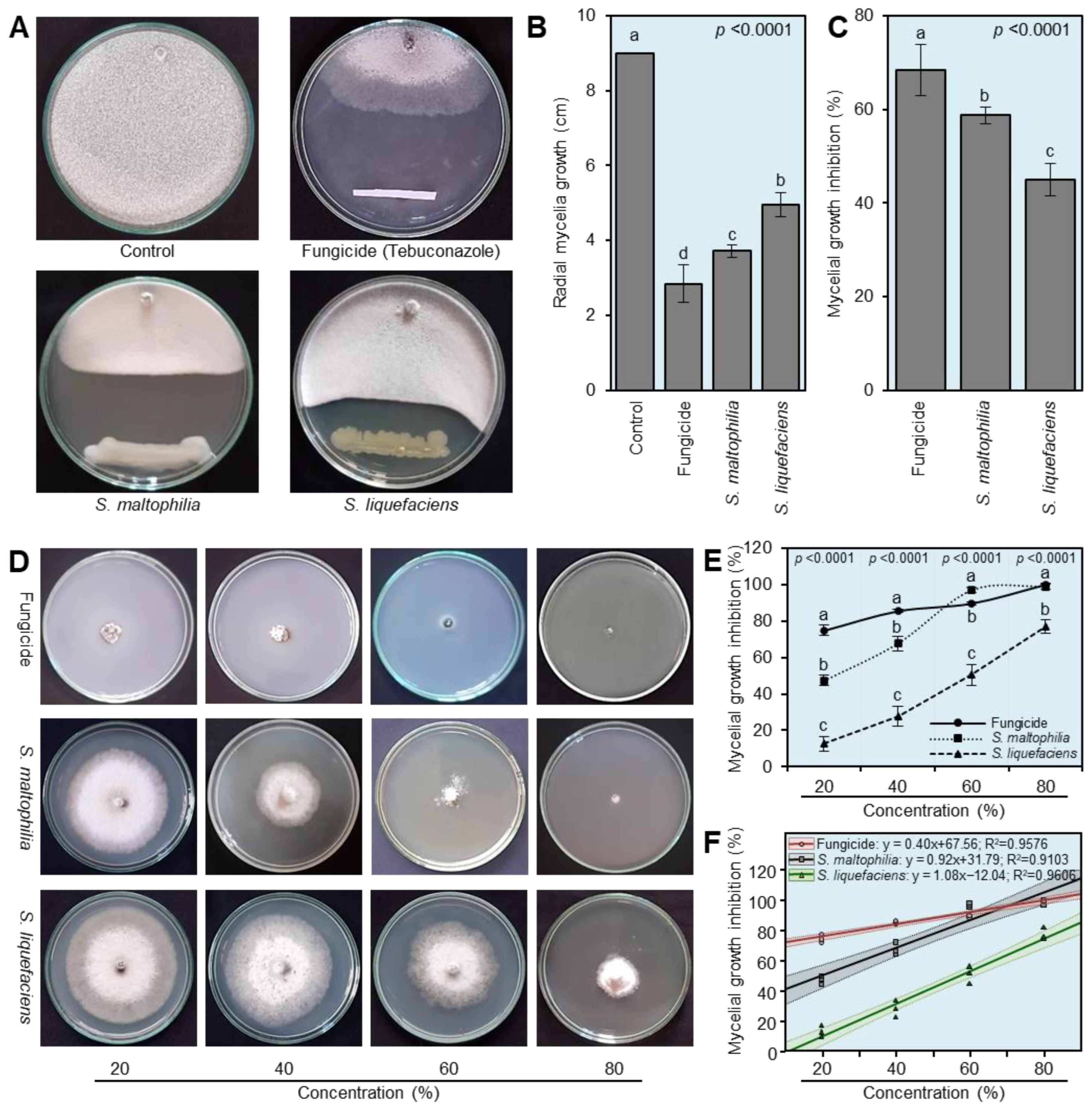
The antifungal activities of both bioagents (S. maltophilia and S. liquefaciens) against S. cepivora were tested in vitro using dual culture and culture filtrate techniques. In the dual culture plates, both bioagents showed strong fungistatic activity against S. cepivora (Figure 2A) and significantly reduced its radial mycelial growth on PDA (Figure 2B). Although the Folicure fungicide (Tebuconazole 25%) had the lowest radial mycelia growth of S. cepivora (2.85 ± 0.50 cm), both S. maltophilia and S. liquefaciens significantly reduced the radial mycelia growth of S. cepivora (3.71 ± 0.16 and 4.95 ± 0.32 cm, respectively) compared with the mock control (Figure 2B). In other words, both S. maltophilia and S. liquefaciens notably inhibited the mycelia growth of S. cepivora by 58.74 ± 1.75% and 44.95 ± 3.51%, respectively, compared with the mock control (Figure 2C).
Moreover, the utilization of bioagent culture filtrates demonstrated strong dose-dependent antifungal activity against S. cepivora (Figure 2D). Briefly, increasing the percentage of the culture filtrate in PDA media from 20 to 80% significantly increased the inhibition of mycelial growth of S. cepivora (Figure 2E). It is worth mentioning that the mycelial growth inhibition (%) due to the utilization of the highest concentration (80%) of the culture filtrate of S. maltophilia was similar to the positive control (Folicure fungicide) and significantly inhibited the mycelial growth of S. cepivora (99.00 ± 1.73%; Figure 2E). Furthermore, the simple linear regression (SLR) showed that the mycelial growth inhibition percentage was positively correlated with the concentrations of culture filtrates of S. maltophilia (y = 0.92x + 31.79; R2 = 0.9103), S. liquefaciens (y = 1.08x − 12.04; R2 = 0.9606), and the Folicure fungicide (y = 0.40x + 67.56; R2 = 0.9576) (Figure 2F).

3.4. Culture filtrates of S. maltophilia and S. liquefaciens Reduced the Development of White Rot Disease

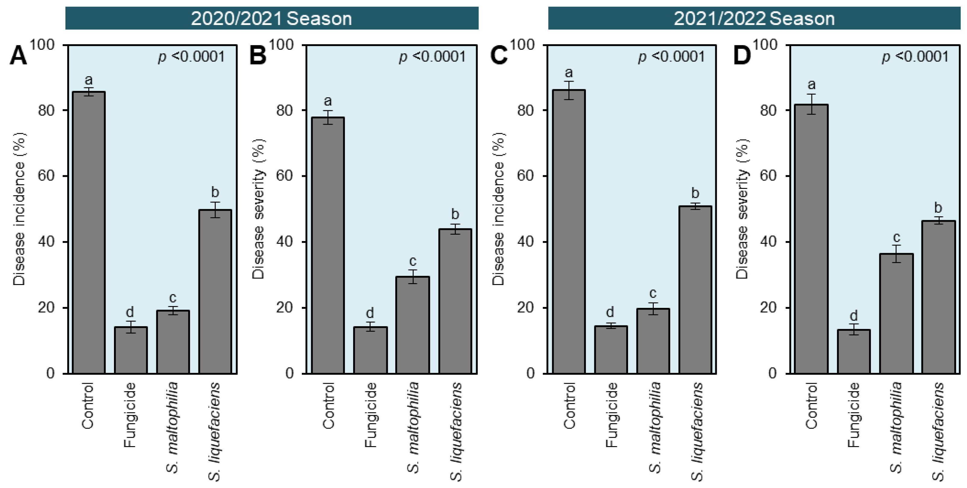
In general, the utilization of the culture filtrates of both bioagents (S. maltophilia and S. liquefaciens) significantly decreased the development of white rot disease on treated onion plants compared with the mock-treated infected plants (control) as expressed by the disease incidence (%) and disease severity (%) during two successive seasons, 2020/2021 and 2021/2022, respectively (Figure 3). Briefly, the white rot disease incidence dropped from 85.58 ± 1.26% in the control plants to 19.08 ± 1.31 and 49.74 ± 2.47% when plants were treated with the culture filtrates of S. maltophilia or S. liquefaciens, respectively, during the 2020/2021 season (Figure 3A). Likewise, the disease severity declined from 77.89 ± 2.18% to 29.39 ± 2.16 and 43.92 ± 1.57% when onions were treated with the culture filtrates of S. maltophilia or S. liquefaciens, respectively, during the same season (Figure 3B). The same trend was observed in the second season, 2021/2022 (Figure 3C,D).

3.5. Cell-Free Culture Filtrates of S. maltophilia and S. liquefaciens Stimulated the Growth of S. cepivora-Infected Onion Plants

During two successive seasons, 2020/2021 and 2021/2022, culture filtrates of S. maltophilia and S. liquefaciens notably increased the number of leaves per plant, plant height, and fresh weight of the shoot system with no significant differences between both bacterial agents but significantly higher differences compared to the mock- and fungicide-treated controls (Figure 4). However, the application of the culture filtrate of S. liquefaciens recorded the longest root length (12.11 ± 0.51 and 12.27 ± 0.96 cm) followed by the culture filtrate of S. maltophilia (7.99 ± 0.85 and 7.70 ± 0.74 cm), which were significantly higher than the mock-treated control (2.02 ± 0.60 and 2.22 ± 0.83 cm) during 2020/2021 and 2021/2022, respectively (Figure 4C,D). Collectively, these findings suggest that the root drench application of both bacterial culture filtrates has no phytotoxicity on the treated onion plants, but they even stimulate their growth.

3.6. Culture Filtrates of S. maltophilia and S. liquefaciens Enhanced the Yield Components of S. cepivora-Infected Onion Plants

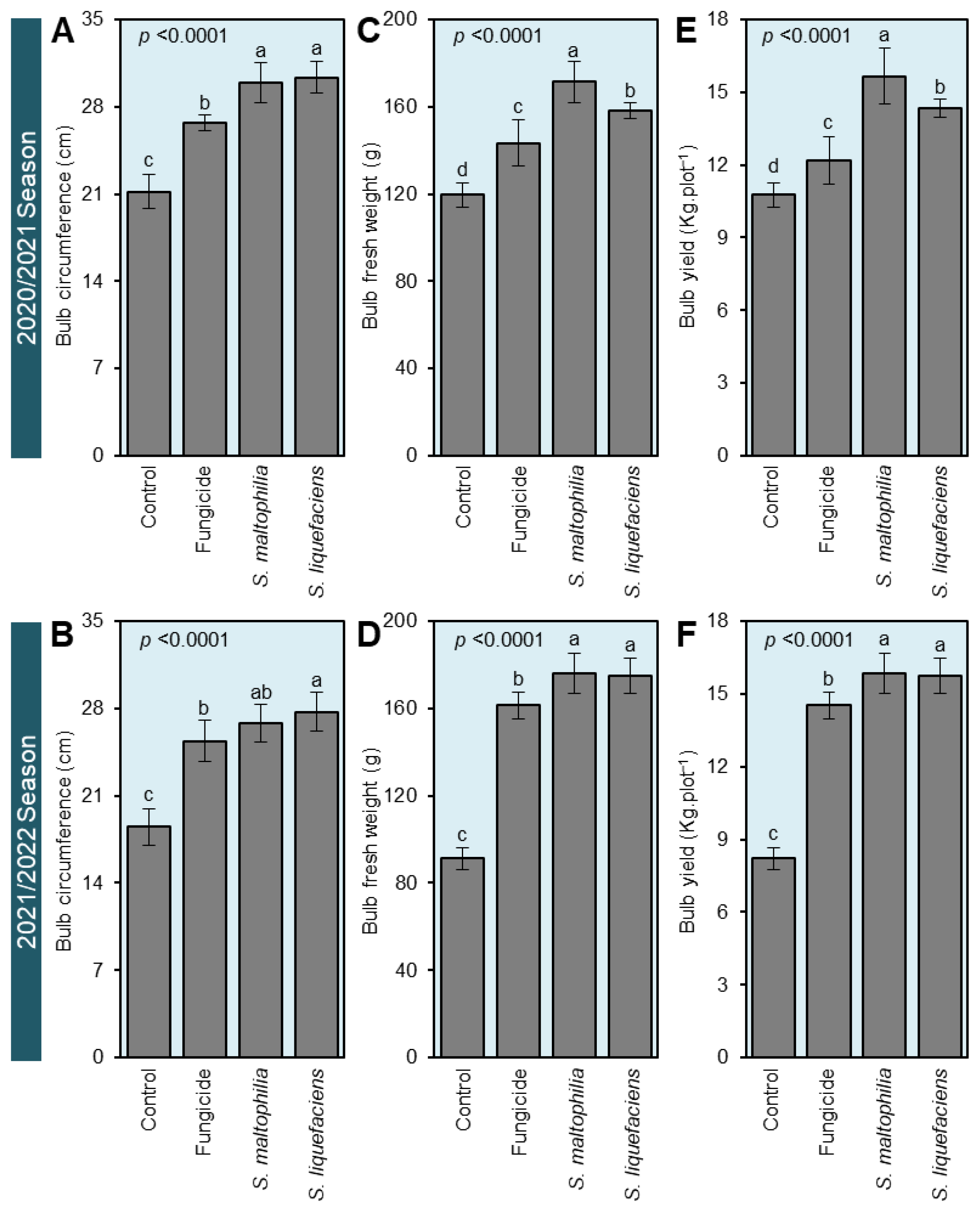
Generally, the root drench application of culture filtrates of both bacterial bioagents remarkably enhanced all yield attributes of S. cepivora-infected onion plants including the bulb diameter (Figure 5A,B), bulb fresh weight (Figure 5C,D), and bulb yield per plot (Figure 5E,F) compared with the mock-treated control during two successive seasons, 2020/2021 and 2021/2022, respectively. Although there were no significant differences between the bulb circumference of S. maltophilia- and S. liquefaciens-treated onions (29.92 ± 1.62 and 30.34 ± 1.28 cm, respectively), the culture filtrate of S. maltophilia had the highest bulb fresh weight (171.27 ± 9.58 g) and bulb yield per plot (15.66 ± 1.17 kg·plot−1), which was comparable to the positive control (Folicure fungicide) during the first season. However, there were no significant differences between S. maltophilia- and S. liquefaciens-treated onion plants in the second season in terms of the bulb fresh weight and bulb yield per plot.

3.7. Cell-Free Culture Filtrates of S. maltophilia and S. liquefaciens Stimulate the Antioxidant Defense Machinery of S. cepivora-Infected Onion Plants

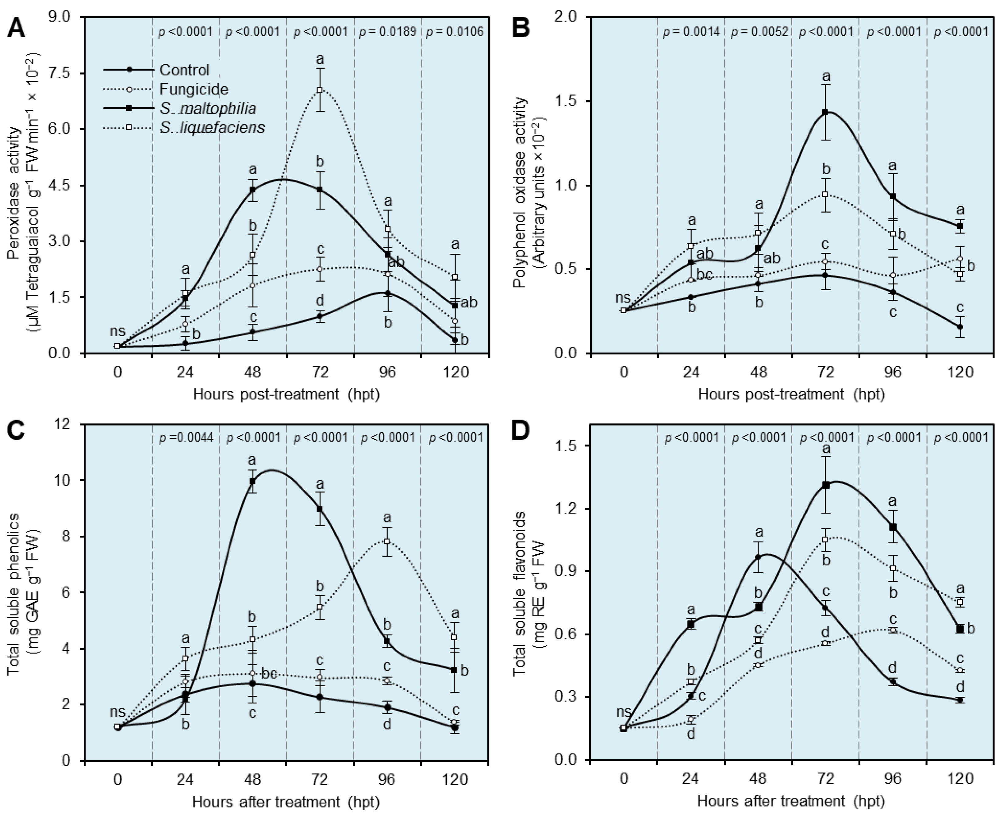
Both enzymatic (peroxidase (POX) and polyphenol oxidase (PPO) activity) and non-enzymatic (total soluble phenolics and flavonoids) antioxidant defense machineries were further investigated to better understand the physiological and biochemical mechanisms of S. maltophilia and S. liquefaciens (Figure 6). Briefly, the root drench application of culture filtrates of both bacterial bioagents gradually enhanced the enzymatic activity of POX (Figure 6A) and PPO (Figure 6B) until they reached their highest peak at 48 or 72 h post treatment (hpt), then they dropped slowly. Onion plants that were treated with the culture filtrate of S. maltophilia recorded their highest peak of POX (3.42 ± 0.29 × 10⁻² μM of tetraguaiacol g−1 FW min−1) at 48 hpt but had the highest peak of PPO (1.43 ± 0.16 × 10⁻² arbitrary units) at 72 hpt. On the contrary, onion plants that were treated with the culture filtrate of S. liquefaciens had their highest peak of POX (7.05 ± 0.57 × 10⁻² μM of tetraguaiacol g−1 FW min−1) at 72 hpt but were the second highest for PPO (0.94 ± 0.1 × 10⁻² arbitrary units) at the same time point, 72 hpt (Figure 6A,B).
Likewise, the non-enzymatic antioxidant defense machinery as expressed by total soluble phenolics (Figure 6C) and flavonoids (Figure 6D) was progressively enhanced due to the root drench application of culture filtrates of both bacterial bioagents until they reached their highest peak then steadily decreased. Briefly, onion plants treated with the culture filtrate of S. maltophilia had the highest peak of total soluble phenolics (9.96 ± 0.42 mg GAE g−1 FW; Figure 6C) and total soluble flavonoids (1.31 ± 0.14 mg RE g−1 FW; Figure 6D) at 48 and 72 hpt, respectively. However, S. liquefaciens-treated plants recorded their highest peak of total soluble phenolics (7.82 ± 0.52 mg GAE g−1 FW; Figure 6C) later at 96 hpt but reached their highest peak of total soluble flavonoids (1.05 ± 0.06 mg RE g−1 FW; Figure 6D) at 72 hpt.

3.8. Culture Filtrates of S. maltophilia and S. liquefaciens and the Endogenous Auxin Content of S. cepivora-Infected Onion Plants

To better understand the physiological and biochemical mechanisms of how culture filtrates of both S. maltophilia and S. liquefaciens stimulate the growth of S. cepivora-infected onion plants, their effect on the endogenous levels of the main auxin, IAA, and its precursor, the amino acid tryptophan, in onion leaves was further investigated. Although the application of the Folicure fungicide did not change the endogenous tryptophan level (600.75 ± 114.52 ng.g−1 FW; Figure 7A) and even reduced the IAA content (182.83 ± 11.45 ng.g−1 FW; Figure 7B) compared with the non-treated control (669.83 ± 159.15 and 214.42 ± 19.39 ng.g−1 FW, respectively), the root drench application of culture filtrates of both bacterial bioagents significantly enhanced the accumulation of IAA and its precursor tryptophan. Onion plants treated with the culture filtrate of S. maltophilia had the highest tryptophan (1086.92 ± 90.46 ng.g−1 FW) and IAA (420.42 ± 36.86 ng.g−1 FW) levels, followed by S. liquefaciens-treated plants (904.33 ± 75.84 and 321.42 ± 32.54 ng.g−1 FW, respectively).

3.9. Chemical Composition of Culture Filtrates of S. maltophilia and S. liquefaciens

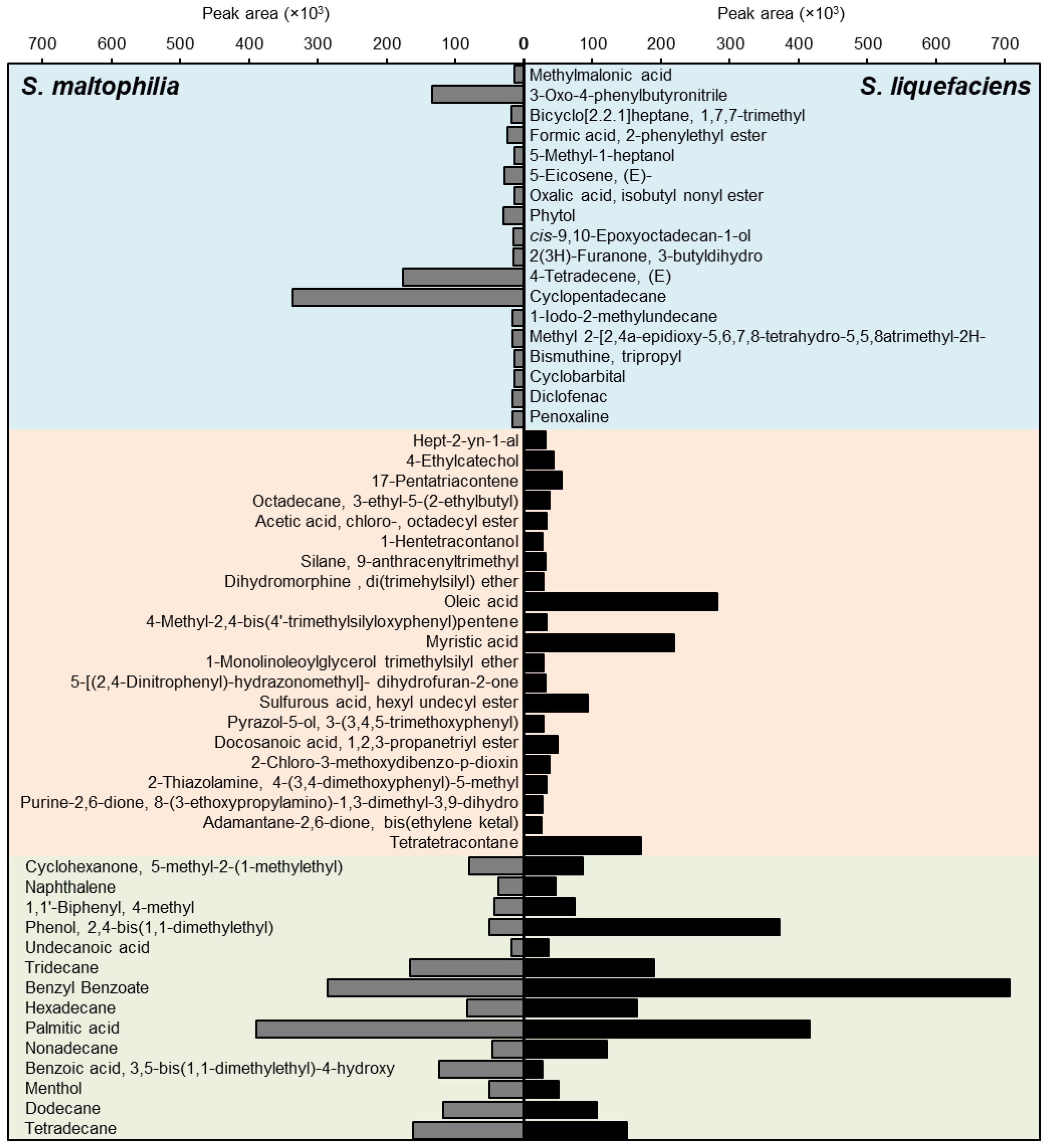
In general, 53 compounds were detected and tentatively identified in the culture filtrates of S. maltophilia and S. liquefaciens (Figure 8). Out of these 53 compounds, 18 compounds were detected only in the culture filtrate of S. maltophilia, and 21 compounds were detected specifically in the culture filtrates of S. liquefaciens. However, only 14 compounds were detected in both culture filtrates including cyclohexanone, 5-methyl-2-(1-methylethyl), naphthalene, 1,1’-biphenyl, 4-methyl, phenol, 2,4-bis(1,1-dimethylethyl), undecanoic acid, tridecane, benzyl benzoate, hexadecane, palmitic acid, nonadecane, benzoic acid, 3,5-bis(1,1-dimethylethyl)-4-hydroxy, menthol, dodecane, and tetradecane (Figure 8).

4. Discussion

White rot is a serious disease that restricts the commercial production of Allium species and causes substantial yield losses worldwide. Although the ascomycetous causal agent, S. cepivora, does not produce any asexual spores, it forms a large proportion of infective sclerotia that survive and remain viable in the soil for up to 20 years in the absence of a susceptible host plant [8]. Unfortunately, there has been no effective control mainly for white rot disease until now; however, chemical control using various agrichemicals, particularly fungicides, is the most commonly used strategy to combat this disease. Nevertheless, the extensive use of chemical fungicides has several health and environmental hazards to humans, animals, and non-target organisms. Moreover, pathogens might develop resistance against the repeatedly used fungicides. Due to the efficiency breakdown of chemical fungicides, the search for safer eco-friendly alternative control measures has become a necessity [50]. Biological control might be a promising control strategy against white rot disease, particularly if it is incorporated into integrated pest management programs [13,50,51].
In the current study, S. maltophilia and S. liquefaciens suppressed the growth of S. cepivora and inhibited the development of white rot disease under field conditions. Several studies have previously reported the antagonistic activity of S. maltophilia against different soil-borne phytopathogens including bacteria such as Ralstonia solanacearum, the causal agent of potato brown rot [20], oomycetes such as Pythium ultimum [52,53], fungi such as Rhizoctonia solani [23,54,55], Fusarium sp. [54,56], Verticillium dahliae [23], and the sclerotia-producing fungus Sclerotium rolfsii [24]. Likewise, numerous S. liquefaciens strains exhibited a strong biocontrol efficacy against soil-borne pathogens including bacteria such as R. solanacearum [57] and fungi such as R. solani [29], F. oxysporum [29,58], and the sclerotia-producing fungus Sclerotinia sclerotiorum [29]. However, to the best of our knowledge, this is the first report about the potential application of S. maltophilia and S. liquefaciens as effective biocontrol agents against S. cepivora, the causal agent of onion white rot disease.
Several hypotheses were proposed to explain the antifungal activity of both bioagents. These hypotheses include the production of several extracellular metabolites such as antibiotics, siderophores, quorum-sensing molecules, N-acyl homoserine lactones (AHLs), and cell wall-degrading enzymes [59]. It is worth mentioning that our GC-MS analysis of the culture filtrates of both bacteria showed that both filtrates contain several bioactive compounds with antimicrobial activity such as menthol [60]; benzyl benzoate [61]; oleic, myristic, and palmitic fatty acids [62,63,64]; tridecane, tetradecane, and hexadecane compounds [65,66]; and phenol,2,4-bis(1,1-diethylethyl) [67]. Taken together, our findings suggest that the antifungal activity of S. maltophilia and S. liquefaciens might be due to the presence of these compounds in their culture filtrate. However, further studies are required to better understand how the internal mechanisms of menthol operate within a fungal cell.
Another hypothesis that could explain the antifungal activity of S. maltophilia and S. liquefaciens is due to their ability to produce cell wall-degrading enzymes [59]. It was previously reported that the fungistatic activity of S. maltophilia W81 against Pythium ultimum is mediated by extracellular proteolytic activity [52] including an inducible extracellular serine protease [53]. Furthermore, the antagonistic activity of S. maltophilia against fungal phytopathogens such as Fusarium sp., Rhizoctonia sp., Alternaria sp., and Bipolaris sorokiniana is associated with its ability to produce chitinase [54,68].
Additionally, our field experiments showed that both bacterial culture filtrates significantly reduced the disease incidence and disease severity of white rot on treated onion plants compared with the non-treated plants. It is worth noting that none of the bacterial culture filtrates caused any phytotoxicity in treated plants as indicated by a stimulated growth performance (number of leaves per plant, root length, plant height, and fresh weight of the shoot system). However, the biochemical and physiological mechanisms behind these roles are poorly understood. This might be due to the enhancement of enzymatic antioxidants and non-enzymatic antioxidant defense machinery [69]. Peroxidase (POX) and polyphenol oxidase (PPO) are two main components in the enzymatic antioxidant system. POX maintains redox homeostasis via the regulation of H2O2 levels [70], whereas PPO is involved in the oxidation of phenolics into highly reactive quinones. Interestingly, our findings proved that the root application of culture filtrates of S. maltophilia and S. liquefaciens considerably boosted the enzymatic activities of both POX and PPO.
Moreover, the non-enzymatic antioxidant defense machinery relies on phenolics, flavonoids, and lipophilic antioxidants such as carotenoids [71]. Our findings showed that the application of culture filtrates of S. maltophilia and S. liquefaciens notably enriched the endogenous levels of total soluble phenolics and flavonoids in treated onion leaves. These findings propose that the application of culture filtrates of S. maltophilia and S. liquefaciens induces multilayered antioxidant defense machinery in S. cepivora-infected onion plants to alleviate the risky consequences of reactive oxygen species (ROS) and preserve their homeostasis. Additionally, Serratia sp. was previously reported to trigger a plant’s defensive mechanisms against miscellaneous phytopathogens via the activation of induced systemic resistance (ISR) after a proper stimulation [59].
Furthermore, culture filtrates of S. maltophilia and S. liquefaciens might reduce the disease incidence and severity of white rot on treated onion plants by prolonging the sclerotial differentiation period. It was previously reported that menthol delays sclerotial differentiation by up to 145 h [60]. As we mentioned above, the GC-MS analysis showed that both culture filtrates were rich in menthol, which might have prolonged the sclerotial differentiation period and resulted in a reduced disease incidence and severity. Moreover, the culture filtrate of S. maltophilia was rich in fatty acids such as oleic and myristic acids. Oleic acid was previously reported to inhibit the growth of numerous phytopathogenic fungi such as R. solani, P. ultimum, Pyrenophora avenae, and Crinipellis perniciosa [62]. Likewise, myristic acid negatively affected mycelial growth and spore germination of four phytopathogenic fungi including A. solani, Colletotrichum lagenarium, F. oxysporum f. sp. Cucumerinum, and F. oxysporum f. sp. lycopersici [63]. Although some oxylipins (fatty acid derivatives) such as (±)-cis-12,13-Epoxy-9(Z)-octadecenoic acid, (±)-threo-12,13-Dihydroxy-9(Z)-octadecenoic acid, and (±)-threo-9,10-Dihydroxy-12(Z)-octadecenoic acid showed potent antifungal activity against S. sclerotiorum [72], the antifungal activity of fatty acids and their derivatives are poorly studied. Collectively, these findings suggest that extracellular metabolites in culture filtrates of S. maltophilia and S. liquefaciens inhibit the growth of S. cepivora or delay the sclerotial differentiation of its sclerotia, resulting in a reduced disease incidence and disease severity. However, further studies are required to better understand the potential role(s) of extracellular metabolites of S. maltophilia and S. liquefaciens.
In addition to their protective role against white rot disease, culture filtrates of S. maltophilia and S. liquefaciens showed strong bio-stimulant properties. Our field experiments showed that both culture filtrates stimulated the growth of S. cepivora-infected onion plants compared with the mock-treated control as expressed by more leaves per plant, longer roots, a higher plant height, and a heavier fresh weight of the shoot system. Moreover, both culture filtrates enhanced the yield components of S. cepivora-infected onion plants as expressed by a bigger bulb diameter, higher bulb fresh weight, and bulb yield per plot during two successive seasons, 2020/2021 and 2021/2022, respectively. Bio-stimulant and growth promotion properties of S. maltophilia and S. liquefaciens might be due to their ability to induce the accumulation of auxins and their precursor tryptophan within treated onion plants. Both S. maltophilia and S. liquefaciens embrace several mechanisms to facilitate plant growth promotion such as enhancing nutrient uptake, producing siderophores, and synthesizing stimulatory phytohormones like IAA [59,73,74,75,76,77,78,79]. Both bacteria have complete metabolic pathways linked to their plant growth promotion properties, including the IAA biosynthesis pathway [73,74,75,76,77,78,79]. Production of IAA by different strains of S. Maltophilia [74,75,76], as well as S. liquefaciens [77,78,79], was previously reported. These characteristics highlight the enrichment of the culture filtrates of both bacteria with growth-promoting phytohormone IAA. Moreover, our findings showed that the root drench application of culture filtrates of both bacterial bioagents significantly enhanced the accumulation of IAA and its precursor tryptophan and stimulated the growth of S. cepivora-infected onion plants.

5. Conclusions

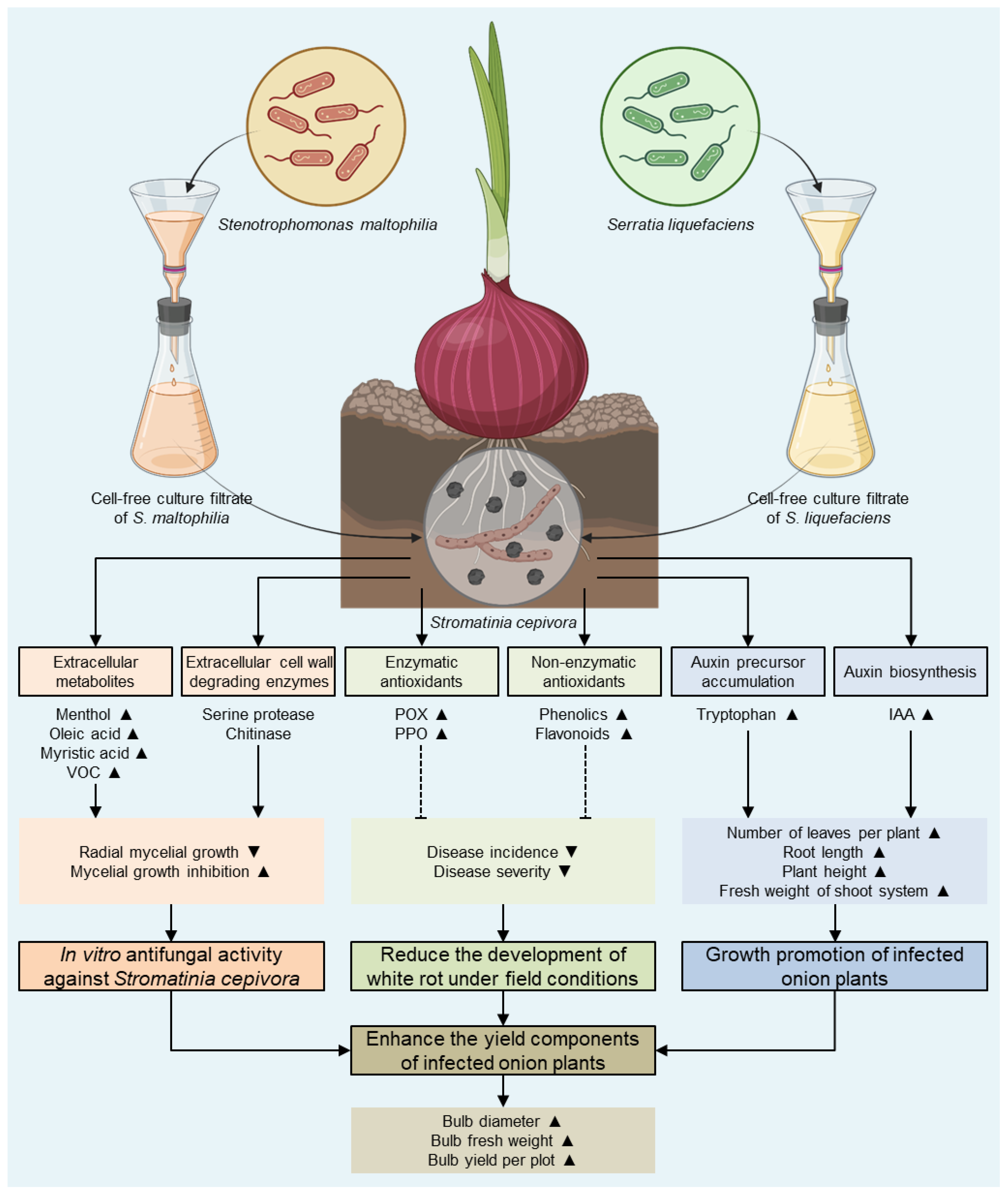
In conclusion, our findings highlight the potential importance of S. maltophilia and S. liquefaciens as plant growth-promoting bacteria with potent bio-control properties against S. cepivora, the causal agent of onion white rot disease (Figure 9). Our findings showed that these two bacteria and their cell-free culture filtrates notably inhibited the mycelial growth of S. cepivora, which might be due to the enriched chemical composition of these filtrates, particularly the high levels of menthol, oleic acid, myristic acid, and other volatile organic compounds. Moreover, culture filtrates of S. maltophilia and S. liquefaciens significantly reduced the development of white rot disease, which might be due to their antifungal activities and/or due to the activation of both enzymatic (POX and PPO) and non-enzymatic (phenolics and flavonoids) antioxidant defense machineries of S. cepivora-infected onion plants. Last but not least, culture filtrates of S. maltophilia and S. liquefaciens promoted the growth of S. cepivora-infected onion plants and enhanced their yield components, which might be a result of a reduced disease severity and/or the induced IAA accumulation. Although further studies are required to better understand the molecular, biochemical, and physiological mechanisms involved in the fungistatic activity of S. maltophilia and S. liquefaciens or their cell-free culture filtrates against S. cepivora, our findings suggest that both bioagents might be eco-friendly alternative control measures to reduce the utilization of chemical fungicides entirely or partially for the safer production of onions in S. cepivora-infested soils.

Supplementary Materials

The following supporting information can be downloaded at: https://www.mdpi.com/article/10.3390/horticulturae9070780/s1, Table S1: Sequences from S. cepivora and some other fungal species that produce significant alignments with S. cepivora—Isolate 2023 using NCBI database.; Table S2: Sequences from S. maltophilia and some other bacteria species that produce significant alignments with S. maltophilia—Isolate YN-2023 using NCBI database.; Table S3: Sequences from S. liquefaciens and some other bacterial species that produce significant alignments with S. liquefaciens—Isolate AE-2023 using NCBI database.

Author Contributions

Conceptualization, Y.N., A.A.E. and A.E.-N.; methodology, Y.N. and A.E.-N.; software, Y.N.; validation, H.E.M.O., Y.N., A.A.E., M.H.E.-M. and A.E.-N.; formal analysis, Y.N.; investigation, Y.N. and A.E.-N.; resources, H.E.M.O., A.A.E. and M.H.E.-M.; data curation, Y.N., A.A.E. and A.E.-N.; writing—original draft preparation, Y.N. and A.E.-N.; writing—review and editing, Y.N. and A.A.E.; visualization, Y.N.; supervision, Y.N. and A.A.E.; project administration, Y.N., A.A.E. and A.E.-N.; funding acquisition, H.E.M.O. and M.H.E.-M. All authors have read and agreed to the published version of the manuscript.

Funding

The authors extend their appreciation to the Deanship for Research & Innovation, Ministry of Education in Saudi Arabia for funding this research work through project number: IFP22UQU4320730DSR030.

Data Availability Statement

The datasets generated and/or analyzed during the current study are available from the corresponding author upon reasonable request.

Conflicts of Interest

The authors declare that there is no conflict of interest and that they have no known competing financial interests or personal relationships that could appear to have influenced the work reported in this paper.

References

  1. Shultz, S. Onions. J. Agric. Food Inf. 2010, 11, 8–15. [Google Scholar]
  2. Javadzadeh, A.; Ghorbanihaghjo, A.; Bonyadi, S.; Rashidi, M.R.; Mesgari, M.; Rashtchizadeh, N.; Argani, H. Preventive Effect of Onion Juice on Selenite-Induced Experimental Cataract. Indian J. Ophthalmol. 2009, 57, 185. [Google Scholar] [PubMed]
  3. FAOSTAT. Food and Agriculture Organization of the United Nations. Available online: https://www.fao.org/faostat/en/#data/QCL (accessed on 2 June 2023).
  4. Dutta, R.; Nadig, S.M.; Manjunathagowda, D.C.; Gurav, V.S.; Singh, M. Anthracnose of Onion (Allium cepa L.): A Twister Disease. Pathogens 2022, 11, 884. [Google Scholar] [PubMed]
  5. Darwesh, O.M.; Elshahawy, I.E. Silver Nanoparticles Inactivate Sclerotial Formation in Controlling White Rot Disease in Onion and Garlic Caused by the Soil Borne Fungus Stromatinia cepivora. Eur. J. Plant Pathol. 2021, 160, 917–934. [Google Scholar]
  6. Elshahawy, I.E. Reduced Sclerotial Viability of Stromatinia cepivora and Control of White Rot Disease of Onion and Garlic by Means of Soil Bio-Solarization. Eur. J. Plant Pathol. 2021, 160, 519–540. [Google Scholar]
  7. Valdés-Santiago, L.; Vargas-Bernal, R.; Herrera-Pérez, G.; Colli-Mull, J.G.; Ordaz-Arias, A. Application of Two-Photon Microscopy to Study Sclerotium cepivorum Berk Sclerotia Isolated from Naturally Infested Soil and Produced In Vitro. Curr. Microbiol. 2021, 78, 749–755. [Google Scholar]
  8. Coley-Smith, J.R.; Mitchell, C.M.; Sansford, C.E. Long-term Survival of Sclerotia of Sclerotium cepivorum and Stromatinia gladioli. Plant Pathol. 1990, 39, 58–69. [Google Scholar]
  9. Amin, M.; Tadele, S.; Selvaraj, T. White Rot (Sclerotium cepivorum Berk)—An Aggressive Pest of Onion and Garlic in Ethiopia: An Overview. J. Agric. Biotechnol. Sustain. Dev. 2014, 6, 6–15. [Google Scholar] [CrossRef]
  10. Melero-Vara, J.M.; Basallote-Ureba, A.M.P.-L.; Basallote-Ureba, M.J. Comparison of Physical, Chemical and Biological Methods of Controlling Garlic White Rot. Eur. J. Plant Pathol. 2000, 106, 581–588. [Google Scholar]
  11. Coventry, E.; Noble, R.; Mead, A.; Whipps, J.M. Suppression of Allium White Rot (Sclerotium cepivorum) in Different Soils Using Vegetable Wastes. Eur. J. Plant Pathol. 2005, 111, 101–112. [Google Scholar]
  12. Clarkson, J.P.; Payne, T.; Mead, A.; Whipps, J.M. Selection of Fungal Biological Control Agents of Sclerotium cepivorum for Control of White Rot by Sclerotial Degradation in a UK Soil. Plant Pathol. 2002, 516, 735–745. [Google Scholar]
  13. Shalaby, M.E.; Ghoniem, K.E. Biological and Fungicidal Antagonism of Sclerotium cepivorum for Controlling Onion White Rot Disease. Ann. Microbiol. 2013, 63, 1579–1589. [Google Scholar] [CrossRef]
  14. Coley-Smith, J.R. White Rot Disease of Allium: Problems of Soil-Borne Diseases in Microcosm. Plant Pathol. 1990, 39, 214–222. [Google Scholar]
  15. Zubrod, J.P.; Bundschuh, M.; Arts, G.; Brühl, C.A.; Imfeld, G.; Knäbel, A.; Payraudeau, S.; Rasmussen, J.J.; Rohr, J.; Scharmüller, A.; et al. Fungicides: An Overlooked Pesticide Class? Environ. Sci. Technol. 2019, 53, 3347–3365. [Google Scholar] [PubMed]
  16. Deresa, E.M.; Diriba, T.F. Phytochemicals as Alternative Fungicides for Controlling Plant Diseases: A Comprehensive Review of Their Efficacy, Commercial Representatives, Advantages, Challenges for Adoption, and Possible Solutions. Heliyon 2023, 9, e13810. [Google Scholar] [CrossRef] [PubMed]
  17. Chet, I.; Inbar, J. Biological Control of Fungal Pathogens. Appl. Biochem. Biotechnol. 1994, 48, 37–43. [Google Scholar] [CrossRef]
  18. Sharma, I. Phytopathogenic Fungi and Their Biocontrol Applications. In Fungi Bio-Prospects in Sustainable Agriculture, Environment and Nano-Technology, Volume 1: Fungal Diversity of Sustainable Agriculture; Sharma, V.K., Shah, M.P., Parmar, S., Kumar, A., Eds.; Academic Press: Cambridge, MA, USA, 2021; Volume 1, pp. 155–188. ISBN 9780128213940. [Google Scholar]
  19. Aliye, N.; Fininsa, C.; Hiskias, Y. Evaluation of Rhizosphere Bacterial Antagonists for Their Potential to Bioprotect Potato (Solanum Tuberosum) against Bacterial Wilt (Ralstonia solanacearum). Biol. Control 2008, 47, 282–288. [Google Scholar]
  20. Messiha, N.A.S.; van Diepeningen, A.D.; Farag, N.S.; Abdallah, S.A.; Janse, J.D.; van Bruggen, A.H.C. Stenotrophomonas Maltophilia: A New Potential Biocontrol Agent of Ralstonia solanacearum, Causal Agent of Potato Brown Rot. Eur. J. Plant Pathol. 2007, 118, 211–225. [Google Scholar] [CrossRef]
  21. Debette, J.; Blondeau, R. Presence of Pseudomonas maltophilia in the Rhizosphere of Several Cultivated Plants. Can. J. Microbiol. 1980, 26, 460–463. [Google Scholar]
  22. Garbeva, P.; Van Overbeek, L.S.; Van Vuurde, J.W.L.; Van Elsas, J.D. Analysis of Endophytic Bacterial Communities of Potato by Plating and Denaturing Gradient Gel Electrophoresis (DGGE) of 16S rDNA Based PCR Fragments. Microb. Ecol. 2001, 41, 369–383. [Google Scholar] [PubMed]
  23. Berg, G.; Marten, P.; Ballin, G. Stenotrophomonas maltophilia in the Rhizosphere of Oilseed Rape—Occurrence, Characterization and Interaction with Phytopathogenic Fungi. Microbiol. Res. 1996, 151, 19–27. [Google Scholar] [CrossRef]
  24. Sultana, F.; Hossain, M.M. Assessing the Potentials of Bacterial Antagonists for Plant Growth Promotion, Nutrient Acquisition, and Biological Control of Southern Blight Disease in Tomato. PLoS ONE 2022, 17, e0267253. [Google Scholar]
  25. Badri Fariman, A.; Abbasiliasi, S.; Akmar Abdullah, S.N.; Mohd Saud, H.; Wong, M.Y. Stenotrophomonas maltophilia Isolate UPMKH2 with the Abilities to Suppress Rice Blast Disease and Increase Yield a Promising Biocontrol Agent. Physiol. Mol. Plant Pathol. 2022, 121, 101872. [Google Scholar] [CrossRef]
  26. Lodewyckx, C.; Vangronsveld, J.; Porteous, F.; Edward, R.B.; Taghavi, S.; Mezgeay, M.; Van Der Lelie, D. Endophytic Bacteria and Their Potential Applications. Crit. Rev. Plant Sci. 2002, 21, 583–606. [Google Scholar]
  27. Santoyo, G.; Moreno-hagelsieb, G.; Orozco-mosqueda, C.; Glick, B.R. Plant Growth-Promoting Bacterial Endophytes. Microbiol. Res. 2016, 183, 92–99. [Google Scholar] [CrossRef]
  28. Liu, Y.; Liu, W.; Liang, Z. Endophytic Bacteria from Pinellia ternata, a New Source of Purine Alkaloids and Bacterial Manure. Pharm. Biol. 2015, 53, 1545–1548. [Google Scholar]
  29. Michail, G.; Reizopoulou, A.; Vagelas, I. Evaluation of The Biocontrol Efficacy of Serratia proteamaculans and S. liquefaciens Isolated From Bats Guano Pile From a Subterrestrial Cave (Greece). Agric. Biol. (Sel’skokhozyaistvennaya Biol.) 2022, 57, 566–578. [Google Scholar] [CrossRef]
  30. Tian, S.P.; Bertolini, P. Effects of Low Temperature on Mycelial Growth and Spore Germination of Botrytis allii in Culture and on Its Pathogenicity to Stored Garlic Bulbs. Plant Pathol. 1995, 44, 1008–1015. [Google Scholar] [CrossRef]
  31. Zewide, T.; Fininsa, C.; Sakhuja, P.K. Management of White Rot (Sclerotium cepivorum) of Garlic Using Fungicides in Ethiopia. Crop Prot. 2007, 26, 856–866. [Google Scholar] [CrossRef]
  32. El-Nagar, A.; Elzaawely, A.A.; Xuan, T.D.; Gaber, M.; El-Wakeil, N.; El-Sayed, Y.; Nehela, Y. Metal Complexation of Bis-Chalcone Derivatives Enhances Their Efficacy against Fusarium Wilt Disease, Caused by Fusarium equiseti, via Induction of Antioxidant Defense Machinery. Plants 2022, 11, 2418. [Google Scholar] [CrossRef]
  33. Nehela, Y.; Mazrou, Y.S.A.; Taha, N.A.; Elzaawely, A.A.; Xuan, T.D.; Makhlouf, A.H.; El-Nagar, A. Hydroxylated Cinnamates Enhance Tomato Resilience to Alternaria alternata, the Causal Agent of Early Blight Disease, and Stimulate Growth and Yield Traits. Plants 2023, 12, 1775. [Google Scholar] [CrossRef] [PubMed]
  34. Tamura, K.; Nei, M. Estimation of the Number of Nucleotide Substitutions in the Control Region of Mitochondrial DNA in Humans and Chimpanzees. Mol. Biol. Evol. 1993, 10, 512–526. [Google Scholar] [CrossRef] [PubMed] [Green Version]
  35. Tamura, K.; Stecher, G.; Kumar, S. MEGA11: Molecular Evolutionary Genetics Analysis Version 11. Mol. Biol. Evol. 2021, 38, 3022–3027. [Google Scholar] [CrossRef] [PubMed]
  36. Johnson, L.F.; Curl, E.A. Methods for Research on the Ecology of Soil-Borne Plant Pathogens. In Methods for Research on the Ecology of Soil-Borne Plant Pathogens; Burgess Publishing Co.: Clayton, NC, USA, 1972. [Google Scholar]
  37. Bulgari, D.; Bozkurt, A.I.; Casati, P.; Çağlayan, K.; Quaglino, F.; Bianco, P.A. Endophytic Bacterial Community Living in Roots of Healthy and ‘Candidatus Phytoplasma Mali’-Infected Apple (Malus domestica, Borkh.) Trees. Antonie Van Leeuwenhoek 2012, 102, 677–687. [Google Scholar] [CrossRef] [PubMed]
  38. Al-Hussini, H.S.; Al-Rawahi, A.Y.; Al-Marhoon, A.A.; Al-Abri, S.A.; Al-Mahmooli, I.H.; Al-Sadi, A.M.; Velazhahan, R. Biological Control of Damping-off of Tomato Caused by Pythium aphanidermatum by Using Native Antagonistic Rhizobacteria Isolated from Omani Soil. J. Plant Pathol. 2019, 101, 315–322. [Google Scholar]
  39. Amin, M.M.; Ahmed, M.F.A. Biological Control of Onion White Rot Disease Using Potential Bacillus subtilis Isolates. Egypt. J. Biol. Pest Control 2023, 33, 27. [Google Scholar] [CrossRef]
  40. Kähkönen, M.P.; Hopia, A.I.; Vuorela, H.J.; Rauha, J.P.; Pihlaja, K.; Kujala, T.S.; Heinonen, M. Antioxidant Activity of Plant Extracts Containing Phenolic Compounds. J. Agric. Food Chem. 1999, 47, 3954–3962. [Google Scholar] [CrossRef]
  41. Djeridane, A.; Yousfi, M.; Nadjemi, B.; Boutassouna, D.; Stocker, P.; Vidal, N. Antioxidant Activity of Some Algerian Medicinal Plants Extracts Containing Phenolic Compounds. Food Chem. 2006, 97, 654–660. [Google Scholar]
  42. Harrach, B.D.; Fodor, J.; Pogány, M.; Preuss, J.; Barna, B. Antioxidant, Ethylene and Membrane Leakage Responses to Powdery Mildew Infection of near-Isogenic Barley Lines with Various Types of Resistance. Eur. J. Plant Pathol. 2008, 121, 21–33. [Google Scholar] [CrossRef]
  43. Malik, C.P.; Singh, M.B. Plant Enzymology and Histo-Enzymology; Kalyani Publishers: New Delhi, India, 1980. [Google Scholar]
  44. Hijaz, F.; Nehela, Y.; Killiny, N. Possible Role of Plant Volatiles in Tolerance against Huanglongbing in Citrus. Plant Signal. Behav. 2016, 11, e1138193. [Google Scholar] [CrossRef] [Green Version]
  45. Nehela, Y.; Hijaz, F.; Elzaawely, A.A.; El-Zahaby, H.M.; Killiny, N. Phytohormone Profiling of the Sweet Orange (Citrus sinensis (L.) Osbeck) Leaves and Roots Using GC-MS-Based Method. J. Plant Physiol. 2016, 199, 12–17. [Google Scholar] [CrossRef]
  46. Killiny, N.; Nehela, Y. Metabolomic Response to Huanglongbing: Role of Carboxylic Compounds in Citrus sinensis Response to ‘Candidatus Liberibacter asiaticus’ and Its Vector. Diaphorina citri. Mol. Plant-Microbe Interact. 2017, 30, 666–678. [Google Scholar] [CrossRef] [PubMed] [Green Version]
  47. Nehela, Y.; Hijaz, F.; Elzaawely, A.A.; El-Zahaby, H.M.; Killiny, N. Citrus Phytohormonal Response to Candidatus Liberibacter Asiaticus and Its Vector. Diaphorina citri. Physiol. Mol. Plant Pathol. 2018, 102, 24–35. [Google Scholar] [CrossRef]
  48. Nehela, Y.; Killiny, N. Melatonin Is Involved in Citrus Response to the Pathogen Huanglongbing via Modulation of Phytohormonal Biosynthesis. Plant Physiol. 2020, 184, 2216–2239. [Google Scholar] [CrossRef] [PubMed]
  49. Nehela, Y.; Taha, N.A.; Elzaawely, A.A.; Xuan, T.D.; Amin, M.A.; Ahmed, M.E.; El-Nagar, A. Benzoic Acid and Its Hydroxylated Derivatives Suppress Early Blight of Tomato (Alternaria solani) via the Induction of Salicylic Acid Biosynthesis and Enzymatic and Nonenzymatic Antioxidant Defense Machinery. J. Fungi 2021, 7, 663. [Google Scholar] [CrossRef]
  50. Hussain, W.A.; Elzaawely, A.A.; El Sheery, N.I.; Ismail, A.A.; El-Zahaby, H.M. Biological Control of Onion White Rot Disease Caused by Sclerotium cepivorum. Environ. Biodivers. Soil Secur. 2017, 1, 101–107. [Google Scholar] [CrossRef]
  51. Rivera-Méndez, W.; Obregón, M.; Morán-Diez, M.E.; Hermosa, R.; Monte, E. Trichoderma asperellum Biocontrol Activity and Induction of Systemic Defenses against Sclerotium cepivorum in Onion Plants under Tropical Climate Conditions. Biol. Control 2020, 141, 104145. [Google Scholar] [CrossRef]
  52. Dunne, C.; Crowley, J.J.; Moënne-Loccoz, Y.; Dowling, D.N.; De Bruijn, F.J.; O’Gara, F. Biological Control of Pythium ultimum by Stenotrophomonas maltophilia W81 Is Mediated by an Extracellular Proteolytic Activity. Microbiology 1997, 143, 3921–3931. [Google Scholar] [CrossRef]
  53. Dunne, C.; Moenne-Loccoz, Y.; de Bruijn, F.J.; O’Gara, F. Overproduction of an Inducible Extracellular Serine Protease Improves Biological Control of Pythium ultimum by Stenotrophomonas maltophilia Strain W81. Microbiology 2000, 146, 2069–2078. [Google Scholar] [CrossRef]
  54. Jankiewicz, U.; Brzezinska, M.S.; Saks, E. Identification and Characterization of a Chitinase of Stenotrophomonas maltophilia, a Bacterium That Is Antagonistic towards Fungal Phytopathogens. J. Biosci. Bioeng. 2012, 113, 30–35. [Google Scholar] [CrossRef]
  55. Giesler, L.J.; Yuen, G.Y. Evaluation of Stenotrophomonas maltophilia Strain C3 for Biocontrol of Brown Patch Disease. Crop Prot. 1998, 17, 509–513. [Google Scholar] [CrossRef]
  56. Dewi, R.R.; Rahmah, S.M.; Taruna, A.; Aini, L.Q.; Fernando, I.; Abadi, A.L.; Syib’li, M.A. The Effectiveness Comparison Between Application of Indigenous Arbuscular Mycorrhizal Fungal Community and Stenotrophomonas maltophilia to Suppress Fusarium Wilt Incidence on Local Garlic Plant (Lumbu Hijau). AGRIVITA J. Agric. Sci. 2023, 45, 131–146. [Google Scholar] [CrossRef]
  57. Antonio Saministrado-Fudalan, P.; Sendaydiego, J.P.; Sinco, A.L. Serratia liquefaciens from IMO: Antibacterial and Chitinolytic Potentials. Int. J. Sci. Basic Appl. Res. 2017, 36, 306–317. [Google Scholar]
  58. Sneh, B.; Agami, O.; Baker, R. Biological Control of Fusarium-Wilt in Carnation with Serratia liquefaciens and Hafnia Alvei Isolated from Rhizosphere of Carnation. J. Phytopathol. 1985, 113, 271–276. [Google Scholar] [CrossRef]
  59. Kshetri, L.; Naseem, F.; Pandey, P. Role of Serratia sp. as Biocontrol Agent and Plant Growth Stimulator, with Prospects of Biotic Stress Management in Plant. In Plant Growth Promoting Rhizobacteria for Sustainable Stress Management: Volume 2: Rhizobacteria in Biotic Stress Management; Springer: Singapore, 2019; pp. 169–200. [Google Scholar] [CrossRef]
  60. Lucini, E.I.; Zunino, M.P.; López, M.L.; Zygadlo, J.A. Effect of Monoterpenes on Lipid Composition and Sclerotial Development of Sclerotium cepivorum Berk. J. Phytopathol. 2006, 154, 441–446. [Google Scholar] [CrossRef]
  61. Diastuti, H.; Chasani, M. Suwandri Antibacterial Activity of Benzyl Benzoate and Crotepoxide from Kaempferia rotunda L. Rhizome. Indones. J. Chem. 2020, 20, 9–15. [Google Scholar] [CrossRef] [Green Version]
  62. Walters, D.; Raynor, L.; Mitchell, A.; Walker, R.; Walker, K. Antifungal Activities of Four Fatty Acids against Plant Pathogenic Fungi. Mycopathologia 2004, 157, 87–90. [Google Scholar] [CrossRef]
  63. Liu, S.; Ruan, W.; Li, J.; Xu, H.; Wang, J.; Gao, Y.; Wang, J. Biological Control of Phytopathogenic Fungi by Fatty Acids. Mycopathologia 2008, 166, 93–102. [Google Scholar] [CrossRef]
  64. Casillas-Vargas, G.; Ocasio-Malavé, C.; Medina, S.; Morales-Guzmán, C.; Del Valle, R.G.; Carballeira, N.M.; Sanabria-Ríos, D.J. Antibacterial Fatty Acids: An Update of Possible Mechanisms of Action and Implications in the Development of the next-Generation of Antibacterial Agents. Prog. Lipid Res. 2021, 82, 101093. [Google Scholar] [CrossRef]
  65. Yogeswari, S.; Ramalakshmi, S.; Neelavathy, R.; Muthumary, J. Identification and Comparative Studies of Different Volatile Fractions from Monochaetia Kansensis by GCMS. Glob. J. Pharmacol. 2012, 6, 65–71. [Google Scholar]
  66. Faridha Begum, I.; Mohankumar, R.; Jeevan, M.; Ramani, K. GC–MS Analysis of Bio-Active Molecules Derived from Paracoccus pantotrophus FMR19 and the Antimicrobial Activity Against Bacterial Pathogens and MDROs. Indian J. Microbiol. 2016, 56, 426–432. [Google Scholar] [CrossRef] [Green Version]
  67. Wagay, N.A.; Mohiuddin, Y.G.; Khan, N.A. Phytochemical Evaluation and Identification of Bioactive Phytochemical Evaluation and Identification of Bioactive Compounds in Camel Thorn Alhagi Pseudalhagi (M. Bieb.). Int. J. Adv. Res. Sci. Eng. 2018, 7, 300–311. [Google Scholar]
  68. Zhang, Z.; Yuen, G.Y. The Role of Chitinase Production by Stenotrophomonas maltophilia Strain C3 in Biological Control of Bipolaris Sorokiniana. Phytopathology 2007, 90, 384–389. [Google Scholar] [CrossRef] [Green Version]
  69. Sharma, P.; Jha, A.B.; Dubey, R.S.; Pessarakli, M. Reactive Oxygen Species, Oxidative Damage, and Antioxidative Defense Mechanism in Plants under Stressful Conditions. J. Bot. 2012, 2012, 217037. [Google Scholar] [CrossRef] [Green Version]
  70. Rajput, V.D.; Harish; Singh, R.K.; Verma, K.K.; Sharma, L.; Quiroz-Figueroa, F.R.; Meena, M.; Gour, V.S.; Minkina, T.; Sushkova, S.; et al. Recent Developments in Enzymatic Antioxidant Defence Mechanism in Plants with Special Reference to Abiotic Stress. Biology 2021, 10, 267. [Google Scholar] [CrossRef] [PubMed]
  71. Racchi, M.L. Antioxidant Defenses in Plants with Attention to Prunus and Citrus spp. Antioxidants 2013, 2, 340–369. [Google Scholar] [PubMed] [Green Version]
  72. Granér, G.; Hamberg, M.; Meijer, J. Screening of Oxylipins for Control of Oilseed Rape (Brassica napus) Fungal Pathogens. Phytochemistry 2003, 63, 89–95. [Google Scholar] [CrossRef]
  73. Desai, M.D.; Suthar, K.P.; Singh, D.; Parekh, V.; Khunt, M.D.; Haidar, A. Molecular Evidence of Phytohormonal Modulatory Effect of Serratia liquefaciens on Rice. Appl. Biol. Res. 2020, 22, 126. [Google Scholar] [CrossRef]
  74. Hassan, T.U.; Bano, A. Comparative Effects of Wild Type Stenotrophomonas maltophilia and Its Indole Acetic Acid-Deficient Mutants on Wheat. Plant Biol. 2016, 18, 835–841. [Google Scholar] [CrossRef] [PubMed]
  75. Ambawade, M.S.; Pathade, G.R. Production of Indole Acetic Acid (IAA) by Stenotrophomonas maltophilia BE25 Isolated from Roots of Banana (Musa spp). Int. J. Sci. Res. 2013, 4, 2644–2650. [Google Scholar]
  76. Singh, R.P.; Jha, P.N. The PGPR Stenotrophomonas maltophilia SBP-9 Augments Resistance against Biotic and Abiotic Stress in Wheat Plants. Front. Microbiol. 2017, 8, 1945. [Google Scholar] [CrossRef] [PubMed]
  77. Caneschi, W.L.; Sanchez, A.B.; Felestrino, É.B.; de Carvalho Lemes, C.G.; Cordeiro, I.F.; Fonseca, N.P.; Villa, M.M.; Vieira, I.T.; Moraes, L.Â.G.; de Almeida Barbosa Assis, R.; et al. Serratia liquefaciens FG3 Isolated from a Metallophyte Plant Sheds Light on the Evolution and Mechanisms of Adaptive Traits in Extreme Environments. Sci. Rep. 2019, 9, 18006. [Google Scholar] [CrossRef] [PubMed] [Green Version]
  78. Cheng, C.; Nie, Z.W.; He, L.Y.; Sheng, X.F. Rice-Derived Facultative Endophytic Serratia liquefaciens F2 Decreases Rice Grain Arsenic Accumulation in Arsenic-Polluted Soil. Environ. Pollut. 2020, 259, 113832. [Google Scholar] [CrossRef] [PubMed]
  79. Han, H.; Wang, Q.; He, L.Y.; Sheng, X.F. Increased Biomass and Reduced Rapeseed Cd Accumulation of Oilseed Rape in the Presence of Cd-Immobilizing and Polyamine-Producing Bacteria. J. Hazard. Mater. 2018, 353, 280–289. [Google Scholar] [CrossRef] [PubMed]
Figure 1. The pathogenicity, morphological characterization, and molecular identification of the isolates of the phytopathogenic fungus S. cepivora and the bacterial bioagents S. maltophilia and S. liquefaciens. (A) The disease severity (%) of various isolates of S. cepivora on onion plants (cultivar Giza 20) under greenhouse conditions. Bars denote the means ± standard deviations (means ± SD). Different letters signify statistically significant differences among isolates using the Tukey HSD test (p < 0.05). (B) The growth and morphological characteristics of the phytopathogenic fungus S. cepivora on potato dextrose agar (PDA) media after 7 days of incubation at 20 ± 2 °C. (C) Sclerotia of S. cepivora formed on PDA. (D) The evolutionary analysis with the Maximum Likelihood method and Tamura-Nei model using the ITS-5.8S rDNA sequence of S. cepivora—Isolate 2023 (GenBank Accession No. OQ392600) in comparison with 20 reference strains/isolates retrieved from the recent available data in National Center for Biotechnology Information (NCBI) GenBank (https://www.ncbi.nlm.nih.gov/; accessed on 7 February 2023). (E,F) The growth and morphological characteristics of the biocontrol agent S. maltophilia on the nutrient agar medium from the top and the bottom of the Petri dish, respectively, after 7 days of incubation at 28 ± 2 °C. (G) The evolutionary analysis with the Maximum Likelihood method and Tamura-Nei model using the 16S rRNA sequence of S. maltophilia—Isolate YN-2023 (GenBank Accession No. OQ975839) in comparison with 20 reference strains/isolates retrieved from the recently available data in NCBI GenBank. (H,I) The growth and morphological characteristics of the biocontrol agent S. liquefaciens on the nutrient agar medium from the top and the bottom of the Petri dish, respectively, after 7 days of incubation at 28 ± 2 °C. (J) The evolutionary analysis with the Maximum Likelihood method and Tamura-Nei model using the 16S rRNA sequence of S. liquefaciens—Isolate AE-2023 (GenBank Accession No. OQ975841) in comparison with 20 reference strains/isolates retrieved from the recently available data in NCBI GenBank. In panels D, G, and J, the query sequence is bolded, and its closest reference strains/isolates are highlighted in light blue.
Horticulturae 09 00780 g001
Figure 2. In vitro antifungal activity of the bacterial bioagents S. maltophilia and S. liquefaciens or their cell-free culture filtrates against the phytopathogenic fungus S. cepivora. (A) Antifungal activity of S. maltophilia and S. liquefaciens against S. cepivora using the double culture assay. (B,C) The radial mycelia growth (cm) and mycelial growth inhibition (%) of S. cepivora after treatment with S. maltophilia and S. liquefaciens using the double culture assay. Bars denote the means ± standard deviations (means ± SD). Different letters signify statistically significant differences among isolates using the Tukey HSD test (p < 0.05). (D) Antifungal activity of different concentrations of cell-free culture filtrates of S. maltophilia and S. liquefaciens against S. cepivora. (E) The mycelial growth inhibition (%) of S. cepivora after treatment with different concentrations (20, 40, 60, 80% (v/v)) of cell-free culture filtrates of S. maltophilia and S. liquefaciens or the positive control (Folicure fungicide). Dots denote the means ± standard deviations (means ± SD). Different letters signify statistically significant differences among isolates using the Tukey HSD test (p < 0.05). (F) The simple linear regression (SLR) analysis of the relationship between the mycelial growth inhibition (%) and different concentrations of culture filtrates of S. maltophilia, S. liquefaciens, and the Folicure fungicide.
Horticulturae 09 00780 g002
Figure 3. The effect of cell-free culture filtrates of S. maltophilia and S. liquefaciens on onion white rot disease, caused by S. cepivora under field conditions during two successive seasons (2020/2021 and 2021/2022). (A,B) The disease incidence (%) and severity (%) of onion white rot disease, respectively, during the 2020/2021 season. (C,D) The disease incidence (%) and severity (%) of onion white rot disease, respectively, during the 2021/2022 season. Bars denote the means ± standard deviations (means ± SD). Different letters signify statistically significant differences among isolates using the Tukey HSD test (p < 0.05).
Horticulturae 09 00780 g003
Figure 4. The effect of cell-free culture filtrates of S. maltophilia and S. liquefaciens on the growth parameters of S. cepivora-infested onion plants under field conditions during two successive seasons (2020/2021 and 2021/2022). (A,B) The number of leaves per plant, (C,D) root length (cm), (E,F) plant height (cm), and (G,H) fresh weight of the shoot system (g) of S. cepivora-infested onion plants during 2020/2021 and 2021/2022 seasons, respectively. Bars denote the means ± standard deviations (means ± SD). Different letters signify statistically significant differences among isolates using the Tukey HSD test (p < 0.05).
Horticulturae 09 00780 g004
Figure 5. The effect of cell-free culture filtrates of S. maltophilia and S. liquefaciens on the yield components of S. cepivora-infested onion plants under field conditions during two successive seasons (2020/2021 and 2021/2022). (A,B) The bulb circumference (cm), (C,D) bulb fresh weight (g), and (E,F) bulb yield (kg·plot−1) of S. cepivora-infested onion plants during 2020/2021 and 2021/2022 seasons, respectively. Bars denote the means ± standard deviations (means ± SD). Different letters signify statistically significant differences among isolates using the Tukey HSD test (p < 0.05).
Horticulturae 09 00780 g005
Figure 6. The effect of cell-free culture filtrates of S. maltophilia and S. liquefaciens on the enzymatic and non-enzymatic antioxidant defense system of S. cepivora-infested onion plants under field conditions. (A) The peroxidase activity (μM of tetraguaiacol g−1 FW min−1), (B) polyphenol oxidase activity (arbitrary units), (C) total soluble phenolics (mg GAE g−1 FW), and (D) total soluble flavonoids (mg RE g−1 FW). Dots denote the means ± standard deviations (means ± SD). Different letters signify statistically significant differences among isolates using the Tukey HSD test (p < 0.05).
Horticulturae 09 00780 g006
Figure 7. The effect of cell-free culture filtrates of S. maltophilia and S. liquefaciens on the endogenous levels of the main auxin, IAA, and its precursor, the amino acid tryptophan, in S. cepivora-infested onion leaves under field conditions. (A) Tryptophan content (ng.g−1 FW) and (B) IAA content (ng.g−1 FW). Bars denote the means ± standard deviations (means ± SD). Different letters signify statistically significant differences among isolates using the Tukey HSD test (p < 0.05).
Horticulturae 09 00780 g007
Figure 8. The chemical composition of cell-free culture filtrates of S. maltophilia and S. liquefaciens. Metabolites were analyzed using GC–MS after solving in n-hexane. Bars represent the peak area of each compound.
Horticulturae 09 00780 g008
Figure 9. A schematic representation of the potential importance of S. maltophilia and S. liquefaciens as plant growth-promoting bacteria with potent bio-control properties against S. cepivora, the causal agent of onion white rot disease. Briefly, S. maltophilia and S. liquefaciens and their cell-free culture filtrates have antifungal activities and can inhibit the mycelial growth of S. cepivora due to their enriched chemical composition of menthol, oleic acid, myristic acid, and other volatile organic compounds (VOC). As a result, cell-free culture filtrates of both bioagents might reduce the development of white rot disease due to the activation of both enzymatic (POX and PPO) and non-enzymatic (phenolics and flavonoids) antioxidant defense machineries of S. cepivora-infected onion plants. Finally, culture filtrates of S. maltophilia and S. liquefaciens might promote the growth of S. cepivora-infected onion plants and enhance their yield components because of a reduced disease severity and/or the induced IAA accumulation. The up-arrow indicates increased levels, whereas down-arrow indicates decreased levels. Solid lines with arrows indicate positive reactions, while dashed lines with whiskers signify negative reactions. For more details, see the main text.
Horticulturae 09 00780 g009
Disclaimer/Publisher’s Note: The statements, opinions and data contained in all publications are solely those of the individual author(s) and contributor(s) and not of MDPI and/or the editor(s). MDPI and/or the editor(s) disclaim responsibility for any injury to people or property resulting from any ideas, methods, instructions or products referred to in the content.