Article

The Morpho-Anatomy of Nectaries and Chemical Composition of Nectar in Pear Cultivars with Different Susceptibility to Erwinia amlylovora

by
Milica Fotirić Akšić
1,
Marina Mačukanović-Jocić
1,
Radenko Radošević
1,
Nebojša Nedić
1,
Uroš Gašić
2,
Tomislav Tosti
3,
Živoslav Tešić
3 and
Mekjell Meland
4,*
1
Faculty of Agriculture, University of Belgrade, Nemanjina 6, Zemun, 11080 Belgrade, Serbia
2
Department of Plant Physiology, Institute for Biological Research “Siniša Stanković”, National Institute of Republic of Serbia, University of Belgrade, Bulevar Despota Stefana 142, 11060 Belgrade, Serbia
3
Faculty of Chemistry, University of Belgrade, Studentski Trg 12-16, 11158 Belgrade, Serbia
4
Department of Horticulture, NIBIO Ullensvang, Norwegian Institute of Bioeconomy Research, Ullensvangvegen 1005, N-5781 Lofthus, Norway
*
Author to whom correspondence should be addressed.
Horticulturae 2023, 9(4), 424; https://doi.org/10.3390/horticulturae9040424
Submission received: 18 February 2023 / Revised: 20 March 2023 / Accepted: 22 March 2023 / Published: 25 March 2023

Abstract

:
The topography and morpho-anatomical characteristics of floral nectaries and the chemical analysis of nectar have been studied in seven pear cultivars with different susceptibility to Erwinia amylovora. The susceptible cultivar Williams, the moderately resistant cultivars Bella di guigno, Poire de Cure and the low susceptible cultivar Alexander Lucas originated from Pyrus communis, while highly resistant cultivars Chojuro and Nijisseiki from P. pyraster and Kieffer as interspecies hybrid P. communis × P. pyraster were included in this experiment and studied for the first time. Large differences in size and structure of the nectaries were observed in these seven pear cultivars. The epidermal cells were with cuticle being more or less striated in Alexander Lucas, Kieffer and Williams. Resistant cultivars had a narrow, elongated cell shape of epidermal cells while those susceptible had an isodiametric. Stomata were mesomorphic in all cultivars except in Poire de Cure and Williams, being slightly xeromorphic since they were situated in deep hollows. Guard cells of the modified stomata were much larger in resistant cultivars. Hypanthium cells were larger in resistant compared to susceptible cultivars. The most abundant sugars were glucose, fructose, sorbitol and sucrose in nectar of all studied pear cultivars. The resistant cultivars (Chojuro, Kieffer and Nijisseiki) had a ~2-fold higher level of sorbitol and galactose, ~2.2-fold higher isomaltose, ~2.7-fold turanose, ~3.35-fold maltose, ~4.4-fold melibiose and ~12.7-fold higher melesitose compared to susceptible cultivars. The sum of quantified phenolic acids varied from 0.049 (Williams) up to 4.074 µg CAE/mL (Kieffer), while flavonoid glycosides levels ranged from 1.224 (Williams) up to 11.686 µg RE/mL (Nijisseiki). In the nectar of the resistant cultivars, rutin, apigetrin, together with patuletin and luteolin glycosides were detected but not in susceptible cultivars, which could be considered as the markers of resistance.

1. Introduction

The pear belongs to the genus Pyrus (Pomoideae, Rosaceae) with 24 species, several natural interspecific hybrids and at least three artificial hybrids [1]. The most widely known and cultivated species is P. communis L., where the center of origin is Caucasus and Asia Minor. This species gave rise to almost 5000 cultivars that are grown today in the world. On the other side, China is the home of the species P. pyrifolia and P. ussuriensis which are internationally known by name as “nashi” pears [2]. The estimated total world production for pears in 2021 was more than 25.6 million metric tons. China was by far the largest producer with 18.8 million tons (approximately 70%), followed by USA and Argentina where almost 50% of the Southern Hemisphere’s pears are produced [3]. Representatives of the Pyrus are genetically diverse, with noticeable variability in morphology, physiology and adaptability to all kinds of stresses [4,5,6]. European pears are distinguished by their juicy, delicate flavor and aroma, while Oriental pears (Asiatic or Nashi) are known for their crispness and sweet taste [7]. Besides creating new cultivars with large fruits and an attractive appearance aimed for fresh consumption or for processing, breeders are trying to produce new genotypes adapted to edaphoclimatic conditions and tolerance to all kind of pests [8].
Erwinia amylovora is the causative agent of fire blight, one of the most devastating bacterial diseases existing as an unsolved problem in most countries where pome fruits such as apple (Malus domestica) and pear (P. communis) or ornamental plants of Rosaceae are grown. It is the major factor that significantly limits the cultivation of the European pear, especially in regions where springtime weather is warm and wet during blossoming [9]. Resistance to fire blight in the European pear was found by Hevesi et al. [10] in Hungarian Sikulai and Szemes Alma cultivars, and in P. pyrifolia, P. ussuriensis or in hybrids between P. communis and these species [11].
The pathogen overwinters in cankers and when air temperature rises, the bacteria emerge in polysaccharide ooze [12]. The ooze serves to protect the bacteria from abiotic stresses [13]. E. amylovora produces exopolysaccharides biofilm which is necessary for the inoculation of the flower [14]. Bacteria disseminate by insects, via wind and rain. The usual place for developing a large epiphytic population is the stigma which is wet and nutrient rich. The actual infection will be attained by the external washing of rain and heavy dew of bacteria from the stigma to the hypanthium. It is assumed that the bacteria penetrate the tissue through the stomata and these openings are the main entrance sites for them. Thus, nectar is an excellent medium for the growth of fire blight bacteria [15].
Pear flowers are fragrant, 2.5–4 cm in diameter, are gathered in corymbs and unfold when the first leaves appear [16]. A pear flower contains 20 to 30 stamens with red anthers. Initially bent inwards, the stamen filaments do not bend outwards until 2–4 days after the corolla have opened, thus giving access to nectar. In pear flowers, the stigma becomes receptive before the anthers mature (protogyny). The basal portions of the petals, calyx and stamen are fused into hypanthium tissue and attached to the ovary. The ovary is inferior, made up of five fused carpels with five independent styles, each one leading to one of the ovary locules containing two ovules.
The investigation of floral nectaries in the Pyrus genus is important not only due to its role in sexual reproduction, but because it is the place for primary infection of Erwinia amylovora in the flowers [17]. Characteristics, micromorphology and ultrastructure of floral nectaries were studied in many cultivated and ornamental members of the Rosaceae family, such as representatives of the genuses Malus, Pyrus, Cydonia, Prunus, Rubus, Chaenomeles, Cotoneaster and Rosa [18,19,20,21,22,23,24,25,26,27,28].
A detailed characterization of floral nectaries of the Pyrus species, including glandular tissue structure, the surface patterns of cuticles and the stomata position, was described by many authors [17,29,30]. Pear nectar is produced in the intrafloral nectary, situated at the apical part of the ovary and lines the adaxial surface of the receptacle. Its nectaries are automorphic, the glandular tissue is protruding out of the receptacular tissue and the glands can have various shapes if observed in the median longitudinal section of the flower [30]. In the Pyrus genus, an additional waxy layer can be observed on the cuticle surface. The glandular surface of the pear is totally smooth [19]. The guard cells of nectary stomata can be found either at the level of epidermal cells (mesomorphic type) or sunken a few cell rows below the epidermis (xeromorphic type). Open receptacles in pears with a fully exposed nectarial region can influence the possible entry site of the bacterium E. amylovora. In pear flowers, invasion of fire blight bacteria can occur more rapidly through nectaries, because the secretory product flows down the glandular surface of the cup-like structure of hypanthium, accumulating in the gap between the style and the nectary, especially if the nectar is abundant and dilute [31].
Floral nectar is an essential link in the interaction between insect-pollinated plants and their pollinators [32]. The sugar composition and the concentration depend on the flower’s age, blooming stage and various ecological factors [33]. Nectar is rich in sugars, predominantly sucrose, glucose and fructose in different proportions, but it also contains some other sugars in smaller quantities such as mannose, arabinose, xylose, maltose, melibiose, raffinose, melezitose, stachyose and sorbitol. It also contains, in small amounts, other organic and inorganic compounds such as free amino acids, water and polyphenols, vitamins, organic acids, metal ions (K, Na, Ca, Mg), proteins, lipids, alkaloids, hormones, coumarins, amino-acids, volatiles, alkaloids and saponins [34,35]. High sugar quantity inhibits growth and multiplication of some bacteria, especially E. amylovora, but some sugars cannot be metabolized and utilized by some bacteria to produce energy [36]. Secondary metabolites, especially chlorogenic acid, phloretin, naringenin, quercetin and catechin and hydroquinone in floral nectar have been associated with antimicrobial defensive functions [31,37,38]. Flower nectars frequently contain antimicrobial proteins (nectarins) in very high concentrations [39]. Kurilla et al. [40] proved that acidic chitinase III protein (Machi 3-1) accumulates to a high level in the nectar and the stigma of the tolerant apple cultivar ‘Freedom’, which enables the development of the disease. Secondary metabolites are involved in regulating the feeding behavior of herbivores; sometimes they prevent these animals from feeding, but sometimes they act as feeding stimulants [41]. Since they are influencing interaction between organisms, they are important for the functioning of the ecosystems [42].
So far, several studies have been done to connect the morphology and anatomy of nectaries in pears and resistance to fire blight [15,18,19,29,31], and some experiments [36,37,40] dealt with the chemical composition of pear and apple nectar and its relationship with E. amylowora. The present study was conducted with the aim to investigate and describe the topography, morphology and anatomy of the nectaries, then to discuss the role of modified stomata in bacterial infection, as well as to analyze and conduct the profile of sugars and polyphenols in the nectar of seven pear cultivars (Williams, Bella di guigno, Poire de Cure, Alexander Lucas, Chojuro, Nijisseiki and Kieffer) with different susceptibilities to E. amylovora for the first time. Such research would help us to understand the resistance and/or susceptibility of pear cultivars to biotic stress caused by this dangerous pathogen. Additionally, the results could have taxonomic importance in differentiation between intraspecies and interspecies pear genotypes.

2. Materials and Methods

2.1. Plant Material

The plant material for this study was obtained from an eleven-year-old pear orchard in Grocka (44°66′ N and 20°72′ E) near Belgrade, Serbia. The cultivars were selected according to their susceptibility/resistance to Erwinia amylovora. The study was performed on seven pear cultivars: Williams’ (susceptible), Bella di Giugno and Poire de Cure (moderately susceptible), Alexander Lucas (low susceptible) all originating from P. communis; Nijisseiki and Chojuro (very high resistant) originated from P. pyrifolia and finally interspecies hybrid P. communis × P. pyrifolia Kieffer, also with very high resistance. The orchard’s design and maintenance are described in Fotirić Akšić et al. [43]. The study was done over two consecutive years (2018–2019).
Each cultivar was represented with three trees. On each tree, four branches were selected cardinally orientated around the tree and isolated with the tulle nets. The branches with flowers that were used for nectar analysis were isolated during the bud opening and nectar gathering was done three consecutive days starting from the full flowering stage. The nectar was individually sucked from the bottom of the flower into calibrated glass micropipettes (10 μL, Color code “Brand”, Germany) and transferred to the conical test tubes. From the orchards, samples were immediately transferred to the laboratory in the hand fridge and then kept frozen at −20 °C until the chemical analysis.

2.2. Light Microscopy (LM)

Morpho-anatomical characteristics of floral nectaries (the receptacle with ovary), which encompass the defining of their position in the flower, and the analysis of size, shape, surface structure and glandular tissue structure, have been analyzed by LM. The axial length and thickness of nectariferous tissue as well as cell dimensions, thickness of nectary cuticle, size and shape of epidermal cells, as well as those of glandular and sub-glandular tissue and size of the nectar chamber, were measured from longitudinal sections of flowers prepared from fixed (fresh) material under LM.
For the LM study, flowers were prepared by removing the outer floral parts (the apical portions of the sepals, petals, stamens). The tissue was processed using a standard paraffin method [44]. The remaining receptacles with the ovaries were fixed in formalin–acetic acid–alcohol (FAA) for 24 h and post-fixed in 70% ethanol, dehydrated through a graded series of ethanol and then xylol before embedding in histowax (paraffin) and dissected to various degrees. The longitudinal sections (3–5 μm thick) were made on LEICA SM 2000 R microtome, mounted serially and stained with alcian blue-safranin. The samples were examined and measured by a Leica DMSL microscope equipped with a digital camera (Leica DC 300) and Leica IM1000 software. Observations and measurements were done on a sample of 25 or more longitudinal sections of each cultivar for each character (circa 100 repetitions), and in addition, arithmetic means were calculated.

2.3. Scanning Electron Microscopy (SEM)

For the SEM study, flower receptacles from unprepared specimens were mounted directly on a metal stubs using double sided adhesive tape. To prevent charge buildup on the specimen surface, samples were sputter-coated with gold as an electrically-conducting metal, for 100 s, at 30 mA (BAL-TEC SCD 005 Sputter Coater) in a controlled manner, and were subsequently viewed using a JEOL JSM-6460LV electron microscope at a 20 kV acceleration voltage. Samples were not prepared beforehand by chemical fixation with glutaraldehyde and osmium tetroxide followed by dehydration in ethanol and air-drying. SEM micrographs were used to obtain more detailed information on the nectary surface structure, as well as for studying the stomata appearance (number shape and size). The diameter of modified stomata was measured and their number per sample area (25 µm2) was counted.

2.4. Chemical Analysis of Nectar

2.4.1. Reagents and Standards

Acetonitrile, formic acid (both MS grade) and phenolic standards (5-O-caffeoylquinic acid, p-hydroxybenzoic acid, aesculetin, caffeic acid, p-coumaric acid, rutin, and apigetrin) were purchased from Sigma-Aldrich (Steinheim, Germany). Sugar standards were purchased from the Tokyo Chemical Industry, TCI, (Zwijndrecht, Antwerp, Belgium), while sugar alcohols were purchased from Sigma-Aldrich (Steinheim, Germany). Ultrapure water (ThermoFisher TKA MicroPure, Dublin, Ireland, water purification system, 0.055 μS/cm) was used to prepare standard solutions and blanks. Syringe filters (13 mm, PTFE membrane 0.45 μm) were purchased from Supelco (Bellefonte, PA, USA).

2.4.2. Determination of Sugars and Sugar Alcohols by High-Performance Anion-Exchange Chromatography/Pulsed Amperometric Detection (HPAEC/PAD)

The concentration of glucose, sucrose and fructose standard solutions was 1000 ng/mL whereas the concentration of the rest of compounds was 100 ng/mL. Diluting these standards with ultra-pure water calibration standards was obtained. The quality control mixture used for monitoring instrument performance was prepared by diluting standards to concentrations in the range of 0.9–100 ng/mL (depending on the concentration in samples).
DIONEX ICS 3000 equipped with the DP liquid chromatography system (Dionex, Sunnyvale, CA, USA) equipped with a quaternary gradient pump (Dionex, Sunnyvale, CA, USA) was used for the chromatographic analysis. The stationary phase was a Carbo Pac®PA10 pellicular anion-exchange column (4 × 250 mm) (Dionex, Sunnyvale, CA, USA) at 30 °C, whereas the mobile phase consisted of the following linear gradients (flow rate, 0.7 mL/min): 0–10 min, 95% A, 5% B; 10.0–10.1 min, 90% A, 10% B; 10.1–20.0 min, 90% A, 10% B; 20.0–20.1 min, 85% A, 15% B; 20.1–25.0 min 85% A, 15% B, 25.0–25.1 min 75% A, 25% B; 25.1–30.0 min 75% A, 25% B, 30.0–30.1 min 95% A, 5% B, 30.1–35.0 min 95% A, 5% B; the eluent A is ultrapure water whereas B represents 600 mM sodium hydroxide. Before, the analysis system was preconditioned at 95% A, 5% B; for 30 min.
The samples were injected with an ICS AS-DV 50 autosampler (Dionex, Sunnyvale, CA, USA). The injection volume was 25 µL. The electrochemical detector with Ag/AgCl as the reference and gold as the working electrode was used for detection of analytes.
The linearity of the method was assessed for mixtures of standards using ordinary least-square regression. The concentration in the calibration curve for each analyte was adjusted according to its responses on the electrochemical detector.
Limits of detection (LOD) and limits of quantification (LOQ) were calculated from the regression line for points near the expected limit, using the following equations:
L O D = ( 3.3 × S D ) / a
L O Q = ( 10 × S D ) / a
where SD is the standard deviation of the response (standard error value for the coefficient a) and a is the slope value obtained from the linear regression [45].
The nectar was fortified with standard solutions of sucrose, glucose, fructose, (50, 100 or 150 ng in 10 mL) and galactitol, erythritol, sorbitol, mannitol, arabinose, rhamnose trehalose, melezitose, maltose, isomaltose, ribose, raffinose and panose (5, 10 and 20 ng in 10 mL). The analysis was performed before and after addition so that the recovered amount could be calculated. The analytical recoveries of sugars and sugar alcohols are given by C/(C0 + CS)·100%, where C is the concentration of sugars or sugar alcohols in the spiked sample, C0 is the concentration of sugars or sugar alcohols in the unspiked sample and CS is the spiked concentration.

2.4.3. UHPLC–Orbitrap MS Analysis of Polyphenolic Compounds

Chromatographic separations were performed using an Accela 600 UHPLC system coupled to a linear ion trap-OrbiTrap (LTQ OrbiTrap) hybrid mass spectrometer (MS). A Syncronis C18 column (100 × 2.1 mm, 1.7 µm particle size) at 40 °C was used as the analytical column for separation. The mobile phase consisted of (A) water + 0.1% formic acid and (B) acetonitrile. A linear gradient program at a flow rate of 0.300 mL/min was used: 0.0–1.0 min 2% B, 1.0–14.0 min from 2% to 98% (B), 14.0–14.1 min from 98% to 2% (B), then 2% (B) for 6 min. The mass spectrometer was operated in negative ionization mode. A full scan (FS) analysis was employed to detected monoisotopic masses and molecular formulas of unknown compounds, while MS fragmentation was used as a tool to predict their structures. Detail chromatography setting and MS parameters were the same, as were previously described in Šuković et al. [46].
A 1000 mg/L stock solution of polyphenolic standards was prepared in the form of a methanol solution. This solution was used to make a 10 mg/L solution, which was diluted with methanol to working concentrations of 0.025, 0.050, 0.100, 0.250, 0.500, 0.750 and 1.000 mg/L. With the aim of semi-quantitative comparison of individual polyphenolic compounds (in the absence of standards), quantification of identified phenolic acid derivatives (compounds 112) were done using the calibration curve of caffeic acid and the results were expressed as µg of caffeic acid equivalents (CAE) per mL of nectar (µg CAE/mL). Quantification of flavonoid glycosides (compounds 1329) were done using the calibration curve of rutin (quercetin 3-O-(6″-rhamnosyl)glucoside) and the results were expressed as µg of rutin equivalents (RE) per mL of nectar (µg RE/mL). Calibration curves were formed by plotting the peak areas of the standard solutions against their concentration.

2.5. Statistical Analysis

The analysis of variance (ANOVA) was performed, in order to estimate the differences in the anatomy of nectaries and chemical content of nectar between the cultivars. All traits were presented as the two-year mean, where anatomy properties were studied on three trees × four branches per year, while samples for the chemical analysis were collected in triplicates per year. A one-way analysis of variance (ANOVA) was used to evaluate the experimental data, followed by the Least Significant Test (LSD) to detect significant differences (p ≤ 0.05) between the mean values. The data were explored using StatSoft Statistica 12 (StatSoft Inc., Tulsa, OK, USA).

3. Results and Discussion

3.1. Topography and Micromorphology of Pear Nectary

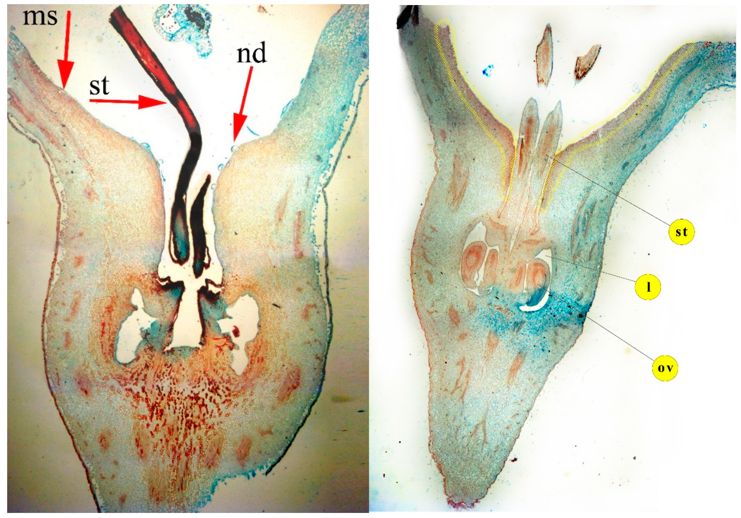
Floral nectary of Pyrus communis was situated on the adaxial surface of a funnel-like floral receptacle, lining the adaxial side of the plate-like hypanthium protruding out of the receptacular tissue at the apical part, almost reaching the circular rim along which the filament bases are attached. Moreover, to the opposite side, its large part extends down the walls of the narrow tubular cavity surrounding the style bases, reaching the apical part of the ovary (Figure 1 and Figure 2).
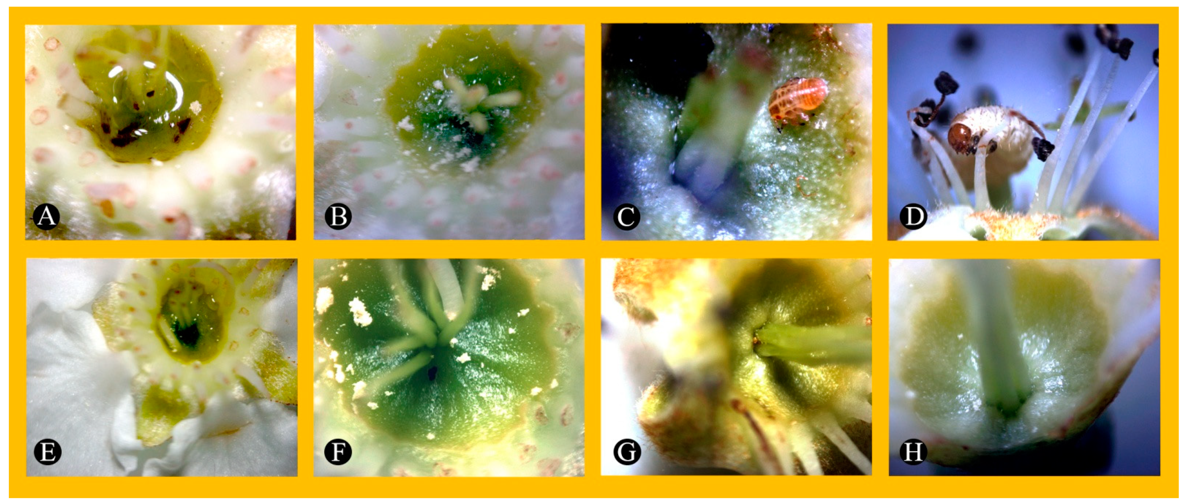
According to Farkas and Orosz-Kovács [30] the nectaries in pear flowers are receptacular–ovarial, and the nectariferous tissue can also extend along the style, even to the very base of the style in some cultivars. Since nectary stomata are abundant all over this glandular surface, the secreted nectar could be stored in this cup-shaped structure of hypanthium (Figure 3). The accumulated secretion product is very attractive for pollinating insects searching for energy food, simultaneously, however, increasing the chance of infection by bacteria, e.g., Erwinia amylovora.
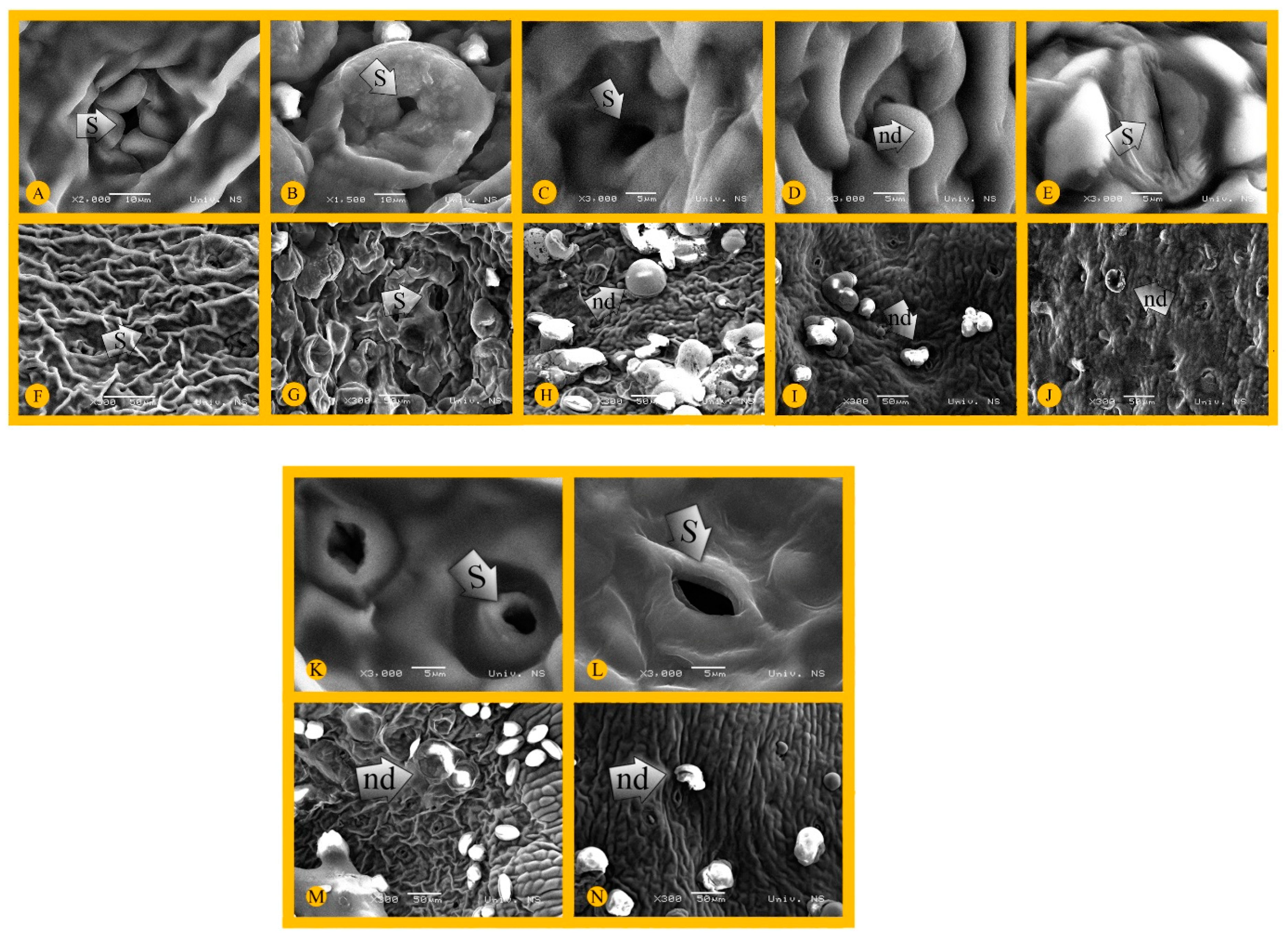
Observation under SEM has revealed the differences among cultivars in the surface structure of nectary relating to the cuticle patterns, the shape of epidermal cells, the appearance and distribution of stomata. Scanning electron microscopy also allowed nectar droplets to be observed in different phases of secretion (Figure 4 and Figure 5).
The nectary is protected by a layer of relatively thin and smooth cuticle covering the outer convex surface of the epidermal cells, usually pentagonal or hexagonal in shape, making the gland surface slightly wrinkled, especially in Alexander Lucas, Kieffer and Nijisseiki (Figure 5F–H,M). The modified stomata, throughout which nectar is secreted, are commonly situated in smaller or larger depressions, and distributed evenly over the entire nectary surface (Figure 4 and Figure 5F–J,M,N). Nectar droplets were often evident in the stomatal area when observed both under LM and SEM (Figure 3, Figure 5 and Figure 6).
In many cases, traces of dried nectar remained on the nectary surface primarily around the stomata in various shapes, usually as deflated balloons, discuss-like forms or rings (Figure 5B,G–J,M,N). Apparently, the viscosity of the nectar contributed to this, which, taking into account the drop volume, the slope of the contact surface and the air humidity, could reduce the speed of the drops flowing down the cup-like glandular surface of the hypanthium. Additionally, the highest amount of nectar droplets covering almost the entire nectar surface was noticeable in Chojuro, compared to the other cultivars. This could be explained, at first, by the presence of numerous stomata responsible for nectar secretion. By observing a freshly picked flower under SEM, it was even possible to witness the process of secretion, which included the appearance of a nectar drop pushed through the stomata opening, increasing to a critical volume, followed by the disintegration of the spherical form and by flowing down the surface (Figure 5D).

3.2. Nectary Anatomy (Histological Analysis)

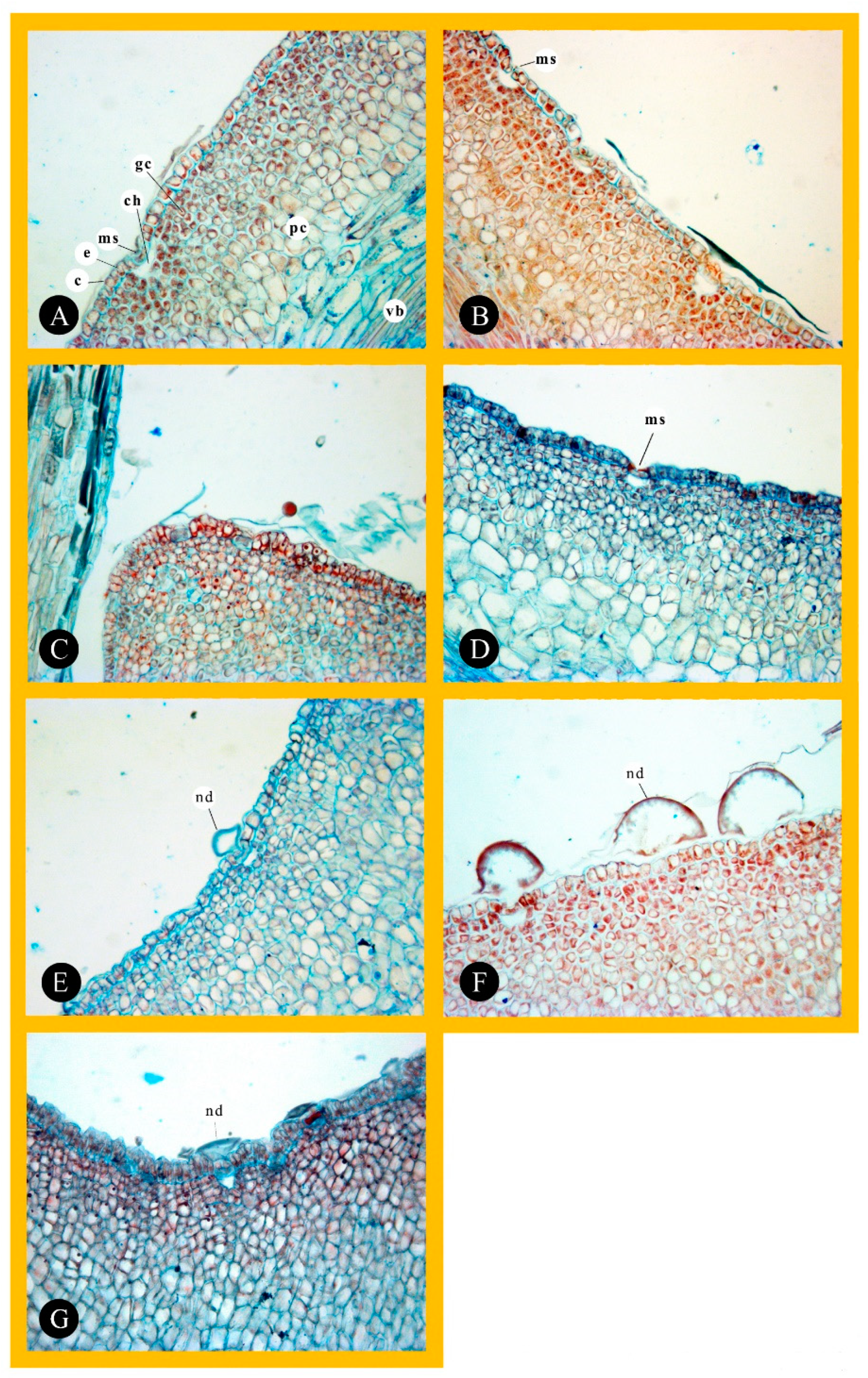
An examination of longitudinal sections of pear flowers allowed the position of the nectaries to be specified and the glandular tissue to be analyzed in detail. The nectariferous tissue is composed of a single-layered epidermis overlying several layers of small polygonal cells forming subepidermal secretory tissue (Figure 2, Figure 3 and Figure 6). The obvious transition in the size of the cells of which the hypanthium tissue is made up indicates the border between the glandular and non-glandular tissue. The measurement results are shown in Table 1.
In the medial longitudinal section of the floral nectary, the cells making up the one-layered epidermis are consistent in shape and palisad-like, and are usually covered by a folded cuticle. These cells are much elongated perpendicularly to the gland surface in Williams’, Chojuro, Alexander Lucas and Kieffer, but this is most evident in Nijisseiki (Figure 6). Hence, of all analyzed pear cultivars, the last one had the longest and most narrow epidermal cells being an average 37.47 μm in length, and 14.09 μm in width. Nevertheless, the length of epidermal cells in Alexander Lucas and Williams’ are nearly the same size, averaging 33.40 and 33.49 μm, respectively. The length/thickness quotient, which also characterizes the shape of the epidermal cells, was always high (above 1), ranging from 1.37 in Williams and Bella di Giugno where cell shape was closest to isodiametric, 2.66, in Nijisseiki indicating the narrow elongated cell shape. Modified stomata are present in the epidermis in all cultivars indicating the type of secretion. They are fairly uniformly distributed over the entire upper hypanthium surface, also descending down the tubular part of the hypanthium that surrounds the base section of the styles (Figure 5 and Figure 6). SEM shows two main types of stomata. The position of the nectary stomata in relation to epidermal cells’ level refers to the ecological type the cultivar belongs to. In the majority of the cultivars, the guard cells were in the same level as the epidermis indicating mesomorphy, but sometimes sunken and located in the funnel-shaped depressions of the nectariferous tissue such as in Poire de Cure and Williams (Figure 5I,J). These cultivars belong to the xeromorphic ecological type. Wavy cuticular ornamentation has been found in Nijisseiki, Poire de Cure and Chojuro, and the cuticle is more striated (wrinkled) in Kieffer, Alexander Lucas, Williams and Bella di Guigno (Figure 5). According to Nagy Tóth et al. [47], the apple cultivars susceptible to E. amylovora have sunken stomata in the epidermis and their nectaries are covered by wrinkled cuticles. The sunken stomata and the folds of the cuticle retain nectar, where bacterium of E. amylovora can multiply. The same authors concluded that if the cultivars have mesomorphic stomata and their nectary surface has less ornamentation, then they are resistant to E. amylovora. In our study, no clear line can be made, because only Williams had sunken stomata and a wrinkled epidermis, but the rest of the cultivars had different resistance combined with different stomata position/nectary ornamentation.
Stomata, composed of a pair of specialized epidermal cells, also varied widely in size, with mean guard cell length ranging from 12.15 (Bella di Giugno) to 37.85 μm (Kieffer). Cultivars Alexander Lucas and Williams’ had stomata nearly the same size, averaging 19.00 and 19.01 μm, respectively. Generally, there is a greater chance of bacteria attack if more and larger stomatal pores are on the surface [48]. Yet, in this study, resistant cultivars had larger guard cells compared to susceptible, which, according to Davis and Gunning [49], has no connection with the quantity of secretion. Stomata are mostly open and only rarely can contract sufficiently to close their pores. The guard cells in modified stomata have no ability to regulate the nectar flow but sometimes may become occluded. Regarding so, maybe the size of the pore and its ability to close makes a clear line between resistant and susceptible cultivars.
Nectar droplets in various stages of secretion could be found above open stomata (Figure 3, Figure 5 and Figure 6). The secretory product in the form of bubble-like appearance moving towards the surface is very often observed. Sub-stomatal nectar chambers of varying sizes were found (Table 1), being triangular or having an irregular shape (Figure 6). These chambers are capable of storing nectar before secreting it to the surface. The accumulated nectar can ensure favorable conditions for Erwinia amylovora, which can potentially contribute to bacterial infection through the modified stomata. Since modified stomata are unable to regulate nectar secretion by changing the size of the stomatal pore, they can potentially be entry sites for bacterial invasion. This is in agreement with the authors who, studying the leaf stomatal response to bacterial infection, stated that apart from hydathodes, stomata and lenticels, natural surface openings through which bacterial pathogens such as E. amylovora have been observed to enter the plant, also include nectarthodes referred to as nectar-secreting pores at the point of emergence of the styles and stamens [50].
The subepidermal secretory tissue is multi-layered, consisting of smaller cells with dark-staining protoplasts and round to oval or irregular in shape, in contrast with some other Rosaceae taxa (e.g., sweet and sour cherry), where the glandular cells are regular, mostly square-shaped and arranged in rows or columns [51]. The nectariferous cells differ from those of the adjacent multi-layered parenchyma that accompany the conductive vessels by their smaller size and denser cytoplasm. However, in most cultivars only a few subepidermal cell rows comprised a continuous glandular tissue, whereas the deeper region below the nectariferous tissue is made of much larger non-glandular parenchymal cells (Figure 6). Non-glandular parenchymal cells of the hypanthium were fairly consistent in shape but not in size. The largest ones were in Nijisseiki, averaging 77.44 μm in length and 47.79 μm in width, while Bella di Giugno (46.31 μm in length and 31.64 μm in width) and Williams (51.03 μm in length and 30.71 μm in width) had the smallest ones. (Table 1). This all means that resistant cultivars had larger hypanthium cells compared to susceptible. In the case of E. amylovora, the bacteria’s population is mostly developed on the stigma, but due to the rain, it migrates towards the hypanthium via the stylar groove, suggesting a conserved infection [52,53]. Rain not only speeds up movement of E. amylovora into the hypanthium, but also makes improper hypanthium surfaces feasible for bacterial growth [54]. In that way, cell wall thickness, the number of layer and compactness of cells in this tissue might be important for the resistance/susceptibility to fire blight.
The glandular tissue in pear cultivars could be well distinguished from the non-glandular parenchyma, compared with some other Maloideae taxa, where small, dark-staining glandular cells are mixed with the larger, light-staining parenchymatous cells, yielding a mosaic-like structure [30].
The vascular bundles were not seen to approach the nectariferous tissue, but are separated from them with several layers of larger subglandular parenchyma cells (Figure 6). It can be assumed that pre-nectar compounds are delivered by some other type of transport, e.g., simplast.

3.3. Sugars Analysis

Nectar is derived from the phloem of the plant, secreted in the nectaries and used to attract insects to flowers and give caloric and nutritional value for pollinators [55]. The pear nectar is a translucent, watery and sugar-rich solution. In this study, the sum of all quantified sugars (Table 2) in all seven pear cultivars ranged from 473 mg/g (Williams’–susceptible) up to 673 mg/g (Nijisseiki–highly resistant). This is much higher than it was when obtained by other authors [56,57] who determined the sugar content of pear nectar to be less than 10%, but more closely to quince (9.0–47.5%), cherry (12–65%) and peach nectar (often >50%) [21]. A statement done by Hevesi et al. [36], who claimed that high concentrations of sugars inhibit the multiplication of E. amylovora, is fully applicable to our results because the most susceptible cultivar has the lowest level of sugars, while resistant ones have a much higher level.
As expected, the most abundant were glucose and fructose (Table 2), followed by sorbitol and sucrose. Glucose ranged from 166.078 (Williams’) up to 212.886 mg/g (Chojuro), while fructose was the lowest in Williams’ (184.544 mg/g) and the highest in Nijisseiki (215.677 mg/g). Sugar alcohol sorbitol ranged from 82.330 mg/g (Bella di guigno) to 186.326 mg/g (Nijisseiki). Sucrose was the lowest in Bella di guigno (10.604 mg/g) and the highest in Poire de Cuire (39.873 mg/g). No matter that Braun and Hildebrand [58] pointed out that low concentrations or an absence of sucrose represent a defense mechanism of R. idaeus against E. amylovora, in this study, Asian pear cultivars that are highly resistant to this pathogen had almost a 2-times higher level of sucrose compared to the susceptible cultivar Williams’. Glucose accounted from 31.32% (Nijisseiki) to 37.29% (Bella di guigno), and fructose from 31.89% (Kieffer) to 39.01% (Williams’). Sorbitol percentage ranged from 17.82% (Williams’) up to 28.88% (Kieffer). Sucrose was around 2–3%, except in Poire de Cure where it was estimated as 6.70%. A low level of sucrose is one of the reasons why pear flowers are not in the favor of bees because these pollinators prefer sucrose over all other sugars, namely: arabinose, xylose, fructose, glucose, galactose, mannose, lactose, maltose, melibiose, trehalose, raffinose and melezitose [59]. The total percentage of the four most abundant sugars (glucose, fructose, sucrose and sorbitol) reached from 93.28% (Chojuro) to 96.14% (Kieffer) of all quantified sugars. These results were quite expected since simple sugars, glucose and fructose and the sugar alcohol sorbitol are the most common in the Rosaceae family [60].
The fructose-to-glucose (F/G) ratio in studied pear cultivars was almost 1:1, while the level of glucose or fructose was from ~5 to ~14-fold higher, respectively, than sucrose level. On the basis of the concentration of sucrose (S) and its relationship with fructose (F) and glucose (G), Baker and Baker [61] distinguished four types of nectar: hexose dominant S/(G + F) < 0.1, hexose rich S/(G + F) = 0.1–0.49, sucrose rich S/(G + F) = 0.5–0.99 and sucrose dominant: S/(G + F) > 1. According to this, all pear nectars from this study were ‘hexose dominant’. The results obtained in the present study coincided with the findings of Farkas et al. [62] who studied the nectar of several pear cultivars, and proved that their secretory product contained high concentrations of hexoses, but no sucrose. Besides, Nagy Toth et al. [33] studied the composition of floral nectar in pome Rosaceae fruits (apple, pear and quince) and proved that the presence of glucose and fructose is a general characteristic, but that sucrose is also present in some cultivars. A similar situation is in apricot and peach [63], raspberry [27] and blackberry nectar [21]. On the contrary, in the sour cherry nectar, glucose, fructose and sucrose were of the ratio 1:1:1 [24], which is most preferable by bees.
Other minor sugars, such as the monosaccharides: arabinose, ri-bose, rhamnose, galactose, xylose; the disaccharides: maltose, isomalt-ose, trehalose, melibiose, gentobiose, turanose; the trisaccharides: pa-nose, raffinose, melezitose, maltotriose; tetrasaccharide: stachyose; as well as the sugar alcohols: erythritol, galactitol and mannitol, were detected in the nectar of all the studied pear cultivars. Some of these minor nectar sugars can have a negative effect to insects by acting as a deterrent, nutritionally unsuitable, or even as toxic [64].
If we compare susceptible (Williams’), moderately resistant (Bella di guigno, Poire de Cuire and Alexander Lucas) and very high resistant (Chojuro, Nijisseiki and Kieffer) cultivars, as the resistance was increasing the levels of sorbitol, galactitol, glucose, fructose, maltose, melibiose, raffinose, melesitose, stachyose and galactose increased too. Galactose, a monosaccharide that is 65% as sweet as sucrose and was already quantified in Asian pears [65], was 2-fold higher in resistant compared to susceptible cultivars. Additionally, isomaltose, maltose and turanose were ~2.2-fold, ~3.35-fold and 2.7 fold, respectively, higher in Asian cultivars. Maltose is pretty rare or absent in nectars, and it is not so attractive for Apis melifera [34]. The level of melezitose was ~4.4 folds higher in highly resistant cultivars compared to Williams’. Melezitose, as an ant attractant or parasitoid repellent, is more common in honey-dews, and is less attractive for nectar-feeding insects [66]. The largest difference was between highly resistant cultivars and Williams’ in regard to melibiose, where Asian cultivars had ~12.8 fold higher than the latter one. According to Lingner et al. [67], melibiose is typically an intracellular carbohydrate and not released into the plant apoplast for the supply of adjacent cells or tissues. Although Hevesi et al. [36] proved that neither melezitose nor melibiose are utilized by E. amylovora, the high concentration of those two sugars is building up the refraction of the nectar and thus inhibiting the growth of bacteria. Oppositely, when the resistance to Erwinia was increasing, the level of trechalose, arabinose, ribose and xylose decreased. In that way, xylose was ~2.1-fold higher in the susceptible cultivar. In addition, almost the same level of panose between resistant and susceptible cultivars was determined too.

3.4. Identification of Pear Nectar Polyphenolics

Phenolic content in nectar has several roles. It can improve reproductive traits, have detrimental effects to pollinators, be repellent to some visitors and defend against attack by herbivores and microorganisms. Since phenolics are having fluorescent properties, those compounds in nectar might serve as a guide for pollinators, especially honeybees [68]. Besides, phenolic substances are volatiles compounds that are attracting pollinators [69,70]. So far, quercetin, kaempferol, luteolin, naringenin, cinnamic, vanillic and caffeic acid have been already reported as the most important phenols of floral nectar [34].
A total of twenty nine polyphenolic compounds were identified, and among them, seven compounds were identified by comparing retention times and MS spectra with available standards, while another twenty two were tentatively identified using previously reported chromatographic and MS fragmentation data found in literature. Identified compounds can be divided into two structurally different groups: (1) phenolic acid derivatives (12 compounds) and flavonoid glycosides (17 compounds). Data on the retention times (tR, min), molecular for-mula, exact and accurate masses ([M–H], m/z), mass accuracy errors (mDa) and major MS3 fragment ions of the identified polyphenolic compounds are summarized in Table 3.
Among phenolic acids, the most common were compounds from the group of hydroxycinnamnic acids, more precisely esters of these acids with quinic acid. Thus, for example, three compounds (1, 8, and 9) with a molecular ion at 337 m/z were identified as p-coumaroylquinic acids. The exact esterification position of these compounds was confirmed by MS fragmentation information [43].
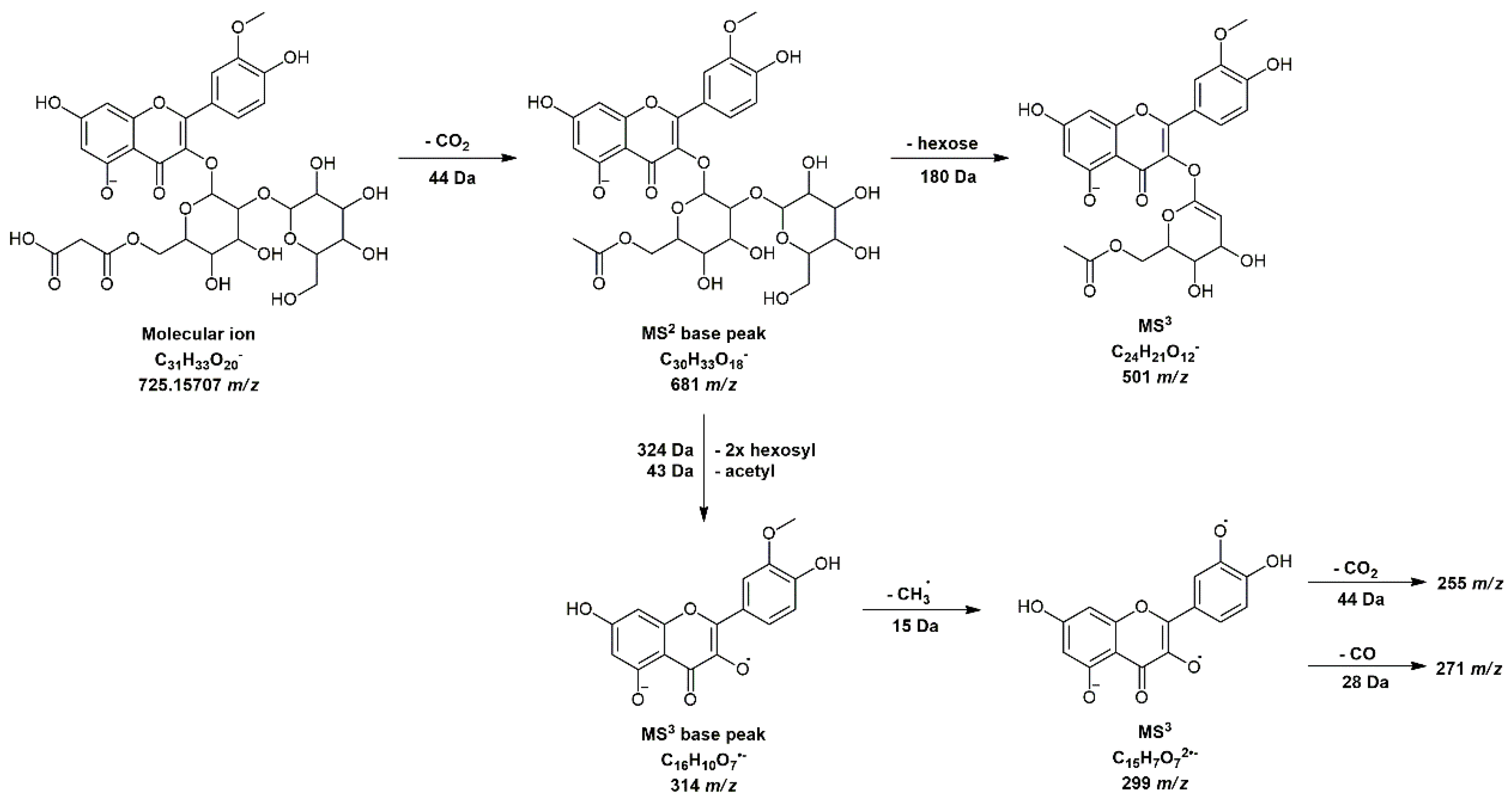
Regarding flavonoid glycosides, 15 compounds were found to be derivatives of the subgroup flavonols (isorhamnetin, patuletin, querce-tin and syringetin), and two (luteolin 7-O-(6″-rhamnosyl)-hexoside-compound 18 and apigenin 7-O-glucoside-compound 24) were from the subgroup of flavones. Flavones are giving yellow coloration of pear fruits and pear nectar [71]. Compound 16 with molecular ion 725 m/z was marked as isorhamnetin 3-O-(2″-hexosyl-6″-malonyl)-hexoside. The MS2 base peak fragment at 681 m/z was generated by the loss of 44 Da (CO2). Evidence that in this case two hexose molecules were bound by 1→2 interglycosidic linkage was a presence of a fragment at 501 m/z (Figure A1), which was generated by the further loss of 180 Da [72]. Several derivatives of pentahydroxy-methoxyflavone were also identified in our pear nectar extracts and such compounds were designated as derivatives of patuletin. For example, compound 20 with 579 m/z was marked as patuletin 3-O-(6″-malonyl)-hexoside. The detailed fragmentation pathway and molecular structure of this compound are shown in Figure A2. In this case, the MS3 fragment at 181 m/z was found to be a significant ion, which is known to be a characteristic RDA fragment for flavonoids that are methoxylated on the A ring, which is also the case with patuletin [73].
Table 4 shows the individual polyphenolic content (µg/mL) determined by LC/MS. Only four components were quantified in all seven pear cultivars and those are: p-hydroxybenzoic acid (from the group of phenolic acids), and isorhamnetin 3-O-hexoside, isorhamnetin 3-O-(6″-malonyl)-hexoside and syringetin 3-O-(6″-malonyl)-hexoside, from the group of flavonoid glycosides. These results were expected since p-hydroxybenzoic acid is a chemical marker for pears [74], while Brahem et al. [75] determined isorhamnetin glycosides in European and Tunisian pear cultivars. The sum of quantified phenolic acids varied from 0.049 (Williams’) up to 4.074 µg CAE/mL (Kieffer). As a matter of fact, resistant pear cultivars to E. amylovora had a ~5.5-fold higher level of phenolic acids. The most abundant phenolic acid was dicaffeoylquinic acid, which was ~9-fold times higher in resistant than in susceptible cultivars. According to Li et al. [76] and Lee et al. [77], dicaffeoylquinic acid has already been detected in immature fruits and fruit peels of Asian pears. Yet, the highest difference between those two groups of pear cultivars was for the 5-O-caffeoylquinic acid isomer, in which resistant cultivars stored ~9.7-fold higher than the susceptible. Caffeic acid and trimethylellagic acid hexoside was absent in susceptible cultivars. According to Horváth et al. [78], pear flowers contain phloretin, rutin, chlorogenic acid and caffeic acid, detected by thin layer chromatography. In this study, cultivar Kieffer (which is an interspecies hybrid between European and Asian pears and resistant to fire blight), stood out due to their high level of chlorogenic (5-O-caffeoylquinic) acid, while other cultivars had much lower and statistically the same level of this compound. Contrary to this, leaves of the same European pear cultivars with moderate resistance to Psylla stored a much higher level of cholorogenic acid than the Asian cultivars which were much more resistant to this insect [43]. Additionally, Cui et al. [79] and Gunen et al. [80] reported that European pears are a much better source of this compound. It seems that the genes responsible for the synthesis of chlorogenic acid have an additive effect, and that is the reason why Kieffer had the highest level of this hydroxycinnamic acid. Similar findings could be concluded for p-hydroxybenzoic acid, which was the highest in Kieffer, compared to the other pear cultivars. Many flavonoids and their glycosides are present in nectar and pollen across many plant species [24,81,82]. In this study, flavonoid glycosides’ levels ranged was from 1.224 (Williams) up to 11.686 µg RE/mL (Nijisseiki), and resistant cultivars stored ~1.6-fold higher of flavonoid glycosides compared to susceptible genotypes. The most abundant was isorhamnetin 3-O-(6″-malonyl)-hexoside, which was already detected in apple fruits [83] and it used to be the main glycoside in Oblačinska sour cherry nectar [24]. Isorhamnetin 3,7-di-O-hexoside was not detected in resistant cultivars while rutin, apigetrin, patuletin 3-O-(6″-hexosyl)-malonyl-hexoside, patuletin 3-O-(6″-malonyl)-hexoside, isorhamnetin 3-O-(2″-rhamnosyl)-hexoside and luteolin 7-O-(6″-rhamnosyl)-hexoside were not detected in susceptible pear cultivars. Rutin is one of the most important flavonoids in pear fruits [76]. The presence of rutin indicates a high biological and nutritional quality of studied nectar due to its high antioxidant activity [84]. Moreover, rutin successfully protects the bees against impairments produced by the acute and chronic application of insecticides [85]. In the previous studies of Fotirić Akšić et al. [42], it was shown that leaves of P. communis cultivars, that are susceptible to Psylla, stored ~90% more rutin than resistant cultivars originated from P. pyrifolia. Apigetrin was previously found in Pyrus communis calluses [86]. It has an antimicrobial effect and it contributes to higher stress tolerance [87]. Flavonoid patuletin and its glycosides are very rare in nature and could be found in spinach and flowers of Tagetes erecta [88,89,90]. Additionally, they have already been identified as the UV absorbing and reflecting compounds in the nectar guides of Rudbeckia hirta and increase the efficiency of pollination [91]. It possesses antibacterial activity against Bacillus spp., Micrococcus sp., Staphylococcus spp. and Streptococcus spp. [92]. In the case of when flavonoid glycosides were present in both groups of pear cultivars, the largest difference was noticed for isorhamnetin 3-O-(6″-rhamnosyl)-hexoside, and susceptible cultivars stored ~9.6-fold higher level than in the other. Isorhamnetin 3-O-(2″-rhamnosyl)-hexoside has been detected in Serbian bee-pollen [93], while luteolin glycosides were already determined in fruits of pear cultivars [94].

4. Conclusions

The initial assumption of this study was that a comparative morpho-anatomical analysis of nectaries would reveal structural changes that could be connected with the tolerance or susceptibility in pear cultivars to fire blight. The results indicated that obtained differences are in the quantitative rather than in the qualitative sense. In that way, the elongated shape of epidermal cells, large hypanthium cells and larger guard cells of the modified stomata were found in resistant cultivars, while the isodiametric shape of epidermal cells, smaller guard cells and smaller hypanthium cells were present in susceptible cultivars. Namely, differences in the measured parameters are small but statistically significant, due to a very large sample size, but are not enough to be attributed to a specific cause. Resistant cultivars had a much higher level of sugars and polyphenols in nectar. Sorbitol, galactose, isomaltose, turanose, maltose, melibiose and melesitose quantity was much lower in susceptible cultivars. Rutin, apigetrin, patuletin and luteolin glycosides were just detected in resistant pear cultivars.
The biggest question is whether resistant cultivars developed such a specific anatomic and/or biochemical answer due to the fire blight attack, or if they are resistant because of their specific anatomy and sugar and polyphenolic metabolism which was developed a long time ago? Because of all of this, further investigations should focus on inter-cultivar specific differences in floral anatomy, and other biochemical components in pear nectar that could be in association with resistance to fire blight. All together, this can help fruit breeders in the selection of promising genotypes.
In addition, this research contributed to the taxonomy by considering that anatomical and micro-morphological features of the floral nectaries could be used as discriminative characters at the species level and represents a significant contribution to differentiation from other congeneric and related species.

Author Contributions

Conceptualization, M.F.A., M.M.-J. and N.N.; methodology, Ž.T., U.G., T.T., M.F.A., R.R. and M.M.-J.; formal analysis, U.G., T.T., M.M.-J. and R.R.; writing, M.F.A., M.M.-J., U.G., T.T. and M.M.; review and editing, Ž.T., M.F.A., M.M.-J. and M.M.; supervision, M.M. All authors have read and agreed to the published version of the manuscript.

Funding

This research was funded by The Research Council of Norway, project no. 244510.

Data Availability Statement

Not applicable.

Acknowledgments

The authors would like to thank The Ministry of Education, Science and Technological Development of the Republic of Serbia (contract number 451-03-47/2023-01/200116). The authors would like to thank Miloš Bokorov from University Center for Electron Microscopy, Novi Sad, for his technical assistance and SEM microscopy.

Conflicts of Interest

The authors declare no conflict of interest.

Appendix A

Figure A1. Fragmentation pathway of isorhamnetin 3-O-(2″-hexosyl-6″-malonyl-hexoside (compound 16).
Horticulturae 09 00424 g0a1
Figure A2. Fragmentation pathway of patuletin 3-O-(6″-malonyl)-hexoside (compound 20).
Horticulturae 09 00424 g0a2

References

  1. Hancock, J.F.; Lobos, G.A. Pears. In Temperate Fruit Crop Breeding: Germplasm to Genomics; Hancock, J.F., Ed.; Springer: Dordrecht, The Netherlands, 2008; pp. 299–335. [Google Scholar] [CrossRef]
  2. Bao, L.; Chen, K.; Zhang, D.; Cao, Y.; Yamamoto, T.; Teng, Y. Genetic diversity and similarity of pear cultivars native to East Asia revealed by SSR (simple sequence repeat) markers. Genet. Resour. Crop Evol. 2007, 54, 959–971. [Google Scholar] [CrossRef]
  3. FAOStat. 2023. Available online: https://www.fao.org/faostat/en/#data/QCL (accessed on 22 January 2023).
  4. Itai, A. Pear. In Fruits and Nuts. Genome Mapping and Molecular Breeding in Plants; Kole, C., Ed.; Springer: Berlin/Heidelberg, Germany, 2007; Volume 4, pp. 157–171. [Google Scholar] [CrossRef]
  5. Jiang, Z.; Tang, F. Assessment of genetic diversity of Chinese sand pear landraces (Pyrus pyrifolia Nakai) using simple sequence repeat markers. Hortic. Sci. 2009, 44, 619–626. [Google Scholar] [CrossRef] [Green Version]
  6. Fotirić Akšić, M.; Cerović, R.; Radošević, R.; Oparnica, Č.; Meland, M. Morphological and anatomical leaf characteristics of some European and Asian pear cultivars. Acta Hortic. 2021, 1303, 63–70. [Google Scholar] [CrossRef]
  7. Reiland, H.; Slavin, J. Systematic review of pears and health. Nutr. Today 2015, 50, 301–305. [Google Scholar] [CrossRef] [PubMed] [Green Version]
  8. Meland, M.; Fotirić Akšić, M.; Cerović, R.; Frøynes, O.; Kaiser, C.; Maas, F. Evaluation of New Promising Norwegian Pear Cultivars in a Nordic Climate. J. Am. Pom. Soc. 2021, 75, 149–156. [Google Scholar]
  9. Przybyla, A.A.; Bokszczanin, K.L.; Schollenberger, M.; Gozdowski, D.; Madry, W.; Odziemkowski, S. Fire blight resistance of pear genotypes from different European countries. Trees 2012, 26, 191–197. [Google Scholar] [CrossRef] [PubMed] [Green Version]
  10. Hevesi, M.; Göndör, M.; Kása, K.; Honty, K.; Tóth, M.G. Traditional and commercial apple and pear cultivars as sources of resistance to fire blight. EPPO Bull. 2004, 34, 377–380. [Google Scholar] [CrossRef]
  11. Thibault, B.; Paulin, J.P. Pear breeding and selection for fire blight resistance. Acta Hortic. 1984, 161, 141–146. [Google Scholar] [CrossRef]
  12. Malnoy, M.; Martens, S.; Norelli, J.L.; Barny, M.; Sundin, G.W.; Smits, T.H.M.; Duffy, B. Fire blight: Applied genomic insights of thepathogen and host. Annu. Rev. Phytopathol. 2012, 50, 475–494. [Google Scholar] [CrossRef] [PubMed] [Green Version]
  13. Griffith, C.S.; Sutton, T.B.; Peterson, P.D. Fire Blight: The Foundation of Phytobacteriology; American Phytopathological Society: St. Paul, MN, USA, 2003; pp. 1–158. [Google Scholar]
  14. Piqué, N.; Miñana-Galbis, D.; Merino, S.; Tomás, J.M. Virulence factors of Erwinia amylovora: A review. Int. J. Mol. Sci. 2015, 16, 12836–12854. [Google Scholar] [CrossRef] [Green Version]
  15. Bubán, T.; Orosz-Kovács, Z. The nectary as the primary site of infection by Erwinia amylovora (Burr.) Winslow et al.: A mini review. Plant Syst. Evol. 2003, 238, 183–194. [Google Scholar] [CrossRef]
  16. Seneta, W.; Dolatowski, J. Dendrologia. Wyd; Naukowe PWN: Warszawa, Poland, 2009; pp. 1–544. [Google Scholar]
  17. Déri, H.; Orosz-Kovács, Z.; Farkas, Á. Morphological characterization of the floral nectary in some apple-shaped and pear-shaped quince cultivars. Acta Bot. Hung. 2007, 49, 171–187. [Google Scholar] [CrossRef]
  18. Farkas, Á.; Orosz-Kovács, Z.; Déri, H.; Chauhan, S.V.S. Floral nectaries in some apple and pear cultivars with special reference to bacterial fire blight. Curr. Sci. 2007, 92, 1286–1289. [Google Scholar]
  19. Farkas, A. Morphology and histology of the nectary in Hungarian local pear cultivars. Acta Hortic. 2005, 671, 127–135. [Google Scholar] [CrossRef]
  20. Radice, S.; Galati, B. Floral nectary ultrastructure of Prunus persica (L.) Batch cv. Forastero (Newcomer), an Argentine peach. Plant Syst. Evol. 2003, 238, 23–32. [Google Scholar] [CrossRef]
  21. Farkas, Á.; Zajácz, E. Nectar production for the Hungarian honey industry. Eur. J. Plant Sci. Biotechnol. 2007, 1, 125–151. [Google Scholar]
  22. Bernardello, G. A systematic survey of floral nectaries. In Nectaries and Nectar; Nicolson, S.W., Nepi, M., Pacini, E., Eds.; Springer: Dordrecht, The Netherlands, 2007; pp. 19–128. [Google Scholar] [CrossRef]
  23. Liu, L.; Zhao, X.M. Ultrastructural study on apricot floral nectary. J. Fruit Sci. 2011, 5, 792–797. [Google Scholar]
  24. Guffa, B.; Nedić, N.; Dabić Zagorac, D.; Tosti, T.; Gašić, U.; Natić, M.; Fotirić Akšić, M. Characterization of Sugar and Polyphenolic Diversity in Floral Nectar of Different ‘Oblačinska’ Sour Cherry Clones. Chem. Biodivers. 2017, 14, e1700061. [Google Scholar] [CrossRef]
  25. Weryszko-Chmielewska, E.; Masierowska, M.; Konarska, A. Characteristics of floral nectaries and nectar in two species of Crataegus (Rosaceae). Plant Syst. Evol. 2003, 238, 33–41. [Google Scholar] [CrossRef]
  26. Weryszko-Chmielewska, E.; Sulborska-Różycka, A.; Sawidis, T. Structure of the nectary in Chaenomeles japonica (Thunb.) Lindl. ex Spach. in different stages of flowering with focus on nectar secretion. Protoplasma 2022, 259, 1467–1476. [Google Scholar] [CrossRef]
  27. Kostryco, M.; Chwil, M. Nectar Secretion, Morphology, Anatomy and Ultrastructure of Floral Nectary in Selected Rubus idaeus L. Varieties. Agriculture 2022, 12, 1017. [Google Scholar] [CrossRef]
  28. Rubtsova, O.L.; Vakulenko, T.B.; Chyzhankova, V.I. Morphological features of nectaries of some species of the genus Rosa (Rosaceae). Ukr. Bot. J. 2022, 79, 103–113. (In Ukrainian) [Google Scholar] [CrossRef]
  29. Konarska, A.; Masierowska, M.; Weryszko-Chmielewska, E. The structure of nectaries and nectar secretion in common pear (Pyrus communis L.). J. Apic. Sci. 2005, 49, 85–92. [Google Scholar]
  30. Farkas, A.; Orosz-Kovács, Z.S. Primary and secondary attractants of flowers in pear Pyrus betulifolia. Acta Hortic. 2004, 636, 317–324. [Google Scholar] [CrossRef]
  31. Farkas, Á.; Orosz Kovács, Z.; Bubán, T. Floral biological studies on pear cultivars in relation to fire blight susceptibility. Int. J. Hortic. Sci. 2004, 10, 25–30. [Google Scholar] [CrossRef]
  32. Canto, A.; Herrera, C.M.; Medrano, M.; Perez, R.; Garcia, I.M. Pollinator foraging modifies nectar sugar composition in Helleborus foetidus (Ranunculaceae): An experimental test. Am. J. Bot. 2008, 95, 315–320. [Google Scholar] [CrossRef] [PubMed] [Green Version]
  33. Nagy Tóth, E.; Szabó, L.G.; Botz, L.; Orosz- Kovács, Z. Effect of rootstocks on floral nectar composition in apple cultivars. Plant Syst. Evol. 2003, 283, 43–55. [Google Scholar] [CrossRef]
  34. Nicolson, S.W.; Thornburg, R.W. Nectar chemistry. In Nectaries and Nectar; Nicolson, S.W., Nepi, M., Pacini, E., Eds.; Springer: Dordrecht, The Netherlands, 2007; pp. 215–264. [Google Scholar] [CrossRef]
  35. Heil, M. Nectar: Generation, regulation and ecological functions. Trends Plant Sci. 2011, 16, 191–200. [Google Scholar] [CrossRef]
  36. Hevesi, M.; Farkas, A.; Kása, A.; Orosz-Kovács, Z. Carbohydrate utilization of Erwinia amylovora in vitro. In. J. Hortic. Sci. 2004, 10, 31–34. [Google Scholar] [CrossRef]
  37. Pontais, I.; Treutter, D.; Paulin, J.P.; Brisset, M.N. Erwinia amylovora modifies phenolic profiles of susceptible and resistant apple through its type III secretion system. Physiol. Plant 2008, 132, 262–271. [Google Scholar] [CrossRef]
  38. González-Teuber, M.; Eilmus, S.; Muck, A.; Svatos, A.; Heil, M. Pathogenesis-related proteins protect extrafloral nectar from microbial infestation. Plant J. 2009, 58, 464–473. [Google Scholar] [CrossRef]
  39. Nogueira, F.C.S.; Farias, A.R.B.; Teixeira, F.M.; Domont, G.B.; Campos, F.A.P. Common Features Between the Proteomes of Floral and Extrafloral Nectar From the Castor Plant (Ricinus communis) and the Proteomes of Exudates From Carnivorous Plants. Front. Plant Sci. 2018, 9, 549. [Google Scholar] [CrossRef] [Green Version]
  40. Kurilla, A.; Toth, T.; Dorgai, L.; Darula, A.; Lakatos, T.; Silhavy, D.; Kerenyi, Z.; Dallmann, G. Nectar and stigma exudate-specific expression of an acidic chitinase could partially protect certain apple cultivars against fire blight disease. Planta 2020, 251, 20. [Google Scholar] [CrossRef] [Green Version]
  41. Nepi, M. Beyond nectar sweetness: The hidden ecological role of non-protein amino acids in nectar. J. Ecol. 2014, 102, 108–115. [Google Scholar] [CrossRef]
  42. Vranova, V.; Rejsek, K.; Skene, K.R.; Formanek, P. Non-proteinamino acids: Plant, soil and ecosystem interactions. Plant Soil 2011, 342, 31–48. [Google Scholar] [CrossRef]
  43. Fotirić Akšić, M.; Dabić, D.; Gašić, U.; Zec, G.; Vulić, T.; Tešić, Ž.; Natić, M. Polyphenolic Profile of Pear Leaves with Different Resistance to Pear Psylla (Cacopsylla pyri). J. Agric. Food Chem. 2015, 63, 7476–7486. [Google Scholar] [CrossRef] [PubMed]
  44. Ruzin, S.E. Plant Microtechnique and Microscopy; Oxford University Press: Oxford, UK, 1999; pp. 1–322. [Google Scholar]
  45. Miller, J.N.; Miller, J.C. Statistics and Chemometrics for Analytical Chemistry, 6th ed.; Pearson Education Limited: Harlow, UK, 2010; pp. 1–278. [Google Scholar]
  46. Šuković, D.; Knežević, B.; Gašić, U.; Sredojević, M.; Ćirić, I.; Todić, S.; Mutić, J.; Tešić, Ž. Phenolic profiles of leaves, grapes and wine of grapevine variety Vranac (Vitis vinifera L.) from Montenegro. Foods 2020, 9, 138. [Google Scholar] [CrossRef] [PubMed] [Green Version]
  47. Nagy Tóth, E.; Bubán, T.; Hevesi, M.; Orosz-Kovács, Z.; Szabó, L.G. Morphological characteristics of the nectary and composition of nectar in flowers of selected apple cultivars. Acta Hortic. 2000, 538, 301–308. [Google Scholar] [CrossRef]
  48. Zeng, W.; Melotto, M.; He, S.Y. Plant stomata: A checkpoint of host immunity and pathogen virulence. Curr. Opin. Biotechnol. 2010, 21, 599–603. [Google Scholar] [CrossRef] [Green Version]
  49. Davis, A.R.; Gunning, B.E.S. The modified stomata of the floral nectary of Vicia faba L. Development, anatomy and ultrastructure. Protoplasma 1992, 166, 134–152. [Google Scholar] [CrossRef]
  50. Melotto, M.; Underwood, W.; He, S.Y. Role of Stomata in Plant Innate Immunity and Foliar Bacterial. Annu. Rev. Phytopathol. 2008, 46, 101–122. [Google Scholar] [CrossRef] [PubMed] [Green Version]
  51. Orosz-Kovács, Z. Nectary structures in cherry cultivars. Acta Agron. Hung. 1993, 42, 239–253. [Google Scholar]
  52. Wilson, M.; Sigee, D.C.; Epton, H.A.S. Erwinia amylovora Infection of hawthorn blossom, II The stigma. J. Phytopath. 1989, 127, 1–14. [Google Scholar] [CrossRef]
  53. Spinelli, F.; Ciampolini, F.; Cresti, M.; Geider, K.; Costa, G. Influence of stigmatic morphology on flower colonization by Erwinia amylovora and Pantoea agglomerans. Eur. J. Plant Pathol. 2005, 113, 395–405. [Google Scholar] [CrossRef]
  54. Wilson, M.; Sigee, D.C.; Epton, H.A.S. Erwinia amylovora infection of hawthorn blossom: III. The Nectary. J. Phytopath. 1990, 128, 62–74. [Google Scholar] [CrossRef]
  55. Davis, A.R. Searching and breeding for structural features of flowers correlated with high nectar-carbohydrate production. Acta Hortic. 2001, 561, 107–121. [Google Scholar] [CrossRef]
  56. Faoro, I.D.; Orth, A.I. Flower visiting insects during the bloom period of Japanese pear orchards in Brazil. Acta Hortic. 2015, 1094, 275–279. [Google Scholar] [CrossRef]
  57. Jacquemart, A.-L.; Michotte-van der, A.A.; Raspé, O. Compatibility and pollinator efficiency tests on Pyrus communis L. cv. Conférence. J. Hortic. Sci. Biotechnol. 2006, 81, 827–830. [Google Scholar] [CrossRef]
  58. Braun, P.G.; Hildebrand, P.D.; Jamieson, A.R. Resistance of raspberry cultivars to fire blight. HortScience 2004, 39, 1189–1192. [Google Scholar]
  59. Seeburger, V.C.; D’Alvise, P.; Shaaban, B.; Schweikert, K.; Lohaus, G.; Schroeder, A.; Hasselmann, I.M. The trisaccharide melezitose impacts honey bees and their intestinal microbiota. PLoS ONE 2020, 15, e0230871. [Google Scholar] [CrossRef] [Green Version]
  60. Colaric, M.; Stampar, F.; Hudina, M. Changes in sugars and phenolics concentrations of Williams pear leaves during the growing season. Can. J. Plant Sci. 2006, 86, 1203–1208. [Google Scholar] [CrossRef]
  61. Baker, H.G.; Baker, I. Floral nectar sugar constituents in relation to pollinator type. In Handbook of Experimental Pollination Biology; Jones, C.E., Little, R.J., Eds.; Van Nostrand Reinhold: New York, NY, USA, 1983; pp. 117–141. [Google Scholar]
  62. Farkas, A.; Orosz-Kovács, Z.; Szabó, L.G. Insect attraction of flowers in pear cultivars. Acta Hortic. 2002, 596, 773–776. [Google Scholar] [CrossRef]
  63. Meheriuk, M.; Lane, W.D.; Hall, J.W. Influence of cultivar on nectar sugar content in several species of tree fruits. HortScience 1987, 22, 448–450. [Google Scholar] [CrossRef]
  64. Romeis, J.; Wäckers, F.L. Nutritional suitability of individual carbohydrates and amino acids for adult Pieris brassicae. Physiol. Entomol. 2002, 27, 148–156. [Google Scholar] [CrossRef]
  65. Li, L.; Wang, T.Q.; Xue, B.; Liu, Z.D.; Zou, L. Chemical Composition and Functional Characteristics of Dietary Fibers from Pingguoli Pear (Pyrus bretschneideri Rehd. cv. Pingguoli Pear). Curr. Top. Nutraceutical Res. 2020, 18, 39. [Google Scholar]
  66. Fischer, M.K.; Shingleton, A.W. Host plant and ants influence the honeydew sugar composition of aphids. Funct. Ecol. 2001, 15, 544–550. [Google Scholar] [CrossRef]
  67. Lingner, U.; Steffen, M.; Sode, B.; Deising, H.B.; Sauer, N. Functional Characterization of a Eukaryotic Melibiose Transporter. Plant Physiol. 2011, 156, 1565–1576. [Google Scholar] [CrossRef] [PubMed] [Green Version]
  68. Soto, V.; Silva, M.F.; Galmarini, C. Effect of Nectar Composition on Bee Attraction for Onion Seed Production. In Nectar—Production, Chemical Composition and Benefits to Animals and Plants, II. Series: Plant Science Research and Practices; Peck, R.L., Ed.; Nova Science Publishers, Inc.: Hauppauge, NY, USA, 2015; pp. 41–58. [Google Scholar]
  69. Deachathai, S.; Mahabusarakam, W.; Phongpaichit, S.; Taylor, W.C.; Zhang, Y.J.; Yang, C.R. Phenolic compounds from the flowers of Garcinia dulcis. Phytochemistry 2006, 67, 464–469. [Google Scholar] [CrossRef]
  70. del Baño, M.J.; Lorente, J.; Castillo, J.; Benavente-Garcia, O.; del Río, J.A.; Ortuño, A.; Quirin, K.W.; Gerard, D. Phenolic diterpenes, flavones, and rosmarinic acid distribution during the development of leaves, flowers, stems, and roots of Rosmarinus officinalis. Antioxidant activity. J. Agric. Food Chem. 2003, 51, 4247–4253. [Google Scholar] [CrossRef] [PubMed]
  71. Ni, J.; Zhao, Y.; Tao, R.; Yin, L.; Gao, L.; Strid, A.; Qian, M.; Li, J.; Li, Y.; Shen, J.; et al. Ethylene mediates the branching of the jasmonate-induced flavonoid biosynthesis pathway by suppressing anthocyanin biosynthesis in red Chinese pear fruits. Plant Biotechnol. 2020, 18, 1223–1240. [Google Scholar] [CrossRef] [Green Version]
  72. Ferreres, F.; Llorach, R.; Gil-Izquierdo, A. Characterization of the interglycosidic linkage in di-, tri-, tetra- and pentaglycosylated flavonoids and differentiation of positional isomers by liquid chromatography/electrospray ionization tandem mass spectrometry. J. Mass Spectrom. 2004, 39, 312–321. [Google Scholar] [CrossRef] [PubMed]
  73. Zengin, G.; Cvetanović, A.; Gašić, U.; Stupar, A.; Bulut, G.; Şenkardes, I.; Dogan, A.; Sinan, K.I.; Uysal, S.; Aumeeruddy-Elalfi, Y.; et al. Modern and traditional extraction techniques affect chemical composition and bioactivity of Tanacetum parthenium (L.) Sch.Bip. Ind. Crops Prod. 2020, 146, 112202. [Google Scholar] [CrossRef]
  74. Andrade, P.B.; Carvalho, A.R.F.; Seabra, R.M.; Ferreira, M.A. A Previous Study of Phenolic Profiles of Quince, Pear, and Apple Purees by HPLC Diode Array Detection for the Evaluation of Quince Puree Genuineness. J. Agric. Food Chem. 1998, 46, 968–972. [Google Scholar] [CrossRef]
  75. Brahem, M.; Renard, C.M.; Eder, S.; Loonis, M.; Ouni, R.; Mars, M.; Le Bourvellec, C. Characterization and quantification of fruit phenolic compounds of European and Tunisian pear cultivars. Food Res. Int. 2017, 95, 125–133. [Google Scholar] [CrossRef]
  76. Li, X.; Zhang, J.-Y.; Gao, W.-Y.; Wang, Y.; Wang, H.-Y.; Cao, J.-G.; Huang, L.-Q. Chemical composition and anti-inflammatory and antioxidant activities of eight pear cultivars. J. Agric. Food Chem. 2012, 60, 8738–8744. [Google Scholar] [CrossRef]
  77. Lee, K.H.; Cho, J.Y.; Lee, H.J.; Park, K.Y.; Ma, Y.-K.; Lee, S.-H.; Cho, J.A.; Kim, W.-S.; Park, K.H.; Moon, J.-H. Isolation and identification of phenolic compounds from an Asian pear (Pyrus pyrifolia Nakai) fruit peel. Food Sci. Biotechnol. 2011, 20, 1539–1545. [Google Scholar] [CrossRef]
  78. Horváth, G.; Farkas, A.; Szabó, G.L. Flavonoids, chalcones and phenyl-propanoids in apple and pear flowers. Int. J. Hortic. Sci. 2004, 10, 35–38. [Google Scholar] [CrossRef]
  79. Cui, T.; Nakamura, K.; Ma, L.; Li, J.; Kayahara, H. Analysis of arbutin and chlorogenic acid the major phenolic constituents in Oriental pear. J. Agric. Food Chem. 2005, 53, 3882–3887. [Google Scholar] [CrossRef] [PubMed]
  80. Gunen, Y.; Misirli, A.; Gulcan, R. Leaf phenolic content of pear cultivars resistant or susceptible to fire blight. Sci. Hortic. 2005, 105, 213–221. [Google Scholar] [CrossRef]
  81. Fotirić Akšić, M.; Gašić, U.; Dragana, D.Z.; Sredojević, M.; Tosti, T.; Natić, M.; Meland, M. Chemical Fingerprint of ‘Oblačinska’ Sour Cherry (Prunus cerasus L.) Pollen. Biomolecules 2019, 9, 391. [Google Scholar] [CrossRef] [Green Version]
  82. Kostić, A.; Milinčić, D.D.; Gašić, U.M.; Nedić, N.; Stanojević, S.P.; Tešić, Ž.L.; Pešić, M.B. Polyphenolic profile and antioxidant properties of bee-collected pollen from sunflower (Helianthus annuus L.) plant. LWT 2019, 112, 108244. [Google Scholar] [CrossRef]
  83. Schieber, A.; Keller, P.; Streker, P.; Klaiber, I.; Carle, R. Detection of isorhamnetin glycosides in extracts of apples (Malus domestica cv. “Brettacher”) by HPLC-PDA and HPLC-APCI-MS/MS. Phytochem. Anal. 2002, 13, 87–94. [Google Scholar] [CrossRef]
  84. Dias, L.G.; Tolentino, G.; Pascoal, A.; Estevinho, L.M. Effect of processing conditions on the bioactive compounds and biological properties of bee pollen. J. Apic. Res. 2016, 55, 357–365. [Google Scholar] [CrossRef] [Green Version]
  85. Riveros, A.J.; Gronenberg, W. The flavonoid rutin protects the bumble bee Bombus impatiens against cognitive impairment by imidacloprid and fipronil. J. Exp. Biol. 2022, 225, jeb244526. [Google Scholar] [CrossRef]
  86. Skočajić, D.; Gašić, U.; Dabić Zagorac, D.; Nešić, M.; Tešić, Ž.; Meland, M.; Fotirić Akšić, M. Analysis of Phenolic Compounds for the Determination of Grafts (in) Compatibility Using In Vitro Callus Cultures of Sato-Zakura Cherries. Plants 2021, 10, 2822. [Google Scholar] [CrossRef] [PubMed]
  87. Genzel, F.; Dicke, M.D.; Junker-Frohn, L.V.; Neuwohner, A.; Thiele, B.; Putz, A.; Usadel, B.; Wormit, A.; Wiese-Klinkenberg, A. Impact of Moderate Cold and Salt Stress on the Accumulation of Antioxidant Flavonoids in the Leaves of Two Capsicum Cultivars. J. Agric. Food Chem. 2021, 69, 6431–6443. [Google Scholar] [CrossRef] [PubMed]
  88. Abdel-Wahhab, M.A.; Said, A.; Antje, H. NMR and radical scavenging activities of patuletin from Urtica urens against aflatoxin B. Pharm. Biol. 2005, 43, 515–525. [Google Scholar] [CrossRef]
  89. Bergman, M.; Varshavsky, L.; Gottlieb, H.E.; Grossman, S. The antioxidant activity of aqueous spinach extract: Chemical identification of active fractions. Phytochemistry 2001, 58, 143–152. [Google Scholar] [CrossRef]
  90. Burlec, A.F.; Pecio, Ł.; Kozachok, S.; Mircea, C.; Corciovă, A.; Vereștiuc, L.; Cioancă, O.; Oleszek, W.; Hăncianu, M. Phytochemical Profile, Antioxidant Activity, and Cytotoxicity Assessment of Tagetes erecta L. Flowers. Molecules 2021, 26, 1201. [Google Scholar] [CrossRef] [PubMed]
  91. Abrol, D.P. Pollination Biology: Biodiversity Conservation and Agricultural Production; Springer: New York, NY, USA, 2012; pp. 413–458. [Google Scholar]
  92. Faizi, S.; Siddiqi, H.; Bano, S.; Naz, A.; Lubna, A.; Mazhar, K.; Nasim, S.; Riaz, T.; Kamal, S.; Ahmad, A.; et al. Antibacterial and Antifungal Activities of Different Parts of Tagetes patula.: Preparation of Patuletin Derivatives. Pharm. Biol. 2008, 46, 309–320. [Google Scholar] [CrossRef] [Green Version]
  93. Mosić, M.; Trifković, J.; Vovk, I.; Gašić, U.; Tešić, Ž.; Šikoparija, B.; Milojković-Opsenica, D. Phenolic Composition Influences the Health-Promoting Potential of Bee-Pollen. Biomolecules 2019, 9, 783. [Google Scholar] [CrossRef] [PubMed] [Green Version]
  94. Lin, L.Z.; Harnly, J.M. Phenolic compounds and chromatographic profiles of pear skins (Pyrus spp.). J. Agric. Food Chem. 2008, 56, 9094–9101. [Google Scholar] [CrossRef] [PubMed]
Figure 1. Longitudinal sections revealing the remaining parts of the reproductive structures and nectary contained within a flower of Kieffer cultivar (left). A comparative diagram of a longitudinal section of a flower’s receptacle showing a position of nectary (green) (right), (×2.5, LM). Abbreviations: s—sepal; st—style; ov—the inferior ovary; fb—filamentum base; h—hypanthium; vb—vascular bundle; ne—nectariferous tissue; o—ovule; p—a long pedicel the flower was carried on.
Horticulturae 09 00424 g001
Figure 2. Longitudinal section of a pear flower receptacle showing the position of nectary, modified stomata and secreted nectar droplets in Bella di Giugno (left) and Alexander Lucas (right) cultivars (×10; LM). Nectary tissue is framed in yellow (right). Abbreviations: nd—nectar droplet; ms—modified stomata; st—style; ov—ovules (2 per locule); l—locule.
Horticulturae 09 00424 g002
Figure 3. Transversally cut flowers of three pear cultivars showing nectar contained in plate-like nectariferous area of hypanthium The remaining basal parts of five petals, numerous stamens and five styles are noticeable. ((A) Chojuro; (B) Nijisseiki; (C,D) Bella di Giugno; (E) Alexander Lucas; (F) Willimas; (G) Poire de Cure; (H) Kieffer) (×1.5; stereomicroscopy).
Horticulturae 09 00424 g003
Figure 4. SEM micrographs showing plate-shaped structure of the hypanthium indicating nectariferous area inside pear cultivar flowers. (A) Kieffer (resistant) (×30), (B) Nijisseiki (resistant) (×50), (C) Poire de Cure (moderately resistant), (D) Bella di guigno (moderately resistant), (E) Williams (susceptible), (F) Alexander Lucas (low susceptible), (G) Chojuro (resistant).
Horticulturae 09 00424 g004
Figure 5. SEM micrographs of the nectary surface showing epidermis covered with cuticle focused on enlarged modified stomata in five cultivars comparatively: (A,F) Alexander Lucas (low susceptible), (B,G) Kieffer (highly resistant), (C,H) Nijisseiki (highly resistant); (D,I) Poire de Cure (moderately resistant); (E,J) Williams (susceptible); (K,M) Bella di guigno (moderately resistant); (L,N) Chojuro (resistant). (AE) close-up of the mostly open stomata; (B) dried nectar droplet; nd—nectar droplet in the initial phase of secretion. (FJ) a larger area of glandular epidermis showing modified stomata and frequent nectar droplets imaged at different stages of secretion.
Horticulturae 09 00424 g005
Figure 6. Fragments of longitudinal sections of a flower receptacle at the level of the nectariferous tissue of the hypanthium showing distinct glandular and subglandular tissue nectaries in eight pear cultivars comparatively; ×20, LM. (A) Aleksander Lucas (low susceptible), (B) Bella di Guigno (moderately resistant), (C) Chojuro (highly resistant), (D) Kieffer (highly resistant), (E) Williams (susceptible), (F) Poire de Cure (moderately resistant), (G) Nijisseiki (highly resistant). Abbreviations: c—cuticle; nd—nectar droplet; ms—modified stomata; e—epidermis; ch—sub-stomatal chamber; gc—glandular cells; pc—non-glandular parenchimal cells; vb—vascular bundle.
Horticulturae 09 00424 g006
Table 1. Measurements of epidermal, secretory, hypanthial and stomatal cell size (µm).
CultivarLength of
Epidermal Cell
(µm)
Width of
Epider-
mal Cell
(µm)
Length of
Glandular Cell (µm)
Width of
Glandular Cell (µm)
Length of
Hypantium Cell (µm)
Width of
Hypantium Cell (µm)
Radius of
Substomatal Chamber (µm)
Stomatal Guard Cells Length (µm)
Williams (SUS) **33.49 a *24.27 de23.79 bc20.48 de51.03 b30.71 a52.91 c19.00 bc
Bella di
Guigno (MS)
34.89 a25.40 e24.13 c19.29 cd46.31 a31.64 a56.75 d12.15 a
Poire de
Cure (MS)
37.30 b26.27 e23.84 bc22.94 e65.39 d33.62 a73.67 f20.93 bc
Alexand.
Lucas (LS)
33.40 a23.54 de22.77 b17.16 b59.61 c37.22 cd59.87 e19.01 bc
Chojuro (HR)33.19 a18.09 bc20.17 a17.16 b66.43 d36.33 b46.62 b20.03 bc
Nijisseiki (HR)37.47 b14.09 a22.31 b14.50 a77.44 e47.79 e24.16 a22.99 c
Kieffer (HR)32.99 a20.54 c24.69 c17.18 b66.02 d39.85 d56.20 de37.85 d
* Different letters in the same column denote a significant difference according to LSD test (p < 0.05). ** SUS—susceptible; MS—moderately resistant; LS—low susceptible; HR—highly resistant.
Table 2. The average content of individual sugars (mg/g) in nectar of seven Pyrus cultivars.
Sugar
Components
WilliamsBella di
Guigno
Poire de CureAlexander
Lucas
ChojuroNijisseikiKieffer
Arabinose1.999 d3.190 g2.326 f1.657 c1.031 a1.256 b2.141 e
Erythritol0.965 e *0.271 a0.999 ef0.887 d0.659 c0.533 b1.023 f
Fructose184.544 a195.603 b204.022 c207.460 c214.435 d215.677 d204.619 c
Galactitol2.875 a3.345 a3.256 a3.487 a5.112 b4.652 b2.145 a
Galactose2.326 b1.595 a3.333 c3.541 c4.985 d5.633 e3.326 c
Gentiobiose0.085 c0.016 a0.054 b0.061 b0.156 e0.145 d0.065 b
Glucose166.078 a188.638 b207.215 c200.340 bc212.886 d210.621 d205.664 c
Isomaltose0.270 ab0.518 c0.299 b0.786 d0.246 a1.023 e0.526 c
Isomaltotriose0.256 a0.906 f0.336 b0.443 c0.453 c0.679 e0.553 d
Maltose0.785 b0.801 bc0.886 c0.934 d3.256 e4.179 f0.457 a
Maltotriose0.112 a0.545 d0.105 a0.253 b0.237 b0.333 c0.253 b
Manitol0.989 c0.690 a0.852 b0.954 c1.690 f1.556 e1.124 d
Melesitose0.052 b0.032 a0.065 b0.099 c0.885 c0.986 c0.124 d
Melibiose0.089 a0.079 a0.124 b0.211 c0.527 e0.411 d0.236 c
Panose0.265 b0.282 b0.279 b0.295 bc0.300 cd0.333 d0.152 a
Raffinose9.652 a13.271 b10.236 a11.067 ab17.653 c16.326 c8.652 a
Ribose0.885 d0.318 a0.899 d0.725 c0.599 b0.326 a0.562 b
Sorbitol84.294 a82.330 a117.365 b121.220 b134.365 c186.326 d185.326 d
Stachyose0.652 ab0.522 a0.771 bc0.877 c1.653 e1.025 d0.790 bc
Sucrose12.666 a10.604 a39.873 c17.812 b21.326 b19.425 a21.198 a
Trechalose1.856 f1.422 d0.652 b0.976 c1.986 g0.443 a1.543 e
Turanose0.074 a0.095 b0.088 ab0.100 b0.237 d0.252 d0.125 c
Xylose1.256 d0.853 b1.385 e1.123 c0.356 a0.445 a0.996 b
* Different letters in the same row denote a significant difference according to LSD test (p < 0.05).
Table 3. High resolution and negative ion MS3 data about identified polyphenolics in pear nectar samples.
NoCompound NametR, minMolecular Formula,
[M–H]
Calculated Mass,
[M–H]
Exact Mass,
[M–H]
Δ mDaMS2 Fragments, (% Base Peak)MS3 Fragments, (% Base Peak)
Phenolic acid derivatives
13-O-p-Coumaroylquinic acid5.00C16H17O8337.09289337.090122.77119(9), 163(100), 173(3), 191(7)119(100)
25-O-Caffeoylquinic acid a5.11C16H17O9353.08781353.084852.96179(4), 191(100), 192(9), 215(4)85(68), 87(25), 93(44), 109(41), 111(24), 127(100), 173(59)
3p-Hydroxybenzoic acid a5.22C7H5O3137.02442137.023590.8393(100), 109(25)
4Ferulic acid hexoside5.38C16H19O9355.10346355.100343.12193(100), 194(15), 293(3)134(77), 149(92), 178(100)
55-O-Caffeoylquinic acid isomer5.57C16H17O9353.08781353.084862.95177(3), 179(3), 191(100), 192(8), 215(8), 307(3)85(97), 87(35), 93(84), 111(43), 127(100), 171(37), 173(78)
6Aesculetin a5.60C9H5O4177.01933177.018770.56131(41), 133(34), 135(100), 147(19)91(100), 107(6)
7Caffeic acid a5.62C9H7O4179.03498179.033891.09135(100)79(4), 91(49), 107(100), 117(39), 135(4)
84-O-p-Coumaroylquinic acid5.75C16H17O8337.09289337.089893.00163(13), 173(92), 174(8), 191(100), 192(8), 298(5), 299(7)85(100), 87(13), 93(27), 111(12), 127(11), 153(9), 173(68)
95-O-p-Coumaroylquinic acid6.15C16H17O8337.09289337.090292.60163(3), 164(3), 191(100), 192(3)71(22), 83(84), 85(37), 111(47), 115(16), 127(100), 171(37)
10p-Coumaric acid a6.51C9H7O3163.04007163.039170.90119(100)91(100)
11Dicaffeoylquinic acid7.00C25H23O12515.11950515.115733.77353(100), 354(17)135(8), 173(3), 179(39), 191(100)
12Trimethylellagic
acid hexoside
8.31C23H21O13505.09876505.094963.80342(37), 343(100), 344(28), 345(3), 425(3), 460(3), 463(3)300(5), 328(100)
Flavonoid glycosides
13Isorhamnetin 3,7-di-O-hexoside5.39C28H31O17639.15667639.151605.07315(20), 316(11), 477(100), 478(17), 479(5), 519(20), 593(9)283(10), 299(34), 314(18), 315(49), 316(6), 342(7), 462(100)
14Patuletin 3-O-(6″-hexosyl)-malonyl-hexoside5.95C31H33O21741.15198741.146885.10697(100), 698(21)209(12), 315(35), 316(13), 330(100), 331(35), 535(14), 655(32)
15Isorhamnetin 3-O-(2″-hexosyl)-hexoside6.01C28H31O17639.15667639.151265.41271(25), 299(43), 300(71), 314(61), 315(72), 459(100), 477(25)138(11), 272(4), 351(4), 354(5), 369(5), 444(100), 445(13)
16Isorhamnetin 3-O-(2″-hexosyl-6″-malonyl-hexoside6.27C31H33O20725.15707725.152394.68681(100), 682(31)255(24), 271(38), 299(66), 300(33), 314(100), 315(87), 501(40)
17Isorhamnetin 3-O-(2″-rhamnosyl)-hexoside6.27C28H31O16623.16176623.157414.35271(19), 299(62), 300(34), 314(100), 315(66), 459(50), 503(16)271(3), 299(100)
18Luteolin 7-O-(6″-rhamnosyl)-hexoside6.31C27H29O15593.15119593.146594.60285(100), 286(16), 550(3)175(57), 197(58), 199(89), 217(100), 241(83), 243(79), 267(37)
19Quercetin 3-O-(6″-rhamnosyl)-glucoside (Rutin) a6.31C27H29O16609.14611609.143432.68255(4), 271(6), 300(33), 301(100), 343(5)151(79), 179(100), 257(16), 273(20)
20Patuletin 3-O-(6″-malonyl)-hexoside6.70C25H23O16579.09916579.095064.10535(100), 536(21)181(5), 315(19), 316(21), 330(48), 331(100), 493(25), 520(6)
21Isorhamnetin 3-O-(6″-rhamnosyl)-hexoside6.81C28H31O16623.16176623.157414.35299(7), 300(17), 315(100), 316(16)300(100), 271(3)
22Isorhamnetin 3-O-hexoside6.89C22H21O12477.10385477.100233.62299(18), 300(27), 314(23), 315(100), 316(21), 462(23), 463(8)300(100), 255(20), 271(14)
23Syringetin 3-O-hexoside7.00C23H23O13507.11441507.110643.77329(73), 330(22), 344(90), 345(43), 346(11), 492(100), 493(20)286(5), 301(20), 314(4), 329(100), 330(5)
24Apigenin 7-O-glucoside (Apigetrin) a7.11C21H19O10431.09837431.094543.83268(18), 269(100), 270(16), 311(4), 401(3)107(13), 117(12), 169(18), 197(13), 201(15), 225(100), 269(10)
25Isorhamnetin 3-O-(6″-malonyl)-hexoside7.19C25H23O15563.10424563.100054.19519(100), 520(19)299(3), 300(39), 314(9), 315(100)
26Isorhamnetin 3-O-(6″acetyl)-hexoside7.20C23H23O13519.11441519.111053.36282(3), 299(4), 300(33), 315(100), 316(13), 317(7)255(15), 271(13), 272(20), 300(100)
27Syringetin 3-O-(6″-malonyl)-hexoside7.34C26H25O16593.11481593.110224.59549(100), 550(19)330(28), 344(6), 345(100)
28Syringetin 3-O-(6″-acetyl)-hexoside7.34C25H25O11549.12498549.120874.11329(37), 330(30), 344(25), 345(100), 346(12), 534(39), 535(14)285(4), 287(5), 301(6), 302(20), 315(3) 330(100)
29Isorhamnetin 3-O-(6″-malonyl)-hexoside isomer7.38C25H23O15563.10424563.100164.08519(100), 520(16)255(5), 300(48), 314(53), 315(100), 357(7), 459(22), 477(6)
“a” Conformed using available standards; The other compounds were identified using HRMS and MS3 data available in literature; tR—retention time; Δ mDa—mean mass accuracy; “−” not detected.
Table 4. The individual polyphenolic content (µg/mL) determined by LC/MS in seven pear cultivars.
NoCompound NameWilliams
(SUS) **
Bella di
Guigno
(MS)
Poire de Cure
(MS)
Alexander Lucas
(LS)
Chojuro
(HR)
Nijisseiki
(HR)
Kieffer
(HR)
Phenolic acid derivatives
13-O-p-Coumaroylquinic acid0.038 a *0.102 b0.400 c
25-O-Caffeoylquinic acid a0.051 a0.047 a0.073 b0.048 a0.199 c
3p-Hydroxybenzoic acid a0.049 c0.004 a0.002 a0.028 b0.048 c0.005 a0.072 d
4Ferulic acid hexoside0.172 c0.038 a0.155 b0.983 e0.199 d0.193 d
55-O-Caffeoylquinic acid isomer0.068 a0.062 a1.266 b
6Aesculetin a0.003 b0.002 ab0.001 a
7Caffeic acid a0.086
84-O-p-Coumaroylquinic acid0.122 a0.465 b
95-O-p-Coumaroylquinic acid0.060 a0.172 c0.105 b
10p-Coumaric acid a0.001 a0.003 b
11Dicaffeoylquinic acid0.051 a0.159 c0.125 b0.214 d0.541 e2.238 f
12Trimethylellagic acid hexoside0.116
Flavonoid glycosides
13Isorhamnetin 3,7-di-O-hexoside0.074
14Patuletin 3-O-(6″-hexosyl)-malonyl-hexoside0.107
15Isorhamnetin 3-O-(2″-hexosyl)-hexoside0.1370.223 c0.423 d0.125 a
16Isorhamnetin 3-O-(2″-hexosyl-6″-malonyl-hexoside0.152 b0.168 c0.279 d0.423 e0.144 a
17Isorhamnetin 3-O-(2″-rhamnosyl)-hexoside0.115 a0.336 c0.145 b
18Luteolin 7-O-(6″-rhamnosyl)-hexoside0.169
19Quercetin 3-O-(6″-rhamnosyl)-glucoside (Rutin) a0.024 a0.072 b
20Patuletin 3-O-(6″-malonyl)-hexoside0.540
21Isorhamnetin 3-O-(6″-rhamnosyl)-hexoside0.037 a0.253 c0.102 b
22Isorhamnetin 3-O-hexoside0.359 c0.760 e0.101 a0.647 d0.194 b1.565 f0.387 c
23Syringetin 3-O-hexoside0.107 b0.155 c0.087 a0.362 d
24Apigenin 7-O-glucoside (Apigetrin)a0.012
25Isorhamnetin 3-O-(6″-malonyl)-hexoside0.608 a1.845 d1.667 b3.128 e1.687 c5.065 g4.107 f
26Isorhamnetin 3-O-(6″acetyl)-hexoside0.253 c0.152 a0.324 de0.184 b0.356 e0.318 d
27Syringetin 3-O-(6″-malonyl)-hexoside0.150 a0.379 c0.209 b0.625 e0.436 d1.476 g0.927 f
28Syringetin 3-O-(6″-acetyl)-hexoside0.286 b0.119 a0.412 d0.348 c0.836 f0.501 e
29Isorhamnetin 3-O-(6″-malonyl)-hexoside isomer0.054 a0.111 b0.197 c0.266 d
“a” Quantified using available standards. “−” not detected. * Different letters in the same row denote a significant difference according to LSD test (p < 0.05). ** SUS—susceptible; MS—moderately resistant; LS—low susceptible; HR—highly resistant.
Disclaimer/Publisher’s Note: The statements, opinions and data contained in all publications are solely those of the individual author(s) and contributor(s) and not of MDPI and/or the editor(s). MDPI and/or the editor(s) disclaim responsibility for any injury to people or property resulting from any ideas, methods, instructions or products referred to in the content.