Review

Pectin, Lignin and Disease Resistance in Brassica napus L.: An Update

1
School of Life Sciences, Zhejiang Normal University, Jinhua 321004, China
2
Zhejiang Provincial Key Laboratory of Biotechnology on Specialty Economic Plants, Zhejiang Normal University, Jinhua 321004, China
3
Key Laboratory of Tropical Fruit Tree Biology of Hainan Province, Haikou 517000, China
4
Jiangsu Collaborative Innovation Center for Modern Crop Production, Institute of Food Crops, Jiangsu Academy of Agricultural Sciences, Nanjing 210014, China
5
Key Laboratory for Conservation and Utilization of Subtropical Bio-Resources, Education Department of Guangxi Zhuang Autonomous Region, College of Agriculture, Yulin Normal University, Yulin 537000, China
*
Authors to whom correspondence should be addressed.
Horticulturae 2023, 9(1), 112; https://doi.org/10.3390/horticulturae9010112
Submission received: 6 November 2022 / Revised: 21 December 2022 / Accepted: 12 January 2023 / Published: 14 January 2023
(This article belongs to the Special Issue Advances in Brassica Crops Genomics and Breeding, 2nd Edition)

Abstract

:
The plant cell wall is dynamically modified during host–pathogen interactions and acts as a crucial factor controlling plant immunity. In the context of recently revised models of plant primary cell walls (PCWs), pectin is considered to be important in determining the mechanical properties of PCWs. A secondary cell wall is present in some cell types and lignin is normally present and acts to strengthen wall rigidity. In this review, we summarize the recent advances in understanding cell-wall-mediated defense responses against pathogens in Brassica napus L. (B. napus). A major part of this response involves pectin and lignin, and these two major cell wall components contribute greatly to immune responses in B. napus. Crosstalk between pectin and lignin metabolism has been detected in B. napus upon pathogen infection, suggesting a synergistic action of pectin and lignin metabolism in regulating cell wall integrity as well as wall-mediated immunity. The transcriptional regulation of cell-wall-mediated immunity in B. napus along with that in Arabidopsis is discussed, and directions for future work are proposed for a better understanding of wall-mediated plant immunity in B. napus.

1. Introduction

Brassica napus L. (B. napus L.) is an economically important crop cultivated worldwide for its edible oil. Being the host to a wide range of pathogens, B. napus suffers from substantial yield and economic loss [1]. Chemical-based pesticides to control disease are often ineffective and pose a threat to environment. Breeding disease-resistant varieties of B. napus using newly developed gene editing technologies such as CRISPR is a promising approach to control disease. An essential prerequisite for creating novel genotypes by targeted gene modification is to identify key regulators involved in disease resistance in B. napus.
Plants have evolved two layers of innate immune systems, including pattern-triggered immunity (PTI) and effector-triggered immunity (ETI). PTI acts as the first layer of immunity, which is triggered when cell surface localized pattern recognition receptors (PRRs) recognize and bind pathogen-associated molecular patterns (PAMPs) or damage-associated molecular patterns (DAMPs). Signaling molecules derived from pathogens are classified as PAMPs, while DAMPs refer to those derived from host cellular structures such as plant cell walls [2]. Pathogens have the ability to suppress PTI by secreting effectors into host plant cells, which can be sensed by the second layer of immunity ETI. ETI involves more robust immune responses that are triggered in the cytoplasm when intracellular receptors recognize pathogen-secreted effectors. It has been suggested that PTI and ETI generally work in a highly coordinated way to protect host plants from pathogen infection [3].
Plant cell walls, the outmost layer of plant cells, are the frontline of defense by acting as a physical barrier. Moreover, the plant cell wall is one of the main sources of DAMPs that can activate PTI [4]. All plant cells are encased in primary cell walls (PCWs) mainly composed of pectin, hemicellulose and cellulose. Some types of plant cells contain secondary cell walls, which are predominately composed of polysaccharides and lignin [5]. Studies have revealed a close relationship between plant cell wall and plant immunity [2]. The contribution of specific cell wall components to disease resistance was mainly examined in cell wall mutants in a range of plant species, especially Arabidopsis thaliana [6]. In this review, we draw together the latest evidence from genetic and transcriptomic studies supporting a role for specific cell wall components, in particular pectin and lignin, in immune response, in B. napus. Pectin–lignin interaction and transcription factors related to cell-wall-mediated immunity in B. napus were also discussed. An overview of plant cell-wall-mediated immunity and its regulation in B. napus is shown in Figure 1. Viral pathogens are out of the scope of discussion of this review as the structure and life cycle of viruses are fundamentally different from those of cellular pathogens.
The plant cell wall is made up of the middle lamella (ML), primary cell wall (PCW) and secondary cell wall (SCW). Middle lamella (ML) is abundant in demethylated homogalacturonan (HG). Pectin has been proposed to be the major component determining the mechanical properties of PCW. Lignification occurs later in SCW production, through the polymerization of the lignin monomers (monolignols). The plant immune system involves PTI (pattern-triggered immunity) and ETI (effector-triggered immunity). Hallmarks of PTI include robust reactive oxygen species (ROS), hormone-signaling pathways, elevations in the cytoplasmic calcium ion concentration, the remodeling of the cell wall structure and the activation of mitogen-activated protein kinases (MAPK) signaling pathways. A range of genes related to pectin metabolism (PMR5, GAUT, PGIP, RWA, PME and PMEI) and lignin biosynthesis and modification (FAH1, C4H, CCR, CAD, PMT5, PAL, COMT1, CCoAOMT and F5H) have been identified to be associated with pathogen-induced immune responses in B. napus [7]. Alterations in the expression levels of these cell-wall-related genes affect the cell wall integrity, as well as cell-wall-derived DAMPs which act as defense signals to trigger PTI. Oligogalacturonides (OGs) are well-described pectin-derived DAMPs that can trigger PTI when perceived by wall-associated kinases (WAKs). ETI occurs when intracellular receptors, mainly nucleotide-binding (NB) and leucine-rich-repeat (LRR)-containing receptors (NLRs), recognize pathogenic effectors, and it involves a set of more robust immune responses than PTI. So far, several transcription factors (TFs) including WRKY15, WRKY28, WRKY33, WRKY70 and MYB43 have been reported to be either positive or negative regulators of immunity in B. napus [8,9,10,11,12]. Among them, only MYB43 was shown to regulate immunity in B. napus via influencing pectin and lignin metabolism. TIR/CC, toll-interleukin 1-like receptor/coiled-coil; MAPKKKs, mitogen-activated protein kinase kinase kinases; PMR, powdery mildew resistance; GAUT, galacturonosyltransferase; PGIP, polygalacturonase inhibitor; RWA, reduced wall acetylation; PME, pectin methylesterase; PMEI, pectin methylesterase inhibitor; FAH, ferulic acid hydroxylase; C4H, cinnamic acid 4-hydroxylase; CCR, cinnamoyl-CoA reductase; CAD, cinnamyl alcohol dehydrogenase; PMT5, p-coumaroyl-CoA:monolignol transferase 5; PAL, phenylalanine ammonia lyase; COMT, caffeic acid o-methyltransferase; CCoAOMT, caffeoyl-coenzyme A 3-O-methyltransferase; F5H, ferulate-5-hydroxylase; SA, salicylic acid; JA, jasmonic acid; ET: ethylene.

2. Pectin Metabolism Is Closely Associated with Disease Resistance in B. napus

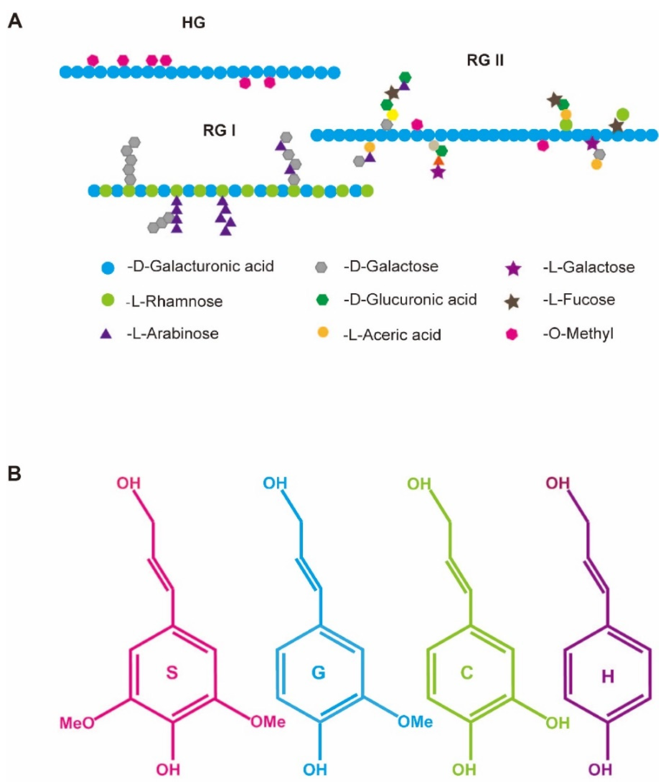
Pectin is the most abundant and structurally complex polysaccharide in plant PCW, which is classified into three main subgroups: homogalacturonan (HG), rhamnogalacturonan I (RG I) and rhamnogalacturonan II (RG II). In contrast to the classical PCW model, pectin has been shown to be closely associated with cell wall mechanics and cell wall integrity (CWI) [13]. Recent studies have also detected an aggregated pectin complex consisting of unbranched HG molecules and branched HGs associated with RG I polymers of lower length [14,15]. A schematic structure of the three major pectic polysaccharides are shown (Figure 2A). Since the role of pectin in regulating cell wall mechanics was revisited and highlighted, emerging evidence has indicated a link between pectin and plant immunity. Much of the evidence from Arabidopsis supports pectin as a vital part of constitutive and inducible defenses. Arabidopsis cell wall mutants where pectin biosynthesis, degradation and modification were impaired showed altered disease resistance phenotypes, suggesting a strong link between pectin metabolism and plant immunity [2,13].
B. napus is closely related to Arabidopsis, and both belong to the Brassicaceae family. B. napus has four to six copies of genes orthologous to each Arabidopsis gene. A diversity of genes involved in pectin metabolism were shown to take part in immunity in Arabidopsis; thus, it is deduced that their orthologous counterpart genes in B. napus are likely to have similar functions. This deduction has been validated by genetic and high-throughput ‘omics’ data in several recent studies. A recent transcriptome study of two B. napus cultivars with contrasting resistance to powdery mildew using RNA-seq showed that genes related to pectin modification and degradation were expressed at significantly higher levels in the resistant line than susceptible lines, including powdery mildew resistant 5 (PMR5), polygalacturonase inhibitor 1 (PGIP1), pectin methylesterase inhibitor 9 (PMEI9) and reduced wall acetylation 2 (RWA2) [16]. As demonstrated recently in Arabidopsis and other crops, members from gene families including PMR, PGIP, PMEI and RWA were closely associated with phenotypes of resistance/susceptibility to pathogens [17,18,19,20]. PGIPs are a group of extracellular leucine-rich repeat proteins that can inhibit the pectin depolymerizing activity of polygalacturonases (PGs) secreted by pathogens and have been shown to protect crop plants against pathogens by maintaining plant CWI [21]. PGIPs in B. napus were found to effectively enhance rapeseed immunity against Sclerotinia sclerotiorum (S. sclerotiorum) infection by transgenic studies, and it was proposed that BnPGIPs confer resistance to S. sclerotiorum by interacting with specific SsPGs and inducing the expression of defense-related genes [22,23]. The pattern and degree of pectin methylesterification have also been shown to influence CWI and plant immunity, which are regulated by pectin methylesterase (PME) and pectin methylesterase inhibitor (PMEI) [24]. PME catalyzes the demethylesterification of the galacturonic acid backbone of pectin, which can be inhibited by PMEI. How the degree of pectin methylesterification affects plant immunity still remains a complex issue. On one hand, de-esterified pectin is supposed to be more susceptible to hydrolysis by other pectic enzymes such as endopolygalacturonases, which might lead to weakened CWI and decreased disease resistance. On the other hand, de-esterified pectin can form a rigid Ca2+-pectate crosslinked “egg box” that could strength CWI, and methanol released due to pectin demethylesterificapectin acts as a signaling molecule to trigger a defense response [25]. Very recently, Wang et al. [26] reported that several PMEI genes in B. napus were strongly responsive to S. sclerotiorum infection and suggested that these BnPMEI genes might act as candidates for breeding novel B. napus with enhanced resistance to sclerotinia stem rot (SSR). However, information on how PME/PMEI in B. napus regulates plant immunity is very limited.
So far, pectin-mediated plant immunity can be, at least partially, explained by its influence on preformed defense and inducible defense. Pectin-derived DAMPs, especially oligogalacturonides (OGs), oligomers of α-1,4–linked galacturonic acid generated through the activity of PG, represent the most described signal molecules that can trigger plant immune responses [20]. In Arabidopsis, OGs generated in vivo could boost disease resistance to necrotrophic and biotrophic pathogens, but the defense responses that were triggered by OGs varied from study to study [20,27,28,29]. However, the excessive accumulation of OGs might have adverse effects on plant growth and development and even lead to plant death, which is known as the trade-off between growth and defense. So far, we are still far from understanding the detailed molecular mechanism of pectin-mediated immunity in B. napus as well as other plants.

3. The Content and Composition of Lignin Determine Disease Resistance of B. napus

Lignification, i.e., the deposition of lignin, is a defining feature of secondary cell wall (SCW) formation. Lignin, an abundant phenylpropanoid polymer, provides mechanical strength and contributes to cell wall structural integrity and rigidity. Alterations in lignin content and composition have also been shown to influence plant disease resistance upon pathogen infection. Increased lignin content was reported to enhance disease resistance by inhibiting the invasion of pathogens in a range of plants including Arabidopsis, maize, tobacco and cotton [30,31,32,33]. More recently, a positive correlation between lignin content and resistance to S. sclerotiorum was observed in transgenic B. napus plants overexpressing Cinnamoyl-CoA Reductase 2, which is required for lignin monomer synthesis [34]. Interestingly, manipulating the lignin content did not compromise plant growth, development and other agronomic traits including seed oil content [34]. Changes in cell wall components associated with clubroot resistance were evaluated using FTIR spectroscopy in both the B. napus susceptible line and resistant lines whose disease resistance was conferred by an R gene, and results revealed a substantial contribution of lignin and phenolics to disease resistance [35]. More recently, Höch et al. compared defense responses between resistant and susceptible B. napus cultivars in response to S. sclerotiorum at both transcriptional and histological levels, and it was suggested that the early timing of lignin deposition and the activation of key genes in the phenylpropanoid pathway required for lignin biosynthesis play a crucial role in disease resistance [36]. In general, a positive correlation was observed between lignin content and resistance to pathogens. The direct reason accounting for this could be that lignin plays a crucial role in thickening the SCW and increasing the cell wall stiffness, which further prevents pathogens from penetrating host cells.
In addition to lignin biosynthesis, lignin composition also influences plant immunity. Lignin is structurally complex as it varies substantially in the levels and types of several major monomeric units from which lignin primarily derives [37]. Four representative monolignols which make up the lignin polymers are shown (Figure 2B). Arabidopsis with strikingly different lignin compositions but similar lignin contents, resulting from the genetic manipulation of F5H encoding ferulate/coniferaldehyde 5-hydroxylase, exhibited different defense responses upon pathogen attack [38]. The difference in biotic stress responses might be attributed to altered elicitors derived from cell wall polysaccharide components, indicating that an alteration in lignin composition might affect interactions with plant cell wall matrix polysaccharides [38]. A strong correlation between the lignin monomer ratio and disease resistance was also observed in wheat and Camelina sativa [39,40]. Very recently, the lignin composition of the B. napus cell wall was reported to affect cell-wall-mediated immunity [41]. Decreasing the S/G lignin ratio in B. napus by knocking out the F5H gene using CRISPR resulted in increased resistance to S. sclerotiorum as well as increased stem strength and altered cell wall structure [41]. This is the first report using genetic engineering tools to profile the effect of the lignin monomer ratio on B. napus’s resistance to S. sclerotiorum. It is assumed that monomer composition impacts the higher order structure of lignin, including lignin branching and its crosslinking with other cell wall polysaccharides.
Alterations in the lignin structure not only influence the spread of pathogens and pathogen-secreted enzymes but also affect the accumulation of defense-related signal molecules that are generated during the process of pathogen infection. Rapid lignin deposition into cell walls was observed in Arabidopsis leaves upon pathogen infection, which was regarded as part of pattern-triggered immunity [42]. Lignin deposition involves three basic steps including monolignol biosynthesis, transport and polymerization, and a diversity of lignin-modifying genes participate in these processes. Although a range of genes involved in lignin deposition have been identified, little is known about their role in regulating plant immunity as well as the underlying molecular mechanism.

4. Pectin and Lignin Act Synergistically to Defend against Pathogens in B. napus

Different components of the plant cell wall are linked and interconnected in a complex way that still remains unclear. Numerous studies have described that pectin and lignin in the plant cell wall represent key players in regulating defense. It is postulated that pectin and lignin act in a synergistic way to defend B. napus against pathogen attack. Current research has indicated crosstalk between pectin and lignin modification during B. napus–pathogen interactions.
Overexpressing a plant mediator gene, B. napus mediator subunit 16 (BnMED16), which is highly responsive to S. sclerotiorum infection, in B. napus resulted in rapid and comprehensive defense responses including the prominent increased expression of the majority of lignin synthesis genes (C4H, CCoAOMT, CCR1, CAD5, CAD2, CAD6, CAD1, CAD7, CAD8, COMT1, FAH1 and PMT5) and pectin synthesis or modification genes (GAUT7, GAUT15, PME3, PME17, PME52, PME10, PME53, PME34, PME16, PME35, PME51 and PGIP1) [7]. This suggests that pectin and lignin might cooperate to confer S. sclerotiorum resistance by mediating the cell wall integrity [7]. So far, a range of cell-wall-associated genes have been linked with disease resistance in B. napus, and they are listed in Table 1.
The most convincing evidence of crosstalk between pectin modification and lignin modification came from transgenic studies in Arabidopsis. Suppressing lignin biosynthetic enzymes including hydroxycinnamoyl CoA:shikimate/quinate hydroxycinnamoyl transferase (HCT) and cinnamoyl CoA reductase 1 (CCR1) in transgenic Arabidopsis induced the ectopic expression of a gene encoding pectin-degrading enzyme (ADPG1), leading to the release of pectic oligosaccharide that further triggered a series of defense responses and enhanced disease resistance [45]. This reveals an impact of lignin modification on CWI through influence on crosslinking to other cell wall components including pectin. Vice versa, it has been reported that pectin modification also affects lignin content and composition. Arabidopsis plants overexpressing a PG gene (PGX2) enhanced lignification through the activation of genes involved in lignin biosynthesis, providing a possible interaction of pectin and/or lignin dynamics [46]. Both pectin and lignin are key cell wall components determining cell wall rigidity and strength, which are supposed to have a direct impact on pathogen penetration and invasion. More importantly, pectin and lignin are a reservoir of signal molecules or metabolites which may regulate plant immune responses indirectly. The modulation of pectin may affect the release of DAMPs as well as the relative location of cell surface receptors, which further influence the PTI [47]. Lignin is a main source of metabolites, particularly soluble phenolic compounds. These metabolites might reduce virulence either by acting as signal molecules that can trigger defense responses or because they are toxic to pathogens [48]. In contrast, lignin-derived metabolites may enhance virulence by serving as a nutrient source for pathogens [49]. Emerging evidence has revealed the concerted role of pectin and lignin involved in pathogen-triggered defense responses in B. napus [45,46]. However, the detailed molecular mechanisms underlining the crosstalk between pectin and lignin dynamics with stress responses in B. napus remains unclear.

5. Transcriptional Regulation of Cell-Wall-Mediated Immunity in B. napus

Cell wall remodeling represents one of the most important defense responses contributing to disease resistance. Cell-wall-mediated immunity is supposed to be regulated by a range of transcription factors (TFs). So far, a range of TFs have been identified as either positive or negative regulators of defense responses against various pathogens in B. napus. The most described TFs associated with immunity belong to the WRKY family. TFs from the WRKY family contain the conserved WRKY domain and have been reported to participate in defense responses caused by biotic stress in many plants [50]. Knocking out WRKY70 using CRISPR/Cas9 resulted in enhanced resistance to S. sclerotinia, and overexpressing WRKY70 showed an opposite effect, suggesting a negative role of WRKY70 in disease resistance to S. sclerotinia in B. napus [8]. In contrast, WRKY33 positively regulated disease resistance to S. sclerotiorum in B. napus by activating salicylic acid (SA)- and jasmonic acid (JA)-mediated defense responses [9]. WRKY15 and WRKY28 were found to increase the susceptibility of B. napus to S. sclerotiorum by repressing the activity of WRKY33, indicating a complex transcriptional network involving multiple WRKYs in regulating immune responses in B. napus [10,11].
However, little is known about the transcriptional regulatory network of cell-wall-mediated plant immunity. In Arabidopsis, a number of MYB TFs have been identified to regulate lignin biosynthesis and modification through activating the expression of the genes involved in the monolignol pathway, including MYB46, MYB58, MYB63 and MYB83 [51,52,53]. Earlier studies on Arabidopsis indicated a link between MYB TFs and lignin metabolism, and whether this regulatory network is associated with defense responses remains unclear. Recently, Chezem and colleagues revealed a regulatory role of MYB15 in promoting defense-induced lignification and basal immunity in Arabidopsis via controlling the synthesis of G-rich lignin and the coumarin metabolite scopoletin [54]. Similarly, MYB was also reported to contribute to disease resistance in B. napus via regulating the remodeling of lignin and pectin. The most recent transgenic study in B. napus indicated a negative role of BnMYB43 in regulating the host plant’s resistance to S. sclerotiorum, probably via the profound remodeling of cell wall ingredients, especially pectin and lignin [12]. Silencing BnMYB43 in B. napus resulted in enhanced disease resistance accompanied with a great increase in pectin content but a significant reduction in the contents of cellulose and lignin [12]. The above studies in Brassicaceae species indicated that MYB TFs are pivotal regulators of cell-wall-mediated plant immunity. The disease resistance not only comes from cell wall remodeling but also from other signal pathways including the hormone-mediated defense signaling pathway. Along with regulating cell wall remodeling, BnMYB43 also greatly affected the expression of genes related to the SA or JA signal pathways [12].
Phytohormones, especially SA, JA and ethylene, have been shown to act in a synergistic manner to trigger defense responses in B. napus [55]. Prior studies have revealed the interplay between cell wall remodeling and phytohormone signaling in the regulation of plant immunity in numerous plants [41]. In Arabidopsis, lignin accumulation induced by cell wall damage was regulated by a genetically complex network involving dynamic interactions between JA and reactive oxygen species (ROS) [56]. A comparison study of two B. napus cultivars with contrasting susceptibility to Xanthomonas campestris pv. Campestris revealed the significance of JA-induced cell-wall-bound hydroxycinnamic acid accumulation that can acquire disease resistance [43]. Cell wall regulation in response to pathogen infection involves the coordination of multiple regulatory layers including a diversity of transcription factors and hormones. The complex transcriptional network responsible for cell-wall-mediated immunity in B. napus still remains unknown.

6. Conclusions and Outlook

This review focused on cell wall components and their role in plant immunity in B. napus. The characterization of the role of several pectin/lignin metabolism-associated genes revealed that in B. napus, pectin/lignin content and modifications were closely associated with plant immunity. Possible explanations for their contribution to plant immunity include the role of pectin/lignin in maintaining CWI as well as their ability to trigger other defense responses. The molecular mechanism underlining cell-wall-mediated plant immunity in B. napus still remains largely unknown. However, recently developed gene editing tools such as CRISPR have served as an effective way to generate targeted cell wall mutants in polyploid crops such as B. napus, enabling investigations of the link between specific cell wall epitopes and disease resistance. Moreover, dynamic changes in specific cell wall epitopes and CWI during plant–pathogen interactions can be visualized with the help of advanced microscopy technologies coupled with plant cell wall probes. These approaches offer new strategies to gain insights into the mechanisms of cell-wall-mediated immunity at a cellular level.
Modification of one cell wall component usually leads to the alteration of other wall components. This was observed in pectin and lignin in Arabidopsis and B. napus upon pathogen infection, indicating that there were close associations between pectin and lignin. The potential crosstalk between the metabolism of the different cell wall polymers during plant–pathogen interactions is a hot topic of discussion. In the future, more attention should be paid to the crosstalk between pectin and lignin modifications. This can be elucidated using strategies including multiomics and cytological technologies. The transcriptional regulation of cell-wall-mediated immunity in B. napus was also reviewed, and so far, MYB TFs were identified as regulating defense-induced lignin/pectin modification. Future work needs to identify more TFs that participate in regulating wall-associated immune responses. This should help reveal the complex regulatory networks that guide cell-wall-mediated immunity in B. napus and other plants. In addition, more pectin/lignin-derived DAMPs are required to be characterized, and their role in triggering immune responses needs elucidation.
Cell wall components including pectin and lignin are of great interest due to their potential utility in developing disease-resistant B. napus crops. Here we argue that identifying pectin/lignin modifying genes in response to biotic stresses as well as revealing the underlining transcription regulatory network that monitors and maintains CWI can help guide breeders to generate novel B. napus crop varieties with enhanced disease resistance via the modulation of cell wall components. In addition, pectin/lignin-derived DAMPs can be utilized as agrochemicals for crop protection. Cell wall engineering can be achieved through traditional and modern strategies, including artificial breeding and selection, transgenic approaches and the recently developed gene editing technologies, in particular CRISPR. Using traditional natural-variation-based approaches, numerous genes or loci related to cell wall traits have been identified, and novel germplasm with modified cell wall components have been developed. However, gene redundancy and extensive periods of time to develop new germplasm by traditional breeding remain major challenges constraining the development of new varieties. To overcome these issues, modern biotechnology-based strategies such as transgenic technologies and gene editing tools can be used for the targeted genetic engineering of specific cell wall components with high efficiency. Using gene editing to modify cell-wall-related genes that alter wall structure and metabolism provides a promising way to test new targets for improving plant immunity that can be rapidly integrated into novel breeding strategies for B. napus and other crops.

Author Contributions

Conceptualization, D.W. and H.L.; resources, Q.L., S.J. and X.F.; writing—original draft preparation, D.W., Q.L., S.J. and X.F.; writing—review and editing, D.W. and H.L.; funding acquisition, H.L. and D.W. All authors have read and agreed to the published version of the manuscript.

Funding

This research was funded by the National Natural Science Foundation of China (grant number 32160435) and the National Natural Science Foundation of China (grant number 32101656).

Data Availability Statement

No new data were created or analyzed in this study. Data sharing is not applicable to this article.

Conflicts of Interest

The authors declare no conflict of interest.

Glossary

Middle lamella (ML)A layer rich in pectic polysaccharides that glues adjacent cells together.
Primary cell wall (PCW)A layer surrounding plant cells and mainly composed of polysaccharides including cellulose, hemicellulose and pectin.
Secondary cell wall (SCW)Synthesized by specialized plant cells when cell growth ceases, mainly composed of polysaccharides (about 65%) and lignin (about 35%).
LigninA principal structural component of secondary cell walls in higher terrestrial plants, which strengths cell wall rigidity. Lignin is typically composed of p-hydroxyphenyl (H), guaiacyl (G) and syringyl (S) units that derive from the polymerization of various aromatic monomers.
XyloglucanThe main component of hemicellulose, which is composed of a 1,4-β-glucan linked backbone. Side chains such as galactosyl and fucosyl-galactosyl residues are linked to the backbone via xylose residues.
CelluloseOne of the three main cell wall polysaccharides, which is made up of β-1,4-linked glucan chains. Multiple linear celluloses are usually organized into crystalline microfibrils in the plant primary cell wall, acting as a major load-bearing cell wall component.
PectinA predominant cell wall polysaccharide of the plant primary cell wall and middle lamella, which is abundant in galacturonic acid and is classified into three major types depending on the composition of the backbone and side chains, including homogalacturonan (HG), rhamnogalacturonan I (RG-I) and rhamnogalacturonan II (RG-II).
Cell wall integrity (CWI)The structural and functional integrity of the plant cell wall.
Microbe-associated molecular pattern (PAMP)Molecules derived from the microbe itself, which can trigger defense responses.
Damage-associated molecular patterns (DAMPs)Endogenous signaling molecules that are derived and released from damaged host cellular structures, which can be perceived by cell surface localized receptors and activate host immune responses.
Pattern recognition receptors (PRRs)Localized on cell surface such as receptor-like kinases/proteins (RLKs/RLPs) with extracellular ligand-binding domain that can monitor and detect nonself and self-derived signaling molecules, triggering immune responses.
Pattern-triggered immunity (PTI)The first layer of the plant immunity, involving a set of induced defenses when pattern recognition receptors perceive signals derived from pathogens or damaged host cellular structures.
Effector-triggered immunity (ETI)The second layer of the plant immunity, involving a series of immune responses that are more sustained and robust than PTI when intracellular receptors recognize pathogen-secreted effectors.
Galacturonosyltransferase (GAUT)An enzyme that is involved in the synthesis of homogalacturonan (HG) by catalyzing the elongation of HG oligogalacturonides in an α-1, 4-configuration.
Pectin methylesterase (PME)A pectin-related enzyme that can decrease the degree of methylesters on the pectin backbone by removing methyl groups from esterified homogalacturonan.
Pectin methylesterase inhibitor (PMEI) Cell wall proteins that can regulate the degree of the methylesterification of pectins by inhibiting the activity of pectin methylesterase.
Polygalacturonase-inhibiting protein (PGIP)Plant cell wall proteins with leucine-rich repeat, which inhibits pectin depolymerization by inactivating the enzymatic activity of polygalacturonase.
Ferulate 5-hydroxylase (F5H)One of the key enzymes that regulate the S/G lignin composition in plants.

References

  1. Neik, T.X.; Amas, J.; Barbetti, M.; Edwards, D.; Batley, J. Understanding Host–Pathogen Interactions in Brassica napus in the Omics Era. Plants 2020, 9, 1336. [Google Scholar] [CrossRef]
  2. Bacete, L.; Mélida, H.; Miedes, E.; Molina, A. Plant cell wall-mediated immunity: Cell wall changes trigger disease resistance responses. Plant J. 2018, 93, 614–636. [Google Scholar] [CrossRef] [PubMed]
  3. Yuan, M.; Ngou, B.P.M.; Ding, P.; Xin, X.-F. PTI-ETI crosstalk: An integrative view of plant immunity. Curr. Opin. Plant Biol. 2021, 62, 102030. [Google Scholar] [CrossRef] [PubMed]
  4. Hou, S.; Liu, Z.; Shen, H.; Wu, D. Damage-Associated Molecular Pattern-Triggered Immunity in Plants. Front. Plant Sci. 2019, 10, 646. [Google Scholar] [CrossRef]
  5. Meents, M.J.; Watanabe, Y.; Samuels, A.L. The cell biology of secondary cell wall biosynthesis. Ann. Bot. 2018, 121, 1107–1125. [Google Scholar] [CrossRef] [Green Version]
  6. Molina, A.; Miedes, E.; Bacete, L.; Rodríguez, T.; Mélida, H.; Denancé, N.; Sánchez-Vallet, A.; Rivière, M.-P.; López, G.; Freydier, A.; et al. Arabidopsis cell wall composition determines disease resistance specificity and fitness. Proc. Natl. Acad. Sci. USA 2021, 118, e2010243118. [Google Scholar] [CrossRef] [PubMed]
  7. Hu, H.; Tang, Y.; Wu, J.; Chen, F.; Yang, Y.; Pan, X.; Dong, X.; Jin, X.; Liu, S.; Du, X. Brassica napus Mediator Subunit16 Induces BnMED25- and BnWRKY33-Activated Defense Signaling to Confer Sclerotinia sclerotiorum Resistance. Front. Plant Sci. 2021, 12, 663536. [Google Scholar] [CrossRef]
  8. Sun, Q.; Lin, L.; Liu, D.; Wu, D.; Fang, Y.; Wu, J.; Wang, Y. CRISPR/Cas9-Mediated Multiplex Genome Editing of the BnWRKY11 and BnWRKY70 Genes in Brassica napus L. Int. J. Mol. Sci. 2018, 19, 2716. [Google Scholar] [CrossRef] [Green Version]
  9. Wang, Z.; Fang, H.D.; Chen, Y.; Chen, K.P.; Li, G.Y.; Gu, S.L.; Tan, X.L. Overexpression of BnWRKY33 in oilseed rape enhances resistance to Sclerotinia sclerotiorum. Mol. Plant Pathol. 2014, 15, 677–689. [Google Scholar] [CrossRef]
  10. Liu, F.; Li, X.; Wang, M.; Wen, J.; Yi, B.; Shen, J.; Ma, C.; Fu, T.; Tu, J. Interactions of WRKY15 and WRKY33 transcription factors and their roles in the resistance of oilseed rape to Sclerotinia infection. Plant Biotechnol. J. 2018, 16, 911–925. [Google Scholar] [CrossRef]
  11. Zhang, K.; Liu, F.; Wang, Z.; Zhuo, C.; Hu, K.; Li, X.; Wen, J.; Bin Yi, B.; Shen, J.; Ma, C.; et al. Transcription factor WRKY28 curbs WRKY33-mediated resistance to Sclerotinia sclerotiorum in Brassica napus. Plant Physiol. 2022, 190, 2757–2774. [Google Scholar] [CrossRef]
  12. Jiang, J.; Liao, X.; Jin, X.; Tan, L.; Lu, Q.; Yuan, C.; Xue, Y.; Yin, N.; Lin, N.; Chai, Y. MYB43 in Oilseed Rape (Brassica napus) Positively Regulates Vascular Lignification, Plant Morphology and Yield Potential but Negatively Affects Resistance to Scle-rotinia sclerotiorum. Genes 2020, 11, 581. [Google Scholar] [CrossRef]
  13. Wang, D.; Yang, L.; Silva, C.; Sarwar, R.; Chen, Y.; Blanco-Ulate, B. Pectin-mediated plant immunity. Annu. Plant Rev. 2022, 5, 55–80. [Google Scholar]
  14. Round, A.N.; Rigby, N.M.; MacDougall, A.J.; Morris, V.J. A new view of pectin structure revealed by acid hydrolysis and atomic force microscopy. Carbohydr. Res. 2010, 345, 487–497. [Google Scholar] [CrossRef] [PubMed]
  15. Paniagua, C.; Kirby, A.R.; Gunning, A.P.; Morris, V.J.; Matas, A.J.; Quesada, M.A.; Mercado, J.A. Unravelling the nanostructure of strawberry fruit pectins by endo-polygalacturonase digestion and atomic force microscopy. Food Chem. 2017, 224, 270–279. [Google Scholar] [CrossRef] [PubMed]
  16. Zhang, M.; Gong, Q.; Su, X.; Cheng, Y.; Wu, H.; Huang, Z.; Xu, A.; Dong, J.; Yu, C. Microscopic and Transcriptomic Comparison of Powdery Mildew Resistance in the Progenies of Brassica carinata × B. napus. Int. J. Mol. Sci. 2022, 23, 9961. [Google Scholar] [CrossRef] [PubMed]
  17. Engelsdorf, T.; Gigli-Bisceglia, N.; Veerabagu, M.; Mckenna, J.F.; Augstein, F.; Does, D.V.D.; Zipfel, C.; Hamann, T. Pat-tern-Triggered Immunity and Cell Wall Integrity Maintenance Jointly Modulate Plant Stress Responses. Cold Spring Harb. Lab. 2017, 11, eaao3070. [Google Scholar]
  18. Lionetti, V.; Fabri, E.; De Caroli, M.; Hansen, A.R.; Willats, W.G.; Piro, G.; Bellincampi, D. Three Pectin Methylesterase Inhibitors Protect Cell Wall Integrity for Arabidopsis Immunity to Botrytis. Plant Physiol. 2017, 173, 1844–1863. [Google Scholar] [CrossRef] [Green Version]
  19. Manabe, Y.; Nafisi, M.; Verhertbruggen, Y.; Orfila, C.; Gille, S.; Rautengarten, C.; Cherk, C.; Marcus, S.E.; Somerville, S.; Pauly, M.; et al. Loss-of-Function Mutation of REDUCED WALL ACETYLATION2 in Arabidopsis Leads to Reduced Cell Wall Acetylation and Increased Resistance to Botrytis cinerea. Plant Physiol. 2011, 155, 1068–1078. [Google Scholar] [CrossRef] [PubMed] [Green Version]
  20. Benedetti, M.; Pontiggia, D.; Raggi, S.; Cheng, Z.; Scaloni, F.; Ferrari, S.; Ausubel, F.M.; Cervone, F.; De Lorenzo, G. Plant immunity triggered by engineered in vivo release of oligogalacturonides, damage-associated molecular patterns. Proc. Natl. Acad. Sci. USA 2015, 112, 5533–5538. [Google Scholar] [CrossRef] [PubMed] [Green Version]
  21. Kalunke, R.M.; Tundo, S.; Benedetti, M.; Cervone, F.; De Lorenzo, G.; D’Ovidio, R. An update on polygalacturonase-inhibiting protein (PGIP), a leucine-rich repeat protein that protects crop plants against pathogens. Front. Plant Sci. 2015, 6, 146. [Google Scholar] [CrossRef] [PubMed] [Green Version]
  22. Wang, Z.; Wan, L.; Zhang, X.; Xin, Q.; Song, Y.; Hong, D.; Sun, Y.; Yang, G. Interaction between Brassica napus polygalac-turonase inhibition proteins and Sclerotinia sclerotiorum polygalacturonase: Implications for rapeseed resistance to fungal in-fection. Planta 2021, 253, 34. [Google Scholar] [CrossRef]
  23. Bashi, Z.D.; Rimmer, S.R.; Khachatourians, G.G.; Hegedus, D.D. Brassica napus polygalacturonase inhibitor proteins inhibit Sclerotinia sclerotiorum polygalacturonase enzymatic and necrotizing activities and delay symptoms in transgenic plants. Can. J. Microbiol. 2013, 59, 79–86. [Google Scholar] [CrossRef] [PubMed]
  24. Coculo, D.; Lionetti, V. The Plant Invertase/Pectin Methylesterase Inhibitor Superfamily. Front. Plant Sci. 2022, 13, 863892. [Google Scholar] [CrossRef]
  25. Komarova, T.V.; Sheshukova, E.V.; Dorokhov, Y.L. Cell wall methanol as a signal in plant immunity. Front. Plant Sci. 2014, 5, 101. [Google Scholar] [CrossRef] [PubMed] [Green Version]
  26. Wang, D.; Jin, S.; Chen, Z.; Shan, Y.; Li, L. Genome-wide identification of the pectin methylesterase inhibitor genes in Brassica napus and expression analysis of selected members. Front. Plant Sci. 2022, 13, 940284. [Google Scholar] [CrossRef]
  27. Galletti, R.; Ferrari, S.; De Lorenzo, G. Arabidopsis MPK3 and MPK6 Play Different Roles in Basal and Oligogalacturonide- or Flagellin-Induced Resistance against Botrytis cinerea. Plant Physiol. 2011, 157, 804–814. [Google Scholar] [CrossRef] [Green Version]
  28. Gravino, M.; Savatin, D.V.; Macone, A.; De Lorenzo, G. Ethylene production in Botrytis cinerea-and oligogalacturonide-induced immunity requires calcium-dependent protein kinases. Plant J. 2015, 84, 1073–1086. [Google Scholar] [CrossRef] [Green Version]
  29. Ferrari, S.; Galletti, R.; Denoux, C.; De Lorenzo, G.; Ausubel, F.M.; Dewdney, J. Resistance to Botrytis cinerea induced in Ara-bidopsis by elicitors is independent of salicylic acid, ethylene, or jasmonate signaling but requires PHYTOALEXIN DEFI-CIENT3. Plant Physiol. 2007, 144, 367–379. [Google Scholar] [CrossRef] [Green Version]
  30. Tronchet, M.; Balaguã, C.; Kroj, T.; Jouanin, L.; Roby, D. Cinnamyl alcohol dehydrogenases-C and D, key enzymes in lignin biosynthesis, play an essential role in disease resistance in Arabidopsis. Mol. Plant Pathol. 2010, 11, 83–92. [Google Scholar] [CrossRef]
  31. Yang, Q.; He, Y.; Kabahuma, M.; Chaya, T.; Kelly, A.; Borrego, E.; Bian, Y.; El Kasmi, F.; Yang, L.; Teixeira, P.; et al. A gene encoding maize caffeoyl-CoA O-methyltransferase confers quantitative resistance to multiple pathogens. Nat Genet. 2017, 49, 1364–1372. [Google Scholar] [CrossRef]
  32. Ma, Q.-H.; Zhu, H.-H.; Han, J.-Q. Wheat ROP proteins modulate defense response through lignin metabolism. Plant Sci. 2017, 262, 32–38. [Google Scholar] [CrossRef] [PubMed]
  33. Shi, H.; Liu, Z.; Zhu, L.; Zhang, C.; Chen, Y.; Zhou, Y.; Li, F.; Li, X. Overexpression of cotton (Gossypium hirsutum) dirigent1 gene enhances lignification that blocks the spread of Verticillium dahliae. Acta Biochim. Biophys. Sin. 2012, 44, 555–564. [Google Scholar] [CrossRef] [Green Version]
  34. Liu, D.; Wu, J.; Lin, L.; Li, P.; Li, S.; Wang, Y.; Li, J.; Sun, Q.; Liang, J.; Wang, Y. Overexpression of Cinnamoyl-CoA Reductase 2 in Brassica napus Increases Resistance to Sclerotinia sclerotiorum by Affecting Lignin Biosynthesis. Front Plant Sci. 2021, 12, 732–733. [Google Scholar] [CrossRef] [PubMed]
  35. Lahlali, R.; Song, T.; Chu, M.; Yu, F.; Kumar, S.; Karunakaran, C.; Peng, G. Evaluating Changes in Cell-Wall Components Associated with Clubroot Resistance Using Fourier Transform Infrared Spectroscopy and RT-PCR. Int. J. Mol. Sci. 2017, 18, 2058. [Google Scholar] [CrossRef] [Green Version]
  36. Höch, K.; Koopmann, B.; von Tiedemann, A. Lignin Composition and Timing of Cell Wall Lignification Are Involved in Brassica napus Resistance to Stem Rot Caused by Sclerotinia sclerotiorum. Phytopathology 2021, 111, 1438–1448. [Google Scholar] [CrossRef] [PubMed]
  37. Liu, Q.; Luo, L.; Zheng, L.Q. Lignins: Biosynthesis and Biological Functions in Plants. Int. J. Mol. Sci. 2018, 19, 335. [Google Scholar] [CrossRef] [Green Version]
  38. Gallego-Giraldo, L.; Posé, S.; Pattathil, S.; Peralta, A.G.; Hahn, M.G.; Ayre, B.G.; Sunuwar, J.; Hernandez, J.; Patel, M.; Shah, J.; et al. Elicitors and defense gene induction in plants with altered lignin compositions. New Phytol. 2018, 219, 1235–1251. [Google Scholar] [CrossRef] [PubMed] [Green Version]
  39. Bhuiyan, N.H.; Selvaraj, G.; Wei, Y.; King, J. Gene expression profiling and silencing reveal that monolignol biosynthesis plays a critical role in penetration defence in wheat against powdery mildew invasion. J. Exp. Bot. 2008, 60, 509–521. [Google Scholar] [CrossRef]
  40. Eynck, C.; Séguin-Swartz, G.; Clarke, W.E.; Parkin, I.A. Monolignol biosynthesis is associated with resistance to Sclerotinia sclerotiorum in Camelina sativa. Mol. Plant Pathol. 2012, 13, 887–899. [Google Scholar] [CrossRef]
  41. Cao, Y.; Yan, X.; Ran, S.; Ralph, J.; Smith, R.A.; Chen, X.; Qu, C.; Li, J.; Liu, L. Knockout of the lignin pathway gene BnF5H decreases the S/G lignin compositional ratio and improves Sclerotinia sclerotiorum resistance in Brassica napus. Plant Cell Environ. 2022, 45, 248–261. [Google Scholar] [CrossRef] [PubMed]
  42. Jeon, H.S.; Jang, E.; Kim, J.; Kim, S.H.; Lee, M.-H.; Nam, M.H.; Tobimatsu, Y.; Park, O.K. Pathogen-induced autophagy regulates monolignol transport and lignin formation in plant immunity. Autophagy 2022, 9, 1–19. [Google Scholar] [CrossRef] [PubMed]
  43. Islam, T.; Lee, B.-R.; Park, S.-H.; La, V.H.; Jung, W.-J.; Bae, D.-W.; Kim, T.-H. Hormonal regulations in soluble and cell-wall bound phenolic accumulation in two cultivars of Brassica napus contrasting susceptibility to Xanthomonas campestris pv. campestris. Plant Sci. 2019, 285, 132–140. [Google Scholar] [CrossRef]
  44. Larkan, N.J.; Ma, L.; Haddadi, P.; Buchwaldt, M.; Parkin, I.A.P.; Djavaheri, M.; Borhan, M.H. The Brassica napus wall-associated kinase-like (WAKL) gene Rlm9 provides race-specific blackleg resistance. Plant J. 2020, 104, 892–900. [Google Scholar] [CrossRef] [PubMed]
  45. Gallego-Giraldo, L.; Liu, C.; Pose-Albacete, S.; Pattathil, S.; Peralta, A.G.; Young, J.; Westpheling, J.; Hahn, M.G.; Rao, X.; Knox, J.P.; et al. ARABIDOPSIS DEHISCENCE ZONE POLYGALACTURONASE 1 (ADPG1) releases latent defense signals in stems with reduced lignin content. Proc. Natl. Acad. Sci. USA 2020, 117, 3281–3290. [Google Scholar] [CrossRef] [Green Version]
  46. Xiao, C.; Barnes, W.J.; Zamil, M.S.; Yi, H.; Puri, V.M.; Anderson, C.T. Activation tagging of Arabidopsis POLYGALAC-TURONASE INVOLVED IN EXPANSION2 promotes hypocotyl elongation, leaf expansion, stem lignification, mechanical stiffening, and lodging. Plant J. 2017, 89, 1159–1173. [Google Scholar] [CrossRef] [Green Version]
  47. Bacete, L.; Hamann, T. The Role of Mechanoperception in Plant Cell Wall Integrity Maintenance. Plants 2020, 9, 574. [Google Scholar] [CrossRef]
  48. Miedes, E.; Vanholme, R.; Boerjan, W.; Molina, A. The role of the secondary cell wall in plant resistance to pathogens. Front. Plant Sci. 2014, 5, 358. [Google Scholar] [CrossRef] [Green Version]
  49. Yadav, V.; Wang, Z.; Wei, C.; Amo, A.; Ahmed, B.; Yang, X.; Zhang, X. Phenylpropanoid Pathway Engineering: An Emerging Approach towards Plant Defense. Pathogens 2020, 9, 312. [Google Scholar] [CrossRef] [Green Version]
  50. Jiang, J.J.; Ma, S.H.; Ye, N.H.; Jiang, M.; Cao, J.S.; Zhang, J.H. WRKY transcription factors in plant responses to stresses. J. Integr. Plant Biol. 2017, 59, 86–101. [Google Scholar] [CrossRef] [Green Version]
  51. Zhou, J.; Lee, C.; Zhong, R.; Ye, Z.-H. MYB58 and MYB63 Are Transcriptional Activators of the Lignin Biosynthetic Pathway during Secondary Cell Wall Formation in Arabidopsis. Plant Cell 2009, 21, 248–266. [Google Scholar] [CrossRef] [PubMed] [Green Version]
  52. Zhong, R.; Richardson, E.A.; Ye, Z.-H. The MYB46 Transcription Factor Is a Direct Target of SND1 and Regulates Secondary Wall Biosynthesis in Arabidopsis. Plant Cell 2007, 19, 2776–2792. [Google Scholar] [CrossRef] [PubMed]
  53. McCarthy, R.L.; Zhong, R.; Ye, Z.-H. MYB83 Is a Direct Target of SND1 and Acts Redundantly with MYB46 in the Regulation of Secondary Cell Wall Biosynthesis in Arabidopsis. Plant Cell Physiol. 2009, 50, 1950–1964. [Google Scholar] [CrossRef] [PubMed] [Green Version]
  54. Chezem, W.R.; Memon, A.; Li, F.S.; Weng, J.K.; Clay, N.K. SG2-Type R2R3-MYB Transcription Factor MYB15 Controls Defense-Induced Lignification and Basal Immunity in Arabidopsis. Plant Cell. 2017, 29, 1907–1926. [Google Scholar] [CrossRef] [Green Version]
  55. Nafisi, M.; Fimognari, L.; Sakuragi, Y. Interplays between the cell wall and phytohormones in interaction between plants and necrotrophic pathogens. Phytochemistry 2015, 112, 63–71. [Google Scholar] [CrossRef] [Green Version]
  56. Denness, L.; McKenna, J.F.; Segonzac, C.; Wormit, A.; Madhou, P.; Bennett, M.; Mansfield, J.; Zipfel, C.; Hamann, T. Cell wall damage-induced lignin biosynthesis is regulated by a reactive oxygen species- and jasmonic acid-dependent process in Ara-bidopsis. Plant Physiol. 2011, 156, 1364–1374. [Google Scholar] [CrossRef] [PubMed]
Figure 1. An overview of plant cell-wall-mediated immunity and its regulation in B. napus.
Horticulturae 09 00112 g001
Figure 2. (A) Schematic structure of the three major pectic polysaccharides including homogalacturonan (HG), rhamnogalacturonan I (RG I) and rhamnogalacturonan II (RG II). HG is unbranched linear chain composed of 1, 4-linked α-D-galactosyluronic acid residues which can be methylesterified. RG I is branched pectic polysaccharides with backbone composed of repeating disaccharide (α-1,4-d-GalA-α-1,2-l-Rha) in which rhamnosyl residues serve as attachment sites for various side chains mainly including galactosyl and arabinosyl residues. RG II has the same backbone as HG and is highly branched with the most complex side chains containing 12 different glycosyl residues linked by over 20 different glycosyl linkages. (B) Chemical structure of the four representative monomeric subunits of lignin, including p-hydroxyphenyl (H), guaiacyl (G) and syringyl (S) units.
Horticulturae 09 00112 g002
Table 1. Selected genes related to cell-wall-mediated immune responses in B. napus.
Gene NamePhenotype Pathogen TestedReference
PMR5, PGIP1, PMEI9, RWA2, PDCB1, C/VIF2Rpowdery mildew [16]
WAK1, WAKL10Spowdery mildew [16]
BnPGIP2, BnPGIP5RSclerotinia sclerotiorum [22]
BnMED16RSclerotinia sclerotiorum [7]
BnaC.CCR2.b, CCRRSclerotinia sclerotiorum [34]
PAL, BnCCR2, BnCAD5, Bn4CLRSclerotinia sclerotiorum [36]
Rcr1RPlasmodiophora brassicae Woronin [35]
MYB43SSclerotinia sclerotiorum [12]
CHS, CAD, COMT1RXanthomonas campestrispv. campestris [43]
F5HSSclerotinia sclerotiorum [41]
Rlm9, WAKLRLeptosphaeria maculans [44]
R: resistant; S: susceptible.
Disclaimer/Publisher’s Note: The statements, opinions and data contained in all publications are solely those of the individual author(s) and contributor(s) and not of MDPI and/or the editor(s). MDPI and/or the editor(s) disclaim responsibility for any injury to people or property resulting from any ideas, methods, instructions or products referred to in the content.