Article

Regeneration of Cotoneaster wilsonii Nakai through Indirect Organogenesis

1
Department of Horticulture, Division of Applied Life Science (BK21 Four), Graduate School, Gyeongsang National University, Jinju 52828, Korea
2
China Agricultural University-Sichuan Chengdu Advanced Agricultural Industrial Institute, Chengdu 611430, China
3
Institute of Agriculture and Life Science, Gyeongsang National University, Jinju 52828, Korea
4
Research Institute of Life Science, Gyeongsang National University, Jinju 52828, Korea
*
Author to whom correspondence should be addressed.
Horticulturae 2022, 8(9), 795; https://doi.org/10.3390/horticulturae8090795
Submission received: 11 July 2022 / Revised: 26 August 2022 / Accepted: 27 August 2022 / Published: 31 August 2022
(This article belongs to the Special Issue Innovation in Propagation and Cultivation of Ornamental Plants)

Abstract

:
The ornamental plant Cotoneaster wilsonii Nakai is a rare endemic species to the Ulleung Island in Korea. There is an urgent need to develop efficient propagation methods to preserve this endangered plant species. The cytokinin thidiazuron (TDZ) at a concentration of 0, 1.0, 2.0, or 3.0 mg·L−1 combined with 0, 0.5, 1.0, or 1.5 mg·L−1 a-naphthaleneacetic acid (NAA) was used to induce calli from young leaf explants on the Murashige and Skoog (MS) medium. The TDZ and/or 6-benzyladenine at a concentration of 0, 0.5, or 1.0 mg·L−1 combined with 0.2 mg·L−1 NAA was supplied to the MS medium to induce shoots from calli. Four auxins at 0, 0.2, 0.4, 0.6, 0.8, or 1.0 mg·L−1 were supplied to half-strength MS medium for root induction. The treatment of TDZ at 1.0 mg·L−1 combined with 1.5 mg·L−1 NAA not only had 100% callus induction ratio, but also induced the heaviest calli. The shoot induction ratio was the greatest with TDZ at 1.0 mg·L−1, and the greatest root induction ratio was obtained with IAA at a concentration of 0.8 mg·L−1. In conclusion, an effective indirect organogenesis system was established for C. wilsonii Nakai.

1. Introduction

The ornamentally important genus Cotoneaster of the Rosaceae is relatively small with about 150 species. This genus is an unarmed shrub which grows mainly in temperate Eurasia [1]. Cotoneaster wilsonii Nakai is a new species found in recent decades, and this species is characterized by a small number of white flowers arranged in a corymb, beautiful red fruits, and deciduous leaves. This woody plant is endemic to the small island of Ulleung-do, Korea. The fact that the genus Cotoneaster hardly occurs in the mainland of South Korea makes the origin of C. wilsonii Nakai on Ulleung Island intriguing [2]. The morphology and identical flavonoid profile of C. wilsonii Nakai are similar to that of C. multiflorus native to China. Researchers suspect that seeds of C. multiflorus floating in the sea or carried by birds or winds may have adapted to the special climate with cool summers and warm winters in Ulleung-do and have differentiated into C. wilsonii Nakai [3]. However, plastid genomes analysis suggested that the species C. schantungensis and C. zabelii were involved in the origin of C. wilsonii according to the latest research, and these two species were endemic to Shandong Province, which is geographically close to the Korean Peninsula [4]. There were only about 100 individuals of C. wilsonii found in sunny cliffs at 110–130 m above the sea, and this plant has been designated as a critically endangered species by the Korean National Institute of Biological Resources [5]. Hence, there is an urgent need to take measures to mass-propagate and preserve this rare plant species.
The propagation of genus Cotoneaster is conventionally done by seeds and cuttings. However, this genus is very difficult to propagate due to the double dormancy of seeds and poor rooting of cuttings [6]. Researchers found that seeds of C. multiflorus should be soaked for 90 min in concentrated sulfuric acid, rinsed, and then stratified for 3–4 months at about 4 °C to break dormancy, and the rooting rate of this genus usually is less than 50% [7]. These difficulties can be overcome by tissue culture techniques. In vitro culture via direct or indirect organogenesis from different plant organs has been used for propagation of many species [8]. There are only few studies on in vitro culture of Cotoneaster, especially C. wilsonii. An efficient direct organogenesis has been established for C. wilsonii through node and shoot tip explants from mature field-grown plants [3]. However, the indirect shoot organogenesis of C. wilsonii through calli has not been discovered until now. Indirect organogenesis is not only used for rapid regeneration, but it is also very important for research related to genetic improvement [9,10]. Moreover, the forest tree genetic transformation and clonal propagation constitute a highly strategic research area nowadays [11]. Thus, developing a new rapid de novo organogenesis system via calli is very meaningful for endangered C. wilsonii.
The role of cytokinins and auxins has been found to be indispensable in plant regeneration in vitro. Cytokinin participates in the maintenance of meristem function and in the modulation of metabolism and morphogenesis in response to environmental stimuli [12]. It positively regulates cell division and also serves a key role in the callus and shoot induction [13,14]. Benzyl adenine (BA) is the most widely used cytokinin in tissue culture [15,16]. However, BA was ineffective for axillary bud sprouting, while another cytokinin thidiazuron (TDZ) promoted shoot proliferation in C. wilsonii in vitro according to previous research [6]. TDZ exhibits strong and long-lasting effects than natural cytokinins do, because it possesses both cytokinin- and auxin-like effects [17]. This result indicated that TDZ can be used for callus and shoot induction in C. wilsonii. Auxin plays important roles in the growth and differentiation of cultured cells and tissues [18]. It is commonly used for callus and root induction [19]. The effect of auxins on callus induction has not been, while on root induction has been studied, in C. wilsonii until now. Different kinds of auxins were tested to induce roots from shoots, and indole-3-butyric acid (IBA) at a concentration of 0.5 mg·L−1 induced the greatest number of roots from shoots, which was proliferates from shoot tips and node explants in C. wilsonii [6].
Since there has been no publication on the indirect organogenesis in C. wilsonii until now, this research was conducted to investigate the callus induction from leaf explants in C. wilsonii, as well as shoot induction and lateral root formation, to develop an indirect organogenesis system.

2. Materials and Methods

2.1. Explant Preparation

Plants were grown in a glasshouse at Gyeongsang National University. Young mature leaves were collected from C. wilsonii trees in April. The leaves were washed with 0.1% teepol solution (Shell Chemicals Ltd., Cheshire, UK) for 5 min, then washed under running tap water for 2 h. Thereafter, the leaves were washed in 70% (v/v) ethanol solution for 30 s on a clean bench, and subsequently washed with sterile water 3 times. Leaves were then soaked in 2% (v/v) sodium hypochlorite for 10 min, followed by 6 rinses with distilled water.

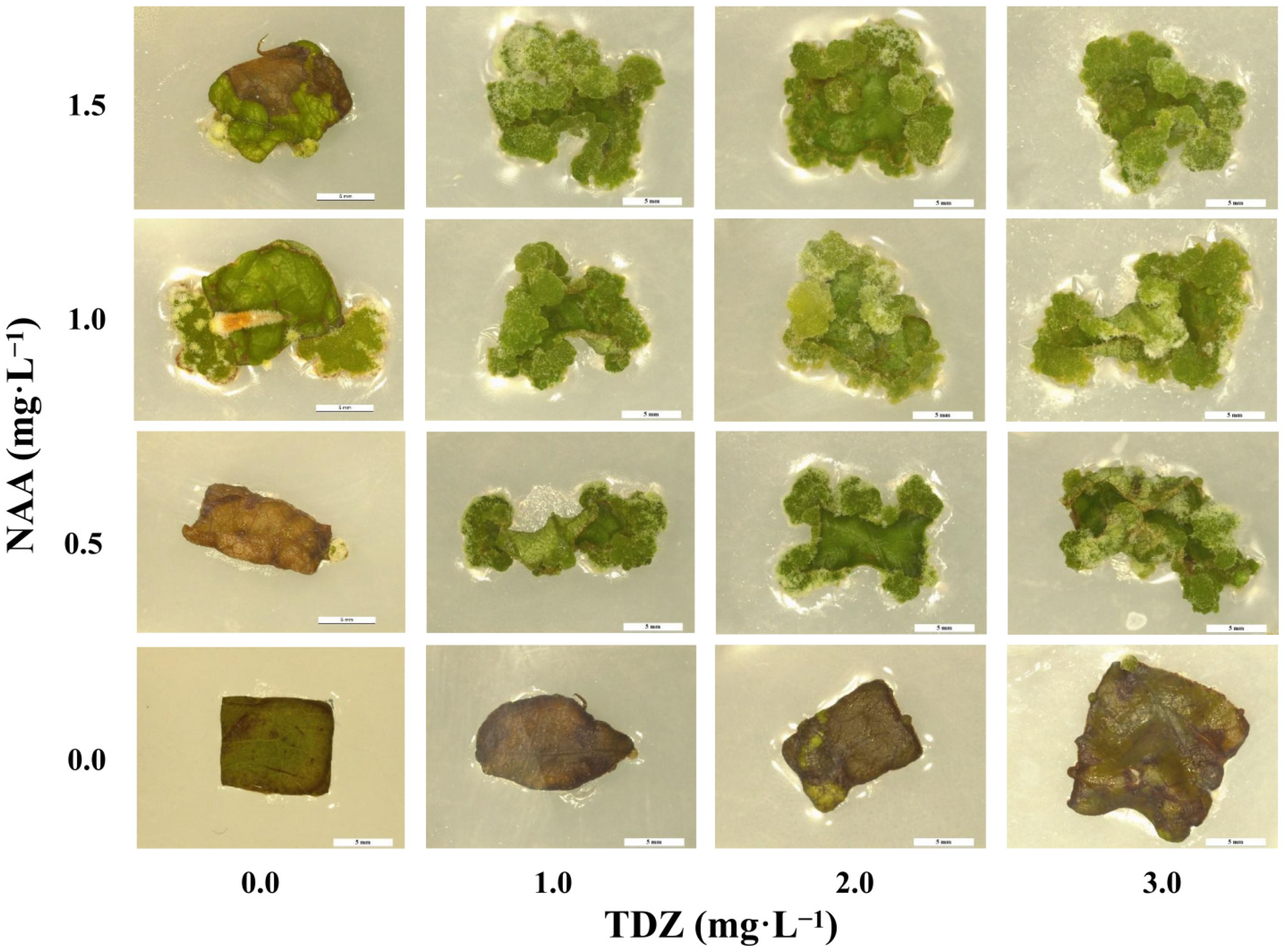
2.2. Callus Induction

The medium used for callus induction was the MS (Murashige and Skoog) medium including vitamins (Duchefa Biochemie, Haarlem, The Netherlands) supplemented with 0, 1.0, 2.0, or 3.0 mg·L−1 TDZ and 0, 0.5, 1.0, or 1.5 mg·L−1 a-naphthaleneacetic acid (NAA). Sucrose (Duchefa Biochemie B.V., Haarlem, The Netherlands) at 30 g·L−1 and agar (Duchefa Biochemie, Haarlem, The Netherlands) at 8 g·L−1 were added into the MS medium, and the pH was adjusted to 5.8 ± 0.1 with NaOH before autoclaving at 121 °C for 30 min. The medium was divided into petri dishes (90.0 mm × 15.0 mm, Cat. No. 11090, SPL Life Sciences, Pocheon, Korea) on a clean bench after autoclaving. Sterilized leaves were cut into 5 mm long squares after cutting off the main veins. Hence, 5 pieces of C. wilsonii leaves were placed on each petri dish. There were three replicates for each treatment. The petri dishes were moved in a dark culture room with a temperature of 24 °C/18 °C (day/night). The callus induction rate and weight of calli were collected one and a half months later. A high accuracy electronic balance (EW 220-3NM, Kern and Sohn GmbH., Balingen, Germany) was placed on a clean bench and sterilized by UV-B light overnight. The weight of the callus was measured in this clean bench next day.

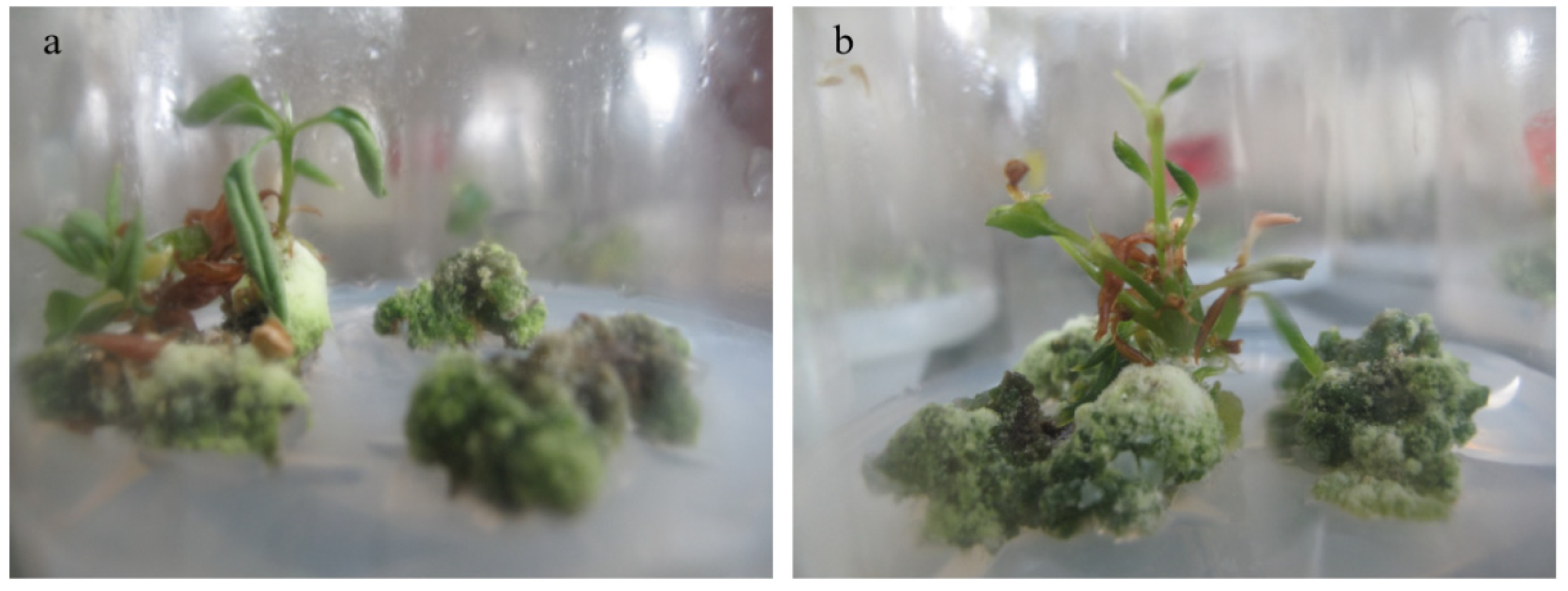
2.3. Shoot Induction
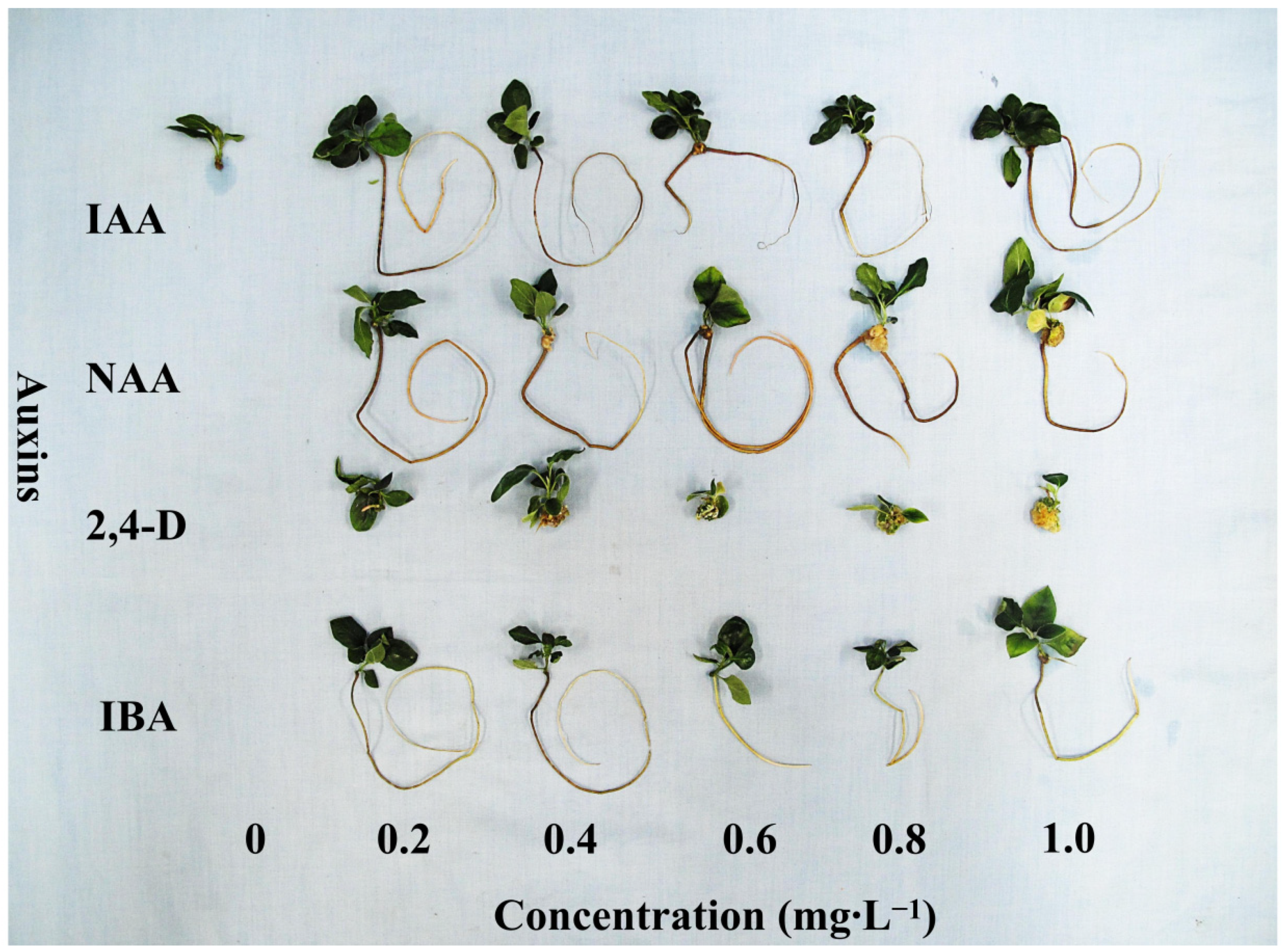
The MS medium for shoot induction was prepared in the same way as the callus induction medium, except for the concentrations of plant growth regulators (PGRs). The calli used for shoot induction were induced by 2.0 mg·L−1 TDZ and 1.5 mg·L−1 NAA, and calli were proliferated by the MS medium supplemented with 2.0 mg·L−1 TDZ and 1.0 mg·L−1 NAA for two passages. The cytokinin TDZ and/or 6-benzyladenine (6-BA) at a concentration of 0, 0.5, or 1.0 mg·L−1 combined with 0.2 mg·L−1 NAA were supplied to the MS medium for shoot induction. Three 0.2 g calli were cultured in one petri dish, and each treatment was repeated three times. All the petri dishes were put into a culture room with a temperature of 24 °C/18 °C (day/night), and with a photoperiod of 16 h with 50 µmol·m−2·s−1 photosynthetic photon flux density (PPFD) from white light emitting diodes (LEDs). The shoot induction rate, number of shoots per callus, and browning rate were collected after a two-month induction.

2.4. Root Induction

The medium used for shoot multiplication was the same as that in Sivanesan et al. [6], 0.5 mg·L−1 TDZ combined with 0.1 mg·L−1 NAA in MS medium. Natural and synthetic auxins indole-3-acetic acid (IAA), NAA, IBA, and 2,4-dichlorophenoxyacetic acid (2,4-D) at a concentration of 0, 0.2, 0.4, 0.6, 0.8, or 1.0 mg·L−1 were supplied to half-strength MS medium for root induction. The C. wilsonii shoots were cut into segments with one node each, and 5 segments were planted on each medium. The root induction rate as well as number and root length of roots per rooted plant were measured two months later.

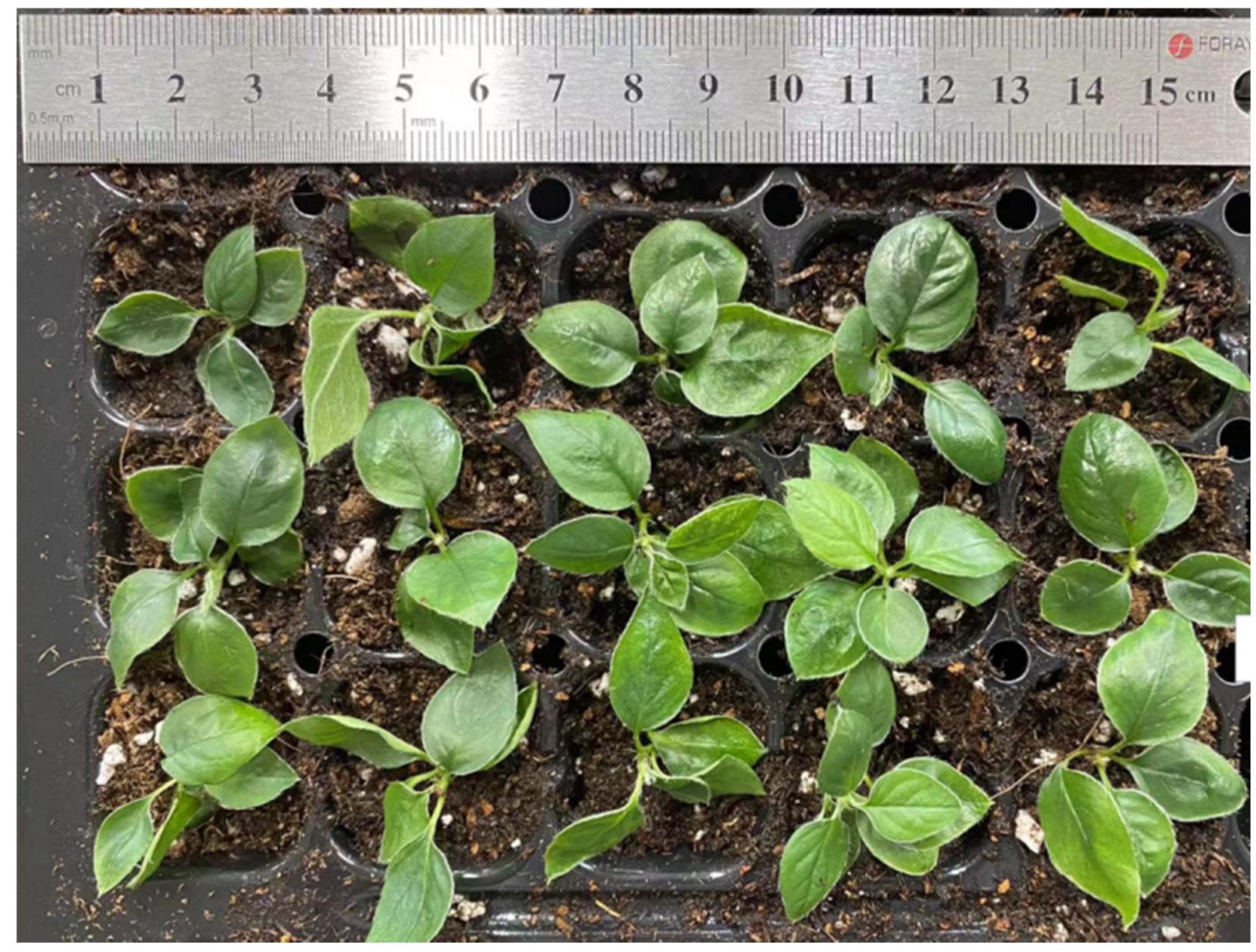
2.5. Acclimatization

The caps of bottles containing rooted shoots or plantlets were open in a culture room for 1 day. Then well-rooted plantlets were taken out and the remaining MS medium was washed away using sterile water. Plantlets were planted in a 128-cell plug tray filled with a commercial growing medium (Bas Van Buuren Substrate, EN-12580, De Lier, The Netherlands). The plug tray was placed in a chamber with 24 °C/18 °C (day/night), a 16 h photoperiod, and 90% relative humidity. Plantlets were moved to a glasshouse after 2 weeks of culture in this growth chamber. Survival rate was collected after another 2 weeks of culture in a glasshouse.

2.6. Statistical Analysis

Significant differences in the data of callus, shoot, and root inductions were analyzed by variance (ANOVA) and Duncan’s multiple range test using the SAS (Statistical Analysis System, version 9.2, Cary, NC, USA) program. The effect of TDZ, NAA, and their interactions on callus induction and weight, as shown in Table 1, were checked through the 2-way ANOVA F-test.

3. Results

3.1. Callus Induction

There was no contamination during callus induction. The explants began to differentiate calli after a two-week cultivation. One and a half months later, the morphology of the calli in each treatment was recorded and is shown in Figure 1. The TDZ alone hardly induced calli (Table 1), while NAA alone, especially at a concentration of 1.0 mg·L−1 successfully induced calli. All combinations of TDZ and NAA induced profuse calli, and the weights of calli increased with the increased concentration of NAA when TDZ was at the same level. The callus weights also increased with higher concentration of TDZ when NAA was at 0.5 or 1.0 mg·L−1. However, there were no significant differences as TDZ concentrations varied when NAA was at a concentration of 1.5 mg·L−1. Moreover, all the treatments, except for treatments without NAA, produced green compact calli, and those calli were covered with a layer of white vigorous cells, indicating that those calli had similar quality regardless of callus weight (Figure 1). The F-test indicated that both NAA and TDZ significantly affected the callus induction in C. wilsonii.
Table 1. Callus induction from the young leaf explants of Cotoneaster wilsonii.
TDZ (A, mg·L−1)NAA (B, mg·L−1)Callus
Induction Ratio (%)Weight (mg)
0.00.00 e 116.9 g
0.576 b35.9 fg
1.096 ab128.4 f
1.548 c57.2 fg
1.00.015 d35.9 fg
0.5100 a213.9 e
1.0100 a282.2 de
1.5100 a471.6 a
2.00.015 d36.5 fg
0.5100 a274.5 de
1.0100 a332.1 cd
1.5100 a400.1 abc
3.00.015 d42.4 fg
0.5100 a373.3 bc
1.0100 a456.2 ab
1.5100 a483.4 a
F-test 2A******
B******
A × B******
1 Lowercase letters indicate significant differences at p ≤ 0.05; 2 *** represent significant at p ≤ 0.001.

3.2. Shoot Induction

Some calli successfully produced shoots after two months of induction period (Figure 2). The shoot induction ratio was the highest when TDZ was at a concentration of 1.0 mg·L−1 (Table 2). BA had little effect on the shoot induction in C. wilsonii. The TDZ at 1.0 mg·L−1 also produced the greatest number of shoots per callus. Approximately 77.7% of the calli cultured on the MS medium without PGRs suffered from browning, and this problem was relieved after PGRs were supplied. BA at 1.0 mg·L−1 caused a more serious browning problem when compared with other treatments. No contaminated plants or calli were found during the whole process.

3.3. Root Induction

The auxins IAA, NAA, and IBA induced, while 2,4-D hardly induced roots in half-strength MS medium (Figure 3 and Table 3). IAA at a concentration of 0.8 mg·L−1 resulted in the greatest root induction ratio (70%). The application of 0.8 mg·L−1 IBA induced the greatest number of roots (1.8) per rooted plant, and IBA at 0.4 mg·L−1 produced the longest root, at an average of 19.6 cm. No contaminated plantlets were found during root induction.

3.4. Acclimatization

The acclimatized plantlets are shown in Figure 4. Approximately 93% plantlets survived, and those plantlets exhibited similar morphological and growth characteristics with their donor plants.

4. Discussion

The combination of cytokinins and auxins was found to be effective for callus induction in many plants [20,21]. High levels of both auxins and cytokinins in the culture medium lead to massive proliferation and dedifferentiation [22]. The great callus induction ratio and callus weights in our study also reflected that high levels of both auxins and cytokinins are necessary for massive callus induction in C. wilsonii. Higher concentrations of auxins than cytokinins are usually required for callus induction. However, a cytokinin/auxin ratio higher that 1 was experimented in this study. Actually, we tried to induce callus using BA and low concentrations of TDZ, and found it was difficult to induce calli in the preliminary experiment. Thus, higher concentrations of TDZ were used to induce callus in this experiment. Some publications also showed that auxin alone successfully induced calli: synthetic auxin 2,4-D alone induced calli from leaf sheath explants of sugarcane [23], and NAA at a concentration of 1 mg·L−1 induced calli from root explants of Dorema ammoniacum [24]. In this study, auxin alone was proven to be effective for callus induction in C. wilsonii, and 1 mg·L−1 NAA resulted in 96% leaf explants forming calli. The auxin-induced callus formation in plants in vitro regeneration represents a typical cell fate change where somatic cells are switched into pluripotent callus cells. Studies related to the molecular control of auxin-induced callus formation have been published in recent years, and several transcription factors contributed to auxin-induced callus formation have been discovered in Arabidopsis [25,26]. However, the potential mechanism of auxin-induced callus formation still needs far more research.
In this study, the cytokinin TDZ was more effective in inducing shoots from calli compared with BA in this study. Explants of some woody species, including C. wilsonii, have naturally strong monopodial growth habits, which causes difficulties in organogenesis in vitro by using common amino purine cytokinins such as BA [27]. The TDZ is known to have higher regeneration rates in comparison to purine-based cytokinins in a number of woody plants [28]. However, the greatest shoot induction ratio was only 44% when the MS medium was supplied with 1 mg·L−1 TDZ in this study. The maximum concentration of TDZ we tested for shoot induction in this study was 1 mg·L−1, so higher concentrations of TDZ can be tested to improve the shoot induction ratio in the future. The precise mechanism of how TDZ affects different scenarios during in vitro woody plant regeneration is still not clear. Physiological-biochemical, genetic researches and the expansion of microscopic approaches are required to clarify the mechanism [29].
Auxins exert direct or indirect effects on root induction and are required for root development [30]. The auxins IAA, NAA, and IBA induced, while 2,4-D hardly induced roots from shoots in this study. Although 2,4-D presents high rooting activity in some plants, it also has high phytotoxicity [31], and it was registered as an herbicide [32]. Moreover, a synthetic auxin 2,4-D is more potent by perhaps 10–1000 times than natural auxins such as IAA [33]. Thus, 2,4-D failed to induce roots in this study, probably due to its toxic effect and the concentrations used in this study might have been too high. The greatest root induction ratio was 70% in this study, much lower than in a previous research (100%), because the shoots used in the previous research were directly from shoot tips or node explants [6]. Rhizogenesis is related to the physiological state of the explant. Perhaps the explants in previous experiment were in different physiological states. Since endogenous auxins are usually synthesized in the shoot tips and tender leaves, and then transported to the action site [34], new leaves on shoots induced from shoot tips or node explants may have a better auxin synthesis ability than calli-induced shoots.

5. Conclusions

Consequently, an effective indirect organogenesis system from young leaves of C. wilsonii Nakai was established. The TDZ at 1.0 mg·L−1 and NAA at 1.5 mg·L−1 not only had 100% callus induction ratio, but also induced the heaviest calli. The TDZ at 1.0 mg·L−1 and NAA at 0.2 mg·L−1 had the greatest shoot induction ratio (44.4%). The greatest root induction ratio (70%) was obtained when IAA at 0.8 mg·L−1. The acclimated plants grew well in a glasshouse with a survival ratio of 93%.

Author Contributions

Conceptualization, B.R.J. and Y.L.; methodology, B.R.J. and Y.L.; software, Y.L.; validation, B.R.J.; investigation, Y.L. and J.X.; resources, B.R.J.; data curation, B.R.J., Y.L. and J.X.; writing—original draft preparation, Y.L.; writing—review and editing, B.R.J., Y.L. and J.X.; supervision, B.R.J. All authors have read and agreed to the published version of the manuscript.

Funding

This research received no external funding. J.X. was supported by a scholarship from the BK21 Program, the Ministry of Education, Republic of Korea.

Institutional Review Board Statement

Not applicable.

Informed Consent Statement

Not applicable.

Data Availability Statement

The data presented in this study are available from the corresponding author upon request.

Conflicts of Interest

The authors declare no conflict of interest.

References

  1. Dickoré, W.B.; Kasperek, G. Species of Cotoneaster (Rosaceae, Maloideae) indigenous to, naturalising or commonly cultivated in Central Europe. Willdenowia 2010, 40, 13–45. [Google Scholar]
  2. Chang, C.S.; Jeon, J.I. Leaf flavonoids in Cotoneaster wilsonii (Rosaceae) from the island Ulleung-do, Korea. Biochem. Syst. Ecol. 2003, 31, 171–179. [Google Scholar]
  3. Yoo, N.H.; Kim, H.K.; Lee, C.O.; Park, J.H.; Kim, M.J. Comparison of anti-oxidant and anti-inflammatory activities of methanolic extracts obtained from different parts of Cotoneaster wilsonii Nakai. Korean J. Med. Crop Sci. 2019, 27, 194–201. [Google Scholar]
  4. Yang, J.; Kim, S.H.; Pak, J.H.; Kim, S.C. Infrageneric Plastid Genomes of Cotoneaster (Rosaceae): Implications for the Plastome Evolution and Origin of C. wilsonii on Ulleung Island. Genes 2022, 13, 728. [Google Scholar]
  5. Yang, J.Y.; Pak, J.H.; Kim, S.C. Chloroplast genome of critically endangered Cotoneaster wilsonii (Rosaceae) endemic to Ulleung Island, Korea. Mitochondrial DNA B 2019, 4, 3892–3893. [Google Scholar]
  6. Sivanesan, I.; Song, J.Y.; Hwang, S.J.; Jeong, B.R. Micropropagation of Cotoneaster wilsonii Nakai—A rare endemic ornamental plant. Plant Cell Tissue Organ Cult. 2011, 105, 55–63. [Google Scholar]
  7. Marković, M.; Grbić, M.; Skočajić, D.; Đukić, M.; Đunisijević-Bojović, D. Propagation of Cotoneaster multiflorus Bunge. By softwood cuttings. In Proceedings of the IX International Scientific Agriculture Symposium, Jahorina, Bosnia and Herzegovina, 4–7 October 2018; pp. 339–342. [Google Scholar]
  8. Mallón, R.; Rodríguez-Oubiña, J.; González, M.L. Shoot regeneration from in vitro-derived leaf and root explants of Centaurea ultreiae. Plant Cell Tissue Organ Cult. 2011, 106, 523–530. [Google Scholar]
  9. Ahmad, N.; Faisal, M.; Anis, M.; Aref, I.M. In vitro callus induction and plant regeneration from leaf explants of Ruta graveolens L. S. Afr. J. Bot. 2010, 76, 597–600. [Google Scholar]
  10. Gantait, S.; Mahanta, M. Picloram-induced enhanced callus-mediated regeneration, acclimatization, and genetic clonality assessment of gerbera. J. Genet. Eng. Biotechn. 2021, 19, 175. [Google Scholar]
  11. Kumar, G. Forest biotechnology: Developments and prospects. Plant Cell Biotechnol. Mol. Biol. 2000, 1, 13–28. [Google Scholar]
  12. Sakakibara, H.; Takei, K.; Hirose, N. Interactions between nitrogen and cytokinin in the regulation of metabolism and development. Trends Plant Sci. 2006, 11, 440–448. [Google Scholar]
  13. Khanam, N.; Khoo, C.; Khan, A. Effects of cytokinin/auxin combinations on organogenesis, shoot regeneration and tropane alkaloid production in Duboisia myoporoides. Plant Cell Tissue Organ Cult. 2000, 62, 125–133. [Google Scholar]
  14. Schaller, G.E.; Street, I.H.; Kieber, J.J. Cytokinin and the cell cycle. Curr. Opin. Plant Biol. 2014, 21, 7–15. [Google Scholar]
  15. Abdouli, D.; Plackova, L.; Doležal, K.; Bettaieb, T.; Werbrouck, S.P. Topolin cytokinins enhanced shoot proliferation, reduced hyperhydricity and altered cytokinin metabolism in Pistacia vera L. seedling explants. Plant Sci. 2022, 322, 111360. [Google Scholar]
  16. Bairu, M.W.; Stirk, W.A.; Dolezal, K.; Van Staden, J. Optimizing the micropropagation protocol for the endangered Aloe polyphylla: Can meta-topolin and its derivatives serve as replacement for benzyladenine and zeatin? Plant Cell Tissue Organ Cult. 2007, 90, 15–23. [Google Scholar]
  17. Murthy, B.; Murch, S.; Saxena, P.K. Thidiazuron: A potent regulator of in vitro plant morphogenesis. Vitr. Cell. Dev. Biol.-Plant 1998, 34, 267–275. [Google Scholar]
  18. Ngomuo, M.; Mneney, E.; Ndakidemi, P. The effects of auxins and cytokinin on growth and development of Musa sp. var. ‘Yangambi’ explants in tissue culture. Am. J. Plant Sci. 2013, 4, 2174. [Google Scholar]
  19. Yu, J.; Liu, W.; Liu, J.; Qin, P.; Xu, L. Auxin control of root organogenesis from callus in tissue culture. Front. Plant Sci. 2017, 8, 1385. [Google Scholar]
  20. Lima, E.C.; Paiva, R.; Nogueira, R.C.; Soares, F.P.; Emrich, E.B.; Silva, Á.A.N. Callus induction in leaf segments of Croton urucurana Baill. Ciênc. Agrotec. 2008, 32, 17–22. [Google Scholar]
  21. Yan, M.M.; Xu, C.; Kim, C.H.; Um, Y.C.; Bah, A.A.; Guo, D.P. Effects of explant type, culture media and growth regulators on callus induction and plant regeneration of Chinese jiaotou (Allium chinense). Sci. Hortic. 2009, 123, 124–128. [Google Scholar]
  22. Wybouw, B.; De Rybel, B. Cytokinin–a developing story. Trends Plant sci. 2019, 24, 177–185. [Google Scholar]
  23. Gopitha, K.; Bhavani, A.L.; Senthilmanickam, J. Effect of the different auxins and cytokinins in callus induction, shoot, root regeneration in sugarcane. Int. J. Pharma Bio Sci. 2010, 1, 1–7. [Google Scholar]
  24. Irvani, N.; Solouki, M.; Omidi, M.; Zare, A.R.; Shahnazi, S. Callus induction and plant regeneration in Dorem ammoniacum D., an endangered medicinal plant. Plant Cell Tissue Organ Cult. 2010, 100, 293–299. [Google Scholar]
  25. Xu, C.; Cao, H.; Zhang, Q.; Wang, H.; Xin, W.; Xu, E.; Zhang, S.; Yu, R.; Yu, D.; Hu, Y. Control of auxin-induced callus formation by bZIP59–LBD complex in Arabidopsis regeneration. Nat. Plants 2018, 4, 108–115. [Google Scholar]
  26. Fan, M.; Xu, C.; Xu, K.; Hu, Y. Lateral organ boundaries domain transcription factors direct callus formation in Arabidopsis regeneration. Cell Res. 2012, 22, 1169–1180. [Google Scholar]
  27. Huetteman, C.A.; Preece, J.E. Thidiazuron: A potent cytokinin for woody plant tissue culture. Plant Cell Tissue Organ Cult. 1993, 33, 105–119. [Google Scholar]
  28. Jones, M.; Yi, Z.; Murch, S.J.; Saxena, P.K. Thidiazuron-induced regeneration of Echinacea purpurea L.: Micropropagation in solid and liquid culture systems. Plant Cell Rep. 2007, 26, 13–19. [Google Scholar]
  29. Novikova, T.I.; Zaytseva, Y.G. TDZ-induced morphogenesis pathways in woody plant culture. In Thidiazuron: From Urea Derivative to Plant Growth Regulator; Ahmad, N., Faisal, M., Eds.; Springer: Singapore, 2018; pp. 61–94. [Google Scholar]
  30. Negishi, N.; Nakahama, K.; Urata, N.; Kojima, M.; Sakakibara, H.; Kawaoka, A. Hormone level analysis on adventitious root formation in Eucalyptus globulus. New Forest. 2014, 45, 577–587. [Google Scholar]
  31. Waite, D.T.; Cessna, A.J.; Grover, R.; Kerr, L.A.; Snihura, A.D. Environmental concentrations of agricultural herbicides: 2,4-D and triallate. J. Environ. Qual. 2002, 31, 129–144. [Google Scholar]
  32. Tofanelli, M.B.D.; Freitas, P.L.; Pereira, G.E. 2, 4-dichlorophenoxyacetic acid as an alternative auxin for rooting of vine rootstock cuttings. Rev. Bras. Frutic. 2014, 36, 664–672. [Google Scholar]
  33. Phillips, G.C.; Garda, M. Plant tissue culture media and practices: An overview. Vitr. Cell. Dev. Biol.-Plant 2019, 55, 242–257. [Google Scholar]
  34. Ljung, K.; Bhalerao, R.P.; Sandberg, G. Sites and homeostatic control of auxin biosynthesis in Arabidopsis during vegetative growth. Plant J. 2001, 28, 465–474. [Google Scholar]
Figure 1. Morphology of the calli induced from the leaf explant of Cotoneaster wilsonii. Bars indicate 5 mm.
Horticulturae 08 00795 g001
Figure 2. Shoots induced from calli in Cotoneaster wilsonii: (a) treatment with 0 mg·L−1 TDZ and 1.0 mg·L−1 BA; and (b) treatment with 1.0 mg·L−1 TDZ and 0 mg·L−1 BA.
Horticulturae 08 00795 g002
Figure 3. Root induced by different concentrations of auxins in Cotoneaster wilsonii.
Horticulturae 08 00795 g003
Figure 4. Acclimatized Cotoneaster wilsonii in a glasshouse.
Horticulturae 08 00795 g004
Table 2. Shoot regeneration from calli in Cotoneaster wilsonii.
TDZ
(mg·L−1)
BA
(mg·L−1)
NAA (mg·L−1)Induction (%)No. of Shoots/CallusCallus Browning (%)
0.00.00.00.0 c 10.0 e77.7 a
0.50.211.1 bc0.7 de0.0 c
1.00.222.2 bc2.5 d33.3 b
0.50.00.211.1 bc0.3 de0.0 c
0.50.233.3 ab2.7 bc0.0 c
1.00.222.2 bc3.0 cd22.2 bc
1.00.00.244.4 a5.3 a11.1 bc
0.50.233.3 ab3.3 b0.0 c
1.00.233.3 ab4.8 b22.2 bc
1 Lowercase letters indicate significant difference at p ≤ 0.05.
Table 3. The effects of auxins on the root induction in Cotoneaster wilsonii.
AuxinConcentration (mg·L−1)Rooting (%)No. of Roots/PlantRoot Length (cm)
-0.00 e 10.0 c0.0 f
IAA0.210 de1.0 bc10.2 ef
0.440 cd1.3 abc17.1 ab
0.650 bcd1.4 ab11.4 cde
0.870 a1.1 abc15.5 bcd
1.030 cde1.3 abc15.4 bcd
NAA0.260 abc1.0 bc10.2 def
0.460 abc1.7 ab10.3 def
0.640 cd1.3 abc15.9 bcd
0.830 cde1.7 ab7.9 ef
1.050 bc1.6 ab6.9 ef
2,4-D0.210 de1.0 bc0.3 ef
0.410 de1.0 bc1.3 ef
0.60 e0.0 c0.0 f
0.80 e0.0 c0.0 f
1.00 e0.0 c0.0 f
IBA0.210 de1.0 bc12.3 cde
0.450 bcd1.2 abc19.6 a
0.640 cd1.3 ab11.4 cde
0.830 cde1.0 bc9.8 def
1.040 cd1.8 a13.7 cde
1 Lowercase letters indicate significant difference at p ≤ 0.05.
Publisher’s Note: MDPI stays neutral with regard to jurisdictional claims in published maps and institutional affiliations.

