Article

Effect of Flavonoid Dynamic Changes on Flower Coloration of Tulipa gesneiana ‘Queen of Night’ during Flower Development

1
School of Design, Shanghai Jiao Tong University, Shanghai 200240, China
2
Instrumental Analysis Center, Shanghai Jiao Tong University, Shanghai 200240, China
*
Author to whom correspondence should be addressed.
Horticulturae 2022, 8(6), 510; https://doi.org/10.3390/horticulturae8060510
Submission received: 19 April 2022 / Revised: 27 May 2022 / Accepted: 7 June 2022 / Published: 9 June 2022
(This article belongs to the Special Issue Advances in Ornamental Plant Cultivation and Physiology)

Abstract

:
Tulipa gesneriana has rich cultivars with abundant flower colors; among them, black tulips have become precious cultivars for their rareness in nature. It is of great significance to understand its color formation mechanism for breeding new cultivars with a blackish flower color in the future. In this present study, petals at five developmental stages of Tulipa gesneriana ‘Queen of Night’ (shorted for ‘QN’), a typical black tulip cultivar, were collected to identity the flavonoid composition and compare the accumulation along with the flower development process, aiming to explore the metabolic mechanism of its flower coloration. By using UPLC-Q-TOF-MS, three anthocyanins and nineteen anthoxanthins (including eighteen flavonols and one flavone) were detected in the petals of ‘QN’. The anthocyanins were identified as delphinidin 3-o-rutinoside, cyanidin 3-o-rutinoside and pelargonidin 3-o-rutinoside, respectively. The main flavonols were identified as quercetin, kaempferol, isorhamnetin, naringin and their glycosides. The only one flavone substance was identified as an apigenin derivative. By comparing the content, anthocyanins were the most abundant substance in the petals of ‘QN’, and showed obvious regularity in the development process. With the flower opening, the anthocyanin content accumulated continuously, and reached the highest level at 3575.9 μg·g−1·FW at S5, accounting for 80% of the total flavonoids. Among them, delphinidin 3-o-rutinoside and cyanidin 3-o-rutinoside made the main contribution to the coloration of ‘QN’. The content of anthoxanthins increased first and then decreased, reaching the highest to 1114.8 μg·g−1·FW at S3. Quercetin and its glycosides were the most important flavonol substances in the petals of ‘QN’, accounting for more than 60% of the total anthoxanthins in the five stages. The content of a flavone identified was extremely low, suggesting a limited role in the flower color of ‘QN’. Taken together, the flower color presentation of ‘QN’ was closely related to the composition and accumulation of anthocyanins and flavonols; anthocyanins were the main substances that determine the petal coloration of ‘QN’, and the high content of flavonols played a role of co-pigmentation with these anthocyanins.

1. Introduction

Flower color is one of the most important ornamental traits of floricultural plants. The metabolic mechanism of flower color formation has been well characterized [1]. Research shows that flower color is the result of the accumulation of metabolites (pigments) in flower epidermal cells, and the relationships among pigment composition and content are the main internal factors that determine the flower color [2,3]. The pigments of affecting flower color are fundamentally divided into three main classes: flavonoids, carotenoids and betalains [4,5]. Flavonoids, as a water-soluble secondary metabolites, widely accumulated in plant cell vacuoles and distributed throughout the plant kingdom, are the most important pigment type [6,7]. Anthocyanin is an important subclass of flavonoids in plant flower organs, which can make flowers show different colors such as red, pink, blue and purple. Other flavonoid substances reflect the color from light yellow to dark yellow [8,9]. They are usually called anthoxanthins, which generally act as the co-pigment of anthocyanins [10,11].
With the continuous improvement of people’s aesthetic requirements, the rare black flower variety in nature has attracted the attention of the floral market because of its special flower color. However, the color mechanism of black flowers is complex. Some scholars suggested that the appearance of black flowers was caused solely by the accumulation of high anthocyanins, such as the findings in Alcea rosea [12] and Thermopsis barbata [13]. Other scholars believed that there was a certain kind of flavonoid related to the high content of anthocyanins or components, which can darken or deepen the black of flowers. For example, in Cosmos atrosanguineus, it was reported that the change of flower color from red to black was caused by the different relative contents of anthocyanins and chalcone [14]. Markham et al. found that the coloration of Lisianthius nigrescens was caused by the molar ratio of the total anthocyanin content to flavone content of 1:1 [15]. In the case of Centaurea cyanus, this seemed to be the color difference of blue, purple and black flowers caused by the difference between the components of cyanidin derivatives and flavone [16]. The study on the color of Zantedeschia hybrida showed that it did not accumulate flavones, but the high content of flavonol played a role of co-pigmentation with the anthocyanins [17]. It was also reported that the coloration of black plants was caused by the interaction between anthocyanins and proanthocyanidins [18]. Therefore, it is necessary to further verify which flavonoid substance may act with anthocyanins to cause this black coloration. Studying more black flower plants and accumulating evidence will help to better explain the formation mechanism of black flowers.
Tulip (Tulipa gesneriana) is a perennial bulbous flower of Liliaceae. For its beautiful flower shapes and abundant flower colors, the tulip occupies an important position in the international flower market [19]. Among abundant tulip cultivars, the black series has always been the most precious cultivars, with a very high economic value [20]. Previous studies revealed flavonoids and carotenoids were common pigments in diverse tulip cultivars, while no betalain was reported in tulips [21,22]. To date, anthocyanins were detected in many tulip cultivars, and 13 anthocyanin pigments in total were already reported in tulips [23,24]. Meanwhile, the relationship between anthocyanin composition, content and flower color has been established [23,25]. Among these anthocyanins, delphinidin and cyanidin corresponded to the red, purple and blue flower color in tulips [23,25]. However, there are also few reports focused on the pigmentation of black tulip cultivars. In addition, studies on flavonol components and their relationship with anthocyanins were limited. For instance, in Tulipa gesneriana ‘Queen of Night’, Nakayama et al. and Torskangerpoll et al. identified two to three components and determined the content proportion of anthocyanin components, respectively [23,25], while Wang et al. reported four anthocyanins in this cultivar, which were completely different from the components reported by the former [22]. They all lacked attention to anthoxanthins. In addition, the discussion on the regular accumulation of pigment components was also ignored. Therefore, the color metabolism mechanism of the black tulip is not fully elucidated, for which there is still a lot of room for discussion.
Tulipa gesneriana ‘Queen of Night’, a typical black cultivar, was used in the present study. Since its petals show obvious coloring regularity from green to red to, finally, blackish purple, along with the process of flower development, this cultivar is thus a good material to study flower coloration. By identifying the flavonoid composition in its petals and further analyzing their accumulation along with the flower’s development, this study aims to clarify its metabolic mechanism and provide a scientific basis for further studying the molecular mechanism of flower coloration, and also provide a theoretical basis for tulip breeding with a blackish flower color.

2. Materials and Methods

2.1. Plant Materials and Reagents

Tulipa gesneriana ‘Queen of Night’ (shortened to ‘QN’ in following texts, tables and figures), imported from the Netherlands, was used as the research material. The healthy bulbs were planted in the Modern Agricultural Engineering Training Center of Shanghai Jiao Tong University (Shanghai, China) in late November 2020. Referring to the criteria mentioned by Momonoi and Nakatsuka et al. [26,27], the flowering period of tulip is divided into five stages (S1–S5, Table S1), and the petals at each stage were collected from certain individual plants, respectively, in April 2021 (Figure 1). The sample at each stage was formed by mixing petals from five individual plant. The samples were frozen in liquid nitrogen immediately and stored in a −80 °C freezer subsequently for pigment extraction.
Several standards and reagents were used for the qualitative and quantitative identification of anthocyanins and anthoxanthins in ‘QN’ petals. All standards were shown in Table S2. The reagents contain 0.1% hydrochloric acid aqueous solution of 70% methanol, 0.1% formic acid H2O and 0.1% formic acid acetonitrile.

2.2. Methods

2.2.1. Color Reactions of Petal Pigment

The 0.2 g petals at the blooming stage (S5) were weighed and put into the mortar with clean stainless steels tweezers. Then 10 mL of petroleum ether, 10% hydrochloric acid and 30% ammonia were added, respectively, to the mortar, to observe the color of the liquid after grinding which was then recorded [28].

2.2.2. Identification and Measurement of Flavonoids

Extraction of Flavonoids

Fresh samples (0.5 g) at five stages, respectively, were fully ground into powder in a mortar, transferred to a centrifugal tube, then 5 mL extraction solution (0.1% hydrochloric acid aqueous solution of 70% methanol) was added and this was vortexed for 30 s. Next, the mixture was added to ultrasonic oscillation wave for 40 min, placed at 4 °C away from light for 24 h and shaken several times. Then, the extracts were centrifuged at 12,000 rpm for 10 min. Finally, the 800 μL of supernatant were taken for qualitative and quantitative analysis of flavonoids.

Qualitative and Quantitative Analysis of Flavonoids

The instruments used were ultra-high-performance liquid chromatography and VION ion mobility quadrupole time-of-flight mass spectrometer (UPLC-Q-TOF-MS) (Waters Corporation, Milford, MA, USA), which configured with Optima LC/MS-grade methanol and trifluoroacetic acid (Thermo Fisher Scientific, Waltham, MA, USA), for identifying the components and contents of anthocyanin and anthoxanthins in petal extractions of ‘QN’. The analysis conditions of UPLC-Q-TOF-MS were referred to previous research of our group [29]. Among them, the PDA detector was used for full UV scanning, then 520 nm and 350 nm were selected for separate scanning modes. The electrospray ion scan mode of anthocyanin was positive, while that of anthoxanthin was negative. Additionally, anthocyanins and anthoxanthins were detected at 520 nm and 350 nm, respectively. The compounds were estimated comprehensively according to the mass spectrometry information, molecular mass, molecular formula, standard compound information and relevant literature [30].
The flavonoid components in petals of ‘QN’ were quantified by the external standard method [31]. Eight working solutions with concentrations of 0.1, 0.5, 1, 5, 10, 50, 100 and 500 μg·mL−1 were prepared by diluting the standard stock solution to verify the linearity of the flavonoids’ curves. The linearity coefficients were determined by the analytical curves generated by the above-mentioned concentration levels [31].
In the method for quantification of the anthocyanin and anthoxanthin components, the analytical curves were constructed by using European Pharmacopoeia Reference Standard (a mixed standard of anthocyanin) and Rutin as reference, respectively. The total anthocyanin content (TAC) and total anthoxanthin content (TAX) were calculated with standard curves. The standard curves constructed were shown in Table S3. The results were calculated by fresh weight.

3. Results

3.1. Color Reaction of Petal Pigment of ‘QN’

In the petroleum ether extract of ‘QN’, it was colorless, indicating that no carotenoid or a very small amount of carotenoids was present in its petals. In the hydrochloric acid test, it was deep red, indicating that the petals of this cultivar contained a large amount of anthocyanins [28]. As for the ammonia reaction, the liquid was yellow, suggesting that it contained flavone or flavonol substances [28]. The above color reaction revealed that the main pigment in the petals of ‘QN’ contained flavonoids, including anthocyanins, flavone or flavonol substances (Figure S1) [28].

3.2. Analysis of Flavonoid Composition in the Petals of ‘QN’

3.2.1. Identification of Anthocyanins

Mass spectrometry (MS) data showed that three components (named A1, A2 and A3) were detected in the petals of ‘QN’, which were observed in all five stages (Figure S2).
There was no direct match between the peak component and the anthocyanin mixed standard component. Therefore, these substances were then estimated according to the comprehensive information such as peak time and MS data (Table 1). The aglycone ion peaks of components A1, A2 and A3 are m/z 303 ([Y0+]), m/z 287 ([Y0+]) and m/z 271 ([Y0+]), respectively, which represent the derivatives of delphinidin aglycone, cyanidin aglycone and pelargonidin aglycone, respectively. The molecular mass difference between their molecular ions and aglycone ions was 308, which was consistent with that of rutinoside [32]. Combined with the comparison of the relevant literature and MS information [23,24], the three components were identified as delphinidin 3-o-rutinoside (Dp-3-rut), cyanidin 3-o-rutinoside (Cy-3-rut) and pelargonidin 3-o-rutinoside (Pg-3-rut), respectively.

3.2.2. Identification of Anthoxanthins

Eighteen flavonols and one flavone were identified from the petal extraction solution of ‘QN’ at 350 nm by UPLC-Q-TOF-MS. These anthoxanthin components were labeled as F1-F19 according to the retention time (Figure S3). The UPLC data, including retention time, molecular ions, fragment ions and aglycone ions, were listed in Table 2. The research has shown that in the negative ion mode, the anthoxanthin substances were generally in position three and position seven [24,33].
The identification of anthoxanthins was similar with that of anthocyanins in the analysis method. F5, F7, F10, F13 and F19 had fragment ions m/z 301 ([Y0]), corresponding to the mass spectrum characteristics of quercetin, thus indicating that they were quercetin derivatives. MS showed that F10 had a quasi-molecular ion m/z 595 [M-H] and fragment ions m/z 301 [M-294], 294 lost were usually the characteristic mass-to-charge ratio of sambubiose, thus tentatively identified as Quercetin-3-sambubiose [34]. According to the correlative literature, it can be also found that 308 lost was consistent with a rutinoside, and 178 and 164 usually corresponded to acetylpentose and galactose, respectively [32,33,34,35,36]. Thus, by identifying characteristic aglycone ions and calculating the lost value of fragment ions, F5, F7 and F19 were inferred as Quercetin-3-acetylpentose-7-sambubiose, Quercetin-3-galactose-sambubiose and Quercetin-3-sophoroside, respectively [32,33,34,35,36]. F14 had a quasi-molecular ion m/z 609 [M-H] and fragment ions m/z 301 [M-308], and the elution retention time of F14 was the same as that of the standard compound, Rutin (Quercetin-3-rutinoside). Therefore, F14 was identified to be Rutin. The mass spectrum data of F13 is unanimous with that of F14, thus indicating F13 was Quercetin-7-rutinoside.
F6, F9, F12, F15, F16 and F17 were suggested to be three sambubiose, one sophoroside, one malonylgalactose and one rutinoside of the flavonol with its fragment ions at m/z 285 ([Y0]), which corresponded to the kaempferol derivatives. F16 had a quasi-molecular ion m/z 579 [M-H] and fragment ions m/z 285 [M-294], thus it can be tentatively identified as Kaempferol-3-sambubiose [34]. F17 had a quasi-molecular ion m/z 593 [M-H] and fragment ions m/z 285 [M-308], and thus can be inferred as Kaempferol-3-rutinoside [32,34]. In a similar manner, F6 and F15 were inferred as Kaempferol-3-sophoroside and Kaempferol-7-malonylgalactose, respectively. Since there was a distinction between the different fragments about the relative abundance, it can be identified that some components were modified with the glycosidation of a disaccharide, for which the connection mode of the glycosidic bond could be different [32,33,34]. According to the fragment value of the components, it can be found that F9 and F12 were modified by the disaccharide. F9 had a quasi-molecular ion m/z 726 [M-H]- and fragment ions m/z 579 [M-147], 285 [M-147-294], 147 lost which may correspond to rhamnoside and 294 was usually the characteristic mass-to-charge ratio of sambubiose, thus, F9 was tentatively identified as Kaempferol-3-rhamnoside-sambubiose [32,33,34]. Similarly, F12 was tentatively identified as Kaempferol-3-galactose-sambubiose [32,33,34].
F1, F2, F4 and F18 had fragment ions m/z 315 ([Y0]), indicating that these three substances were isorhamnetin derivatives. By direct comparison with collected standards of isorhamnetin glycosides, F1, F2, F4 and F18 can be inferred as Isorhamnetin-7-succinylrutinoside-rhamnoside, Isorhamnetin-3-succinylrutose-galactoside, Isorhamnetin-3-sophoroside-7-gallicylglucoside and Isorhamnetin-3-succinylsophoroside, respectively [32,33,34].
The fragment ions of F8 and F11 are m/z 271 ([Y0]), corresponding to naringin derivatives. MS showed that F11 had a quasi-molecular ion m/z 595 [M-H] and fragment ions m/z 271 [M-324], 324 corresponding to a disaccharide, usually the characteristic mass-to-charge ratio of sophoroside [32], thus F11 can be tentatively identified as Naringenin-3-sophorosid. Similarly, F8 can be speculated as Naringin-3-acetylrutinoside [32,34].
Since F3 was the only flavone detected in the extract, with fragment ions m/z 269 ([Y0]), corresponding to the apigenin derivative. F3 had the molecular ion m/z 745 [M-H] and fragment ions m/z 451 [M-294] and m/z 269 [M-294-182], and 182 lost may correspond to acetylrhamnose, thus it can be tentatively identified as Apigenin-3-acetylrhamnose-7-sambubiose [32,33,34].

3.3. The Accumulation of Flavonoids along with the Process of Flower Opening

3.3.1. Dynamic Changes of Anthocyanins

The total amount of anthocyanins accumulated continuously during flower opening and reached the highest value to 3575.9 μg·g−1·FW at S5, which was 42.5 times of S1 (Figure 2A). Among them, the accumulation of Dp-3-rut was consistent with that of the total anthocyanins and peaked at S5 with the content of 3126.9 μg·g−1·FW, corresponding to 118.1 times of S1. The accumulation of Pg-3-rut and Cy-3-rut both increased firstly, which was then followed by a decrease, and reached the highest at S3, which were 17.9 times and 38.9 times of S1, respectively.
During different development stages, the proportion of each component was different. It has been reported that the proportion of the pigment components may affect the appearance of the flower color [14,15,16]. Therefore, in our study, the columnar percentage accumulation diagram was constructed by calculating the proportion of each component in the total anthocyanin contents with the development of ‘QN’. The content proportion of three anthocyanin components were varied greatly, as shown in Figure 2B. Although the content of Pg-3-rut increased in the early flowering stages (S1–S3), its percentage in the total anthocyanins presented an overall decrease up to S5, its proportion was equal to about 10% from S3 to S4 and a minimum percentage (2%) was present at S5. Interestingly, the percentage of Cy-3-rut accounts for about 50% from S1 to S4. The proportion of Cy-3-rut and Dp-3-rut were the same at S4, about 45%. The percentage of Cy-3-rut decreased by 35% at S5, while Dp-3-rut was as high as 87%. On the whole, the proportion of Cy-3-rut was highest (45–56%), followed by that of Dp-3-rut (29–45%) in the early stages (from S1 to S4); these two components accounted for the vast majority of total anthocyanins, and only 10–22% of Pg-3-rut was present within these stages. However, at the flower opening stage (S5), Dp-3-rut became the only majority component of anthocyanins, reaching 87%, accompanied with only 11% of Cy-3-rut and less than 2% of Pg-3-rut.

3.3.2. Dynamic Changes of Anthoxanthin Components

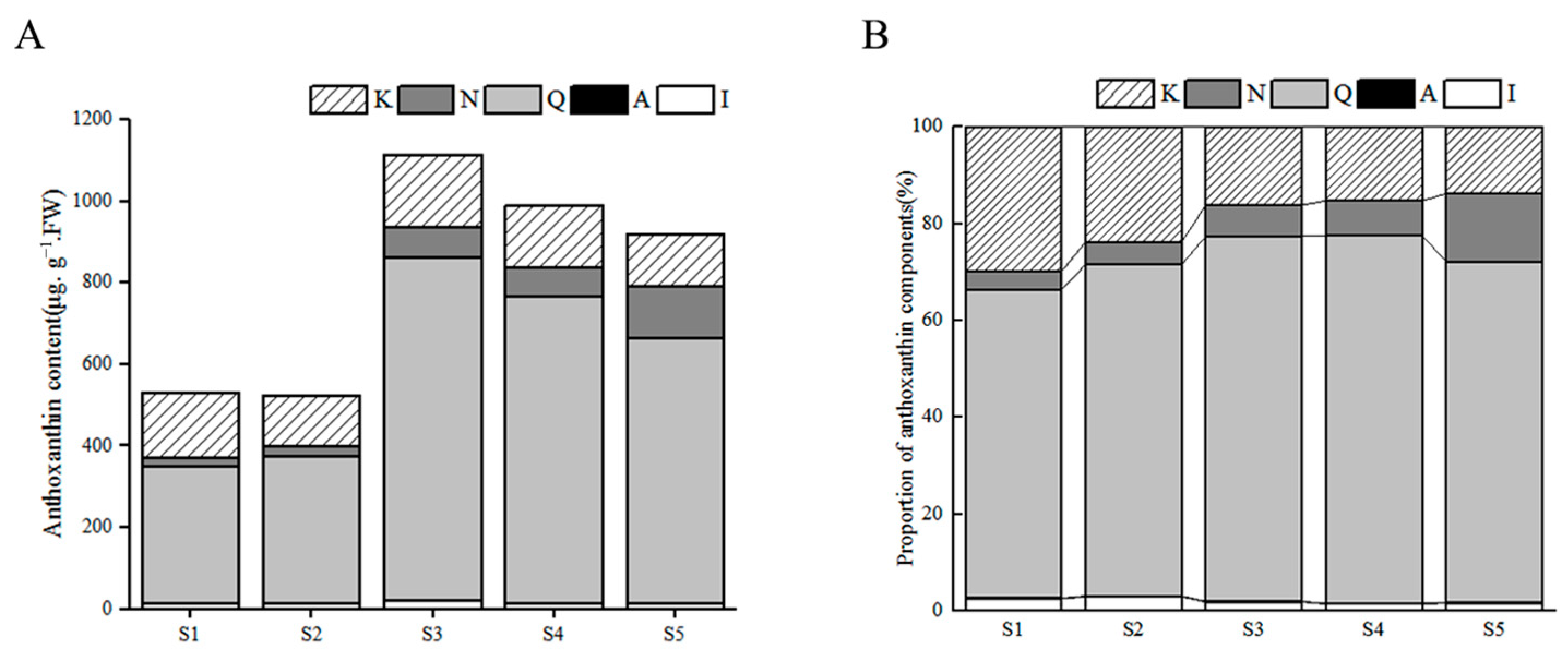
Anthoxanthins were detected in the petals of ‘QN’ by identification, which were a kaempferol derivative (K), naringin derivative (N), quercetin derivative (Q), apigenin derivative (A) and isorhamnetin derivative (I). As shown in Figure 3A, the total amount of anthoxanthins increased first and then decreased, and accumulated to a maximum of 1114.8 μg·g−1·FW at S3, 2.1 times of S1. The accumulation trends of I and Q were consistent with that of the total anthoxanthins. Among all anthoxanthins, component Q contributes the most, followed by component K. The content of the only flavone, component A, was below the detection limit, with almost no contribution to the anthoxanthins.
The percentages of the individual anthoxanthin components were calculated and the results were shown in Figure 3B. Among all the components, Q had an important proportion within the whole flowering process, which was more than 60%. Component K generally accounted for the second highest proportion in anthoxanthins, especially when at 30% in S1, and then gradually decreased, where the minimum proportion was present at S5. Therefore, kaempferol and quercetin derivatives accounted for the absolute majority of anthoxanthins. An increase trend of the proportion was observed in the components N and I, which were different to that of Q. Component I had very low proportion of about less than 3%. The proportion of A was almost 0 because of its very low content.

3.3.3. Dynamic Changes of Total Flavonoids

The contents of anthocyanins and anthoxanthins in each stage were accumulated and the dynamic accumulation pattern of the total flavonoids in ‘QN’ were presented in Figure 4. The accumulation of flavonoids showed a rising trend, along with the flower developmental process. The total flavonoid content reached 4494.3 μg·g−1·FW at S5, 7.3 times of S1. During the early developmental stages (S1–S2), the contents of both anthocyanins and anthoxanthins were at a low level, and the content of anthoxanthins was higher than that of anthocyanins. However, the content of anthocyanins increased dramatically later and was much higher than that of anthoxanthins during the late developmental stages (from S3 to S5).
In terms of the content proportion, an increasing trend of the anthocyanin proportion was observed along with the opening of flowers, peaked at S5 with a percentage of 80%, and its proportion was equal to about 75% during S3–S4. However, an opposite trend of anthoxanthins was present, the highest (86%) was present at S1, then followed a continuous decrease up to S5 with the minimum value of 20%. At the early stages of flower development, the petals were almost colorless, and anthoxanthins accounted for the main part of the flavonoids. When the inner side of the petal was completely colored (S3), although the anthoxanthin content was higher than that of S1 and S2, the anthoxanthin percentage had rapidly declined from 76% at S2 to 28% at S3, accompanied with a fast increase of the anthocyanin proportion which had risen from 24% to 72%. Subsequently, this low proportion of anthoxanthins and high proportion of anthocyanins were maintained up to S5. Surprisingly, the proportion ratio of TAC and TAX was close to three during S2–S4, even to four at S5, and so it can be found that the proportion of TAC and TAX were basically reciprocals to each other in the early or late stages.

4. Discussion

In this study, the flavonoid components in the petals of ‘QN’ were identified and analyzed by UPLC-Q-TOF-MS. A total of three anthocyanin components and nineteen anthoxanthins (including eighteen flavonols and one flavone) were detected in the petals of ‘QN’. Meanwhile, the accumulation of these substances was obviously related to the coloring of petals in the process of flower development in ‘QN’.
The anthocyanins were identified as delphinidin 3-o-rutinoside, cyanidin 3-o-rutinoside and pelargonidin 3-o-rutinoside, respectively, which were previously reported in certain tulips [23]. However, in ‘QN’, the different scholars have had different identification results about the quantity and structure of anthocyanins components. Wang et al. considered that there were four substances in ‘QN’, among these components are different from our study [22]. They did not give a clear basis for component identification and analysis. We thought this may be related to the determination method of the anthocyanin components. Wang et al. used spectral characteristics to identify the components by high-performance liquid chromatography (HPLC). Studies have shown that the UPLC method is more sensitive, faster and consumes less eluent than HPLC [38,39]. In terms of sensitivity with UPLC, analyte-dependency increased by ten times and the method speed improved by five times under conditions of comparable peak separations with HPLC [40]. In earlier studies, Nakayama et al. and Torskangerpoll et al. identified two and three components, respectively, through identification by HPLC, for which the identified components were consistent with the results of this study [23,25]; this can further confirm our conclusion. Furthermore, in our study, we can demonstrate that there were only three peaks in the mass spectra of the petals of five different developmental stages in ‘QN’. Consequently, it is clear that the anthocyanin components of ‘QN’ are delphinidin 3-o-rutinoside, cyanidin 3-o-rutinoside and pelargonidin 3-o-rutinoside. As for anthoxanthin components, four isorhamnetin derivatives, one apigenin derivative, six quercetin derivatives, six kaempferol derivatives and two naringin derivatives were identified. Previous studies have reported that these components were closely related to the color appearance of ornamental flowers, such as in water lilies (Nymphaea spp.), Zantedeschia hybrida, Paeonia suffruticosa et al. [17,41,42]. This is the first report of these compounds in Tulipa gesneriana, except for Rutin. Among them, the apigenin derivative is only one flavone substance with a very low content. The rest of the anthoxanthin components are all flavonol substances. Therefore, anthocyanins and flavonols are the main flavonoid substances in petals of ‘QN’ that may affect its coloration.
The accumulation of total flavonoids showed an obvious increase along with the flower development process in petals of ‘QN’. The accumulation of anthocyanins was consistent with that of flavonoids, and it was the main contributor of flavonoids in the important period of petal coloring (S3–S5). This result reveals that anthocyanins play a key role in petal coloration in ‘QN’. The increase trend of anthocyanins was consistent with the previous reports of Zhao et al. [43] and Kitamura et al. [18]. A similar finding of the high anthocyanins was also reported in other black flowers such as Cosmos atrosanguineus [14] and Dahlia variabilis [44,45]. Among the three identified components, Dp-3-rut was the highest component at the full flowering stage (S5), accounting for 87% of the total anthocyanins, which was consistent with the results of Nakayama et al. [25]. Studies have shown that the coloration appearance of black flowers was caused by the absorption of visible light under the action of the combination of Dp and cyanidin (Cy) [16,46,47], which can also be verified in this study. The high levels of Dp and Cy derivatives were observed in petals of ‘QN’, but their accumulation patterns were different, suggesting they may play a role in different stages of the petal coloration. Before the flower full opening (S1-S3), the proportion of Cy-3-rut in the total anthocyanins was greater than that of Dp 3-rut, revealing that Cy-3-rut played a major role within these stages in petals of ‘QN’. At S4, the content of Dp 3-rut was basically the same as that of Cy-3-rut, which can be seen in that after the inner perianth was completely dark colored and the outer perianth petals were just colored, the effects of Cy-3-rut and Dp 3-rut kept the balance, approximately. During the flower’s full opening (S5), Dp 3-rut increased dramatically and became the absolutely dominant pigment in the petals, revealing its significant role in this stage. The above accumulation patterns were well consistent with its color characteristics: at first, the petals appeared slightly pink, then turn reddish purple and further to blackish. Corresponding to the accumulation and interaction of various pigments, the petal color deepened and tended to black at last. This finding was consistent with the studies of Nieuwhof et al. and Van raamsdonk et al., who suggested that the petals of the dark red, magenta, orange and pink varieties were mainly cyanidin; the petals of purplish red to blue–purple were mainly Dp [21]. It was also found that the accumulation of Cy was one of the main reasons for its color in the black Centaurea cyanus and Zantedeschia hybrida [16,17]. In a black Dahlia, it was found that Cy derivatives had a greater impact on the flower color than pelargonidin (Pg) derivatives [45]. Therefore, Dp and Cy really play an important role in the petal coloration of ‘QN’. In the flavonoid synthesis pathway, there are three branches to synthesising the anthocyanins [48]. Some plants often lack a certain synthesis branch, resulting in different anthocyanin metabolites, and then affecting the flower color [2,11]. However, Cy, Pg and Dp derivatives were all observed in the petals of ‘QN’, revealing that all three branches of anthocyanin synthesis exist in ‘QN’, and mainly two branches of Cy and Dp are catalyzed by Flavonoid 3′-hydroxylase (F3’H) and flavonoid-3′, 5′-hydroxylase (F3’5’H), respectively. Research showed that the F3’H transcript was only expressed in the early stage of flower development [49], which was consistent with our result on the high Cy accumulation during the early stages. F3’5’H, known as the blue gene, has important expression in blue–purple flowers [50,51,52]. In ‘QN’, it was confirmed that the bottom of the inner petal was blue, therefore, F3’5’H should have an important role in ‘QN’, and the color of black and blue petals may be closely related. Therefore, due to its comprehensive synthetic branching pathway and differences among branches, this cultivar can become a good material for the study of the flower color formation mechanism, that would be able to further explore the molecular mechanism of ‘QN’ based on our results. Therefore, it is beneficial to achieve the objective of cultivating as many black flower varieties as possible with molecular techniques in the future.
Anthoxanthins, including flavonols and flavones as common flavonoid substances, can not only make the petals appear from ivory white to yellow, but also often affect the appearance of the plant flower color as co-pigments of anthocyanin [4,53]. In ‘QN’, we detected 19 anthoxanthins, and their accumulation pattern was different with that of anthocyanins. The total content of anthoxanthins increased first and then decreased, which was consistent with the results of Magnolia biondii and Cymbidium cv. Mystique [54,55]. In the petals of ‘Queen of Night’, quercetin derivatives accounted for the highest proportion, followed by kaempferol derivatives, which was similar to that in Freesia [29]. Quercetin, kaempferol and their derivatives are common flavonols in plants. They form a complex with anthocyanins to produce a blue shadow and form co-pigments to increase the color intensity and stability of anthocyanins, so as to affect the coloring of the petals [56]. Park et al. showed that increasing the level of flavonols by exogenous supplementation of quercetin 3-o-rutoside changed the flower color of tobacco [57]. In seven plants with red and white flowers, the accumulation of anthocyanins was inhibited by increasing the content of flavonols through genetic engineering, resulting in the production of white flowers [58]. Therefore, flavonols may compete with anthocyanins. On the other hand, they cannot be absent as their co-pigment effect with anthocyanins affects the coloring mediated by the anthocyanins, which has also been verified in some black flowers. For example, the coloration of black Zantedeschia hybrida was caused by the co-pigmentation of flavonol compounds and the high content of anthocyanins, but it was strange that the content of the total anthocyanins (TAC) was less than that of the total anthoxanthins (TAX) [17], which was inconsistent with our results of ‘QN’. It may be that the mechanism is different for different black-color flowers. We deemed that in the early or late stage of flower development in ‘QN’, anthocyanins and anthoxanthins have always played different roles, with primary and secondary roles. In addition, it was interesting to find that the proportion of the two flavonoid components (TAC and TAX) in the early or late stage were basically reciprocal to each other, and the value was maintained at approximately three. When the flower color reached its heyday, it even reached four. According to the above, it is undeniable that anthocyanins and anthoxanthins, especially flavonols, do interact and affect the petal color of black tulips. At present, there is no report on the proportion of the flavonoids components in black flowers, so our results can provide some good insight into the relationship between the proportion of flavonoid components and coloration, so as to improve the metabolic coloration mechanism of black flowers.

5. Conclusions

Our results showed that the main pigment substances in the petals of ‘QN’ were anthocyanins and flavonols, which showed an obvious accumulation regularity with the petal coloration. This study has not only clarified the metabolic regular that affects flower color formation, but also has provided a way of focusing on the synthetic branching pathway, mainly the two branches of Cy and Dp catalyzed by Flavonoid 3′-hydroxylase (F3’H) and flavonoid-3′, 5′-hydroxylase (F3’5’H). Therefore, it will provide a scientific basis for the further study on the molecular mechanism of regulating flower color formation in the petals of ‘QN’. Further, being combined with the metabolic bases and the molecular mechanism will provide a theoretical basis for breeding new cultivars with blackish flowers in the future.

Supplementary Materials

The following supporting information can be downloaded at: https://www.mdpi.com/article/10.3390/horticulturae8060510/s1, Table S1: Morphological characteristics of ‘QN’ at different stages of development. Table S2: Standards used in UPLC-Q-TOF-MS. Table S3: Standard curves constructed by relevant standard compounds. Figure S1: Color reaction of petal pigment of ‘QN’. (a) Color reaction in petroleum ether. (b) Color reaction in the hydrochloric acid. (c) Color reaction in the ammonia. Figure S2: UPLC separation diagram at 520 nm of ‘QN’ at different stages of development. (A) Anthocyanin stand compounds. (B) The petal exaction of ‘QN’ at S1. (C) The petal exaction of ‘QN’ at S2. (D) The petal exaction of ‘QN’ at S3. (E) The petal exaction of ‘QN’ at S4. (F) The petal exaction of ‘QN’ at S5. The same for below. Figure S3: UPLC separation diagram at 350 nm of ‘QN’ at different stages of development.

Author Contributions

Conception and design, D.T.; experiment implementation, data analysis and writing of the manuscript, X.G.; manuscript revision, D.T. and X.F.; UPLC-Q-TOF-MS analysis, X.L. All authors have read and agreed to the published version of the manuscript.

Funding

This research was funded by the Shanghai Municipal Agricultural Commission, China, Grant number Hu Nong Ke Tui Zhi (2020) no. 1-1.

Institutional Review Board Statement

Not applicable.

Informed Consent Statement

Not applicable.

Data Availability Statement

The data that support the findings of this study are available in the insert article or the Supplementary Materials here.

Conflicts of Interest

The authors declare no conflict of interest.

References

  1. Zhang, Y.; Zhou, T.; Dai, Z.; Dai, X.; Li, W.; Cao, M.; Li, C.; Tsai, W.-C.; Wu, X.; Zhai, J. Comparative transcriptomics provides insight into floral color polymorphism in a Pleione limprichtii orchid population. Int. J. Mol. Sci. 2019, 21, 247. [Google Scholar] [CrossRef] [PubMed] [Green Version]
  2. Zhao, D.; Tao, J. Recent advances on the development and regulation of flower color in ornamental plants. Front. Plant Sci. 2015, 6, 261. [Google Scholar] [CrossRef] [PubMed] [Green Version]
  3. Yin, X.; Lin, X.; Liu, Y.; Irfan, M.; Chen, L.; Zhang, L. Integrated metabolic profiling and transcriptome analysis of pigment accumulation in diverse petal tissues in the lily cultivar ‘Vivian’. BMC Plant Biol. 2020, 20, 446. [Google Scholar] [CrossRef] [PubMed]
  4. Tanaka, Y.; Sasaki, N.; Ohmiya, A. Biosynthesis of plant pigments: Anthocyanins, betalains and carotenoids. Plant J. 2008, 54, 733–749. [Google Scholar] [CrossRef] [PubMed]
  5. Tanaka, Y.; Brugliera, F.; Chandler, S. Recent progress of flower colour modification by biotechnology. Int. J. Mol. Sci. 2009, 10, 5350–5369. [Google Scholar] [CrossRef] [PubMed]
  6. Samanta, A.; Das, G.; Das, S.K. Roles of flavonoids in plants. Carbon 2011, 100, 12–35. [Google Scholar]
  7. Iwashina, T. The structure and distribution of the flavonoids in plants. J. Plant Res. 2000, 113, 287. [Google Scholar] [CrossRef]
  8. Xue, L.; Wang, Z.; Zhang, W.; Li, Y.; Wang, J.; Lei, J. Flower pigment inheritance and anthocyanin characterization of hybrids from pink-flowered and white-flowered strawberry. Sci. Hortic. 2016, 200, 143–150. [Google Scholar] [CrossRef]
  9. Wang, J.; Lewis, D.; Shi, R.; McGhie, T.; Wang, L.; Arathoon, S.; Schwinn, K.; Davies, K.; Qian, X.; Zhang, H. The colour variations of flowers in wild Paeonia delavayi plants are determined by four classes of plant pigments. N. Z. J. Crop Hortic. Sci. 2022, 50, 69–84. [Google Scholar] [CrossRef]
  10. Zhao, D.; Jiang, Y.; Ning, C.; Meng, J.; Lin, S.; Ding, W.; Tao, J. Transcriptome sequencing of a chimaera reveals coordinated expression of anthocyanin biosynthetic genes mediating yellow formation in herbaceous peony (Paeonia lactiflora Pall.). BMC Genom. 2014, 15, 689. [Google Scholar] [CrossRef] [Green Version]
  11. Tanaka, Y.; Brugliera, F. Flower colour and cytochromes P450. Philos. Trans. R. Soc. B Biol. Sci. 2013, 368, 20120432. [Google Scholar] [CrossRef] [PubMed] [Green Version]
  12. Hosaka, H.; Mizuno, T.; Iwashina, T. Flavonoid pigments and color expression in the flowers of black Hollyhock (Alcea rosea ‘nigra’). Bull. Natl. Mus. Nat. Sci. Ser. B 2012, 38, 69–75. [Google Scholar]
  13. Murai, Y.; Yangzom, R.; Gyeltshen, C.; Dorji, K.; Wangmo, C.; Iwashina, T. Flower pigments of black pea Thermopsis barbata (Fabaceae) in Bhutan. Bull. Natl. Mus. Nat. Sci. Ser. B 2017, 43, 87–91. [Google Scholar]
  14. Amamiya, K.; Iwashina, T. Qualitative and quantitative analysis of flower pigments in chocolate Cosmos, Cosmos atrosanguineus, and its hybrids. Nat. Prod. Commun. 2016, 11, 77–78. [Google Scholar] [CrossRef] [PubMed] [Green Version]
  15. Markham, K.R.; Bloor, S.J.; Nicholson, R.; Rivera, R.; Shemluck, M.; Kevan, P.G.; Michener, C. Black flower coloration in wild Lisianthius nigrescens: Its chemistry and ecological consequences. Z. Nat. C 2004, 59, 625–630. [Google Scholar] [CrossRef]
  16. Deng, C.; Li, S.; Feng, C.; Hong, Y.; Huang, H.; Wang, J.; Wang, L.; Dai, S. Metabolite and gene expression analysis reveal the molecular mechanism for petal colour variation in six Centaurea cyanus cultivars. Plant Physiol. Biochem. 2019, 142, 22–33. [Google Scholar] [CrossRef]
  17. Lei, T.; Song, Y.; Jin, X.; Su, T.; Pu, Y. Effects of pigment constituents and their distribution on spathe coloration of Zantedeschia hybrida. HortScience 2017, 52, 1840–1848. [Google Scholar] [CrossRef] [Green Version]
  18. Kitamura, S.; Shikazono, N.; Tanaka, A. TRANSPARENT TESTA 19 is involved in the accumulation of both anthocyanins and proanthocyanidins in Arabidopsis. Plant J. 2004, 37, 104–114. [Google Scholar] [CrossRef]
  19. Bashir, M.; Asif, M.; Naveed, M.; Qadri, R.W.K.; Faried, N.; Anjum, F. Postharvest exogenous application of various bacterial satrains improves the longevity of cut ‘royal virgin’ tulip flowers. Pak. J. Agric. Sci. 2019, 56, 1–6. [Google Scholar] [CrossRef]
  20. Barra, A.; Mogollón, N. Propagación in vitro de los cultivares de Etlingera hemisphaerica ‘Red Tulip’ y ‘Black Tulip’. Rev. Fac. Agron. Univ. Zulia 2007, 24, 101–109. [Google Scholar]
  21. Van Raamsdonk, L. Flower pigment composition in Tulipa. Genet. Resour. Crop Evol. 1993, 40, 49–54. [Google Scholar] [CrossRef]
  22. Wang, Y.; Chen, L.; Yang, Q.; Hu, Z.; Guo, P.; Xie, Q.; Chen, G. New insight into the pigment composition and molecular mechanism of flower coloration in tulip (Tulipa gesneriana L.) cultivars with various petal colors. Plant Sci. 2022, 317, 111193. [Google Scholar] [CrossRef] [PubMed]
  23. Torskangerpoll, K.; Norbaek, R.; Nodland, E.; Ovstedal, D.O.; Andersen, O.M. Anthocyanin content of Tulipa species and cultivars and its impact on tepal colours. Biochem. Syst. Ecol. 2005, 33, 499–510. [Google Scholar] [CrossRef]
  24. Yuan, Y.; Ma, X.; Tang, D.; Shi, Y. Comparison of anthocyanin components, expression of anthocyanin biosynthetic structural genes, and TfF3′ H1 sequences between Tulipa fosteriana ‘Albert heijn’ and its reddish sport. Sci. Hortic. 2014, 175, 16–26. [Google Scholar] [CrossRef]
  25. Nakayama, M.; Okada, M.; Taya-Kizu, M.; Urashima, O.; Kan, Y.; Fukui, Y.; Koshioka, M. Coloration and anthocyanin profile in tulip flowers. JARQ-Jpn. Agric. Res. Q. 2004, 38, 185–190. [Google Scholar] [CrossRef] [Green Version]
  26. Momonoi, K.; Yoshida, K.; Mano, S.; Takahashi, H.; Nakamori, C.; Shoji, K.; Nitta, A.; Nishimura, M. A vacuolar iron transporter in tulip, TgVit1, is responsible for blue coloration in petal cells through iron accumulation. Plant J. 2009, 59, 437–447. [Google Scholar] [CrossRef]
  27. Nakatsuka, T.; Nishihara, M.; Mishiba, K.; Yamamura, S. Temporal expression of flavonoid biosynthesis-related genes regulates flower pigmentation in gentian plants. Plant Sci. 2005, 168, 1309–1318. [Google Scholar] [CrossRef]
  28. Fan, J.; Zhang, H.; Miao, M.; Che, D. Components of flower pigments in petals of lily. J. Northeast Agric. Univ. 2016, 23, 10–22. [Google Scholar]
  29. Zhu, J.; Guo, X.; Li, X.; Tang, D. Composition of flavonoids in the petals of Freesia and prediction of four novel transcription factors involving in Freesia flavonoid pathway. Front. Plant Sci. 2021, 12, 756300. [Google Scholar] [CrossRef] [PubMed]
  30. Zhang, Y.; Zhao, M.; Liu, T.; Zhu, W.; Zhao, C.; Wang, M. Rapid characterization of the chemical constituents of Yinchen Wuling powder by UPLC coupled with fourier transform ion cyclotron resonance mass spectrometry. J. Pharm. Biomed. Anal. 2021, 198, 114022. [Google Scholar] [CrossRef] [PubMed]
  31. Zeraik, M.; Yariwake, J. Quantification of isoorientin and total flavonoids in Passiflora edulis fruit pulp by HPLC-UV/DAD. Microchem. J. 2010, 96, 86–91. [Google Scholar] [CrossRef]
  32. Li, X.; Lu, M.; Tang, D.; Shi, Y. Composition of carotenoids and flavonoids in narcissus cultivars and their relationship with flower color. PLoS ONE 2015, 10, e0142074. [Google Scholar] [CrossRef] [Green Version]
  33. Ma, Y.L.; Cuyckens, F.; Heuvel, H.V.D.; Claeys, M. Mass spectrometric methods for the characterisation and differentiation of isomeric O-diglycosyl flavonoids. Phytochem. Anal. 2001, 12, 159–165. [Google Scholar] [CrossRef] [PubMed]
  34. Zhu, M.; Zheng, X.; Shu, Q.; Li, H.; Zhong, P.; Zhang, H.; Xu, Y.; Wang, L.; Wang, L. Relationship between the composition of flavonoids and flower colors variation in tropical water lily (Nymphaea) cultivars. PLoS ONE 2012, 7, e34335. [Google Scholar] [CrossRef] [PubMed] [Green Version]
  35. Wang, L.-J.; Su, S.; Wu, J.; Du, H.; Li, S.-S.; Huo, J.-W.; Zhang, Y.; Wang, L.-S. Variation of anthocyanins and flavonols in Vaccinium uliginosum berry in Lesser Khingan Mountains and its antioxidant activity. Food Chem. 2014, 160, 357–364. [Google Scholar] [CrossRef]
  36. Sánchez-Rabaneda, F.; Jáuregui, O.; Casals, I.; Andrés-Lacueva, C.; Izquierdo-Pulido, M.; Lamuela-Raventós, R.M. Liquid chromatographic/electrospray ionization tandem mass spectrometric study of the phenolic composition of cocoa (Theobroma cacao). J. Mass Spectrom. 2003, 38, 35–42. [Google Scholar] [CrossRef]
  37. Yang, B.; Dong, W.; Zhang, A.; Sun, H.; Wu, F.; Wang, P.; Wang, X. Ultra-performance liquid chromatography coupled with electrospray ionization/quadrupole-time-of-flight mass spectrometry for rapid analysis of constituents of Suanzaoren decoction. J. Sep. Sci. 2011, 34, 3208–3215. [Google Scholar] [CrossRef]
  38. Swartz, M.E. UPLC™: An introduction and review. J. Liq. Chromatogr. Relat. Technol. 2005, 28, 1253–1263. [Google Scholar] [CrossRef]
  39. Klimczak, I.; Gliszczyńska-Świgło, A. Comparison of UPLC and HPLC methods for determination of vitamin C. Food Chem. 2015, 175, 100–105. [Google Scholar] [CrossRef]
  40. Churchwell, M.I.; Twaddle, N.C.; Meeker, L.R.; Doerge, D.R. Improving LC–MS sensitivity through increases in chromatographic performance: Comparisons of UPLC–ES/MS/MS to HPLC–ES/MS/MS. J. Chromatogr. B 2005, 825, 134–143. [Google Scholar] [CrossRef] [Green Version]
  41. Wu, Q.; Wu, J.; Li, S.-S.; Zhang, H.-J.; Feng, C.-Y.; Yin, D.-D.; Wu, R.-Y.; Wang, L.-S. Transcriptome sequencing and metabolite analysis for revealing the blue flower formation in waterlily. BMC Genom. 2016, 17, 897. [Google Scholar] [CrossRef] [PubMed] [Green Version]
  42. Li, C.; Du, H.; Wang, L.; Shu, Q.; Zheng, Y.; Xu, Y.; Zhang, J.; Zhang, J.; Yang, R.; Ge, Y. Flavonoid composition and antioxidant activity of tree peony (Paeonia section Moutan) yellow flowers. J. Agric. Food Chem. 2009, 57, 8496–8503. [Google Scholar] [CrossRef] [PubMed]
  43. Zhao, D.; Tang, W.; Hao, Z.; Tao, J. Identification of flavonoids and expression of flavonoid biosynthetic genes in two coloured tree peony flowers. Biochem. Biophys. Res. Commun. 2015, 459, 450–456. [Google Scholar] [CrossRef] [PubMed]
  44. Deguchi, A.; Ohno, S.; Hosokawa, M.; Tatsuzawa, F.; Doi, M. Endogenous post-transcriptional gene silencing of flavone synthase resulting in high accumulation of anthocyanins in black dahlia cultivars. Planta 2013, 237, 1325–1335. [Google Scholar] [CrossRef] [Green Version]
  45. Deguchi, A.; Tatsuzawa, F.; Hosokawa, M.; Doi, M.; Ohno, S. Quantitative evaluation of the contribution of four major anthocyanins to black flower coloring of Dahlia petals. Hortic. J. 2016, 85, 340–350. [Google Scholar] [CrossRef] [Green Version]
  46. Gitonga, V.W.; Stolker, R.; Koning-Boucoiran, C.F.; Aelaei, M.; Visser, R.G.; Maliepaard, C.; Krens, F.A. Inheritance and QTL analysis of the determinants of flower color in tetraploid cut roses. Mol. Breed. 2016, 36, 143. [Google Scholar] [CrossRef] [Green Version]
  47. Abdel-Aal, E.-S.M.; Young, J.C.; Rabalski, I. Anthocyanin composition in black, blue, pink, purple, and red cereal grains. J. Agric. Food Chem. 2006, 54, 4696–4704. [Google Scholar] [CrossRef]
  48. Koes, R.; Verweij, W.; Quattrocchio, F. Flavonoids: A colorful model for the regulation and evolution of biochemical pathways. Trends Plant Sci. 2005, 10, 236–242. [Google Scholar] [CrossRef]
  49. Ueyama, Y.; Suzuki, K.-I.; Fukuchi-Mizutani, M.; Fukui, Y.; Miyazaki, K.; Ohkawa, H.; Kusumi, T.; Tanaka, Y. Molecular and biochemical characterization of torenia flavonoid 3′-hydroxylase and flavone synthase II and modification of flower color by modulating the expression of these genes. Plant Sci. 2002, 163, 253–263. [Google Scholar] [CrossRef]
  50. Shoji, K.; Momonoi, K.; Tsuji, T. Alternative expression of vacuolar iron transporter and ferritin genes leads to blue/purple coloration of flowers in Tulip cv. Murasakizuisho. Plant Cell Physiol. 2010, 51, 215–224. [Google Scholar] [CrossRef] [Green Version]
  51. Shoji, K.; Miki, N.; Nakajima, N.; Momonoi, K.; Kato, C.; Yoshida, K. Perianth bottom-specific blue color development in Tulip cv. Murasakizuisho requires ferric ions. Plant Cell Physiol. 2007, 48, 243–251. [Google Scholar] [CrossRef] [PubMed] [Green Version]
  52. To, K.Y.; Wang, C.K. Molecular breeding of flower color. Floric. Ornam. Plant Biotechnol. 2006, 1, 300–310. [Google Scholar]
  53. Gao, F.; Yang, C.-L.; Jiang, G. Effects of the coupling between electrode and GQD-anthoxanthin nanocomposites for dye-sensitized solar cell: DFT and TD-DFT investigations. J. Photochem. Photobiol. A Chem. 2021, 407, 113080. [Google Scholar] [CrossRef]
  54. Wang, N.; Zhang, C.; Bian, S.; Chang, P.; Xuan, L.; Fan, L.; Yu, Q.; Liu, Z.; Gu, C.; Zhang, S. Flavonoid components of different color Magnolia flowers and their relationship to cultivar selections. HortScience 2019, 54, 404–408. [Google Scholar] [CrossRef] [Green Version]
  55. Nakatsuka, T.; Suzuki, T.; Harada, K.; Kobayashi, Y.; Dohra, H.; Ohno, H. Floral organ-and temperature-dependent regulation of anthocyanin biosynthesis in Cymbidium hybrid flowers. Plant Sci. 2019, 287, 110173. [Google Scholar] [CrossRef]
  56. Houghton, A.; Appelhagen, I.; Martin, C. Natural blues: Structure meets function in anthocyanins. Plants 2021, 10, 726. [Google Scholar] [CrossRef]
  57. Park, S.; Kim, D.H.; Yang, J.H.; Lee, J.Y.; Lim, S.H. Increased flavonol levels in Tobacco expressing AcFLS affect flower color and root growth. Int. J. Mol. Sci. 2020, 21, 1011. [Google Scholar] [CrossRef] [Green Version]
  58. Luo, P.; Ning, G.; Wang, Z.; Shen, Y.; Jin, H.; Li, P.; Huang, S.; Zhao, J.; Bao, M. Disequilibrium of flavonol synthase and dihydroflavonol-4-reductase expression associated tightly to white vs. red color flower formation in plants. Front. Plant Sci. 2016, 6, 1257. [Google Scholar] [CrossRef] [Green Version]
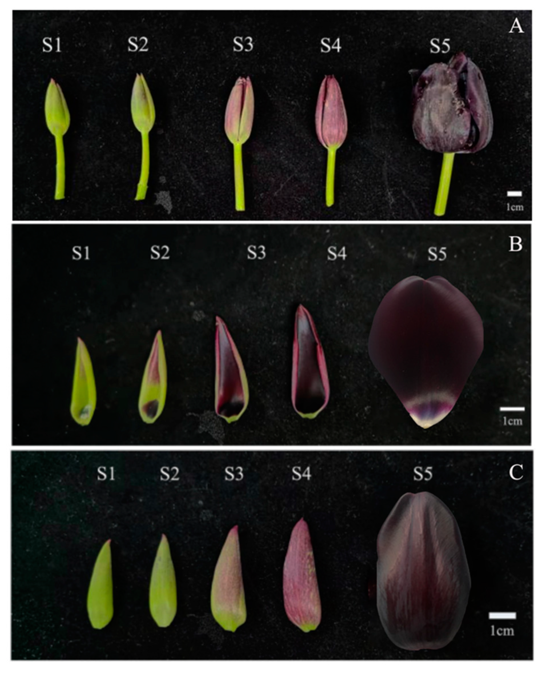
Figure 1. The phenotype of flower color in five different stages of ‘QN’. (A) Whole flower, (B) Inner petal, (C) Outer petal. S1: Green bud stage; S2: Coloring stage; S3: Budding stage; S4: Complete coloring stage; S5: Blooming stage.
Horticulturae 08 00510 g001
Figure 2. The accumulation of anthocyanins in petals of ‘QN’ at different development stages. (A) Content of three individual anthocyanin components. (B) Percentage of three individual anthocyanin components.
Horticulturae 08 00510 g002
Figure 3. The accumulation of anthoxanthins in petals of ‘QN’ at different development stages. (A) Content of individual anthoxanthin component. (B) Percentage of individual anthoxanthin component. K stands for kaempferol and its derivatives; N stands for naringin and its derivatives; Q stands for quercetin and its derivatives; A stands for apigenin and its derivatives; I stands for isorhamnetin and its derivatives.
Horticulturae 08 00510 g003
Figure 4. The accumulation of flavonoids in petals of ‘QN’ at different development stages. (A) Content of flavonoids. (B) Percentage of flavonoids.
Horticulturae 08 00510 g004
Table 1. Anthocyanin components in petals of ‘QN’.
No.IdentificationtR (min)ESI-(+)-MS (m/z)References
A1Delphinidin 3-o-rutinoside5.26611 [M + H]+, 303 [Y0+] [23]
A2Cyanidin 3-o-rutinoside5.73595 [M + H]+, 287 [Y0+][23,24]
A3Pelargonidin 3-o-rutinoside6.20579 [M + H]+, 271 [Y0+][23,24]
Table 2. Anthoxanthin components in petals of ‘QN’.
No.Identification/Tentative IdentificationtR (min)ESI-(+)-MS (m/z)References
F1Isorhamnetin-7-succinylrutinoside-rhamnoside0.79869 [M-H], 461, 315 [Y0][32,33,34]
F2Isorhamnetin-3-succinylrutose-galactoside1.17881 [[M-H], 473, 315 [Y0][32,33,34]
F3Apigenin-3-acetylrhamnose-7-sambubiose1.31745 [M-H], 451, 269 [Y0][32,33,34]
F4Isorhamnetin-3-sophoroside-7-gallicacylglucoside3.63953 [M-H], 627, 315 [Y0][32,33,34]
F5Quercetin-3-acetylpentose-7-sambubiose4.16771 [M-H], 593, 301 [Y0][32,33,34]
F6Kaempferol-3-sophoroside4.42769 [M-H], 447, 285 [Y0][32,35,36]
F7Quercetin-3-galactose-sambubiose5.12755 [M-H], 591, 301 [Y0][32,33,34]
F8Naringin-3-acetylrutinoside5.22627 [M-H], 316, 271 [Y0][32,34]
F9Kaempferol-3- rhamnoside- sambubiose5.32726 [M-H], 579, 285 [Y0][32,33,34]
F10Quercetin-3-sambubiose5.42595 [M-H], 301 [Y0][32,37]
F11Naringenin-3-sophoroside5.54595 [M-H], 271 [Y0][32,35,36]
F12Kaempferol-3-galactose-sambubiose5.60741 [M-H], 575, 285 [Y0][32,33,34]
F13Quercetin-7-rutinoside5.85609 [M-H], 301 [Y0][32,37]
F14Rutin(Quercetin-3-rutinoside)5.92609 [M-H], 301 [Y0]std
F15Kaempferol-7-malonylgalactose6.09691 [M-H], 447, 285 [Y0][32,34]
F16Kaempferol-3-sambubiose6.21579 [M-H], 285 [Y0][32,37]
F17Kaempferol-3-rutinoside6.36593 [M-H], 285 [Y0][32,37]
F18Isorhamnetin-3-succinylsophoricoside6.74737 [M-H], 415, 315 [Y0][32,34]
F19Quercetin-3-sophoroside6.84624 [M-H], 301 [Y0][32,35,36]
Note: std: The components are identified by the standard compound.
Publisher’s Note: MDPI stays neutral with regard to jurisdictional claims in published maps and institutional affiliations.