Review

Hormonal Signaling in the Progamic Phase of Fertilization in Plants

by
Ekaterina V. Zakharova
1,*,
Marat R. Khaliluev
1,2,* and
Lidia V. Kovaleva
3
1
All-Russia Research Institute of Agricultural Biotechnology, Timiryazevskaya 42, 127434 Moscow, Russia
2
Department of Biotechnology, Institute of Agrobiotechnology, Russian State Agrarian University—Moscow Timiryazev Agricultural Academy, Timiryazevskaya 49, 127434 Moscow, Russia
3
K. A. Timiryazev Institute of Plant Physiology, Russian Academy of Sciences, Botanicheskaya 35, 127276 Moscow, Russia
*
Authors to whom correspondence should be addressed.
Horticulturae 2022, 8(5), 365; https://doi.org/10.3390/horticulturae8050365
Submission received: 4 March 2022 / Revised: 15 April 2022 / Accepted: 19 April 2022 / Published: 21 April 2022

Abstract

:
Pollen–pistil interaction is a basic process in the reproductive biology of flowering plants and has been the subject of intense fundamental research that has a pronounced practical value. The phytohormones ethylene (ET) and cytokinin (CK) together with other hormones such as auxin, gibberellin (GA), jasmonic acid (JA), abscisic acid (ABA), and brassinosteroids (BRs) influence different stages of plant development and growth. Here, we mainly focus on the information about the ET and CK signaling in the progamic phase of fertilization. This signaling occurs during male gametophyte development, including tapetum (TAP) cell death, and pollen tube growth, including synergid programmed cell death (PCD) and self-incompatibility (SI)-induced PCD. ET joins the coordination of successive events in the developing anther, including the TAP development and cell death, anther dehiscence, microspore development, pollen grain maturation, and dehydration. Both ET and CK take part in the regulation of pollen–pistil interaction. ET signaling accompanies adhesion, hydration, and germination of pollen grains in the stigma and growth of pollen tubes in style tissues. Thus, ET production may be implicated in the pollination signaling between organs accumulated in the stigma and transmitted to the style and ovary to ensure successful pollination. Some data suggest that ET and CK signaling are involved in S-RNase-based SI.

1. Introduction

An underlying mechanism of male gametophyte interaction with sporophyte-derived tissues is one of the current topics in developmental biology. The cell-to-cell interplay in the pollen–pistil system largely determines the possibility of mutual gamete assimilation under compatible pollination or its impossibility under a genetically determined barrier to self-fertilization, which has both theoretical (the cell-to-cell interactions as a general biological problem) and practical significance (the crop yield and quality). Interspecific incompatibility is closely related to the applied problems of plant distant hybridization as well as central evolutionary botany problems, such as the origin of species [1,2]. Pollen–pistil interaction begins with the entry and germination of pollen grains (PGs) in the stigma and subsequent pollen tube (PTs) growth in the transmitting tract (intercellular space of transmitting tissues or open style channel) [1,3]. This process is completed by the fusion of male and female gametes in the ovary [1,2,3]. Many interactions are involved in this complex process, such as cellular recognition, extracellular and intracellular signaling, and other yet unknown factors. In the last 20 years, significant progress has been made in underlying the key mechanisms involved in the regulation of PTs growth, including molecular signaling in the pollen–pistil interaction, as well as their physiological responses [1,2,3,4,5,6,7,8,9,10,11].
Sexual reproduction of angiosperms is a highly selective process. Maternal tissues of pistil are able to distinguish self and same-species non-self PGs. This selectivity is accompanied by an enormous diversity in the cell surfaces of male and female reproductive structures. The pistil is well organized not only for PGs acceptance and PTs growth but also has mechanisms that blocked their growth at different stages: hydration and germination on the stigma surface, growth inhibition in the stigma, style, or ovary [12,13,14].
Firstly, the role of phytohormones in the process of fertilization in plants was suggested by Barendse and Linskens (1970), who found the presence of indole-3-acetic acid (IAA) and gibberellic acid (GA3) in the pollen of Petunia hybrida L. and Lilium sp. [15]. Later, in order to study the phytohormonal regulation of male gametophyte growth, Sondheimer and Linskens (1974) treated in vitro germinated pollen of P. hybrida L. with exogenous phytohormones (IAA, GA3, abscisic acid (ABA) and zeatin). IAA, GA3, and zeatin treatments at 10−7–10−4 M suppressed pollen germination, whereas ABA stimulated pollen growth. The authors suggested that phytohormones may be involved in the regulation of pollen maturation, germination, and growth of PTs along with the transmitting tissues of style, as well as fertilization and post-fertilization [16]. To date, the coordination at cellular, tissue, organ, and organismal levels, as well as different physiological and morphological responses by phytohormones has been clearly established [17,18]. There are much data on the modulation in the transcription of various genes and translation by phytohormones in plants [19]. However, the initial stages of hormonal signal transduction pathways associated with perception and transmission to gene responses are much less known [20,21,22,23].
Generally, despite advances in the underlying hormonal regulation in reproduction and development processes, their regulatory role in individual stages of plant fertilization has been poorly understood. Therefore, the purpose of this review is the least studied hormonal aspect of intercellular interactions, which includes the possible hormone participation as a trigger for the pollination and control of male gametophyte development in the sporophyte pistil tissues, as well as the progamic phase of fertilization.

2. Phytohormones in Tapetum (TAP) and Pollen Wall Development Programs

The male gametophyte development in flowering plants is a complex biological phenomenon comprising a set of events, such as cell division, differentiation, and cell death. It is totally dependent on the sporophytic tissues of the anther, where PG are formed and maturate (Figure 1) [24,25,26,27]. Microsporocytes develop in the anther loculi, which consist of four cell layers: TAP, middle layer, endothecium, and epidermis [28,29,30] (Figure 1B).
Normal stamen formation is essential for male gametophyte development. PGs (male gametophytes) are formed as a result of the meiotic division of microsporocytes with the tetrad formation. Each microspore forms a PG covered with dense ectexine, endexine, and inner intine (Figure 1C). The microspore divides asymmetrically to form a larger vegetative cell and a smaller generative cell by mitotic division. When the pollen has matured, the anthers open to release the pollen [31]. Primary parietal cells of a young pollen sac undergo a series of further periclinal divisions to form endothelial cells and secondary parietal cells; then the secondary parietal cells divide to form the middle cell layer and TAP (Figure 1B) [32,33,34,35].
IAA plays a decisive role in the initiation of stamen development; the auxin flow also enhances the independent regulation of stamen filament elongation [36]. The development and function of stamens are also modulated by the GA and JA phytohormones [37,38].
ABA and JA play a crucial role in the coordination of pollen maturation as well as the correct timing of pollen release [36,39,40]. JA mainly controls the late stages of anther development [41].
GA is necessary for the development and function of TAP cells [42,43]. GA is an important player in the formation of the pollen exine [38,42].
BRs control the anther development, including the formation of microspore mother cells, development of microspores, and TAP development [36,44,45]. BRs are also participated in the pollen wall formation by regulating the synthesis or transport of sporopollenin [45].
The CK synthesis and subsequent signal transduction actively take place at the early stages of TAP development and decrease at later stages [36]. An enhanced CK expression induces the impaired formation of the exine in pollen [46].
ET is included in the coordination of the successive events at all phases of male gametophyte development, including the initiation of TAP PCD [36,47,48,49,50,51,52,53].
The TAP is a short-lived tissue, forming the inner layer of the anther; it directly fits the sporogenic tissue. The TAP plays a key role in the development of PG by contributing to enzymes, nutrients, and other inclusions [54,55]. The TAP storage substances (proteins, lipids, and carbohydrates) are secreted to the loculi, providing the progress of meiosis, development of microspores, and PG maturation. The TAP cells are interconnected by cytoplasmic bridges, allowing for the synchronization of their activities [56] (Figure 1B).
Programmed cell death (PCD) is an essential part of the plant reproductive development and the final differentiation stage of the anther TAP [57]. TAP PCD is a precisely controlled process that takes place at the late stages of microsporogenesis. In flowering plants, the TAP PCD is a typical type of cell death with characteristic cell shrinkage, DNA degradation, and caspase-like proteolytic activity. The TAP is degraded with the maturation of microspores to be completed by the moment when binucleate pollen grains are formed [39,58,59,60,61,62].
Transcriptomic studies of Arabidopsis anther have demonstrated tight coordination of the TAP cell death activity with pollen development [44]. The precise timing of TAP PCD is decisive for pollen maturation. Any disturbance (delay or promotion) of the TAP PCD often adversely influences pollen development. The factors involved in TAP PCD form an intricate regulated network. In model plants (Arabidopsis, rice, tomato, and rapeseed), the TAP PCD regulatory network is generated by manifold factors [37].
The pollen wall is a complex multilayer outer coat. The mature pollen grains generally contain ectexine, endexine, and inner intine (Figure 1C). The exine is the most complicated and vital outer layer of the pollen wall [37,63,64,65,66]. Microspores synthesize the pectin–cellulose primexine, acting as a basis for the deposition of sporopollenin precursors [67]. The TAP is the major tissue for synthesizing sporopollenin precursors [54]. Various fatty acids and phenolic compounds are the major components of sporopollenin [68]. Ariizumi and Toriyama [69] have analyzed the Arabidopsis genes associated with the biosynthesis and transport of the lipidic and phenolic precursors for exine formation. Genome-wide coexpression analysis has detected 98 specific candidate Arabidopsis genes expressed in the anther likely to be involved in phenolic and lipidic metabolism as well as transport [70]. The pollen coat formation is completed in the later microgametogenesis stages when degenerating TAP are deposited on the surface of PGs [71]. The final stage in PG maturation is dehydration [64,65].
In the flowering plants, a mature PG contains two (vegetative and generative cells) or three (two small sperm and one vegetative) cells (Figure 1C); the vegetative cell generated the PT; in the former case, the generative cell divides in the growing PT to give two sperm cells, and the PT delivers two sperm cells for fertilization [28,29,72]. The PT germinates through the transmitting tract, enters the ovule through the micropyle, and ruptures. One of the sperm cells fertilizes the ovule, while the other sperm cell fuses with the central cell to produce the embryo and the endosperm (double fertilization, discovered by S.G. Navashin) [72,73].
The effects of phytohormones are quite complex and each of them performs not one, but several functions, depending on the target site exposure and type of plant tissue, as well as external conditions. Therefore, we limited ourselves to a brief overview of the most important aspects of the participation of all phytohormones and focused mainly on the role of cytokinin (CK) and ethylene (ET) in the progamic phase of fertilization, since they play an essential role in the male gametophyte development, pollination, pollen recognition during the arrest of self-incompatible (SI) PTs, including SI-induced PCD.
Hirano et al. [36] investigated the global gene expression in rice related to phytohormones in the TAP and showed that auxin, GA, CK, brassinosteroids (BRs), ET, ABA, and jasmonic acid (JA), affect the pollen wall development. So far, 102 gene encoding transcription factors, 57 signaling-related genes, 48 and 111 genes related to protein modification and degradation, subsequently, as well as 18 genes associated with hormone metabolism have been detected in the developing rice anther. The expression profile of the genes related to the regulation of hormone metabolism comprises four genes associated with IAA; two, with ABA; nine, with ET; one, with CK; one, with JA, and two, with GA [74].

2.1. Auxin (IAA)

Auxin is a key regulator of plant growth and development during ontogenesis. The cell-to-cell differences in auxin concentration coordinate innumerable certain developmental responses [75,76,77]. Expression of biosynthetic, signaling and transport auxin-mediated genes occurs during pollen development [78,79]. It has been clearly demonstrated that auxins are essential for the anther development and pollen production [80,81,82,83,84]. IAA accumulates in PGs, endothecium, epidermis, and TAP cells [79]. Alani et al. postulate that auxins produced by the TAP are essential for pollen development in Arabidopsis [79]. However, Yao et al. showed that the TAP provides cells with nutrients, but the auxin produced by the TAP is not sufficient to support Arabidopsis pollen development at early stages. The early stages of microspore development require auxin produced by diploid sporophyte microsporocytes, which is sufficient for male gametophyte development [84] (Figure 1A).
Additionally, the local biosynthesis, transport, and signaling of auxin are critical for stamen initiation [36] (Figure 1A). The auxin synthesized in anthers plays an essential role in anther dehiscence and pollen maturation, while the auxin transport independently contributes to the regulation of filament elongation [84,85]. Auxin acts through JA to regulate the anther dehiscence and pollen maturation, whereas the auxin-regulated stamen initiation and filament elongation may be attributed to other signals. The auxin response factor17 (ARF17) controls the primexine deposition and callose biosynthesis by directly modulating the expression of the CalS5 gene [86].

2.2. GA and JA Are Indispensable for Stamen Development

The development and function of stamen are modulated by various phytohormones, with a key role of GAs and JAs [87,88] (Figure 1A). The deficiencies in the signal transduction pathways of GAs and JAs retard stamen development, pollen maturation, or anther dehiscence.
GA plays a critical role in the development and function of the TAP and pollen Arabidopsis [42,43]. The genes involved in the GA signaling are preferentially expressed throughout the TAP development, i.e., anthers may be the main organ of GA biosynthesis in flowers [36]. The GAMYB MYB transcription factor (TF) is a main TF in the GA signaling pathway, being essential for pollen exine formation [42].
The JA synthesis and signaling are active in the TAP at all developmental stages [36]. JA mainly controls the late stages of anther development. JAs are important for pollen development and stamen elongation, as well as regulated the correct timing of pollen release [41]. The JA mutants of Arabidopsis are sterile due to the arrest of stamen development [89]. An excess level of JAs induces expression genes encoding TFs crucial for normal stamen development [88].

2.3. ABA

Although ABA is associated with pollen wall development [36,45,74], their roles in male gametophyte development remain poorly understood. Exogenous ABA suppressed anther development and caused pollen abortion in tomatoes [90]. During the early anther development, ABA is localized mainly in microsporocytes and the TAP, but later it is bordered by the TAP [91]. Until now, the function of ABA at the early stage of anther development remains poorly understood. ABA might control the gene expression involved in cell separation during the early stage of anther development [92]. The authors also suggested that ABA may regulate the TAP separation from microsporocytes during early anther development in Brassica napus L. Since ABA is known to suppress the PCD of aleurone cells, induction of its deactivation enzymes at the late stages of TAP development may play a critical role in PCD induction in TAP cells [36]. The ABA distribution in developing anthers of fertile and male sterile lines of petunia (P. hybrida L.) was analyzed by the immunohistochemical method [40]. It has been established that the fertile male gametophyte development is accompanied by a gradual increase in the ABA level in reproductive cells and, on the contrary, a gradual decrease in the TAP cells and middle layers. Abortion of sterile microsporocytes in the prophase I of meiosis caused by premature TAP degeneration with complete preservation of the middle layers was accompanied by a sharp, two-fold increase in ABA level in the reproductive cells. These data indicate that ABA is involved in the PCD of microsporocytes at the meiosis stage [40].

2.4. ET Signaling in Male Gametophyte Development

ET is involved in the coordination of successive responses in the anther [47,48,49,50] (Figure 1). The correct formation of stamens is important for the male gametophyte development, and hence for the successful fruit setting. Dynamics of gene expression encoding various components of signal transduction and ethylene responses in male reproductive organs were revealed, suggesting an active role of ET in pollen development [50,51,52,53]. In 44K LM (laser microdissection) microarray, the gene expression profile of developing rice anther shows the presence of ET signaling in pollen development at the late stages and in TAP throughout the anther development [36].
Transgenic tobacco plants expressing the anther-specific mutated melon ET receptor Cm-ERS1/H70A gene from melon were characterized by reduced level of ET and abnormal stamen development, altered floral architecture, as well as decreased pollen production due to late TAP degeneration [93].
In P. hybrida L., ET is involved in the coordination of the sequence of responses in the developing anther, including the initiation of TAP PCD [50]. The fertile male gametophyte formation has two maximums of ET production. The first peak coincides with the TAP degeneration during microspore development. A putative factor that causes the TAP destruction is a threefold increase in the ET content at the tetrad stage. The second peak accompanies PG maturation and dehydration. The application of 2,5-norbornadiene (NBD) to flower buds arrests the anther development, whereas exogenous ET (10–100 µL/L) leads to TAP PCD and degradation of male reproductive cells.
The death of petunia sterile male gametophyte takes place in the meiotic prophase because of the TAP premature destruction and is accompanied by an upsurge in ET production [50]. In this process, the ET level is tenfold higher as compared with the ET level of fertile anthers during TAP PCD; this correlates with both microsporocyte and TAP tissue degeneration. In addition, the microsporocyte abortion in the meiotic prophase is also accompanied by a twofold elevation in the ABA content in reproductive cells [69]. The ABA signaling in the rice anthers acts at the tetrad and tricellular stages [74]. ABA can be a potential inductor of the stress-induced male sterility (MS) and the regulation of sugar transport from the TAP to anther apoplast [94]. ET-IAA interactions are also observed [49]. Additionally, ET has promoted the anther opening [95].

2.5. CK Signaling Is Involved in TAP and Pollen Development

CKs are involved in the regulation of several developmental processes (including stimulation of cell division and cell differentiation, as well as morphogenesis) [46,96,97,98,99,100,101]. At high concentrations, CKs have a growth-inhibitory and even an apoptotic effect [101]. However, their functions in the formation of reproductive organs have not yet been studied in any detail. The CK synthesis and subsequent signaling actively occur during the early TAP developmental stages and significantly decrease in the later stages in rice [36]. There is some evidence that CKs are involved in male gametophyte development [46].
The stamenless-2 (sl-2) mutant of S. lycopersicum L. [102] and a genetic MS line of rapeseed (Brassica napus L.) [103] characterized lower endogenous CK content. Accumulation of cytokinin oxidase/dehydrogenase in the male reproductive tissues of transgenic maize (Zea mays L.) resulted in MS plants [104]. The CK synthesis and subsequent signaling actively occur during the early TAP developmental stages and significantly decrease at the later stages [36]. The loss of ROCK1 (repressor of cytokinin deficiency 1) function enhances the CK response and induces defective exine formation in Arabidopsis pollen [105].
In Arabidopsis, the CK receptor genes in the sporophyte are required for male and female functions [106]. In a model of the triple Arabidopsis mutant (cre1-12 ahk2-2tk ahk3-3), it was shown that CK receptors in the sporophyte are required for anther opening, pollen maturation, induction of PG germination on the pistil stigma, and maturation of the female gametophyte [107].

3. Pollen–Pistil Interactions in the Progamic Phase of Fertilization

3.1. Growth

Once a PG has reached the papilla cells of the stigma, it is rehydrated and activated [104]. The lipids in the stigma exudate mediate pollen hydration [108]. The polar (tip) growth of PT is provided by a fine-tuned network of cellular responses, including a dynamic organization of the actin cytoskeleton (AC), vesicle, and protein trafficking, establishing intracellular tip-localized Ca2+ and pH gradients and signal transduction pathways [109,110,111,112,113,114,115,116,117,118,119,120].
pH and Ca2+ concentration control the actin polymerization together with actin-binding proteins [118,119]. Winship et al. [119] believe that the pH gradient regulates the PT growth rather than the tip-focused Ca2+ gradient. Ca2+ as a central second messenger in plants coordinates varying physiological responses. The PT tip-focused Ca2+ gradient serves as a signal for Ca2+/calmodulin protein kinases and is key for the regulation of AC dynamics. The Ca2+/CPK (calcium-dependent PK) signaling pathway may regulate the PT growth by maintaining the intracellular ion concentrations at the apex via ion channels [120]. The ROP (Rho of plants) signaling network, depending on Rho GTPase (ROP1), provides a molecular linkage between the AC, vesicular trafficking, and polarity formation [121]. The Ca2+/CPK signaling pathway may crosstalk with reactive oxygen species (ROS) [122].

3.2. Pollen–Pistil Interaction

PT growth is provided for by an elaborate mechanism, requiring the integration and coordination with other signaling systems and numerous pistil factors [123]. Currently, the molecular mechanisms underlying these interactions are actively studied [29,118,120,124,125,126]. The interaction of the pollen tube with the pistil tissues activates a specific range of 1254 genes that have not been detected in in vitro cultured pollen tubes. Transcriptomic studies have relived the peptides involved in the pollen–pistil interactions. Among them, there are arabinogalactan proteins (AGPs), cysteine-rich polypeptides (CRPs), defensin-like proteins (DEFL), S-RNases, transmitting tissue-specific (TTS) proteins, extensin-like proteins (PELPIII), and lipid transfer proteins (LTPs) [124]. Female gametophyte plays a valuable role in the PT attraction towards the ovule by secretion of ovular attractants, including LURE and ZmEA1 proteins, belonging to the protective defensin-like subfamily of CRPs [125]. As has been shown, all LURE genes are expressed by the synergid cells. Tip-localized pollen-specific receptor-like kinase 6 (PRK6) acts as a major membrane receptor for external AtLURE 1 attractants in A. thaliana L. [127].

3.3. BRs Are Essential for Male Fertility

The BRs act in the pollen wall formation through the regulation of sporopollenin synthesis or transport [36] (Figure 1B,C). BRs control the various developmental stages of the anther, including the formation of the microspore mother cell, and its development, as well as TAP development and pollen coat formation [45]. In the Solanum lycopersicum L., the BR signaling regulator (BZR1) directly binds to the promoter of RBOH1, thereby inducing the promotion of pollen development via the RBOH1-mediated ROS signaling pathway by triggering PCD and tapetal cell degradation (see the review by [44]).

3.4. ET Signaling in Pollen–Pistil System

The studies involving the orchid [128], tobacco [47], and petunia [129] demonstrate that the pollination-induced ET production in the pistil tissues and its release is critical for PT growth and successful fertilization. As has been shown, the pollination in orchids triggers an interorgan regulation of the ET biosynthesis genes; the authors assume that it is ACC, the soluble ET precursor, that acts as a secondary transmissible ET signal coordinating the development of floral organs after pollination [128]. The combinatorial interplay among the ACC isoforms modulates the ET biosynthesis in A. thaliana L. [130].
In P. inflata R.E.Fr., both compatible and incompatible pollinations result in a significant increase in ET synthesis, which peaks 3 h after pollination. The second burst in ET production begins 18 h after compatible pollination [131].
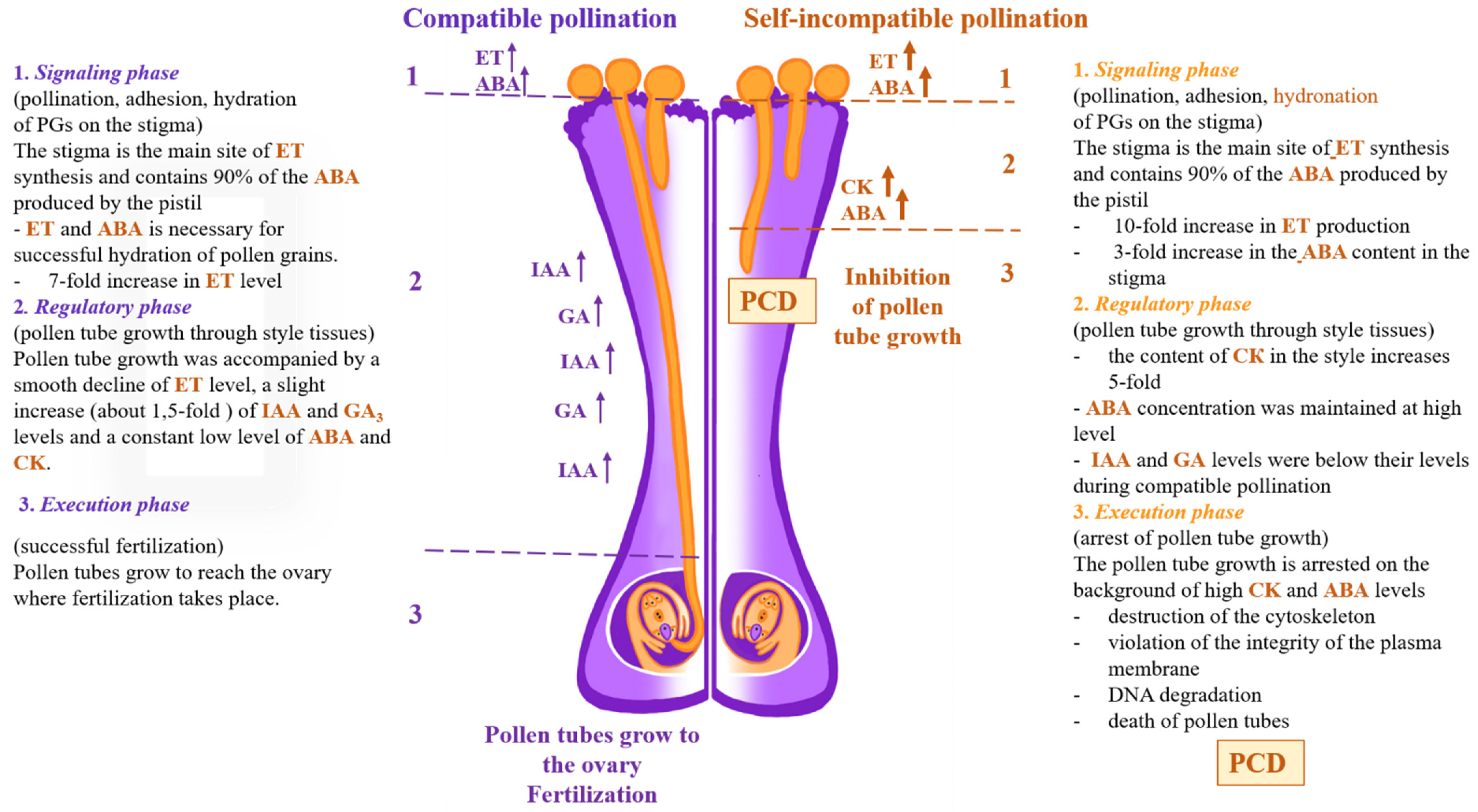
The germination of PGs on the stigma of P. hybrida L. is accompanied by a 7-fold increase in the ET content [50,132] (Figure 2). It is shown that the stigma is the main part of ET synthesis. Accumulated (100-fold) ACC in PGs triggers the autocatalytic ET biosynthesis in the pollen–pistil system. The ACC content in the stigma tissue reaches its maximum 2 h after pollination. The ACC concentration in styles and ovaries is 100-fold lower as compared with the stigmas. NBD blocks the male gametophyte development and growth.
The pollination signal is generated on the surface of the stigma and is transduced by a certain signal resulting from the hormonal imbalance in the pistil and PT cell [47,128,129]. The stigma is the main part of ET synthesis and contains 90% of ABA synthesized by the pistil [50,132]. The peak of ET production by pistil tissues belongs to earlier events, which accompany adhesion, hydration, and pollen grain germination in the stigma tissues [50,131,132].
The hormonal content in the petunia pollen–pistil system under SI pollination differed dramatically from that under compatible pollination and changed during PT growth [133].
In a compatible pollination event (left part of the scheme, colored violet), PTs grow to reach the ovary, where fertilization takes place. Pollen grain germination on the stigma (within 4 h after pollination) was accompanied by a seven-fold increase in ET level [47,132,133]. During the subsequent 4 h, ET production by pistil tissues decreased. The growth of PT by 8 h was accompanied by a constant low level of ABA and CK. Herewith, IAA and GAs levels increased about 1.5-fold within 8 h [133].
In the case of SI pollination (right part of the scheme, colored orange), the pattern is completely different. PG germination on the stigma (within 4 h of pollination) was accompanied by a 10-fold and 3-fold increase in ET and ABA content in the stigma and style, subsequently [47,132,133]. During the subsequent 4 h, the ABA concentration was maintained at the same high level. One may speculate that this is related to a SI mechanism and connected with ET activity. The inhibition of PT growth by 8 h was accompanied by a 5-fold increase in CK content in style tissues [133]. Thus, the PT growth is inhibited in the background of high CK and ABA levels [133]. This altered hormonal level activates several signaling cascades and transcription regulation. The expression of S-RNAse, proteases, and nucleases is commenced. CK together with CLPs and S-RNAse disarrange the AC in the SI PTs, thereby causing membrane disorganization, destruction of organelles, and possible DNA degradation as the final stage in the SI-induced PCD in P. hybrida L. [134,135].
The maximum ET production by pistil tissues is associated with the earlier events accompanying the adhesion, hydration, and subsequently germination of PGs in the stigma tissues. This indicates that the ET synthesis is stimulated by pollen–pistil interaction and may be implicated in the interorgan pollination signal generated in the stigma and transmitted to the style, ovary, and other floral organs (calyx and corolla) for successful pollination [50,132].
Additionally, ET induces the synergid PCD and disrupts pollen tube PT [128]. In successful fertilization, ET signaling is induced by the ER-localized EIN2 and EIN3 and perceived by the synergids. ACC introduction into the female gametophyte by microinjection or a constitutive ET response leads to premature synergid disorganization. Mou et al. [129] have shown that ACC signaling in Arabidopsis ovular is involved in PT attraction and promotes secretion of the pollen tube PT chemoattractant LURE1.2.

3.5. ET as a Regulator of Hormonal Interplay in Pollen–Pistil System

A diverse group of phytohormones modulates the ET level in various plants acting at the ACS gene expression [136,137,138,139,140]. EIN3, one of the regulators of a feedback ET response, is a link in the interaction with the other phytohormone signaling pathways.
In vitro germinated petunia PG is accompanied by changes at the levels of plant hormones, such as IAA, ABA, GAs, and CKs, and is sensitive to the treatment with exogenous phytohormones. The membrane potential on the PT and cytosolic pH, lateral membrane allocation of PM H+–ATPase, and organization of PT AC are sensitive to exogenous hormones. Exogenous applications of IAA, ABA, and GA3 display the growth-stimulating action accompanied by orthovanadate-sensitive polarization of the PM. GA has the maximum stimulatory effect; IAA induces alkalinization of the cytosol; kinetin, in contrast, causes cytosol acidification; and exogenous CK inhibits pollen tube germination and growth. All these facts suggest the existence of extremely complex interactions between ET, IAA, ABA, and CK [140].

3.6. ET–IAA Interplay

Auxin plays a critical role in the maintenance of PT polar growth, complying with its similar response in the other organs [141]. The ET’s ability to modulate the effect of auxin biosynthesis and transport of its determines a wide range of physiological manifestations in plants [142].
In P. hybrida L., ET controls the PG germination and PT growth by interacting with auxin, a likely key regulator of plant cell polarization and morphogenesis and one of the factors controlling the ET biosynthesis at the level of ACC synthase gene expression (Figure 2). The male gametophyte–stigma tissues interaction leads to an increase in ET and IAA production by a 7- to 10-fold and a 1.5- to 2.0-fold content in the pollen–pistil system over 0–4 h, subsequently.
Exogenous IAA and ET stimulate the PT germination and growth. 1-MCP (methylcyclopropene), a blocker of ET reception, and TIBA (triiodobenzoic acid), a blocker of IAA transport, inhibit pollen germination, while IAA removes the inhibitory of both 1-MCP and AOA (aminoxyacetic acid), an inhibitor of ACC synthesis, and Ethrel, a plant growth regulator, partially removed the inhibitory effect of TIBA. The pollen pollination preliminarily treated with 1-MCP causes a 2.5-fold decline in both the rate of PT growth and ACC level. IAA inhibits the action of 1-MCP recovering the synthesis of ACC and PT growth compared with control values [50,132,142].
These results taken together suggest the interaction of the ET-IAA interplay signal transduction pathways at the level of ACC biosynthesis during the germination and growth of petunia male gametophyte.

3.7. ET-ABA Interplay

The germination and growth of petunia male gametophyte are characterized by a high level of ET and ABA production in the pollen–pistil system. The stigma is the main part of ET synthesis and contains about 90% of ABA. ABA abolishes the inhibitory effects of 1-MCP (an ET receptors blocker), AOA (a blocker of ACC), and Fluridone (a blocker of ABA synthesis) on the PT development, whereas ethrel arrest has an inhibitory effect of fluridone on the PT growth. In stigmas pretreated with AOA and ABA, ABA suppresses the inhibitory effect of AOA on the ACC synthesis in the petunia pollen–pistil system before pollination [50,132,143,144].
ABA is involved in the osmoregulation in petunia male gametophyte in the progamic phase of fertilization by interacting with ET at the ACC synthesis level [144]. Two potential targets of the ABA in a PT are identified, namely, (1) PM H+-ATPase, an electrogenic proton pump, and (2) Ca2+-dependent K+ channels [140]. A stimulatory effect of ABA on the H+-ATPase electrogenic activity is mediated by an increase in the free Ca2+ level in the PT cytosol and ROS generation. The ET/ABA content of the stigma may control adhesion, hydration, and germination of PGs.

3.8. IAA-CK Interplay

The pistil developmental program is under the control of both IAA and CK. The CK response integrates auxin and CK pathways for the development of the female reproductive organ. In a triple mutant, both pistil length and the ovule numbers are reduced [145].
IAA and CK play a key role in the AC regulation during petunia PT growth via their effects on actin polymerization [133]. The AC in growing PTs is sensitive to exogenous auxins. The IAA growth stimulatory effect correlates with an increased amount of actin filaments (AFs) in both apical and subapical zones of PTs. In contrast, CK decreases the total AFs content in PTs and inhibits their development. The CK inhibitory effect on the growth of petunia male gametophyte is associated with the degradation of F-actin along the PT, disarranging the apically directed vesicular transport required for its growth.
In vitro cultured pollen on the medium supplemented with latrunculin B (Latr B), an inhibitor of actin polymerization, arrests the PT growth because of the AC disturbance. This effect is accompanied by a dramatic decrease (almost to zero) in the endogenous IAA content. IAA causes monotonous alkalinization of the cytoplasm in PTs and has a significant effect on F-actin assembly, thereby suggesting the involvement of IAA in the interplay with AFs organization through the corresponding modulation of the activity of pH-sensitive actin-binding proteins (ABPs). As for CK, its content significantly increases during the first 60 min of culture. These facts suggest that both IAA and CK play an important role in the regulation of the AC during PT growth via their effects on F-actin polymerization. These data suggest the hypothesis that the AC in male gametophytes acts here as an integrator of auxin and CK signaling pathways and that the mechanism of their interplay may include both ABPs and PIN proteins [140].

3.9. ET-IAA-CK Interplay

The ET content is regulated by other plant hormones, for example, auxin, GA3, and CK [139]. The hormones can interact at the level of their metabolism, transport, and transduction of hormonal signals. Signaling pathways of auxin, ET, and CKs are united via the peptide Polaris (PLS), which inhibits ET and CK responses and positively regulates auxin transport [146]. An important factor in the ET response regulation and the target for other hormones is the controlled degradation of the proteins involved in ET signaling pathway. In particular, the induction of ET biosynthesis by CKs is performed by stabilizing ACC synthases (ACS5 and ACS9) [147]. An ET-induced regulatory module delays the flower senescence in rose by controlling the CK synthesis [148].

3.10. BRs and PT Growth

In Arabidopsis, the pistil BRs promote PT growth [149,150]. The CYP90A1/CPD promoter of the key enzymes in BR biosynthesis is highly active in the cells of the tract for PTs. The in vitro grown PTs respond to BRs in a dose-dependent manner. Pollen germination and growth increase nine- and fivefold, respectively, when the media are supplemented with 10 µM epibrassinolide.

4. Self-Incompatibility (SI)-Induced PCD

In flowering plants, the pollen–pistil interplay followed by the transfer of the recognition function to the sporophytic tissue of the pistil has led to a successful establishment of SI.
SI is the genetically related reproductive barrier that inhibits autogamy and allows the pistil to reject “self” pollen and to accept “non-self” pollen. SI is the kind of pollen–pistil interplay best understood at a molecular level. SI is specified by S-determinant genes at a highly polymorphic S-locus [151]. The pollen–pistil interaction and three different types of the molecular control of pollen and pistil recognition have been characterized for three families—Brassicaceae, Papaveraceae, and Solanaceae. The system of self-recognition characteristic of two families (Brassicaceae and Papaveraceae) is due to specific reactions between the male and female S-determinants belonging to the same S-haplotype. The growth inhibition of incompatible PTs is carried out on the stigma [152].

4.1. Papaver SI System

In Papaver, PrsS-PrpS represents the protein–ligand/receptor interacting pairs. The stigma-specific polypeptide ligand is produced in the stigma that causes rejection of incompatible PTs. The downstream signaling events triggered by the S-specific interaction of PrsS with incompatible pollen have been well characterized [153,154]. SI is implemented via a signaling cascade with an increase in Ca2+, ROS, and NO, as well as mitogen-activated protein kinase (MAPK) activity and protein phosphorylation. Rapid inhibition of PT growth is achieved by AC depolymerization and inhibition of soluble inorganic pyrophosphatase. An incompatible interaction triggered PCD involving the activation of a DEVDase/caspase-3–like activity. The SI-induced cytosol acidification of the PT is likely to be fundamental for the induction of a caspase-like activity and F-actin foci formation [155].

4.2. S-RNase–Based SI

SI is the system of “non-self-recognition” in the Solanaceae, Rosaceae, and Plantaginaceae families [156,157] (Figure 2). It is genetically determined by a single S-locus with multiple haplotypes and fundamentally differing from the Papaver SI. The S-locus encodes the S-RNases expressed in the pistil and multiple SLF (S-locus F-box) proteins in pollen controlling the female and male specificity of SI, respectively. The S-RNases function as a cytotoxin to inhibit self-pollen. The SLF proteins collaboratively defuse the non-self S-RNases via the ubiquitin–26S proteasome system.
In Pyrus pyrifolia (Burm.f.) Nakai (Rosaceae), S-RNase specifically degrades the ROS in the incompatible PT tip, depolymerizes AC, and triggers the changes in mitochondria and DNA degradation in SI PTs [158].
Nicotiana spp. has an S-RNase–based SI. Three genes (HT-B, 120K, and NaStEP), unrelated to the S-locus that have been identified in the pistil, suggest that this process is even more complex [159]. These results imply that NaStEP and NaSIPP destabilize mitochondria, thereby blocking PT growth.
In P. hybrid L., PCD markers, such as DNA fragmentation (a violation of the plasma membrane integrity and DNA degradation), in the in vivo growing SI PTs suggest that PCD is a factor of S-RNase–based SI [134]. Using transmission electron microscopy shows a complete degradation of the PT content.
The AC is turned on in the S-RNase–based SI-induced PCD in P. hybrida L. In vivo, the pretreatment of petunia stigma with Latr B completely inhibits the germination of SI PTs [158]. An in vitro cultured of petunia PTs on the medium supplemented with 0.2 nM Latr B inhibits their growth via a disturbance of the AC organization [135]. Earlier, Roldán et al. indicated the F-actin disorganization in the incompatible PTs of Nicotiana. alata Link and Otto [160].
In P. hybrida L., the SI-induced PCD of PTs is associated with caspase-like protease (CLP) activity [134]. 90% of in vivo growing SI PTs show CLP activity.
CLP activities are detectable in 90% of the in vivo growing SI PTs. A high CLP activity within 2–4 h is characteristic of the growing PTs and coincides with the S-RNase activation. The Latr B-induced f-actin depolymerization decreases the CLP activity in the SI PTs. These results suggest that the CLP activity is specific to the cytoplasm and, most likely, to the nuclei of the PT suggested by its intense glow. Any CLP activity is absent in in vivo grown compatible PTs [135].

4.3. ET, ABA, and CK as the Factors of SI-Induced PCD in P. hybrida L.

The phytohormone ET has been implicated as an important regulator of the PCD in plants, including senescence of generative and reproductive organs; aerenchyma formation; leaf and petal abscission; and endosperm cell death in fertilized ovules [161,162,163]. Currently, ET is recognized as one of the positive triggers of PCD. [164,165]. In some species, ET acts as a key hormone for floral senescence [161]. Shibuya et al. has shown the involvement of two proteins of the ET biosynthesis in petal senescence in Japanese morning glory (Ipomea nil L.) [166].
ABA may play a protective reaction defining the timing and extent of PCD in cereals. In particular, the ABA inhibition during the development of maize and wheat endosperm stimulates ET synthesis, while the inhibition of ET biosynthesis retards PCD. The treatment with 1-MCP decreases the degree of DNA degradation while Fluridone (an inhibitor of ABA synthesis) stimulates ET synthesis 2–2.5-fold [167]. ABA has a regulatory effect on the ET biosynthetic machinery in Hibiscus flower development [168]. Autophagy, a variant of PCD, has been implicated in the responses to various environmental stresses through interplaying with ABA, JA, and salicylic acid (SA) [101].

4.3.1. ET and ABA

Higher ET and ABA levels, exceeding 2–2.5-fold their levels after cross-compatible pollination, are observed in the inhibition of PT growth after SI pollination in P. hybrida L. [133].
Pretreatment of the petunia stigmas with ethrel and ABA, as well as inhibitors of their synthesis, AOA (an inhibitor of ACC) and fluridone, respectively, to different degrees influences the growth of incompatible PT [134]. The fluridone treatment stimulates their growth 1.5-fold. AOA stimulates the PT growth threefold so that the PTs almost reach the ovary (90% of the style length). At the same time, degradation and fragmentation of DNA in PTs is almost completely absent. These results suggest that ET controls the progress of PCD at the DNA degradation in the SI pollen tubes during functioning of SI mechanism.
Summing up, the above-mentioned data favor the assumption that ET and ABA, exerting antagonistic/synergetic effects, most likely control the progress of SI-induced PCD at the DNA degradation in PTs. It is suggested that the ABA-induced changes in cytoplasmic pH (pHc) are involved in the cascade response in the progamic phase of fertilization, including the function of pH-dependent K+-channels. A stimulatory effect of ABA on the H+-ATPase electrogenic activity is mediated by an increase in the cytosolic Ca2+ content and ROS generation in the PT [139]. In addition, there is evidence that ABA is a putative determinant of PCD in the TAP [93].
In the Brassicaceae, pollen triggers the downstream signaling pathways to prevent self-pollination. Note that a reorganization of actin filaments is observed in the papilla cells, similar to the early events in the poppy SI PT [169,170]. Su et al. showed that ET negatively mediates the SI response of B. rapa L. ssp. pekinensis via PCD in the papilla cells [171]. An ET treatment of the stigmas induces PCD in the papilla cells and breaks down the SI, whereas the treatment of the stigmas with ET inhibitors suppresses PCD and compatible pollination.

4.3.2. CK as a Factor of SI-Induced PCD

The natural CK (kinetin) and its synthetic analog (6-benzylaminopurine (BAP)) at high concentrations induce PCD in plants. Note that BAP induces PCD in the cultured plant cells, whereas kinetin induces this process in living tissues. All CK-dependent phenomena, including PCD, are associated with a multistep phosphorelay signaling pathway, comprising histidine kinase receptors (HKs), histidine phosphotransfer proteins (HPs), and RRs [172].
CKs play a crucial role in the AC regulation during PT growth via their effects on F-actin polymerization. Kinetin decreases the total AFs content in PTs and inhibits their growth. In vitro pollen germination on the kinetin-containing culture medium leads to a decrease in the density of AFs along the entire PT. This is most clearly seen in the apical zone of PTs. The CK content increases during the first 60 min of culture [140].
Acidification of cytosol in the PT plays an important role in the SI-induced PCD in the Papaver PTs by creating favorable conditions for CLP activation [153]. In P. hybrida L., CK inhibits PT growth due to acidification of the cytoplasm and disorganization of AC. [140]. Zakharova et al. have shown that exogenous CK (zeatin) stimulates the CLP activity in compatible petunia PTs and blocked their growth. The actin depolymerization with Latr B significantly reduces the CLP activity in the SI PTs and, on the contrary, its increase in the compatible pollen tubes. The authors assume that CK is an assumed activator of the CLPs [135]. According to their hypothesis, CK at high concentrations acidifies the cytosol SI PTs, thus creating favorable conditions for CLP activation and, perhaps, for AC reorganization. Correspondingly, CK together with CLPs and S-RNase trigged AC destruction of SI PTs, disruption of the membrane integrity and organelles, as well as eventually degrading DNA as the final stage of the S-RNase–based SI-induced PCD in P. hybrida L. S-RNase destroys the AC breaking it down into point foci [173]. A similar pattern was observed after treating the in vitro growing PTs with CK [140].
The above-mentioned effects of CK evidently comply with its growth-inhibitory effect, in particular, a dramatic (5-fold) rise in the CK level 4–8 h after SI pollination during the arrest of SI PTs in styles. Exogenous CKs (kinetin and zeatin) inhibit PT growth both in vitro and in vivo [133].
Thus, the identified inhibitory effect of CK on the PT growth and the actin polymerization, and the opposite stimulatory effect on the caspase-like activity give reason to believe that CK, similar to CLPs, plays a key role in the arrest of PT growth in the course of the S-RNase-based SI-induced PCD in P. hybrida L.

5. Conclusions and Future Prospects

In this review, we summarized the data on the involvement of ET and CK along with other phytohormones (auxin, GA, ABA, JA, and BRs) in the male gametophyte development and pollen–pistil interplay in the progamic phase of fertilization and SI-induced PCD.
(1) ET joins the coordination of successive events in the developing anther, including the TAP development and cell death, anther dehiscence, microspore development PG maturation, and dehydration.
(2) Hormonal signaling during male gametophyte development and PT growth play one of the key roles along with polymorphic secreted peptides and small proteins, ROS/NO signaling, and second messenger Ca2+.
(3) The physiological effect on phytohormones in PG/PT–female gametophyte communications comprise the modulation of pH, namely, a temporary disturbance of the homeostatic regulation of the PT cytosol pH, which may act as a signal in the initiation of further responses triggered by phytohormones.
(4) ET is a regulator of gametophyte–sporophyte interplay in the progamic stage of fertilization and controls PT germination via the interaction with IAA, GA, ABA, and CK at the level of ACC. ET is regarded as a positive SI-induced PCD regulator.
(5) CK is a factor of the S-RNase-based SI-induced PCD in P. hybrida L. along with CLPs and cytoskeleton depolymerization. CK inhibits the growth of PTs, causing acidification of the cytoplasm and destroying the AC.
Two main questions will have to be answered in the future. Firstly, what is the regulatory mechanism for the involvement of hormonal signals in the pollination, germination, and growth of PTs? What signals play a central role in this cascade? A vision for the next decade is the integration of advanced genetic, molecular, physiological, biochemical, and cellular-biological approaches for a comprehensive study of plant fertilization. The data obtained will serve as a basis for the development of technologies for overcoming the hybridization barriers between species and for obtaining new varieties of crops resistant to the effects of climate change and diseases.

Author Contributions

Conceptualization, E.V.Z. and L.V.K.; Writing and original draft preparation, E.V.Z. and L.V.K.; reviewing, and editing of the manuscript; approved the final manuscript for publication, and agreed to be accountable for all aspects of the manuscript, M.R.K.; funding acquisition, M.R.K. All authors have read and agreed to the published version of the manuscript.

Funding

This article was funded by the Russian Scientific Foundation, grant No. 22-24-01148 via the research project “Role of the cytoskeleton in the S-RNase-based self-incompatibility under normal and stressful conditions in vitro and in vivo in a model of transgenic petunia plants (Petunia hybrida L.) with lifetime identification of actin and tubulin”.

Institutional Review Board Statement

Not applicable.

Informed Consent Statement

Not applicable.

Data Availability Statement

Not applicable.

Acknowledgments

The authors are deeply grateful to Victoria Lukina for help in preparing the drawings for the article.

Conflicts of Interest

The authors declare no conflict of interest.

References

  1. Lord, E.M.; Russell, S.D. The mechanisms of pollination and fertilization in plants. Annu. Rev. Cell Dev. Biol. 2002, 18, 81–105. [Google Scholar] [CrossRef] [PubMed] [Green Version]
  2. Franklin-Tong, V.E. Signaling and the modulation of pollen tube growth. Plant Cell 1999, 11, 727–738. [Google Scholar] [CrossRef] [PubMed] [Green Version]
  3. Lord, E.M. Adhesion and guidance in compatible pollination. J. Exp. Bot. 2003, 54, 47–54. [Google Scholar] [CrossRef] [PubMed]
  4. Li, H.; Lin, Y.; Heath, R.M.; Zhu, M.X.; Yang, Z. Control of pollen tube tip growth by a Rop GTPase-dependent pathway that leads to tip-localized calcium influx. Plant Cell 1999, 11, 1731–1742. [Google Scholar] [PubMed] [Green Version]
  5. Pruitt, R.E.; Vielle-Calzada, J.P.; Ploense, S.E.; Grossniklaus, U.; Lolle, S.J. FIDDLEHEAD, a gene required to suppress epidermal cell interactions in Arabidopsis, encodes a putative lipid biosynthetic enzyme. Proc. Nat. Acad. Sci. USA 2000, 97, 1311–1316. [Google Scholar] [CrossRef] [Green Version]
  6. Nasrallah, M.E.; Kandasamy, M.K.; Chang, M.-C.; Stadler, Z.; Lim, S.; Nasrallah, J.B. Identifying genes for pollen-stigma recognition in crucifers. Ann. Bot. 2000, 85 (Suppl. A), 125–132. [Google Scholar] [CrossRef] [Green Version]
  7. Geitmann, A.; Emons, A.M.C. The cytoskeleton in plant and fungal cell tip growth. J. Microsc. 2000, 198, 218–245. [Google Scholar] [CrossRef] [Green Version]
  8. Blasiak, J.; Mulcahy, D.L.; Musgrave, M.E. Oxytropism: A new twist in pollen tube orientation. Planta 2001, 213, 318–322. [Google Scholar] [CrossRef]
  9. Geitmann, A.; Snowman, B.; Franklin-Tong, V.E.; Emons, A.M.C. Alterations in the actin cytoskeleton of the pollen tube are induced by the self-incompatibility reaction in Papaver rhoeas. Plant Cell 2000, 12, 1239–1251. [Google Scholar] [CrossRef] [Green Version]
  10. de Graaf, B.H.; Derksen, J.W.M.; Mariani, C. Pollen and pistil in the progamic phase. Sex. Plant Reprod. 2001, 14, 41–55. [Google Scholar] [CrossRef]
  11. Cheung, A.Y.; Wu, H.-M. Over-expression of an Arabidopsis formin stimulates supernumerary actin cable formation from pollen tube cell membrane. Plant Cell 2004, 16, 257–269. [Google Scholar] [CrossRef] [PubMed] [Green Version]
  12. Wheeler, M.J.; Franklin-Tong, V.E.; Franklin, F.C.H. The molecular and genetic basis of pollen-pistil interactions. New Phytol. 2001, 151, 565–584. [Google Scholar] [CrossRef] [PubMed]
  13. Dumas, C.; Knox, R.B. Callose and determination of pistil viability and incompatibility. Theor. Appl. Genet. 1983, 67, 1–10. [Google Scholar] [CrossRef] [PubMed]
  14. Kovaleva, L.V.; Zakharova, E.V.; Minkina, Y.V.; Voronkov, A.S. Effects of flavonols and phytohormones on germination and growth of petunia male gametophyte. Allelopath. J. 2009, 23, 51–62. [Google Scholar]
  15. Barendse, G.W.M.; Pereira, A.S.R.; Berkers, P.A.; Driessen, F.M.; van Eyden-Emons, A.; Linskens, H.F. Growth hormons in pollen, styles and ovaries of Petunia hybrida and Lilium species. Acta Bot. Neerl. 1970, 19, 175–185. [Google Scholar] [CrossRef]
  16. Sondheimer, E.; Linskens, H.F. Control of in vitro germination and tube extension of Petunia hybrida pollen. Proc. Konin. Neder. Akad. Wetensch. 1974, C77, 116–124. [Google Scholar]
  17. Liang, X.; Abel, S.; Keller, J.A.; Shen, N.F.; Theologis, A. The 1-aminocyclopropane-1-carboxylate synthase gene family of Arabidopsis thaliana. Proc. Nat. Acad. Sci. USA 1992, 89, 11046–11050. [Google Scholar] [CrossRef] [Green Version]
  18. Theologis, A.; Zarembinski, T.I.; Oeller, P.W.; Liang, X.; Abel, S. Modification of fruit ripening by suppressing gene expression. Plant Physiol. 1992, 100, 549–551. [Google Scholar] [CrossRef]
  19. Nemhauser, J.L.; Hong, F.; Chory, J. Different plant hormones regulate similar processes through largely nonoverlapping transcriptional responses. Cell 2006, 126, 467–475. [Google Scholar] [CrossRef] [Green Version]
  20. Brault, M.; Amiar, Z.; Pennarun, A.M.; Monestiez, M.; Zhang, Z.; Cornel, D.; Dellis, O.; Knight, H.; Bouteau, F.; Rona, J.-P. Plasma membrane depolarization induced by abscisic acid in Arabidopsis suspension cells involves reduction of proton pumping in addition to anion channel activation, which are both Ca2+ dependent. Plant Physiol. 2004, 135, 231–243. [Google Scholar] [CrossRef] [Green Version]
  21. He, X.-J.; Mu, R.-L.; Cao, W.-H.; Zhang, Z.-G.; Zhang, J.-S.; Chen, S.-Y. AtNAC2, a transcription factor downstream of ethylene and auxin signaling pathways, is involved in salt stress response and lateral root development. Plant J. 2005, 44, 903–916. [Google Scholar] [CrossRef] [PubMed]
  22. Zalejski, C.; Zhang, Z.; Quettier, A.L.; Maldiney, R.; Bonnet, M.; Brault, M.; Demandre, C.; Miginiac, E.; Rona, J.-P.; Sotta, B.; et al. Diacylglycerol pyrophosphate is a second messenger of abscisic acid signaling in Arabidopsis thaliana suspension cells. Plant J. 2005, 42, 145–152. [Google Scholar] [CrossRef] [PubMed]
  23. Bögre, L.; Henriques, R.; Magyar, Z. TOR tour to auxin. EMBO J. 2013, 32, 1069–1071. [Google Scholar] [CrossRef] [PubMed]
  24. Mascarenhas, J.P. The male gametophyte of flowering plants. Plant Cell 1989, 1, 657–664. [Google Scholar] [CrossRef] [Green Version]
  25. McCormick, S. Male gametophyte development. Plant Cell 1993, 5, 1265–1275. [Google Scholar] [CrossRef]
  26. Liu, L.; Wang, T. Male gametophyte development in flowering plants: A story of quarantine and sacrifice. J. Plant Physiol. 2021, 258, 153365. [Google Scholar] [CrossRef]
  27. Huang, J.; Dong, J.; Qu, L.-J. From birth to function: Male gametophyte development in flowering plants. Curr. Opin. Plant Biol. 2021, 63, 102118. [Google Scholar] [CrossRef]
  28. Bedinger, P. The remarkable biology of pollen. The Plant Cell 1992, 4, 879–887. [Google Scholar]
  29. Hafidh, S.; Fíla, J.; Honys, D. Male gametophyte development and function in angiosperms: A general concept. Plant Reprod. 2016, 29, 31–51. [Google Scholar] [CrossRef]
  30. Dukowic-Schulze, S.; van der Linde, K. Oxygen, secreted proteins and small RNAs: Mobile elements that govern anther development. Plant Reprod. 2021, 34, 1–19. [Google Scholar] [CrossRef]
  31. Wilson, Z.A.; Zhang, D.B. From Arabidopsis to rice: Pathways in pollen development. J. Exp. Bot. 2009, 60, 1479–1492. [Google Scholar] [CrossRef] [PubMed] [Green Version]
  32. Luo, X.D.; Dai, L.F.; Wang, S.B.; Wolukau, J.N.; Jahn, M.; Chen, J.F. Male gamete development and early tapetal degeneration in cytoplasmic male-sterile pepper investigated by meiotic, anatomical and ultrastructural analyses. Plant Breed. 2006, 125, 395–399. [Google Scholar] [CrossRef]
  33. Varnier, A.L.; Mazeyrat-Gourbeyre, F.; Sangwan, R.S.; Clément, C. Programmed cell death progressively models the development of anther sporophytic tissues from the tapetum and is triggered in pollen grains during maturation. J. Struct. Biol. 2005, 152, 118–128. [Google Scholar] [CrossRef] [PubMed]
  34. Zhu, J.; Lou, Y.; Xu, X.; Yang, Z.N. A genetic pathway for tapetum development and function in Arabidopsis. J. Integr. Plant Biol. 2011, 53, 892–900. [Google Scholar] [CrossRef]
  35. Zhang, D.; Yang, L. Specification of tapetum and microsporocyte cells within the anther. Curr. Opin. Plant Biol. 2014, 17, 49–55. [Google Scholar] [CrossRef] [PubMed]
  36. Hirano, K.; Aya, K.; Hobo, T.; Sakakibara, H.; Kojima, M.; Shim, R.A.; Hasegawa, Y.; Ueguchi-Tanaka, M.; Matsuoka, M. Comprehensive transcriptome analysis of phytohormone biosynthesis and signaling genes in microspore/pollen and tapetum of rice. Plant Cell Physiol. 2008, 49, 1429–1450. [Google Scholar] [CrossRef]
  37. Heslop-Harrison, J. Pollen: Development and Physiology; Butterworth: London, UK, 2013; p. 337. [Google Scholar]
  38. Mutasa-Göttgens, E.; Hedden, P. Gibberellin as a factor in floral regulatory networks. J. Exp. Bot. 2009, 60, 1979–1989. [Google Scholar] [CrossRef] [Green Version]
  39. Wang, M.; Hoekstra, S.; van Bergen, S.; Lamers, G.E.; Oppedijk, B.J.; van der Heijden, M.W.; de Priester, W.; Schilperoort, R.A. Apoptosis in developing anthers and the role of ABA in this process during androgenesis in Hordeum vulgare L. Plant Mol. Biol. 1999, 39, 489–501. [Google Scholar] [CrossRef]
  40. Kovaleva, L.V.; Voronkov, A.C.; Zakharova, E.V.; Andreev, I.M. ABA and IAA control microsporogenesis in Petunia hybrida. Protoplasma 2018, 255, 751–759. [Google Scholar] [CrossRef]
  41. Huang, H.; Liu, B.; Liu, L.; Song, S. Jasmonate action in plant growth and development. J. Exp. Bot. 2017, 68, 1349–1359. [Google Scholar] [CrossRef] [Green Version]
  42. Qian, Q.; Yang, Y.; Zhang, W.; Hu, Y.; Li, Y.; Yu, H.; Hou, X. A novel Arabidopsis gene RGAT1 is required for GA-mediated tapetum and pollen development. New Phytol. 2021, 231, 137–151. [Google Scholar] [CrossRef] [PubMed]
  43. Avanci, N.C.; Luche, D.D.; Goldman, G.H.; Goldman, M.H.S. Jasmonates are phytohormones with multiple functions, including plant defense and reproduction. Genet. Mol. Res. 2010, 9, 484–505. [Google Scholar] [CrossRef] [PubMed]
  44. Heslop-Harrison, J. The pollen wall: Structure and development. In Pollen; Butterworth-Heinemann: Oxford, WI, USA, 1971; pp. 75–98. [Google Scholar]
  45. Zhang, F.P.; Sussmilch, F.; Nichols, D.S.; Cardoso, A.A.; Brodribb, T.J.; McAdam, S.A. Leaves, not roots or floral tissue, are the main site of rapid, external pressure-induced ABA biosynthesis in angiosperms. J. Exp. Bot. 2018, 69, 1261–1267. [Google Scholar] [CrossRef]
  46. Terceros, G.C.; Resentini, F.; Cucinotta, M.; Manrique, S.; Colombo, L.; Mendes, M.A. The Importance of Cytokinins during Reproductive Development in Arabidopsis and Beyond. Int. J. Mol. Sci. 2020, 21, 8161. [Google Scholar] [CrossRef]
  47. Rieu, I.; Wolters-Arts, M.; Derksen, J.; Mariani, C.; Weterings, K. Ethylene regulates the timing of anther dehiscence in tobacco. Planta 2003, 217, 131–137. [Google Scholar] [CrossRef] [PubMed]
  48. Rogers, H.J. Programmed cell death in floral organs: How and why do flowers die? Ann. Bot. 2006, 97, 309–315. [Google Scholar] [CrossRef]
  49. Wang, Y.; Kumar, P.P. Characterization of two ethylene receptors PhERS1 and PhETR2 from petunia: PhETR2 regulates timing of anther dehiscence. J. Exp. Bot. 2007, 58, 533–544. [Google Scholar] [CrossRef] [PubMed] [Green Version]
  50. Kovaleva, L.V.; Dobrovolskaya, A.; Voronkov, A.; Rakitin, V. Ethylene is involved in the control of male gametophyte development and germination in petunia. J. Plant Growth Reg. 2011, 30, 64–73. [Google Scholar] [CrossRef]
  51. Hua, J.; Sakai, H.; Nourizadeh, S.; Chen, Q.G.; Bleecker, A.B.; Ecker, J.R.; Meyerowitz, E.M. EIN4 and ERS2 are members of the putative ethylene receptor gene family in Arabidopsis. Plant Cell 1998, 10, 1321–1332. [Google Scholar] [CrossRef] [Green Version]
  52. Jegadeesan, S.; Beery, A.; Altahan, L.; Meir, S.; Pressman, E.; Firon, N. Ethylene production and signaling in tomato (Solanum lycopersicum) pollen grains is responsive to heat stress conditions. Plant Reprod. 2018, 31, 367–383. [Google Scholar] [CrossRef]
  53. An, J.; Almasaud, R.A.; Bouzayen, M.; Zouine, M.; Chervin, C. Auxin and ethylene regulation of fruit set. Plant Sci. 2020, 292, 110381. [Google Scholar] [CrossRef] [PubMed]
  54. Lou, Y.; Xu, X.F.; Zhu, J.; Gu, J.N.; Blackmore, S.; Yang, Z.N. The tapetal AHL family protein TEK determines nexine formation in the pollen wall. Nat. Comm. 2014, 5, 3855. [Google Scholar] [CrossRef] [PubMed] [Green Version]
  55. Pacini, E.; Guarnieri, M.; Nepi, M. Pollen carbohydrates and water content during development, presentation, and dispersal: A short review. Protoplasma 2006, 228, 73–77. [Google Scholar] [CrossRef] [PubMed]
  56. Daneva, A.; Gao, Z.; Van Durme, M.; Nowack, M.K. Functions and regulation of programmed cell death in plant development. Ann. Rev. Cell Dev. Biol. 2016, 32, 441–468. [Google Scholar] [CrossRef] [PubMed]
  57. Papini, A.; Mosti, S.; Brighigna, L. Programmed-cell-death events during tapetum development of angiosperms. Protoplasma 1999, 207, 213–221. [Google Scholar] [CrossRef]
  58. Uzair, M.; Xu, D.; Schreiber, L.; Shi, J.; Liang, W.; Jung, K.-H.; Chen, M.; Luo, Z.; Zhang, Y.; Yu, J.; et al. PERSISTENT TAPETAL CELL2 is required for normal tapetal programmed cell death and pollen wall patterning. Plant Physiol. 2020, 182, 962–976. [Google Scholar] [CrossRef] [Green Version]
  59. Parish, R.W.; Li, S.F. Death of a tapetum: A programme of developmental altruism. Plant Sci. 2010, 178, 73–89. [Google Scholar] [CrossRef]
  60. Solís, M.T.; Chakrabarti, N.; Corredor, E.; Cortés-Eslava, J.; Rodríguez-Serrano, M.; Biggiogera, M.; Risueño, E.M.; Testillano, P.S. Epigenetic changes accompany developmental programmed cell death in tapetum cells. Plant Cell Physiol. 2014, 55, 16–29. [Google Scholar] [CrossRef] [Green Version]
  61. Zheng, S.; Li, J.; Ma, L.; Wang, H.; Zhou, H.; Ni, E.; Jiang, D.; Liu, Z.; Zhuang, C. OsAGO2 controls ROS production and the initiation of tapetal PCD by epigenetically regulating OsHXK1 expression in rice anthers. Proc. Nat. Acad. Sci. USA 2019, 116, 7549–7558. [Google Scholar] [CrossRef] [Green Version]
  62. Huang, M.D.; Hsing, Y.I.C.; Huang, A.H. Transcriptomes of the anther sporophyte: Availability and uses. Plant Cell Physiol. 2011, 52, 1459–1466. [Google Scholar] [CrossRef] [Green Version]
  63. Wang, Y.; Ye, H.; Bai, J.; Ren, F. The regulatory framework of developmentally programmed cell death in floral organs: A review. Plant Physiol. Biochem. 2021, 158, 103–112. [Google Scholar] [CrossRef] [PubMed]
  64. Knox, R.B. The pollen grain. In Embryology of Angiosperms; Springer: Berlin/Heidelberg, Germany, 1984; pp. 197–271. [Google Scholar]
  65. Jiang, J.; Zhang, Z.; Cao, J. Pollen wall development: The associated enzymes and metabolic pathways. Plant Biol. 2013, 15, 249–263. [Google Scholar] [CrossRef] [PubMed]
  66. Goldberg, R.B.; Beals, T.P.; Sanders, P.M. Anther development: Basic principles and practical applications. Plant Cell 1993, 5, 1217–1229. [Google Scholar] [PubMed] [Green Version]
  67. Dobritsa, A.A.; Shrestha, J.; Morant, M.; Pinot, F.; Matsuno, M.; Swanson, R.; Møller, B.L.; Preuss, D. CYP704B1 is a long-chain fatty acid ω-hydroxylase essential for sporopollenin synthesis in pollen of Arabidopsis. Plant Physiol. 2009, 151, 574–589. [Google Scholar] [CrossRef] [Green Version]
  68. Ariizumi, T.; Toriyama, K. Genetic regulation of sporopollenin synthesis and pollen exine development. Ann. Rev. Plant Biol. 2011, 62, 437–460. [Google Scholar] [CrossRef]
  69. Shi, J.; Cui, M.; Yang, L.; Kim, Y.J.; Zhang, D. Genetic and biochemical mechanisms of pollen wall development. Trends Plant Sci. 2015, 20, 741–753. [Google Scholar] [CrossRef]
  70. Quilichini, T.D.; Grienenberger, E.; Douglas, C.J. The biosynthesis, composition and assembly of the outer pollen wall: A tough case to crack. Phytochemistry 2015, 113, 170–182. [Google Scholar] [CrossRef]
  71. Stanley, R.G.; Linskens, H.F. Pollen: Biology Biochemistry and Management; Springer: Berlin, Germany, 1974. [Google Scholar]
  72. Navashin, S. Resultate einer Revision der Befruchtungsvorgange bei Lilium martagon und Fritillaria tenella. Bull. Acad. St. Petersbourg 1898, 9, 377–382. [Google Scholar]
  73. Kordyum, E.L. Double fertilization in flowering plants: 1898–2008. Cytol. Genet. 2008, 42, 147–158. [Google Scholar] [CrossRef]
  74. Hanamata, S.; Sawada, J.; Ono, S.; Ogawa, K.; Fukunaga, T.; Nonomura, K.I.; Kimura, S.; Kurusu, T.; Kuchitsu, K. Impact of autophagy on gene expression and tapetal programmed cell death during pollen development in rice. Front. Plant Sci. 2020, 11, 172. [Google Scholar] [CrossRef] [Green Version]
  75. Teale, W.D.; Paponov, I.A.; Palme, K. Auxin in action: Signalling, transport and the control of plant growth and development. Nat. Rev. Mol. Cell Biol. 2006, 7, 847–859. [Google Scholar] [CrossRef] [PubMed]
  76. Brumos, J.; Robles, L.M.; Yun, J.; Vu, T.C.; Jackson, S.; Alonso, J.M.; Stepanova, A.N. Local auxin biosynthesis is a key regulator of plant development. Dev. Cell 2018, 47, 306–318. [Google Scholar] [CrossRef] [PubMed] [Green Version]
  77. Hirano, V.; Altamura, M.M.; Falasca, G.; Costantino, P.; Cardarelli, M. Auxin regulates Arabidopsis anther dehiscence, pollen maturation, and filament elongation. Plant Cell 2008, 20, 1760–1774. [Google Scholar]
  78. Feng, X.L.; Ni, W.M.; Elge, S.; Mueller-Roeber, B.; Xu, Z.H.; Xue, H.W. Auxin flow in anther filaments is critical for pollen grain development through regulating pollen mitosis. Plant Mol. Biol. 2006, 61, 215–226. [Google Scholar] [CrossRef]
  79. Alani, R.; Aloni, E.; Langhans, M.; Ullrich, C.I. Role of auxin in regulating Arabidopsis flower development. Planta 2006, 223, 315–328. [Google Scholar] [CrossRef] [PubMed]
  80. Cheng, Y.; Dai, X.; Zhao, Y. Auxin biosynthesis by the YUCCA flavin monooxygenases controls the formation of floral organs and vascular tissues in Arabidopsis. Genes Dev. 2006, 20, 1790–1799. [Google Scholar] [CrossRef] [Green Version]
  81. Ding, Z.; Wang, B.; Moreno, I.; Duplakova, N.; Simon, S.; Carraro, N.; Reemmer, J.; Pěnčík, A.; Chen, X.; Tejos, R.; et al. ER-localized auxin transporter PIN8 regulates auxin homeostasis and male gametophyte development in Arabidopsis. Nat. Comm. 2012, 3, 941. [Google Scholar] [CrossRef] [PubMed] [Green Version]
  82. Cecchetti, V.; Brunetti, P.; Napoli, N.; Fattorini, L.; Altamura, M.M.; Costantino, P.; Cardarelli, M. ABCB1 and ABCB19 auxin transporters have synergistic effects on early and late Arabidopsis anther development. J. Integr. Plant Biol. 2015, 57, 1089–1098. [Google Scholar] [CrossRef] [Green Version]
  83. Cecchetti, V.; Celebrin, D.; Napoli, N.; Ghelli, R.; Brunetti, P.; Costantino, P.; Cardarelli, M. An auxin maximum in the middle layer controls stamen development and pollen maturation in Arabidopsis. New Phytol. 2017, 213, 1194–1207. [Google Scholar] [CrossRef] [Green Version]
  84. Yao, X.; Tian, L.; Yang, J.; Zhao, Y.N.; Zhu, Y.X.; Dai, X.; Zhao, Y.; Yang, Z.N. Auxin production in diploid microsporocytes is necessary and sufficient for early stages of pollen development. PLoS Genet. 2018, 14, e1007397. [Google Scholar] [CrossRef] [PubMed]
  85. Cecchetti, V.; Altamura, M.M.; Brunetti, P.; Petrocelli, V.; Falasca, G.; Ljung, K.; Costantino, P.; Cardarelli, M. Auxin controls Arabidopsis anther dehiscence by regulating endothecium lignification and jasmonic acid biosynthesis. Plant J. 2013, 74, 411–422. [Google Scholar] [CrossRef]
  86. Yang, J.; Tian, L.; Sun, M.-X.; Huang, X.-Y.; Zhu, J.; Guan, Y.-F.; Jia, Q.-S.; Yang, Z.-N. AUXIN RESPONSE FACTOR17 is essential for pollen wall pattern formation in Arabidopsis. Plant Physiol. 2013, 162, 720–731. [Google Scholar] [CrossRef] [PubMed] [Green Version]
  87. Marciniak, K.; Przedniczek, K. Comprehensive insight into gibberellin-and jasmonate-mediated stamen development. Genes 2019, 10, 811. [Google Scholar] [CrossRef] [PubMed] [Green Version]
  88. Plackett, A.R.; Thomas, S.G.; Wilson, Z.A.; Hedden, P. Gibberellin control of stamen development: A fertile field. Trends Plant Sci. 2011, 16, 568–578. [Google Scholar] [CrossRef]
  89. Yea, Q.; Zhu, W.; Li, L.; Zhang, S.; Yin, Y.; Ma, H.; Wang, X. Brassinosteroids control male fertility by regulating the expression of key genes involved in Arabidopsis anther and pollen development. Proc. Nat. Acad. Sci. USA 2010, 107, 6100–6105. [Google Scholar] [CrossRef] [Green Version]
  90. Chandra Sekhar, K.N.; Sawney, V.K. Role of ABA in stamen and pistil development in the normal and solanifolia mutant of tomato (Lycopersicom esculentum). Sex. Plant Reprod. 1991, 4, 279–283. [Google Scholar] [CrossRef]
  91. Peng, Y.B.; Zou, C.; Wang, D.H.; Gong, H.Q.; Xu, Z.H.; Bai, S.N. Preferential localization of abscisic acid in primordial and nursing cells of reproductive organs of Arabidopsis and cucumber. New Phytol. 2006, 170, 459–466. [Google Scholar] [CrossRef]
  92. Zhu, Y.; Dun, X.; Zhou, Z.; Xia, S.; Yi, B.; Wen, J.; Shen, J.; Ma, C.; Tu, J.; Fu, T. A separation defect of tapetum cells and microspore mother cells results in male, sterility in Brassica napus: The role of abscisic acid in early anther development. Plant Mol. Biol. 2010, 72, 111–123. [Google Scholar] [CrossRef]
  93. Takada, K.; Ishimaru, K.; Kamada, H.; Ezura, H. Anther-specific expression of mutated melon ethylene receptor gene Cm-ERS1/H70A affected tapetum degeneration and pollen grain production in transgenic tobacco plants. Plant Cell Rep. 2006, 25, 936–941. [Google Scholar] [CrossRef]
  94. De Storme, N.; Geelen, D. Cytokinesis in plant male meiosis. Plant Signal. Behav. 2013, 8, e23394. [Google Scholar] [CrossRef] [Green Version]
  95. Wang, F.; Cheng, Z.; Wang, J.; Zhang, F.; Zhang, B.; Luo, S.; Lei, C.; Pan, T.; Wang, Y.; Zhu, Y.; et al. Rice STOMATAL CYTOKINESIS DEFECTIVE2 regulates cell expansion by affecting vesicular trafficking in rice. Plant Physiol. 2022. [Google Scholar] [CrossRef]
  96. Kakimoto, T. Perception and signal transduction of cytokinins. Ann. Rev. Plant Biol. 2003, 54, 605–627. [Google Scholar] [CrossRef]
  97. Matsumoto-Kitano, M.; Kusumoto, T.; Tarkowski, P.; Kinoshita-Tsujimura, K.; Vaclavikova, K.; Miyawaki, K.; Kakimoto, T. Cytokinins are central regulators of cambial activity. Proc. Nat. Acad. Sci. USA 2008, 105, 20027–20031. [Google Scholar] [CrossRef] [Green Version]
  98. Mok, M.C. Cytokinins and plant development—An overview. In Cytokinins, 2nd ed.; Mok, D.W.S., Mok, M.C., Eds.; CRC Press: Boca Raton, FL, USA, 2019; pp. 155–166. [Google Scholar]
  99. Kieber, J.J.; Schaller, G.E. Cytokinin signaling in plant development. Development 2018, 145, dev149344. [Google Scholar] [CrossRef] [Green Version]
  100. Hirose, N.; Takei, K.; Kuroha, T.; Kamada-Nobusada, T.; Hayashi, H.; Sakakibara, H. Regulation of cytokinin biosynthesis, compartmentalization and translocation. J. Exp. Bot. 2008, 59, 75–83. [Google Scholar] [CrossRef] [PubMed] [Green Version]
  101. Li, Y.; Lin, Y.; Li, X.; Guo, S.; Huang, Y.; Xie, Q. Autophagy Dances with Phytohormones upon Multiple Stresses. Plants 2020, 9, 1038. [Google Scholar] [CrossRef]
  102. Sawhney, V.K.; Shukla, A. Male sterility in flowering plants: Are plant growth substances involved? Am. J. Bot. 1994, 81, 1640–1647. [Google Scholar] [CrossRef]
  103. Shukla, A.; Sawhney, V.K. Metabolism of dihydrozeatin in floral buds of wild-type and a genic male sterile line of rapeseed (Brassica napus L.). J. Exp. Bot. 1993, 44, 1497–1505. [Google Scholar] [CrossRef]
  104. Huang, S.; Cerny, R.E.; Qi, Y.; Bhat, D.; Aydt, C.M.; Hanson, D.D.; Malloy, K.P.; Ness, L.A. Transgenic studies on the involvement of cytokinin and gibberellin in male development. Plant Physiol. 2003, 131, 1270–1282. [Google Scholar] [CrossRef] [PubMed] [Green Version]
  105. Niemann, M.C.; Bartrina, I.; Ashikov, A.; Weber, H.; Novák, O.; Spíchal, L.; Strnad, M.; Strasser, R.; Bakker, H.; Schmülling, T.; et al. Arabidopsis ROCK1 transports UDP-GlcNAc/UDP-GalNAc and regulates ER protein quality control and cytokinin activity. Proc. Nat. Acad. Sci. USA 2015, 112, 291–296. [Google Scholar] [CrossRef] [Green Version]
  106. Kinoshita-Tsujimura, K.; Kakimoto, T. Cytokinin receptors in sporophytes are essential for male and female functions in Arabidopsis thaliana. Plant Signal. Behav. 2011, 6, 66–71. [Google Scholar] [CrossRef] [PubMed] [Green Version]
  107. Firon, N.; Nepi, M.; Pacini, E. Water status and associated processes mark critical stages in pollen development and functioning. Ann. Bot. 2012, 109, 1201–1214. [Google Scholar] [CrossRef] [PubMed] [Green Version]
  108. Wolters-Arts, M.; Lush, W.M.; Mariani, C. Lipids are required for directional pollen-tube growth. Nature 1998, 392, 818–821. [Google Scholar] [CrossRef] [PubMed]
  109. Mascarenhas, J.P. Molecular mechanisms of pollen tube growth and differentiation. Plant Cell 1993, 5, 1303. [Google Scholar] [CrossRef] [Green Version]
  110. Camacho, L.; Malhó, R. Endo/exocytosis in the pollen tube apex is differentially regulated by Ca2+ and GTPases. J. Exp. Bot. 2003, 54, 83–92. [Google Scholar] [CrossRef] [PubMed]
  111. Feijó, J.A.; Costa, S.S.; Prado, A.M.; Becker, J.D.; Certal, A.C. Signalling by tips. Curr. Opin. Plant Biol. 2004, 7, 589–598. [Google Scholar] [CrossRef] [Green Version]
  112. Romagnoli, S.; Cai, G.; Faleri, C.; Yokota, E.; Shimmen, T.; Cresti, M. Microtubule-and actin filament-dependent motors are distributed on pollen tube mitochondria and contribute differently to their movement. Plant Cell Physiol. 2007, 48, 345–361. [Google Scholar] [CrossRef]
  113. Iwano, M.; Entani, T.; Shiba, H.; Kakita, M.; Nagai, T.; Mizuno, H.; Miyawaki, A.; Shoji, T.; Kubo, K.; Isogai, A.; et al. Fine-tuning of the cytoplasmic Ca2+ concentration is essential for pollen tube growth. Plant Physiol. 2009, 150, 1322–1334. [Google Scholar] [CrossRef] [Green Version]
  114. Zhang, Y.; McCormick, S. The regulation of vesicle trafficking by small GTPases and phospholipids during pollen tube growth. Sex. Plant Reprod. 2010, 23, 87–93. [Google Scholar] [CrossRef]
  115. Guan, Y.; Guo, J.; Li, H.; Yang, Z. Signaling in pollen tube growth: Crosstalk, feedback, and missing links. Mol. Plant 2013, 6, 1053–1064. [Google Scholar] [CrossRef] [Green Version]
  116. Qin, T.; Liu, X.; Li, J.; Sun, J.; Song, L.; Mao, T. Arabidopsis microtubule-destabilizing protein 25 functions in pollen tube growth by severing actin filaments. Plant Cell 2014, 26, 325–339. [Google Scholar] [CrossRef] [PubMed] [Green Version]
  117. Onelli, E.; Idilli, A.I.; Moscatelli, A. Emerging roles for microtubules in angiosperm pollen tube growth highlight new research cues. Front. Plant Sci. 2015, 6, 51. [Google Scholar] [CrossRef] [PubMed] [Green Version]
  118. Hafidh, S.; Potěšil, D.; Fíla, J.; Čapková, V.; Zdráhal, Z.; Honys, D. Quantitative proteomics of the tobacco pollen tube secretome identifies novel pollen tube guidance proteins important for fertilization. Genome Biol. 2016, 17, 81. [Google Scholar] [CrossRef] [PubMed] [Green Version]
  119. Winship, L.J.; Rounds, C.; Hepler, P.K. Perturbation analysis of calcium, alkalinity and secretion during growth of lily pollen tubes. Plants 2017, 6, 3. [Google Scholar] [CrossRef] [PubMed]
  120. Yang, H.; You, C.; Yang, S.; Zhang, Y.; Yang, F.; Li, X.; Chen, N.; Luo, Y.; Hu, X. The role of calcium/calcium-dependent protein kinases signal pathway in pollen tube growth. Front. Plant Sci. 2021, 12, 633293. [Google Scholar] [CrossRef] [PubMed]
  121. Chen, W.; Gong, P.; Guo, J.; Li, H.; Li, R.; Xing, W.; Yang, Z.; Guan, Y. Glycolysis regulates pollen tube polarity via Rho GTPase signaling. PLoS Genet. 2018, 14, e1007373. [Google Scholar] [CrossRef] [Green Version]
  122. Podolyan, A.; Luneva, O.; Klimenko, E.; Breygina, M. Oxygen radicals and cytoplasm zoning in growing lily pollen tubes. Plant Reprod. 2021, 34, 103–115. [Google Scholar] [CrossRef]
  123. Wu, J.; Qin, X.; Tao, S.; Jiang, X.; Liang, Y.-K.; Zhang, S. Long-chain base phosphates modulate pollen tube growth via channel-mediated influx of calcium. Plant J. 2014, 79, 507–516. [Google Scholar] [CrossRef] [Green Version]
  124. Dresselhaus, T.; Franklin-Tong, N. Male–female crosstalk during pollen germination, tube growth and guidance, and double fertilization. Mol. Plant 2013, 6, 1018–1036. [Google Scholar] [CrossRef] [Green Version]
  125. Higashiyama, T.; Takeuchi, H. The mechanism and key molecules involved in pollen tube guidance. Ann. Rev. Plant Biol. 2015, 66, 393–413. [Google Scholar] [CrossRef]
  126. Higashiyama, T.; Yang, W.C. Gametophytic pollen tube guidance: Attractant peptides, gametic controls, and receptors. Plant Physiol. 2017, 173, 112–121. [Google Scholar] [CrossRef] [PubMed] [Green Version]
  127. Takeuchi, H.; Higashiyama, T. Tip-localized receptors control pollen tube growth and LURE sensing in Arabidopsis. Nature 2016, 531, 245–248. [Google Scholar] [CrossRef] [PubMed]
  128. O’Neill, S.D.; Nadeau, J.A.; Zhang, X.S.; Bui, A.Q.; Halevy, A. Interorgan regulation of ethylene biosynthetic genes by pollination. Plant Cell 1993, 5, 419–432. [Google Scholar] [PubMed] [Green Version]
  129. Holden, M.J.; Marty, J.A.; Singh-Cundy, A. Pollination-induced ethylene promotes the early phase of pollen tube growth in Petunia inflata. J. Plant Physiol. 2003, 160, 261–269. [Google Scholar] [CrossRef] [PubMed]
  130. Tsuchisaka, A.; Yu, G.; Jin, H.; Alonso, J.M.; Ecker, J.R.; Zhang, X.; Gao, S.; Theologis, A. A combinatorial interplay among the 1-aminocyclopropane-1-carboxylate isoforms regulates ethylene biosynthesis in Arabidopsis thaliana. Genetics 2009, 183, 979–1003. [Google Scholar] [CrossRef] [Green Version]
  131. Singh, A.; Evensen, K.B.; Kao, T.-H. Ethylene synthesis and floral senescence following compatible and incompatible pollination in Petunia inflata. Plant Physiol. 1992, 99, 38–45. [Google Scholar] [CrossRef]
  132. Kovaleva, L.V.; Timofeeva, G.V.; Rodionova, G.B.; Zakharova, E.V.; Rakitin, V.Y. Role of ethylene in the control of gametophyte-sporophyte interactions in the course of the progamic phase of fertilization. Russ. J. Dev. Biol. 2013, 44, 69–77. [Google Scholar] [CrossRef]
  133. Kovaleva, L.V.; Zakharova, E.V. Hormonal control of pollen-pistil interactions at the progamic phase of fertilization after compatible and incompatible pollination in petunia (Petunia hybrida L.). Sex. Plant Reprod. 2003, 16, 191–194. [Google Scholar] [CrossRef]
  134. Kovaleva, L.V.; Zakharova, E.V.; Timofeeva, G.V.; Andreev, I.M.; Golivanov, Y.Y.; Bogoutdinova, L.R.; Baranova, E.N.; Khaliluev, M.R. Aminooxyacetic acid (AOA), inhibitor of 1-aminocyclopropane-1-carboxilic acid (ACC) synthesis, suppresses self-incompatibility-induced programmed cell death in self-incompatible Petunia hybrida L. pollen tubes. Protoplasma 2020, 257, 213–227. [Google Scholar] [CrossRef]
  135. Zakharova, E.V.; Timofeeva, G.V.; Fateev, A.D.; Kovaleva, L.V. Caspase-like proteases and the phytohormone cytokinin as determinants of S-RNAse–based self-incompatibility–induced PCD in Petunia hybrida L. Protoplasma 2021, 258, 573–586. [Google Scholar] [CrossRef]
  136. Stepanova, A.N.; Yun, J.; Likhacheva, A.V.; Alonso, J.M. Multilevel interactions between ethylene and auxin in Arabidopsis roots. Plant Cell 2007, 19, 2169–2185. [Google Scholar] [CrossRef] [PubMed] [Green Version]
  137. Völz, R.; Heydlauff, J.; Ripper, D.; von Lyncker, L.; Groß-Hardt, R. Ethylene signaling is required for synergid degeneration and the establishment of a pollen tube block. Dev. Cell 2013, 25, 310–316. [Google Scholar] [CrossRef] [PubMed] [Green Version]
  138. Mou, W.; Kao, Y.T.; Michard, E.; Simon, A.A.; Li, D.; Wudick, M.M.; Lizzio, M.A.; Feijo, J.A.; Chang, C. Ethylene-independent signaling by the ethylene precursor ACC in Arabidopsis ovular pollen tube attraction. Nat. Comm. 2020, 11, 4082. [Google Scholar] [CrossRef] [PubMed]
  139. Yoo, S.-D.; Cho, Y.; Sheen, J. Emerging connections in the ethylene signaling network. Trends Plant Sci. 2009, 14, 270–279. [Google Scholar] [CrossRef] [PubMed] [Green Version]
  140. Kovaleva, L.; Voronkov, A.; Zakharova, E.; Minkina, Y.; Timofeeva, G.; Andreev, I. Regulation of petunia pollen tube growth by phytohormones: Identification of their potential targets. J. Agric. Sci. Technol. A 2016, 6, 239–254. [Google Scholar] [CrossRef] [Green Version]
  141. Dal Bosco, C.; Dovzhenko, A.; Palme, K. Intracellular auxin transport in pollen: PIN8, PIN5 and PILS5. Plant Signal. Behav. 2012, 7, 1504–1505. [Google Scholar] [CrossRef] [Green Version]
  142. Muday, G.K.; Rahman, A.; Binder, B.M. Auxin and ethylene: Collaborators or competitors? Trends Plant Sci. 2012, 17, 181–195. [Google Scholar] [CrossRef]
  143. Kovaleva, L.V.; Zakharova, E.V.; Voronkov, A.S.; Timofeeva, G.V. Auxin abolishes inhibitory effects of methylcyclopropen and amino oxyacetic acid on pollen grain germination, pollen tube growth, and the synthesis of ACC in petunia. Russ. J. Dev. Biol. 2017, 48, 122–129. [Google Scholar] [CrossRef]
  144. Kovaleva, L.V.; Zakharova, E.V.; Voronkov, A.S.; Timofeeva, G.V.; Andreev, I.M. Role of abscisic acid and ethylene in the control of water transport-driving forces in germinating petunia male gametophyte. Russ. J. Plant Physiol. 2017, 64, 782–793. [Google Scholar] [CrossRef]
  145. Cucinotta, M.; Manrique, S.; Guazzotti, A.; Quadrelli, N.E.; Mendes, M.A.; Benkova, E.; Colombo, L. Cytokinin response factors integrate auxin and cytokinin pathways for female reproductive organ development. Development 2016, 143, 4419–4424. [Google Scholar] [CrossRef] [Green Version]
  146. Chilley, P.M.; Casson, S.A.; Tarkowski, P.; Hawkins, N.; Wang, K.L.; Hussey, P.J.; Beale, M.; Ecker, J.R.; Sandberg, G.K.; Lindsey, K. The POLARIS peptide of Arabidopsis regulates auxin transport and root growth via effects on ethylene signaling. Plant Cell 2006, 18, 3058–3072. [Google Scholar] [CrossRef] [PubMed] [Green Version]
  147. Hansen, M.; Chae, H.S.; Kieber, J.J. Regulation of ACS protein stability by cytokinin and brassinosteroid. Plant J. 2008, 57, 606–614. [Google Scholar] [CrossRef] [PubMed] [Green Version]
  148. Wu, L.; Ma, N.; Jia, Y.; Zhang, Y.; Feng, M.; Jiang, C.Z.; Ma, C.; Gao, J. An ethylene-induced regulatory module delays flower senescence by regulating cytokinin content. Plant Physiol. 2017, 173, 853–862. [Google Scholar] [CrossRef] [PubMed] [Green Version]
  149. Iqbal, N.; Khan, N.A.; Ferrante, A.; Trivellini, A.; Francini, A.; Khan, M.I.R. Ethylene role in plant growth, development and senescence, Interaction with other phytohormones. Front. Plant Sci. 2017, 8, 475. [Google Scholar] [CrossRef] [PubMed] [Green Version]
  150. Vogler, F.; Schmalzl, C.; Englhart, M.; Bircheneder, M.; Sprunck, S. Brassinosteroids promote Arabidopsis pollen germination and growth. Plant Reprod. 2014, 27, 153–167. [Google Scholar] [CrossRef] [PubMed]
  151. Takayama, S.; Isogai, A. Self-incompatibility in plants. Ann. Rev. Plant Biol. 2005, 56, 467–489. [Google Scholar] [CrossRef] [Green Version]
  152. Bedinger, P.A.; Broz, A.K.; Tovar-Mendez, A.; McClure, B. Pollen-pistil interactions and their role in mate selection. Plant Physiol. 2017, 173, 79–90. [Google Scholar] [CrossRef]
  153. Bosch, M.; Franklin-Tong, V.E. Self-incompatibility in Papaver: Signalling to trigger PCD in incompatible pollen. J. Exp. Bot. 2008, 59, 481–490. [Google Scholar] [CrossRef] [Green Version]
  154. Bosch, M.; Poulter, N.S.; Vatovec, S.; Franklin-Tong, V.E. Initiation of programmed cell death in self-incompatibility: Role for cytoskeleton modifications and several caspase-like activities. Mol. Plant. 2008, 1, 879–887. [Google Scholar] [CrossRef] [Green Version]
  155. Wilkins, K.A.; Bosch, M.; Haque, T.; Teng, N.; Poulter, N.S.; Franklin-Tong, V.E. Self-incompatibility-induced programmed cell death in field poppy pollen involves dramatic acidification of the incompatible pollen tube cytosol. Plant Physiol. 2015, 167, 766–779. [Google Scholar] [CrossRef] [Green Version]
  156. Kubo, K.; Entani, T.; Takara, A.; Wang, N.; Fields, A.M.; Hua, Z.; Toyoda, M.; Kawashima, S.; Ando, T.; Isogai, A.; et al. Collaborative non-self recognition system in S-RNase-based self-incompatibility. Science 2010, 330, 796–799. [Google Scholar] [CrossRef]
  157. Kubo, K.I.; Tsukahara, M.; Fujii, S.; Murase, K.; Wada, Y.; Entani, T.; Iwano, M.; Takayama, S. Cullin1-P is an essential component of non-self recognition system in self-incompatibility in Petunia. Plant Cell Physiol. 2016, 57, 2403–2416. [Google Scholar] [CrossRef] [PubMed] [Green Version]
  158. Wang, C.L.; Zhang, S.L. A cascade signal pathway occurs in self-incompatibility of Pyrus pyrifolia. Plant Signal. Behav. 2011, 6, 420–421. [Google Scholar] [CrossRef] [PubMed] [Green Version]
  159. García-Valencia, L.E.; Bravo-Alberto, C.E.; Wu, H.M.; Rodríguez-Sotres, R.; Cheung, A.Y.; Cruz-García, F. SIPP, a novel mitochondrial phosphate carrier, mediates in self-incompatibility. Plant Physiol. 2017, 175, 1105–1120. [Google Scholar] [CrossRef] [PubMed] [Green Version]
  160. Roldán, J.A.; Rojas, H.J.; Goldraij, A. Disorganization of F-actin cytoskeleton precedes vacuolar disruption in pollen tubes during the in vivo self-incompatibility response in Nicotiana alata. Ann. Bot. 2012, 110, 787–795. [Google Scholar] [CrossRef] [Green Version]
  161. Bleecker, A.B.; Kende, H. Ethylene: A gaseous signal molecule in plants. Ann. Rev. Cell Dev. Biol. 2000, 16, 1–18. [Google Scholar] [CrossRef] [Green Version]
  162. Rogers, H.J. From models to ornamentals: How is flower senescence regulated? Plant Mol. Biol. 2013, 82, 563–574. [Google Scholar] [CrossRef]
  163. Trobacher, C.P. Ethylene and programmed cell death in plants. Botany 2009, 87, 757–769. [Google Scholar] [CrossRef]
  164. Müller, M.; Munné-Bosch, S. Ethylene response factors: A key regulatory hub in hormone and stress signaling. Plant Physiol. 2015, 169, 32–41. [Google Scholar] [CrossRef] [Green Version]
  165. Rantong, G.; Evans, R.; Gunawardena, A.H. Lace plant ethylene receptors, AmERS1a and AmERS1c, regulate ethylene-induced programmed cell death during leaf morphogenesis. Plant Mol. Biol. 2015, 89, 215–227. [Google Scholar] [CrossRef] [Green Version]
  166. Shibuya, K.; Niki, T.; Ichimura, K. Pollination induces autophagy in petunia petals via ethylene. J. Exp. Bot. 2013, 64, 1111–1120. [Google Scholar] [CrossRef] [PubMed] [Green Version]
  167. Young, T.E.; Gallie, D.R. Regulation of programmed cell death in maize endosperm by abscisic acid. Plant Mol. Biol. 2000, 42, 397–414. [Google Scholar] [CrossRef] [PubMed]
  168. Trivellini, A.; Ferrante, A.; Vernieri, P.; Serra, G. Effects of abscisic acid on ethylene biosynthesis and perception in Hibiscus rosa-sinensis L. flower development. J. Exp. Bot. 2011, 62, 5437–5452. [Google Scholar] [CrossRef] [PubMed]
  169. Huang, X.; Hou, L.; Meng, J.; You, H.; Li, Z.; Gong, Z.; Yang, S.; Shi, Y. The antagonistic action of abscisic acid and cytokinin signaling mediates drought stress response in Arabidopsis. Mol. Plant 2018, 11, 970–982. [Google Scholar] [CrossRef] [PubMed] [Green Version]
  170. Huang, J.; Su, S.; Dai, H.; Liu, C.; Wei, X.; Zhao, Y.; Wang, Z.; Zhang, X.; Yuan, Y.; Yu, X.; et al. Programmed cell death in stigmatic papilla cells is associated with senescence-induced self-incompatibility breakdown in Chinese cabbage and radish. Front. Plant Sci. 2020, 11, 1847. [Google Scholar] [CrossRef]
  171. Su, S.; Dai, H.; Wang, X.; Wang, C.; Zeng, W.; Huang, J.; Duan, Q. Ethylene negatively mediates self-incompatibility response in Brassica rapa. Biochem. Biophys. Res. Comm. 2020, 525, 600–606. [Google Scholar] [CrossRef]
  172. Kunikowska, A.; Byczkowska, A.; Doniak, M.; Kaźmierczak, A. Cytokinins résumé: Their signaling and role in programmed cell death in plants. Plant Cell Rep. 2013, 32, 771–780. [Google Scholar] [CrossRef] [Green Version]
  173. Thomas, S.G.; Huang, S.; Li, S.; Staiger, C.J.; Franklin-Tong, V.E. Actin depolymerization is sufficient to induce programmed cell death in self-incompatible pollen. J. Cell Biol. 2006, 174, 221–229. [Google Scholar] [CrossRef] [Green Version]
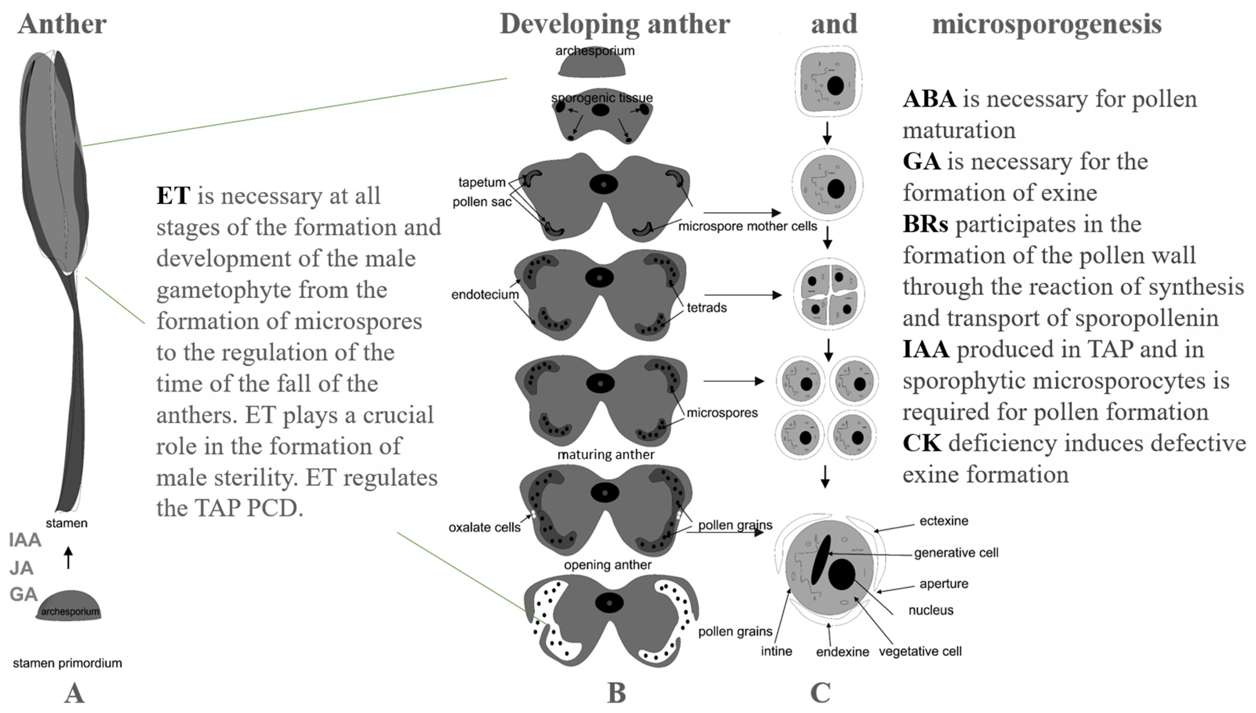
Figure 1. Scheme of microsporogenesis and anther development and the role of phytohormones at different stages. Stamen development (A). Anther development (B). Pollen formation (C).
Horticulturae 08 00365 g001
Figure 2. The germination and growth of petunia PT in the compatible and self-incompatible pollinations and the role of phytohormones at different stages of these processes (scheme).
Horticulturae 08 00365 g002
Publisher’s Note: MDPI stays neutral with regard to jurisdictional claims in published maps and institutional affiliations.